Simple method for long-term recording of electrocardiogram signals in prawns - Inter-Research Science Publisher
←
→
Page content transcription
If your browser does not render page correctly, please read the page content below
Vol. 30: 59–68, 2021                        AQUATIC BIOLOGY
                                                                                               Published March 25
 https://doi.org/10.3354/ab00741                      Aquat Biol

                                                                                               OPEN
                                                                                               ACCESS

              Simple method for long-term recording
              of electrocardiogram signals in prawns
          Han-Tso Lin1,*,#, Tsen-Chien Chen2,#, Jun-Ping Chang3, Zin-Yan Chou3,
                              Shian-Yu Chiou3, Meng-Li Tsai3
                  1
                   Department of Biotechnology, Ming Chuang University, Taoyuan City 33348, Taiwan
   2
    Department of Leisure Management, Minghsin University of Science and Technology, Hsinchu County 30401, Taiwan
           3
            Department of Biomechatronic Engineering, National Ilan University, Yilan County 26047, Taiwan

       ABSTRACT: This paper presents a novel approach to the long-term recording of electrocardio-
       gram (ECG) signals in prawns, including a 3-lead electrode, a lightweight external wires assem-
       bly, a waterproof electrode−wire junction, and a standardized implanting procedure. The pro-
       posed low-cost device is easily constructed, even by untrained undergraduate students, using
       common laboratory materials. Consistent ECG recordings were obtained over a period of 72 h in
       experiments on 6 freely moving prawns (Macrobrachium rosenbergii). A subsequent experiment
       conducted continuously for 38 d revealed that despite gradual attenuation the ECG signals could
       still be identified (signal-to-noise ratio ≥ 3). The prawns survived implantation and later under-
       went natural molting, whereupon the electrode was successfully re-implanted. The proposed
       electrode is a valuable tool by which to gain insight into the long-term physiological state of
       crustaceans.

       KEY WORDS: Electrocardiogram · Crustaceans · Giant river prawn · Macrobrachium rosenbergii ·
       Cardiac arrest

                  1. INTRODUCTION                           between heartbeat patterns in electrocardiogram
                                                            (ECG) signals and external stimuli, thereby making
   Decapod crustaceans (e.g. crabs, prawns, lobsters,       it possible to use cardiac activity (e.g. heart rate or
and crayfish) are a diverse group of more than 14 000       heart rate variability) as a proxy for metabolic
extant species, many of which are exploited commer-         changes (Ern et al. 2014, Tsai et al. 2019). Numerous
cially as a source of protein (Deshmukh 2013). Devel-       researchers have sought to use ECG recordings of
oping a more complete understanding of decapod              crustaceans as a tool to observe underlying physio-
crustaceans is crucial to the sustainability of this        logical functions. Larimer (1962) pioneered the use of
resource.                                                   real-time ECGs to monitor changes in the cardiac
   Crustaceans are sensitive to environmental condi-        activity of crayfish under various oxygen concentra-
tions, including temperature, noise, dissolved oxy-         tions. Florey & Kriebel (1974) recorded ECG patterns
gen content, photoperiod, and the presence of chem-         from unrestrained crabs to elucidate the effects of
ical compounds. Unfortunately, the rigid shell-like         temperature, anoxia, and sensory stimulation on
exoskeleton of crustaceans makes it difficult to ob-        heart rate. Through the analysis of ECG recordings,
serve physiological responses to external conditions.       Mickel & Childress (1982) examined the effects of
Researchers have clearly demonstrated a correlation         pressure and temperature on the heart rate of
                                                            © The authors 2021. Open Access under Creative Commons by
*Corresponding author: htlin@mail.mcu.edu.tw                Attribution Licence. Use, distribution and reproduction are un-
#                                                           restricted. Authors and original publication must be credited.
  These authors contributed equally to this work
                                                            Publisher: Inter-Research · www.int-res.com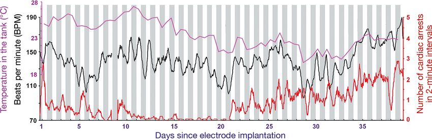
60                                           Aquat Biol 30: 59–68, 2021

Bythograea thermydron (a deep-sea crab living on            mammals, every characteristic of the ECG waveform
hydrothermal vents). Mislin (1966) revealed that            can be attributed to electrophysiological activity.
light levels could affect the heartbeat of animals.         Thus, it is essential that researchers maintain consis-
Burnovicz et al. (2009) examined ECG signals from           tency in the ECG recording process.
Neohelice granulata (previously known as Chasmag-             This paper presents a novel approach to the long-
nathus) under flashing lights, bubble-induced noise,        term recording of ECG signals in prawns, including a
and a moving visual danger stimulus present in              3-lead electrode with lightweight external wires and
experiments.                                                waterproof electrode−wire junctions providing buoy-
   ECG monitoring has revealed some interesting phe-        ancy. In experiments, the proposed electrode implan-
nomena. Yazawa & Katsuyama (2001) identified in-            tation scheme was shown to improve the accuracy of
stances of spontaneous repetitive cardiac arrest in         electrode placement, resulting in highly consistent
freely moving spiny lobsters. Similar research has          ECG signals between individuals with a signal-to-
been used to study the social behavior of crustaceans,      noise ratio remaining at a readable level even after
such as fighting for territory and/or mating rights.        monitoring for an extended duration. Electrode im-
Schapker et al. (2002) used ECG signals to monitor          plantation appears to have only a minor physiologi-
heart rate as an index by which to assess stress asso-      cal impact on the animals, as indicated by continued
ciated with social interactions. Listerman et al. (2000)    normal growth and molting following the operation.
explored the relationship between social interactions
and serotonin levels (5-HT) in the hemolymph of
crayfish. Based on heart rate, Cooper et al. (2011) mon-              2. MATERIALS AND METHODS
itored the physiological conditions in crustaceans of
both sexes during copulation.                                   2.1. Preparation of animals for experiments
   In the studies mentioned above, heart activity was
characterized from infrared signals in the contraction         Live specimens of the giant river prawn Macro-
process of the heart (Ern et al. 2014), changes in the      brachium rosenbergii were purchased from a local
impedance of peripheral components of the heart (Lis-       farm located in Toucheng Township in Yilan County,
terman et al. 2000, Schapker et al. 2002, Burnovicz et      Taiwan. After shipping, the prawns were settled at a
al. 2009, Cooper et al. 2011), or changes in membrane       culture density (total biomass of prawns to water vol-
potential during heart contractions (Larimer 1962,          ume) of 1:500. Water temperature in the tank was
Florey & Kriebel 1974, Mickel & Childress 1982, Ya-         maintained at approximately 25°C using an auto-
zawa & Katsuyama 2001). Overall, changes in mem-            mated aquarium heater, and dissolved oxygen was
brane potential present the richest information, in         provided by continuous air pumping. The hygiene of
terms of calculating heart rate, indicating the electri-    the aquaculture tanks was scrupulously maintained
cal dynamics associated with populations of myo-            via daily cleaning, which included removing feed
cardial cells in various regions of the heart and even      residue from the bottom of the tank, replacing a por-
physiological or pathological conditions. However,          tion of the water, and feeding the animals on com-
no previous study based on membrane potential has           mercial feed twice per day. The animals selected for
been conducted for extended periods (e.g. several           subsequent experiments were specimens with a
weeks). It is therefore difficult to determine whether      carapace length of 30 to 45 mm and no obvious signs
those recording methods provide stable results over         of damage or appendage loss.
the long term. Yazawa & Katsuyama (2001) reported
that their approach to ECG recording could be con-
tinued for several weeks; however, no mention was                         2.2. Electrode assembly
made as to the quality of ECG recordings over time.
   Furthermore, those articles did not provide a clear        We considered 3 points in the design of the elec-
description of the position of the electrode leads im-      trode: (1) preventing short-circuit events during
planted in the animal’s body (i.e. using vague terms,       underwater recordings by protecting the connection
such as cardiac region). The waveforms of the ECG           between the implanted electrode and the external
recording vary as a function of the position of the         conduction wire through the creation of a chamber
leads on the heart. Irisawa & Irisawa (1957) demon-         using a 2 ml microcentrifuge tube; (2) precisely
strated that the ECG waveforms obtained from the            aligning the 3 electrode leads horizontally, such that
surface of peripheral heart regions differ from those       the middle one operates as a grounding reference
obtained from the mid-dorsal region of the heart. In        lead, while the other 2 act as signal leads for voltage
Lin et al.: Long-term ECG recording of prawns 61
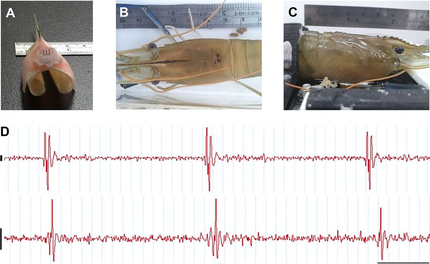
A B C
Fig. 1. Proposed 3-channel electrode. (A) Materials used in the construction of the electrode assembly, including (from left to
right) a stainless syringe needle (3 needles are required for 1 assembly), a Dupont pin header, and a microcentrifuge tube (2 ml
size). Red arrows indicate the portion to be sectioned or removed. (B) Dupont pin header with pins removed, 3 syringe needle
shafts (cut away from the hub) put into the holes left from the removed pins (indicated by white arrows), and a trimmed cap
from a microcentrifuge tube. (C) Pins of female pin header to be removed and replaced by needles
inputs to obtain differential signals; and (3) ensuring 5-pin header (pitch: 2.54 mm), a single-row female 3-
that the electrode assembly is as light as possible to pin header (pitch: 1.27 mm), a section of silicone tub-
avoid placing a physical load (stress) on the animal, ing (no. B07000OD, A-M Systems), a 2 ml microcen-
with a particular focus on the external conduction trifuge tube, and epoxy glue.
wire and quantity of epoxy adhesive. Three sections of enameled wire (60 cm in length)
Fig. 1A presents the materials used in the construc- were clamped together at both ends using 2 hemo-
tion of the electrode assembly, which included three stats. The teeth of the hemostats were covered with a
27 gauge (G) stainless steel syringe needles (to act as short sleeve of silicone tubing (no. 807600, A-M Sys-
leads), the plastic cap cut from a 2 ml microcentrifuge tems) to prevent damage to the insulation around the
tube (to act as an electrode mounting assembly), a wires. The clamped bundle of enameled wires was
Dupont pin header with 1.27 mm pitch (to act as the then hung in a manner that would allow it to droop
base for the leads), and Coltène/Whaledent dental naturally under the effects of gravity (Fig. 2A). With
cement (to mount the needles). The shafts of the nee- the upper hemostat held steady, the lower hemostat
dles were cut away from the mounting hub using was rotated counterclockwise to coil the wires tightly
wire cutters to replace the pins in the Dupont pin together to form a cable, whereupon the lower hemo-
header (Fig. 1B). A thin layer of dental cement was stat was removed. The end of the new cable was then
used to fix the exposed needles in place within the trimmed using scissors and threaded through a PE20
pin header, after carefully adjusting the length of the tube until the end extended out of the tube cap by
shafts to ensure consistency. A hole drilled through approximately 1 cm.
the center of the cap was just large enough to allow Both ends of the wire cable were loosened to sepa-
the 3 leads to pass through. The edges of the cap rate the individual wires, the ends of which were then
were cut in a zigzag shape to facilitate adhesion at stripped of their insulation to a distance of approxi-
the end of the assembly process. The electrode as- mately 5 mm (Fig. 2B). One end of each wire was re-
sembly was cemented within the cap using a small spectively soldered to each of the 3 needles mounted
amount of dental cement, as shown in Fig. 1C. The in the female pin header and tube cap assembly
total cost of the materials was less than US$1, and (Fig. 2C). The tub cap and connection between the
even a novice could construct an electrode inde- wires and pin header were sealed using epoxy glue
pendently after a 1 h demonstration. (Fig. 2D). A segment of silicon tubing (length = 1 cm)
was slipped over the other end of the cable (connect-
ing to the amplifier), whereupon the wires were sol-
2.3. Assembly of external conduction wires dered to the middle 3 pins of a 5-pin male pin header
and encased within epoxy glue (Fig. 2E). A 2 ml
The materials used to construct the light-weight microcentrifuge tube was attached to the epoxy-
external conduction wire assembly included 3 sec- sealed cap to make a waterproofed chamber for the
tions of thin enameled wire (no. 45; 0.07 mm), a sec- electrode connector joint and provide additional
tion of polyethylene tubing (PE20), a single-row male buoyancy (Fig. 2F).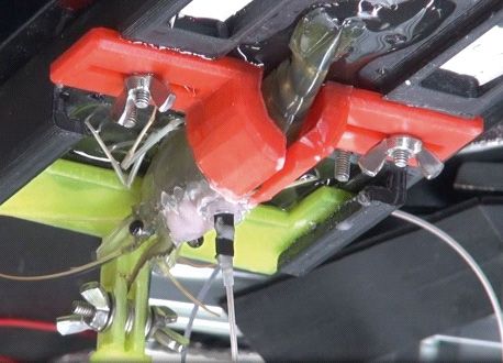
62                                                 Aquat Biol 30: 59–68, 2021

                A                     B                    C

                    D                                      E

                                                                F
Fig. 2. Materials and steps taken to make the electrocardiogram signal conduction wire. (A) Three enameled wires were
clamped on both ends by hemostats, then intertwined to unite them and form a handmade cable. (B) The handmade cable was
insulated with a PE20 tube leaving each end exposed (un-insulated) and unwound. (C) A cap from a microcentrifuge tube was
used as a plug to seal the chamber shown in (F). A hole was drilled in the centre of the cap to allow the cable to pass through it.
(D) The un-insulated ends of the enameled wires were soldered separately to 1 of 3 pins of a 5-pin holder. (E) Joints (and cap)
were sealed with epoxy glue (gray color) for waterproofing. (F) The entire handmade cable for ECG signal conduction is
shown. The end with a small, female type pin holder and a chamber made with a microcentrifuge tube was connected to the
                             electrodes on the prawn; the other end was connected to the amplifier

              2.4. Electrode implantation                           be taken to avoid penetrating the carapace and thereby
                                                                    prevent the loss of excessive body fluids during im-
   Based on our previous experience, we determined                  plantation. The middle lead was aligned with the ori-
that needles of roughly 4 mm in length would be                     entation of the rostrum to ensure accuracy (Fig. 3A).
required for prawns with a body weight of 30 to 50 g.                  With the leads held vertically, they were pushed by
The position at which the leads should be implanted                 hand through the notch in the carapace, whereupon
is at the posterior edge of a distinct square bump on               the wounds were sealed using a small amount of
the carapace, corresponding to the posterior margin                 dental cement. The base of the rostrum was also
of the heart.                                                       coated with cement to increase the strength of the
   With the animal held in place on a clamping frame, a             attachment (Fig. 3B). After the cement dried, the
knife was used to scrape the carapace surrounding the               Dupont female pin header from the external conduc-
heart to facilitate the subsequent adhesion of dental               tion assembly was attached to the male pin header of
cement. A scalpel was then used to scrape a notch                   the electrode, over which was placed the microcen-
(5 mm in length) to weaken the carapace. Care must                  trifuge tube (Fig. 3C).

A                                 B                                          C

Fig. 3. Implantation of electrocardiogram (ECG) electrode in a giant river prawn. (A) Top view of prawn fixed on frame. The
white arrow indicates the notch (black bar) used to locate the site of implantation. The middle of the notch should be aligned
with the axis of the rostrum in the carapace (indicated by the white arrow aligned vertically to the notch) to ensure positional
accuracy. (B) Insertion site coated with dental cement following electrode implantation. (C) Instance of ECG monitoring of a
                          freely moving giant river prawn (9.9 g in weight) after electrode implantation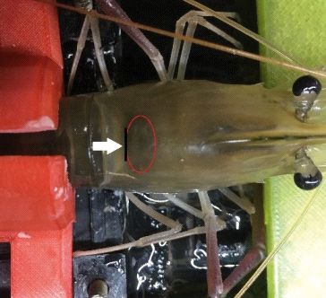
Lin et al.: Long-term ECG recording of prawns 63
2.5. Data acquisition The contraction of the heart (observed directly
through the translucent carapace) produces a group
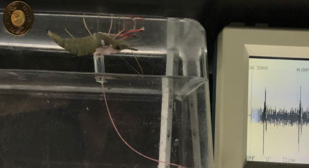
The implanted prawn was moved to the ex- of waveforms in the ECG pattern (see Fig. 4). In cal-
perimental tank (size 30 × 30 × 25 cm), which was culating the heart rate, the time interval between the
held within a Faraday cage to enable the recording largest wavelet in 2 successive waveform groups is
of ECG signals under freely moving conditions. treated as the period between 2 heartbeats.
Throughout the recording period, the prawns were
fed normally every 12 h. The middle lead was
grounded (to act as a reference), and the left and 3. RESULTS
right leads served as voltage inputs to obtain dif-
ferential signals. The signals were band-pass fil- The proposed ECG recording method proved
tered (approx. 100−300 Hz) and amplified 5000× highly stable, based on the uniformity of results ob-
using a differential amplifier (Model 3000, A-M tained from different animals. Fig. 4 presents the
Systems). After processing, the signals underwent ECG recordings from 3 prawns of various sizes in a
analog−digital conversion using a PowerLab 4/25 freely moving state at 1, 36, and 72 h after implanta-
(ADInstruments) at a sampling rate of 1000 Hz tion. Clearly, the patterns in the ECG signals did not
before being stored on a computer. Chart 5 soft- vary noticeably over time, indicating that the pro-
ware (version 5.5.6; ADInstruments) was used for posed method could be used for analysis over an
data analysis. extended duration.
Fig. 5 presents an example ECG
recording obtained over a 38 d period,
1h 36 h 72 h showing the ECG intensity (i.e. maxi-
mum amplitude of the waveform
components) and noise. Despite a no-
ticeable decrease in ECG intensity be-
#1 ginning at 22 d after implantation, the
ECG features remained identifiable
with a signal-to-noise ratio exceeding
3.0.
We also found that changes in am-
bient temperature had a dramatic effect
on the heart rate of 1 prawn in long-
term recording. As shown in Fig. 6,
the trend between heart rate and am-
#2 bient temperature remained consistent
throughout a 38 d recording period.
Under stressful environmental distur-
bances, we observed temporary cessa-
10 µV
tions of the heartbeat (skipping 1 or
more pulses). Manually exchanging
50 ms the water in the observation tank
tended to agitate the prawn, with a re-
sult that the frequency of cardiac ar-
rests exceeded the baseline during the
#3
first 9 d. Between Days 9 and 20, these
events occurred less frequently, indica-
ting that the animal had become accli-
mated to this interference. During this
Fig. 4. Stability and comparability of electrocardiogram (ECG) signals recorded period, the heart rate of the animal
using the proposed system. The weights of the 3 prawns were as follows: 41 g tended to increase during the daytime
(#1), 48 g (#2), and 70 g (#3). Data were recorded at 1, 36, and 72 h after elec-
trode implantation. All of the ECG signals presented similar patterns, with
and decrease when it was dark. After
little interday and intersubject variation. Each heartbeat presented 2 distinct Day 21, this pattern gradually reversed,
larger wavelets followed by a group of smaller wavelets such that the heart rate dropped during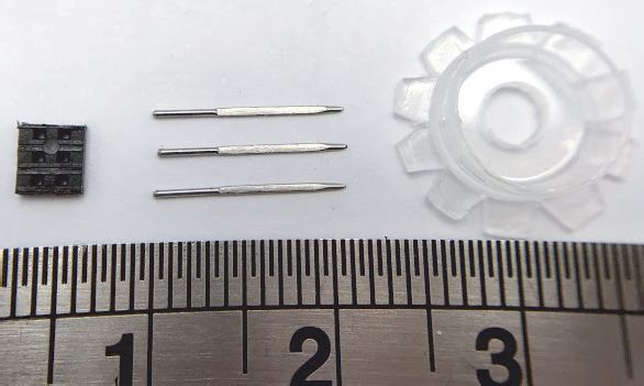
64                                                   Aquat Biol 30: 59–68, 2021

               160                                                                                                   36
                                                                                           ECG Signal
                                                                                           Noise                     30

               120                                                                         S/N ratio

                                                                                                                     24
Voltage (µV)

                                                                                                                          S/N ratio
               80                                                                                                    18

                                                                                                                     12

               40

                                                                                                                     6

                0                                                                                                    0
                     D0   D2   D4   D6   D8 D10 D12 D14 D16 D18 D20 D22 D24 D26 D28 D30 D32 D34 D36 D38
                                                             Time (d)

                               D4                                  D18                                   D38
40 µV

                                    100 ms

Fig. 5. Electrocardiography data from continuous recording of a giant river prawn (44 g) over 38 d. The histograms show the
amplitude of the electrocardiogram (ECG) signal and background noise during recording, whereas the gray line graph shows
the signal-to-noise (S/N) ratio. The day of electrode implantation is designated D0. Also, signal and noise data were averaged
in segments of 2 d. At the time of death on Day 39, the body weight of the prawn was 48 g. The ECG signal began decaying
after Day 22; however, the S/N ratio remained above 3.0. The bottom panels show an example of the actual ECG signals
                         recorded on Days 4 (D4), 18 (D18), and 38 (D38) after electrode implantation

the bright period and rose during the dark period. Af-              pace had formed completely with the required hard-
ter Day 21, the number of cardiac arrests suddenly in-              ness. This made it possible to implant electrodes into
creased, while the signal amplitude decreased (Fig. 5).             the same animal a second time, yielding ECG signals
We posit that this can be attributed to a gradual de-               with a good signal-to-noise ratio.
terioration of the health of the animal, resulting in its             The proposed system uses light and easily deform-
death on Day 39, rather than a decline in the recording             able enameled wires for signal transmission within a
performance of the electrode.                                       PE20 tube to ensure that the apparatus would not
  The implanted electrode was shown not to affect                   affect the movement of the animal. The microcentri-
the growth or molting behavior of the prawns. Even                  fuge tube also provided buoyancy to offset the
during the molting process, the electrode captured                  weight of the other components and thereby reduce
ECG signals until the carapace had lifted off entirely.             physiological stress. As shown in Fig. 8, the continu-
Fig. 7 shows that by Day 10 after molting, there were               ous recording of ECG patterns for 72 h revealed no
some anomalies in the appearance of the carapace at                 notable increases or decreases in heart rate following
the original site of implantation; however, the cara-               electrode implantation.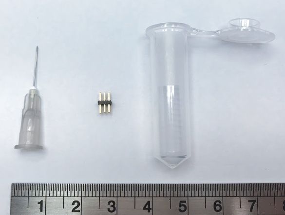
Lin et al.: Long-term ECG recording of prawns                                           65

Fig. 6. Changes in the heart rate of a giant river prawn as a function of temperature over 38 d (same prawn as in Fig. 5). The
horizontal axis shows the timeline of this experiment, with white columns indicating light periods and gray columns indicating
darkness. The averages of heart rate (black line) and number of cardiac arrests (red line) were calculated from 2 min recording
intervals obtained every half hour. These 2 curves are processed by a moving average, which is a calculation used to smooth a
curve by creating a series of averages of different subsets of the full data. For each curve, the first element of the moving aver-
age is obtained by taking the mean of the initial fixed subset (16 data points) of the original series. Then the data points in-
cluded in the subset are modified by shifting forward, that is, excluding the first number of the original series and including
            the next value in the subset. In this way, the calculation continues until the entire data set is averaged

Fig. 7. Giant river prawn (42.4 g) with implanted electrode. (A) Molted cephalothorax carapace section with electrode still at-
tached. (B) Dorsal and (C) lateral views of the prawn 10 d after molting, showing that the carapace of the prawn was affected
by the implanted electrode but still grew to full size. (D) Electrocardiogram (ECG) signals at 2 d prior to molting (upper) and
5 d after a second electrode was implanted (lower). Scale bar in (D) indicates 10 μV vertically and 100 ms horizontally

                     4. DISCUSSION                                  voltage, impedance, and current, and researchers
                                                                    have yet to attain a consensus as to which of these
  Numerous studies have used electrical activity in                 variables is most representative. Several recent stud-
the myocardium of crustaceans to obtain physiologi-                 ies have used impedance detectors (Schapker et al.
cal data, such as heart rate. Importantly, however,                 2002, Bierbower & Cooper 2009, Weineck et al. 2018,
electrical activity can be characterized in terms of                Adams et al. 2019). However, changes in impedance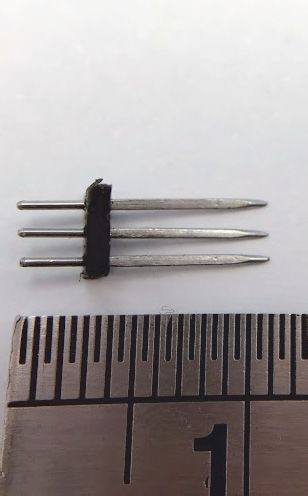
66                                             Aquat Biol 30: 59–68, 2021

                   250                                                          taining to crucial variables, such as the
                                                                                length of the wire inserted into the
                                                                                carapace. The heart of a prawn is very
                                                                                close to the inner surface of the cara-
                                                                                pace, such that inserting a wire of ex-
      50                                                                        cessive length would likely interfere
     250                                                                        with the normal beating of the heart.
                                                                                Even if the wire did not come into
                                                                                direct contact with the heart, it would
                                                                                inevitably contact the connective tissue
                                                                                surrounding the organ. A very soft
      50                                                                        wire would prevent damage to the
     250
                                                                                heart; however, it would also be sus-
                                                                                ceptible to deformation, causing it to
                                                                                extend irregularly across the surface of
Heart rate (BPM)

                                                                                the heart. Furthermore, it is very diffi-
      50
                                                                                cult to standardize and confirm the dis-
     250                                                                        tance between the 2 leads, thereby
                                                                                making it difficult to ensure consistency
                                                                                between implant operations.
                                                                                  An ECG measures changes in the
                                                                                membrane potential of the myocardial
      50                                                                        muscle tissue during each heartbeat,
     250                                                                        including depolarization and repolar-
                                                                                ization. Measuring these changes in
                                                                                membrane potential would make it
                                                                                possible to calculate the heart rate as
                                                                                well as other physiological and patho-
      50
     250
                                                                                logical conditions. Our use of a 3-lead
                                                                                electrode with grounding reference
                                                                                lead and 2 voltage input leads close
                                                                                to the heart was shown to reduce
                                                                                electromagnetic interference from the
      50                                                                        surrounding environment as well as
         0           12        24          36        48          60         72  changes in membrane potential asso-
                                       Time (h)                                 ciated with the contraction of sur-
Fig. 8. Heart rate (beats per minute [BPM]) of 6 prawns over 72 h immediately   rounding muscle cells of the other
after electrode implantation. All data were recorded in a bright environment    organs. The proposed system makes it
                                    for 72 h                                    possible to record the electrophysio-
                                                                                logical details of heart function, even
are actually associated with the environment between              while the animal is swimming freely. This provides
the 2 leads near the heart, as opposed to changes in              information of far greater richness than can be
membrane potential resulting from depolarization or               achieved simply by measuring changes in im-
repolarization; therefore, the impedance signals can              pedance. In the present study, the electrodes were
only be used to calculate heart rate.                             not implanted directly above the heart but rather
  The 2 leads in the studies mentioned above are usu-             were placed close to the posterior side of the heart,
ally implanted directly above the heart. Thus, the                thereby preventing damage to heart tissue. The pro-
strength of the captured signal depends strongly on               posed system also makes it possible to control the
the distance between the heart and the non-insulated              length of the leads inserted into the carapace,
part of the wire buried within the body. Signal                   thereby ensuring uniformity across samples.
strength also depends on the size of the conductor                  Nevertheless, there are still a number of questions
area used to capture the electrical signal. Unfortu-              concerning the standardization of this system. The
nately, previous studies failed to provide details per-           point of implantation is along the posterior edge of a
Lin et al.: Long-term ECG recording of prawns                                      67

distinct square bump on the surface of the carapace,            Tethering the prawns to a 60 cm external wire for
corresponding to the posterior margin of the heart.          signal transmission seldom led to entanglement suf-
Nonetheless, the caudal border of the heart does not         ficient to hinder the animal’s movement. Nonethe-
necessarily coincide with the square carapace feature        less, all assessments were performed with the animal
in every animal. Thus, during implantation, it is nec-       alone in a tank. In the future, it will be necessary
essary to observe the systole of the heart through the       to obtain simultaneous recordings from multiple
translucent carapace. The margin between the elec-           prawns in the same tank. Tethering prawns in this
trode and heart must be small enough to ensure a             way would make it impossible to conduct multi-
strong signal but large enough to avoid injuring heart       animal experiments of extended duration or involv-
tissue. Ideally, the leads inserted into the prawn           ing excessive movement.
should be of sufficient length to reach the desired re-         Ideally, the electrodes should be equipped with a
gion adjacent to the heart without any extraneous ma-        wireless transmission system, perhaps mounted on
terial, which might otherwise compromise the signal-         the carapace at the cephalothorax; however, the size
to-noise ratio. Obviously, the size of the heart varies      and weight of the device would be a key issue. In the
with the size of the body; therefore, the length of the      external wire system in this study, the connection
inserted leads must be adjusted according to the size        between the external wires and the output terminal
of the test animal. The ideal length can be determined       of the electrode was enclosed within a microcentri-
by observing the heart through the translucent cara-         fuge tube. This made it possible to offset the weight
pace from the side. Nonetheless, these observations          of the tether by taking advantage of the buoyancy
are only rough approximations; therefore, it is impor-       provided by the tube. It is very likely that the same
tant to remember that operators will tend to differ in       approach could be adopted to enclose a wireless
terms of electrode placement.                                transmission system.
   The transparent carapace of giant river prawns
makes it possible to observe contractions of the heart
directly during the recording of ECG signals. As             Acknowledgements. The authors thank Mr. Jui-Yu Wu for
                                                             his assistance in completing the experiments in Fig. 7. This
shown in Fig. 4, each heartbeat provides a correspon-
                                                             work was supported by grants MOST-107-2313-B-197-001
ding set of waveforms, containing 1 major wavelet            and MOST-106-2621-B-159-001 from the Ministry of Sci-
(i.e. with the largest amplitude) and 2 minor wavelets       ence and Technology of Taiwan.
(with much smaller amplitudes). Analogous to mam-
mals, these waveforms indicate the changes in poten-
tial associated with ventricular depolarization. How-                           LITERATURE CITED
ever, each ventricle in a mammal is connected to only
                                                             Adams R, Stanley CE, Piana E, Cooper RL (2019) Physiolog-
1 artery, whereas each ventricle in a prawn is con-
                                                                 ical and behavioral indicators to measure crustacean
nected to 7 arteries (McLaughlin 1983): the unpaired             welfare. Animals (Basel) 9:914
anterior median artery, sternal artery and dorsal ab-        Bierbower SM, Cooper RL (2009) Measures of heart and
dominal artery, and the paired anterior lateral arteries         ventilatory rates in freely moving crayfish. J Vis Exp 32:
and hepatic arteries. Furthermore, it is likely that ven-        e1594
                                                             Burnovicz A, Oliva D, Hermitte G (2009) The cardiac re-
tricular depolarization in prawns differs from that in           sponse of the crab Chasmagnathus granulatus as an
mammals, manifesting in multiple QRS complexes.                  index of sensory perception. J Exp Biol 212:313−324
   Invasive recording devices must always be as small        Cooper RM, Schapker H, Adami H, Cooper RL (2011) Heart
and unobtrusive as possible. The electrode structure             and ventilatory measures in crayfish during copulation.
                                                                 Open J Mol Integr Physiol 1:36−42
in the current study uses three 27 G stainless steel
                                                             Deshmukh V (2013) Principles of crustacean taxonomy. In:
needles for insertion into the prawn’s body. The                 Jose J, Pillai SL (eds) Training programme on taxonomy
diameter of these needles (0.4 mm) is small enough               and identification of commercially important crustaceans
to minimize damage to the animal, while large                    of India. Central Marine Fisheries Research Institute,
                                                                 Kochi, p 28−39
enough to resist the rigors of construction and inser-
                                                             Ern R, Huong DTT, Phuong NT, Wang T, Bayley M (2014)
tion. Materials other than 27 G stainless steel needles          Oxygen delivery does not limit thermal tolerance in a
(such as microwire) could theoretically be used to               tropical eurythermal crustacean. J Exp Biol 217:809−814
fabricate the electrode; however, the wire must be of        Florey E, Kriebel ME (1974) The effects of temperature,
sufficient strength to penetrate the flexible membra-            anoxia and sensory stimulation on the heart rate of unre-
                                                                 strained crabs. Comp Biochem Physiol A Comp Physiol
nous connective tissue surrounding the heart without             48:285−300
the need for carapace laceration, which might other-         Irisawa H, Irisawa AF (1957) The electrocardiogram of a
wise cause uncontrollable bleeding.                              stomatopod. Biol Bull 112:358−362
68                                                 Aquat Biol 30: 59–68, 2021

Larimer JL (1962) Responses of the crayfish heart during          Schapker H, Breithaupt T, Shuranova Z, Burmistrov Y,
    respiratory stress. Physiol Zool 35:179−186                      Cooper RL (2002) Heart and ventilatory measures in
Listerman LR, Deskins J, Bradacs H, Cooper RL (2000) Heart           crayfish during environmental disturbances and social
    rate within male crayfish: social interactions and effects       interactions. Comp Biochem Physiol A Mol Integr Physiol
    of 5-HT. Comp Biochem Physiol A Mol Integr Physiol               131:397−407
    125:251−263                                                   Tsai ML, Hsu WP, Chen TC (2019) Evaluation of suitable
McLaughlin PA (1983) Internal anatomy. In: Mantel LH (ed)            temperature range for post-harvest processing of mud
    The biology of Crustacea, Vol. 5. Academic Press, New            crabs through cardiac performance. Aquacult Res 50:
    York, NY, p 1−52                                                 3711−3719
Mickel TJ, Childress JJ (1982) Effects of pressure and tem-       Weineck K, Ray AJ, Fleckenstein LJ, Medley M, Dzubuk N,
    perature on the EKG and heart rate of the hydrothermal           Piana E, Cooper RL (2018) Physiological changes as a
    vent crab Bythograea thermydron (Brachyura). Biol Bull           measure of crustacean welfare under different standard-
    162:70−82                                                        ized stunning techniques: cooling and electroshock. Ani-
Mislin H (1966) Experimenteller Nachweis der Beeinflus-              mals (Basel) 8:158
    sung des Elektrokardiogramms (EKG) dekapoder Krebse           Yazawa T, Katsuyama T (2001) Spontaneous and repe-
    (Astacus fluviatilis F., Astacus leptodactylus E., Carcinus      titive cardiac slowdown in the freely moving spiny
    maenas L.) durch optische Reize (Optocardialer Hemm-             lobster, Panulirus japonicus. J Comp Physiol A 187:
    reflex). Rev Suisse Zool 73:301−312                              817−824

Editorial responsibility: Bernard Sainte-Marie,                   Submitted: June 7, 2020
 Mont-Joli, Quebec, Canada                                        Accepted: December 11, 2020
Reviewed by: 3 anonymous referees                                 Proofs received from author(s): March 23, 2021
You can also read