NOVEL ELISA PROTOCOL LINKS PRE-EXISTING SARS-COV-2 REACTIVE ANTIBODIES WITH ENDEMIC CORONAVIRUS IMMUNITY AND AGE AND REVEALS IMPROVED SEROLOGIC ...
←
→
Page content transcription
If your browser does not render page correctly, please read the page content below
METHODS
                                                                                                                                              published: 09 April 2021
                                                                                                                                     doi: 10.3389/fimmu.2021.614676

                                            Novel ELISA Protocol Links
                                            Pre-Existing SARS-CoV-2
                                            Reactive Antibodies With Endemic
                                            Coronavirus Immunity and Age
                                            and Reveals Improved Serologic
                                            Identification of Acute COVID-19
                                            via Multi-Parameter Detection
                             Edited by:
                     Vitaly V. Ganusov,     Rachel R. Yuen 1, Dylan Steiner 2, Riley M.F. Pihl 3, Elizabeth Chavez 1, Alex Olson 4,
The University of Tennessee, Knoxville,     Erika L. Smith 1, Lillia A. Baird 2, Filiz Korkmaz 2, Patricia Urick 2, Manish Sagar 1,2,4,
                          United States     Jacob L. Berrigan 1, Suryaram Gummuluru 1, Ronald B. Corley 1,5, Karen Quillen 2,
                        Reviewed by:        Anna C. Belkina 6,7, Gustavo Mostoslavsky 8, Ian R. Rifkin 9,10, Yachana Kataria 7,
                        Laura Anfossi,      Amedeo J. Cappione III11, Wenda Gao 12, Nina H. Lin 4, Nahid Bhadelia 2,5
               University of Turin, Italy   and Jennifer E. Snyder-Cappione 1*
                Jerold G. Woodward,
University of Kentucky, United States       1 Department of Microbiology, Boston University School of Medicine, Boston, MA, United States, 2 Department of Medicine,

                  *Correspondence:          Boston University School of Medicine, Boston, MA, United States, 3 PiBS Program, Boston University School of Medicine,
         Jennifer E. Snyder-Cappione        Boston, MA, United States, 4 Section of Infectious Diseases, Department of Medicine, Boston Medical Center, Boston, MA,
                    cappione@bu.edu         United States, 5 National Emerging Infectious Diseases Laboratories (NEIDL), Boston University, Boston, MA, United States,
                                            6 Flow Cytometry Core Facility, Boston University School of Medicine, Boston, MA, United States, 7 Department of Pathology

                                            and Laboratory Medicine, Boston University School of Medicine, Boston, MA, United States, 8 Center for Regenerative
                   Specialty section:
                                            Medicine, Boston University School of Medicine, Boston, MA, United States, 9 Renal Section, Department of Medicine,
         This article was submitted to
                                            Boston University School of Medicine, Boston, MA, United States, 10 Renal Section, Department of Medicine, VA Boston
                      Viral Immunology,
                                            Healthcare System, Boston, MA, United States, 11 MilliporeSigma, Burlington, MA, United States, 12 Antagen
               a section of the journal
                                            Pharmaceuticals, Boston, MA, United States
              Frontiers in Immunology

         Received: 06 October 2020
          Accepted: 03 March 2021           The COVID-19 pandemic has drastically impacted work, economy, and way of life.
           Published: 09 April 2021         Sensitive measurement of SARS-CoV-2 specific antibodies would provide new insight
                            Citation:       into pre-existing immunity, virus transmission dynamics, and the nuances of SARS-CoV-2
       Yuen RR, Steiner D, Pihl RMF,
        Chavez E, Olson A, Smith EL,
                                            pathogenesis. To date, existing SARS-CoV-2 serology tests have limited utility due to
        Baird LA, Korkmaz F, Urick P,       insufficient reliable detection of antibody levels lower than what is typically present after
Sagar M, Berrigan JL, Gummuluru S,
                                            several days of symptoms. To measure lower quantities of SARS-CoV-2 IgM, IgG, and IgA
    Corley RB, Quillen K, Belkina AC,
 Mostoslavsky G, Rifkin IR, Kataria Y,      with higher resolution than existing assays, we developed a new ELISA protocol with a
    Cappione AJ III, Gao W, Lin NH,         distinct plate washing procedure and timed plate development via use of a standard
Bhadelia N and Snyder-Cappione JE
         (2021) Novel ELISA Protocol
                                            curve. Very low optical densities from samples added to buffer coated wells at as low as a
     Links Pre-Existing SARS-CoV-2          1:5 dilution are reported using this ‘BU ELISA’ method. Use of this method revealed
   Reactive Antibodies With Endemic
                                            circulating SARS-CoV-2 receptor binding domain (RBD) and nucleocapsid protein (N)
  Coronavirus Immunity and Age and
          Reveals Improved Serologic        reactive antibodies (IgG, IgM, and/or IgA) in 44 and 100 percent of pre-pandemic
 Identification of Acute COVID-19 via        subjects, respectively, and the magnitude of these antibodies tracked with antibody
           Multi-Parameter Detection.
          Front. Immunol. 12:614676.
                                            levels of analogous viral proteins from endemic coronavirus (eCoV) strains. The disease
   doi: 10.3389/fimmu.2021.614676           status (HIV, SLE) of unexposed subjects was not linked with SARS-CoV-2 reactive

Frontiers in Immunology | www.frontiersin.org                                      1                                          April 2021 | Volume 12 | Article 614676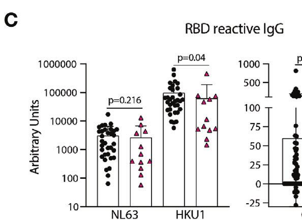
Yuen et al.                                                                                                  Sensitive Detection of SARS-CoV-2 Antibodies

                                          antibody levels; however, quantities were significantly lower in subjects over 70 years of
                                          age compared with younger counterparts. Also, we measured SARS-CoV-2 RBD- and N-
                                          specific IgM, IgG, and IgA antibodies from 29 SARS-CoV-2 infected individuals at varying
                                          disease states, including 10 acute COVID-19 hospitalized subjects with negative serology
                                          results by the EUA approved Abbott IgG chemiluminescent microparticle immunoassay.
                                          Measurements of SARS-CoV-2 RBD- and N- specific IgM, IgG, IgA levels measured by
                                          the BU ELISA revealed higher signal from 9 of the 10 Abbott test negative COVID-19
                                          subjects than all pre-pandemic samples for at least one antibody specificity/isotype,
                                          implicating improved serologic identification of SARS-CoV-2 infection via multi-parameter,
                                          high sensitive antibody detection. We propose that this improved ELISA protocol, which is
                                          straightforward to perform, low cost, and uses readily available commercial reagents, is a
                                          useful tool to elucidate new information about SARS-CoV-2 infection and immunity and
                                          has promising implications for improved detection of all analytes measurable by
                                          this platform.
                                          Keywords: SARS-CoV-2, COVID-19, antibodies, serology, nucleocapsid (N), receptor binding domain (RBD), ELISA

INTRODUCTION                                                                    serological assays have been developed by multiple
                                                                                manufacturers and academic institutes and many are CE-
From the first reported case of COVID-19 caused by the virus                     marked and granted emergency use authorization (EUA) from
SARS-CoV-2 in December 2019 (1, 2) there have been more than                    the US Food and Drug Administration (FDA). Varieties include
127 million reported cases and 2.79 million deaths worldwide as                 point-of-care rapid lateral flow assays, chemiluminescence
of March 29, 2021. Common symptoms of SARS-CoV-2                                immunoassays (CLIA), multi-plex bead/cell based-assays (15,
infection include fever, cough, myalgia, and fatigue and these                  16), and enzyme-linked immunosorbent assays (ELISA) (17–20).
symptoms vary widely in magnitude, nature, and duration                         These tests detect antibodies that primarily target the
between individuals for reasons that are not clear to date (3,                  nucleocapsid protein (N) or the spike (S) protein of SARS-
4), with some individuals with confirmed infections remaining                    CoV-2, and specifically the Receptor Binding Domain (RBD)
asymptomatic (5). Epidemiological evidence indicates silent viral               of spike which is an immunodominant surface protein targeted
spread via asymptomatic individuals within communities and                      by neutralizing antibodies and a main target antigen for vaccine
the extent of this form of transmission is currently unclear (6).               development (20–22). Some of these tests possess high sensitivity
SARS-CoV-2 has homology to other alpha and beta ‘common                         and specificity for detection of SARS-CoV-2 antibodies 14 days
cold’ endemic coronaviruses (eCoVs) in circulation, and cross-                  after diagnosis and/or symptom onset (23–26). However, others
reactive T cell immunity to SARS-CoV-2 spike (S) and                            report negative results from individuals who are asymptomatic,
nucleocapsid (N) proteins are present in a substantial                          mildly symptomatic, or symptomatic for less than 14 days, even
percentage of unexposed individuals (7–10). Also, reactive                      when SARS-CoV-2 infection is confirmed (19, 27, 28); whether
antibodies to SARS-CoV-2 S and N proteins are present in                        such individuals possess antibodies below the limit of the
unexposed individuals, with virus neutralization activity                       detection of the particular test used or lack these antibodies
reported from pre-pandemic pediatric samples (11, 12). It is                    altogether is unresolved.
postulated that this cross-reactive immunity may influence the                       To enable detection of low levels of SARS-CoV-2-reactive
nature and severity of COVID-19 symptoms upon infection and                     antibodies, we modified the standard ELISA procedure,
impact disease course (13) and may impact herd immunity.                        particularly the plate washing method, to improve sensitivity.
    Sensitive and accurate detection of virus-specific immune                    Our protocol (the ‘BU ELISA’) allows clear SARS-CoV-2-
factors, such as antibodies, is imperative in order to measure                  reactive antibody signal resolution at sample dilutions as low
rates of SARS-CoV-2 infections within communities with greater                  as 1:5. Using this protocol we measured the levels of SARS-CoV-
accuracy, to more fully define cross-reactive immunity in                        2-reactive IgM, IgG, and IgA from plasma or serum from three
unexposed individuals, and to gain new understanding about                      groups of individuals: (1) 71 subjects that varied by age, HIV
the nature of effective versus potentially deleterious immune                   infection, and systemic lupus erythematosus (SLE) disease status
responses upon SARS-CoV-2 exposure. Antibody measurements                       with all samples collected before November 8th, 2019 (‘pre-
are of p articular importance, as pathogen-spec i fic                            pandemic’); (2) 20 subjects hospitalized with COVID-19
immunoglobulins are a known first line of defense upon                           (‘Acute’) (3) nine subjects with samples collected two-seven
exposure and can prevent new infections. Antibody titers are                    months after confirmed SARS-CoV-2 infection
used to assess both likelihood of protection from re-infection and              (‘Convalescent’). In addition, the performance of the BU
general vaccine efficacy (14). A variety of SARS-CoV-2                           ELISA, Antagen’s IgM IgG Lateral Flow Device (LFD) test and

Frontiers in Immunology | www.frontiersin.org                               2                                     April 2021 | Volume 12 | Article 614676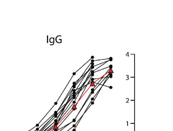
Yuen et al.                                                                                          Sensitive Detection of SARS-CoV-2 Antibodies

the Abbott IgG chemiluminescent microparticle immunoassay                 reactive antibodies (IgG, clone CR3022, gift from the Ragon
(CMIA) were directly compared from samples from the three                 Institute; IgA, clone CR3022, Absolute Antibodies; IgM, clone
subject groups.                                                           BIB116, Creative Diagnostics) were diluted in Thermo Fisher
                                                                          casein blocking buffer, and 50µl of each were added to the plates
                                                                          for 1 hour at room temperature, with dilution buffer only added
                                                                          to blank wells. After incubation, samples were removed by a
MATERIAL AND METHODS                                                      swift flick into a biohazard waste container. The plates were
                                                                          again washed three times with PBS containing 0.05% Tween 20
Participants                                                              (PBST) and banged on absorbent paper towels, and immediately
Pre-Pandemics: Samples were collected for unrelated studies               anti-human horseradish peroxidase (HRP)-conjugated
prior to December 2019; these subjects included samples from              secondary antibodies for IgG (cat#A18817, Thermo Fisher,
a HIV and Aging cohort previously described in detail (29) and            1:2000), IgM (cat#A18841, Thermo Fisher, 1:8000), and IgA
also from a geriatric cohort of subjects all over 60 years old with       (Jackson Immunoresearch, cat#109-035-011, 1:2000) diluted in
the following exclusion criteria: HIV, active hepatitis B or C, or        casein blocking buffer were added to the plates at 50µl per well
any recent active infection within the past six months, diagnosis         for 30 minutes at room temperature. Next, plates were washed
with an autoimmune disease, or treatment with any type of                 four times with 0.05% PBST as described, and 50µl per well of
immunomodulatory therapy within 12 months. All pre-                       3,3’,5,5’-Tetramethylbenzidine (TMB)-ELISA substrate solution
pandemic samples were collected before November 8th, 2019.                (Thermo Fisher Scientific, cat# 34029) was added and incubation
Acute COVID-19: De-identified samples from hospitalized                    occurred in the dark until a visible color difference between
patients at Boston Medical Center with confirmed PCR                       the well with the seventh dilution (1.37ng/ml) of recombinant
positivity for SARS-CoV-2 comprise this group, with 10                    antibody and the diluent only ‘zero’ well appeared, this time
subjects with positive and 10 with negative serology tests                ranged from ~8-30 minutes. The reaction was stopped by
determined by the Abbott CMIA EUA approved assay.                         adding 50µl of stop solution for TMB (Thermo Fisher
Samples were collected at various time points after onset of              Scientific, cat#N600) and the optical density was measured 450
symptoms, (range 3-40 days); all samples were collected during            nm (OD 450nm) on a SpectraMax190 Microplate Reader
the spring and summer of 2020, and all subjects in this group             (Molecular Devices). Seven-point sample dilution curves were
survived and were ultimately discharged from the hospital.                run in uncoated wells and paired antigen coated wells (SARS-
Eighty percent of this group was comprised of males and 60%               CoV-2 RBD and N). An example of a plate map shown in
identified as black. SARS-CoV-2 Infected Convalescent: Subjects            Supplemental Figure 1. Samples were not run in a blinded
were recruited by contacting individuals who had been                     manner as OD measurements are determined via a plate
confirmed to have SARS-CoV-2 infection through their                       reader. To ensure accurate determination of antibody levels
exposure at a biomedical conference in March 2020. None of                between subject groups, samples from pre-pandemic, acute
the individuals were hospitalized. Samples were collected 2-7             COVID-19, and SARS-CoV-2 infected survivors were routinely
months after a positive SARS-CoV-2 PCR test. The average age              run in mixed batches on ELISA plates and/or during
of this group was 52, with 22% male and 33% identified as black.           experimental runs.
All samples collected and/or used in this study with proper
IRB approvals from the Boston University Institutional
Review Board.                                                             Antigens
                                                                          SARS-CoV-2 RBD was a gift from the Schmidt lab at the Ragon
                                                                          Institute and was expressed and purified as previously described
The BU ELISA Protocol                                                     (30). SARS-CoV-2 N (Cat# 40588-V08B) and S (Cat# 40591-
Antibodies reactive to all four eCoV and SARS-CoV-2 RBD or N              V08H), NL63 N (Cat# 40641-V07E), 229E N (Cat# 40640-
were assayed from sera or plasma as described in accompanying             V07E), OC43 N (Cat# 40643-V07E) and HKU1 N (Cat#
SOP (Supplemental Figure 1). Briefly, wells of 96-well plates              40641-V07E) was purchased from Sino Biological. Histidine-
(Pierce 96-Well Polystyrene Plates; cat#15041, Thermo Fisher              tagged NL63 and HKU1 RBD sequences were inserted into
Scientific) were coated with 50µl/well of a 2µg/ml solution of             plasmid vector VRc (gift from the Schmidt lab at the Ragon
each respective protein in sterile PBS (Gibco) or with PBS only           Institute) and was expressed in 293 Freestyle cells (293F,
for 1 hour at room temperature. Coating solution was removed              ThermoFisher) and purified on Ni-NTA resin as previously
manually by a swift flick of the plates into a biohazard waste             described (31).
container. Next, 200µl per well of sterile PBS was added with a
multichannel pipettor and liquid was removed via swift flick and
the plate was banged on absorbent paper towels to remove                  Determination of Arbitrary Units
residual liquid; this washing procedure was performed three               Data were analyzed using GraphPad Prism 8. Arbitrary units
times. Next, 200µl of casein blocking buffer (Thermo Fisher               (AU) on a ng/ml scale were calculated from the optical
Scientific, cat#37528) was added to wells at room temperature for          density (OD) values according to standard curves generated by
1 hour. Next, plates were washed three times as previously                known amounts of monoclonal anti-SARS-CoV-2 RBD IgG,
described. Subject samples and monoclonal SARS-CoV-2 RBD                  IgM, or IgA. The OD values of blank (diluent only) wells

Frontiers in Immunology | www.frontiersin.org                         3                                   April 2021 | Volume 12 | Article 614676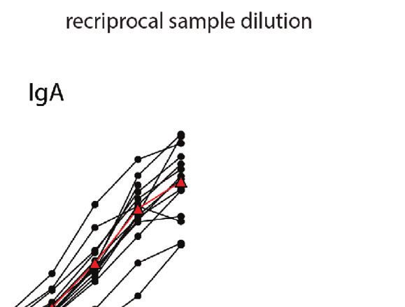
Yuen et al.                                                                                                                Sensitive Detection of SARS-CoV-2 Antibodies

withthe same coat and secondary detection antibody were                                 LFD Tests
averaged and subtracted from the OD values of each respective                           Antagen’s DISCOVID IgM IgG LFD test was used to detect
sample well and then the ODs were logarithmically transformed.                          SARS-CoV-2 RBD specific IgM and IgG antibodies following
Next, a non-linear regression of the sigmoidal standard                                 manufacturer instructions. Briefly, 20µl of plasma or serum was
curve was used to extrapolate a “concentration” for the                                 added to the indicated sample port, immediately followed by
patient samples, which was then inverse log transformed and                             provided diluent, and incubated at room temperature before
multiplied by the respective dilution factor. AU values for each                        reading at 45 minutes. The results were scored as positive or
sample were chosen from the linear portion of the dilution curve                        negative for IgM and IgG by two independent readers blinded to
for the antigen coated wells, and the paired buffer only coat                           donor sample status.
value was subtracted to determine the net AU amount.
                                                                                        Abbot Serology Test
                                                                                        The SARS-CoV-2 IgG assay is a chemiluminescent microparticle
Determination of the Presence Versus                                                    immunoassay (CMIA) used for the detection of SARS-CoV-2
Absence of SARS-CoV-2 Reactive                                                          nucleocapsid protein-specific IgG in human samples. The assays
Antibodies in Samples and of Arbitrary                                                  were performed according to manufacturer’s protocol.
Unit Values:
First, the average ODs of corresponding ‘blank’ wells (sample                           Automated Washer
diluent only in buffer only coated or antigen coated) on a given                        Plates were washed with Molecular Devices SkanWasher 400
plate was subtracted from all wells with samples. ODs for blank                         microplate washer with three rounds of aspiration and wash with
wells was consistently ~0.05 regardless of coat. Metric 1: Signal                       a final aspiration step for each run. This protocol was run twice
was considered positive from a given subject if (1) the OD values                       after the coating, blocking, and sample incubation steps and
from the antigen coated wells was a minimum of 2.5x higher                              three times after the addition of the secondary detection antibody
than that of the paired buffer coated well for at least two sample                      step in the experiment shown in Figure 1A. Plates were rotated
dilutions and (2) one antigen-coated well OD value was over 0.1,                        180° between each run. Residual wash buffer was left in the plates
after the average OD values of the respective blank wells                               (plates were not blotted post-wash) to mimic a fully
were subtracted.                                                                        automated system.

 FIGURE 1 | The modified ELISA (BU ELISA) protocol exhibits low background signal at high sample concentration and use of SARS-Cov-2 RBD-recombinant
 antibody standard curves allows for accurate sample quantification via accounting for OD drift between experimental runs. (A) Dilution curves of buffer only coated
 wells from five donor samples after using an automated plate washer or the BU ELISA method of multichannel plate washing. Experiment was performed once.
 (B) Representative dilution curves of buffer only coated wells from 30 subjects, average and range of 1:5 sample dilution for each isotype from all subjects; IgM, IgG,
 and IgA were detected in individual assays. (C) Representative IgM, IgG, and IgA standard curves from 15 different experimental runs are shown. The average of all
 runs shown as red triangles.

Frontiers in Immunology | www.frontiersin.org                                       4                                           April 2021 | Volume 12 | Article 614676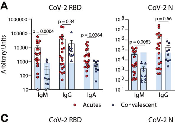
Yuen et al.                                                                                                         Sensitive Detection of SARS-CoV-2 Antibodies

RESULTS                                                                                 the number and placement of washing steps (Methods and
                                                                                        Supplemental Figure 1). This buffer only coat ‘noise’ is
A Modified ELISA Protocol Demonstrates                                                   remarkably consistent between multiple runs of a given sample
Low Noise From High Concentration                                                       (Supplemental Figure 2) and appears to be due to components
Human Serum and Plasma Samples                                                          within the sample, such as IgG and inflammatory factors (32)
The Enzyme-Linked Immunosorbent Assay (ELISA) is a                                      and not due to assay variability. Importantly, when ODs from
commonly used method for the measurement of analytes in a                               uncoated wells with the same dilution of sample are not
suspension sample. While low cost and easy to adapt in most lab                         measured and properly subtracted, incorrect interpretation of
settings, a limitation of this platform is high background from                         results as positive can occur (33); therefore, the no coat values
some biological samples at low sample dilutions. Specifically,                           were subtracted from coated OD results for all results to
optical densities (ODs) from sample dilutions lower than 1:100 is                       determine the true antigen-specific signal. Also, detection
often sizeable and can mask the analyte of interest. This issue is                      antibodies were tested for specificity to confirm accuracy of
particularly germane to serologic testing for SARS-CoV-2, as                            isotype-specific readouts and the ability of our IgG detection
antibodies that are cross-reactive in unexposed individuals,                            reagent to measure all four IgG subclasses (Supplemental
newly generated in asymptomatic and/or recent infections,                               Figure 3).
induced from an encounter with low viral dose, or waned post
convalescence may be missed because levels are below the limit                          Modification of ELISA Development
of detection of current assays. To address this issue, we have                          Duration Based on Standard Curve Signal
developed an ELISA protocol with unique steps to reduce non-                            Detection Enables Accurate Comparison
specific signal at low sample dilutions. One change is the plate                         of Antibody Levels Between Experimental
washing procedure, which is performed manually by an operator                           Runs by Minimizing Impact of OD Drift
using a multichannel pipettor and includes agitation and soaking                        During assay development we noted differences in OD values in
steps with repeated complete removal of residual fluid as                                different experimental runs even with strict adherence to all
described (Supplemental Figure 1). ELISAs were performed                                procedures and length of steps. Therefore, for all sample runs, we
that compared buffer coated well OD values of five human                                 included a standard curve using recombinant monoclonal IgM,
plasma samples with plates washed with our method or an                                 IgG, and IgA antibodies that recognize SARS-CoV-2 RBD for
automated plate washer and the total levels of non-specifically                          each of the respective isotype assays and stopped the
bound IgG was determined. The manual washing procedure                                  development reaction when there was a visible difference
resulted in a notably lower average and range of ODs at 1:5, 1:10,                      between the seventh dilution (1.37ng/ml) of the standard and
and 1:25 dilutions as compared with the automated washer                                the ‘zero’ (sample diluent only) well. Addition of these standards
(Figure 1A). This BU ELISA protocol was run on samples                                  and timing of development in this manner helped to ensure
from a total of 71 pre-pandemic and 29 SARS-CoV-2-infected                              accurate calculation of the relative antibody levels (termed
subjects (Table 1), with paired antigen coated and buffer coated                        ‘Arbitrary Units’ (AU) on a ng/ml scale, calculated as
wells for six or seven sample dilutions (Supplemental Figure 1)                         described in Methods) between samples run on different days,
for all subjects. The average ODs for buffer coated, 1:5 diluted                        plates, and/or by different operators. It is important to note that
sample loaded wells from all subject samples measured at this                           monoclonal antibody curves that recognize SARS-CoV-2 RBD
dilution were 0.16, 0.098, and 0.076 for IgM, IgG, and IgA                              were not used to quantify the exact number of antibodies in
respectively (Figure 1B). Given these low background OD                                 samples, as this cannot be calculated accurately due to the
values and the results from the wash method comparison, it’s                            mixture of reactive immunoglobulins in human samples.
possible that details of our protocol other than the washing                            Alternatively, we used SARS-CoV-2 RBD-specific monoclonal
method may contribute to these low background ODs, such as                              antibodies to determine AU levels all for RBD– and N– reactive
the type of plates, the blocking buffer/sample diluent used, and                        antibodies; an approach used in other studies (34, 35) and
                                                                                        provided reproducible and foreseeable values in this cohort.
                                                                                        The OD values of the standard curves following this
TABLE 1 | Cohort.                                                                       development procedure for the IgM, IgG, and IgA assays for
                                                                                        15 representative runs are shown (Figure 1C). The development
Cohort Characteristics             Age          Sex,         Length of Symptoms
                                (average,       M (%)       (days average, range)       time of these runs to complete visualization of the standard curve
                                  range)                                                development ranged from ~8-30 minutes, demonstrating the
                                                                                        need to adjust substrate incubation time per experimental run to
Pre-pandemics (n = 71)
                                                                                        maximize signal detection.
   Healthy (n = 37)            50   (21 - 96)    78                  N/A
   HIV+ (n = 24)               45   (22 - 79)    96                  N/A
   SLE (n = 10)                39   (23 - 69)    30                  N/A
Acute (n = 20)                 63   (48 - 84)    80              18 (3 - 40)+
                                                                                        SARS-CoV-2 RBD-Reactive Antibodies
Convalescent                   53   (35 - 77)    22              27 (0 - 61)*           Were Detected at Low Levels in 44
(n = 9)                                                                                 Percent of Pre-Pandemic Samples
+current length of symptoms at time of sample collection.                               SARS-CoV-2 RBD IgM, IgG, and IgA ELISA assays were
*total length of symptoms.                                                              performed on 40, 71, and 40 pre-pandemic samples,

Frontiers in Immunology | www.frontiersin.org                                       5                                    April 2021 | Volume 12 | Article 614676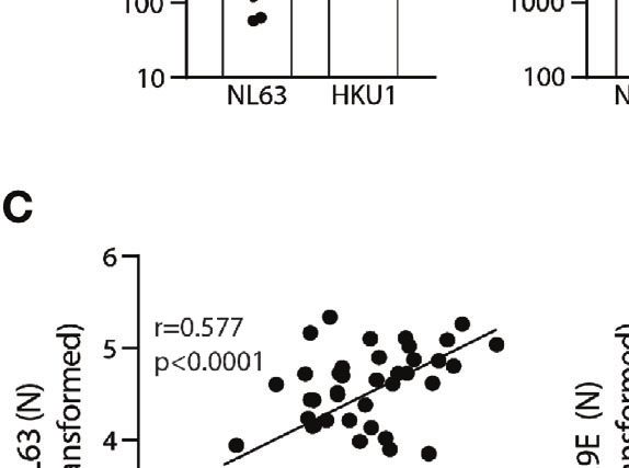
Yuen et al.                                                                                                            Sensitive Detection of SARS-CoV-2 Antibodies

respectively (Table 1) using the BU ELISA protocol. The OD                           respective isotype graph. We defined a subject as positive for a
curves from the BU ELISA for both the buffer coat and SARS-                          given antibody readout as follows: the OD value from the RBD-
CoV-2 RBD coated wells [after first subtracting the blank well(s)                     coated well ≥ 2.5x the uncoated well from the paired sample
with paired coat] from seven representative pre-pandemic                             dilution for at least two dilutions in the series and ≥ 0.1 for at
subjects with positive signal (three subjects per graph) for IgM,                    least one dilution. Following this guideline, 31/71 of the
IgG, and/or IgA is shown (Figure 2A). There is clear RBD-                            unexposed individuals possessed reactive antibodies of at least
specific signal with proportional loss of OD with sample dilution,                    one isotype to SARS-CoV-2 RBD, albeit all at very low levels in
providing evidence of true specific signal (Figure 2A). The                           the circulation (~40 ng/ml) (Figure 2B). We compared the
calculated Arbitrary Units (AUs) from the buffer only and                            calculated AUs from IgG reactive to SARS-CoV-2 Spike (S)
antigen coated wells from these curves is shown beneath each                         and RBD from 14 pre-pandemic subjects and found no

 FIGURE 2 | Detection and quantification of SARS-CoV-2 RBD- and N- reactive antibodies in pre-pandemic samples. (A) Representative dilution curves of seven
 pre-pandemic samples with SARS-CoV-2 RBD-reactive antibodies (three subjects per graph). Open and solid symbols represent buffer only coat and SARS-CoV-2
 RBD coat, respectively. Arbitrary Units (AU) were calculated as described in Methods and shown beneath the respective isotype graph for diluent only and SARS-
 CoV-2 RBD coat. AUs for SARS-CoV-2 RBD (B) and N (C) reactive IgM, IgG, and IgA in pre-pandemic samples. Open and solid symbols represent negative and
 positive results, respectively, as determined by Metric 1. Enumeration of the positive samples for each isotype in the pre-pandemic cohort is shown beneath each
 graph with percentages of total in parentheses. (D) Correlation between AUs for IgG reactive to SARS-CoV-2 RBD and N (n=32). Values were log-transformed to
 obtain a parametric distribution. Statistical analyses were performed using an unpaired non-parametric Mann-Whitney t-test in (B, C) and Pearson’s correlation of
 normally distributed AU values for (D).

Frontiers in Immunology | www.frontiersin.org                                    6                                           April 2021 | Volume 12 | Article 614676
Yuen et al. Sensitive Detection of SARS-CoV-2 Antibodies
significant correlation (Supplemental Figure 4). These results cohort and found no correlation between the two readouts
could be due to differences in portions and/or presentation of the (Figure 2D). This suggests these antibodies to different
RBD antigen in the different tests. portions of SARS-CoV-2 are elicited during distinct
immune responses.
All Pre-Pandemic Subjects Contained
Circulating SARS-CoV-2 Nucleocapsid IgG Reactive With SARS-CoV-2 RBD and
(N) IgG Antibodies With a Wide Range Of N in Pre-Pandemic Samples Correlate
Levels Found Between Individuals With Immunity to NL63 and Both NL63 and
Using the BU ELISA protocol, we measured IgM, IgG, and IgA 229E eCoV Strains, Respectively
levels reactive with SARS-CoV-2 nucleocapsid protein (N) from 20, To determine if antibodies elicited by endemic coronavirus (eCoV)
52, and 20 subjects from our pre-pandemic cohort, respectively. infections are linked to antibodies reactive to SARS-CoV-2 in
Seven dilutions were run for all samples with and without N coated unexposed subjects, IgG specific for NL63 and HKU1 RBD
wells as with the SARS-CoV-2 RBD assays; sample dilution curves proteins, and IgG reactive with the N protein from all four eCoV
were generated, and positive results were determined using Metric 1 strains in the circulation (NL63, HKU1, 229E and OC43) were
and AUs calculated. A wide range of pre-existing antibody levels measured. The levels of antibodies to eCoV RBD and N proteins in
were found; for example, the IgG levels varied by more than four pre-pandemic samples showed general differences, with more IgG
fold, from roughly 0.0134 to 54 mg/ml (Figure 2C). reactive to HKU1 than NL63 RBDs among the subjects and similar
levels of IgG reactive with N proteins of the NL63, 229E, and OC43
No Correlation Between Levels of strains, with lower levels reactive with the HKU1 N (Figure 3A).
Cross-Reactive SARS-COV-2 Antibodies IgG reactive with SARS-CoV-2 RBD significantly correlated with
to RBD and N Antigens NL63 and not HKU1 RBD-specific IgG (Figure 3B) and IgG
We compared the levels of SARS-COV-2 RBD- and N- reactive reactive with SARS-CoV-2 N correlated with N-specific IgG for
antibodies between individual pre-pandemic subjects in our all four eCoV strains (Figure 3C). Taken together, these results
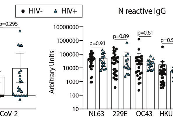
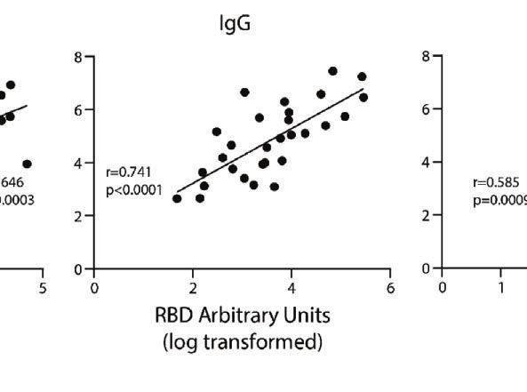
FIGURE 3 | SARS-CoV-2 RBD and N reactive IgG in pre-pandemic samples track with IgG recognizing analogous proteins of eCoV strains. (A) AUs of IgG reactive
to RBD of NL63 and HKU1 and N of all four eCoV strains (NL63, 2293, OC43, and HKU1). (B) Correlation between SARS-CoV-2 RBD IgG levels with NL63, HKU1
RBD IgG levels in individual subjects. (C) Correlation between SARS-CoV-2 N IgG and NL63, 229E, OC43, and HKU1 N IgG levels, n=30-42. Values were log-
transformed to obtain a parametric distribution. Statistical analyses were performed using Pearson’s correlation of normally distributed log transformed AU values in
(B, C) and an unpaired non-parametric Mann-Whitney t-test in (A).
Frontiers in Immunology | www.frontiersin.org 7 April 2021 | Volume 12 | Article 614676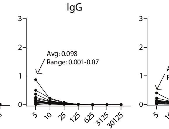
Yuen et al.                                                                                                       Sensitive Detection of SARS-CoV-2 Antibodies

suggest previous circulating endemic coronavirus infections elicit                Comparison of SARS-COV-2 Specific
SARS-CoV-2 cross-reactive antibodies.                                             RBD and N Antibody Levels Between
                                                                                  Hospitalized COVID-19 Subjects With
HIV or SLE Disease Status Does Not                                                Acute Disease and Convalescent
Impact SARS-CoV-2 Reactive RBD and N                                              Survivors of Infection
Antibody Levels in Unexposed Individuals                                          We next used the BU ELISA to measure the IgM, IgG, and IgA
We next compared the levels of eCoV and/or SARS-CoV-2                             RBD- and N-specific antibodies from individuals at different
reactive antibodies in our pre-pandemic cohort with the                           times and magnitudes of severity after SARS-CoV-2 infection. Of
subjects re-classified by HIV and SLE status. We found lower                       the 20 COVID-19 hospitalized subjects (Acutes), 10 scored
levels of NL63 RBD-reactive IgG in HIV+ as compared to                            negative and 10 positive on the EUA approved Abbott SARS-
uninfected subjects (Figure 4A); however, there were no other                     CoV-2 N-specific IgG CMIA. RBD- and N- specific IgM and IgA
significant differences found between the antibody levels reactive                 was higher in all Acutes as compared with the Convalescent
to the RBD or N proteins, for either the eCoV strains or SARS-                    subjects, but IgG levels were similar, suggesting waning of IgM
CoV-2, between groups classified via HIV or SLE status (Figures                    and IgA over time or reduced induction of these isotypes in
4A, B).                                                                           subjects that do not require hospitalization (Figure 5A). Also,
                                                                                  RBD- and N- specific IgM, IgG, and IgA levels significantly
Unexposed Individuals Over 70 Years Old                                           correlate among all COVID-19 subjects in the study (n=29)
Have Significantly Lower Levels of SARS-                                           (Figure 5B).
CoV-2 RBD and N Reactive IgG Than
Younger Counterparts
We next re-categorized our pre-pandemic cohort into two                           Comparison of RBD- and N-Specific
groups by age, 70 yo (n=12). All eCoV                        Antibody Levels Measured by the BU
and SARS-CoV-2 reactive IgG levels measured were lower in the                     ELISA From Hospitalized, Acute COVID-19
>70yo group, with high significance for SARS-CoV-2 RBD                             Subjects With Positive Versus Negative
(p=.0007) and N (p=.0045). It should be noted that                                Abbott Test Results
comparisons of younger (
Yuen et al.                                                                                                            Sensitive Detection of SARS-CoV-2 Antibodies

 FIGURE 5 | Quantification of the relative levels of IgM, IgG, and IgA-reactive SARS-CoV-2-RBD and N antibodies from acute and convalescent SARS-CoV-2
 infected subjects. (A) Arbitrary Units (AUs) of SARS-CoV-2 RBD and N reactive IgM, IgG, and IgA of acute and convalescent subjects. Open and solid symbols
 represent negative and positive results, respectively, as determined by our Metric 1 described in Methods. (B) Correlation between SARS-CoV-2 RBD and N IgM,
 IgG, and IgA log transformed AUs. Values were log-transformed to obtain a parametric distribution. (C) Quantification of SARS-CoV-2 RBD and N reactive IgM, IgG,
 and IgA of acute subjects regrouped based on results from Abbott’s SARS-CoV-2 IgG CMIA. Correlation between SARS-CoV-2 RBD (D) and N (E) IgM, IgG, and
 IgA AUs (log transformed) with the number of days post symptom (dps) onset at time of sample collection for acute subjects. Quantification of SARS-CoV-2 RBD
 reactive IgM and N reactive IgA (F) and RBD & N reactive for IgM, IgG, and IgA (G) for pre-pandemics (n = 19) and Acutes re-classified based on Abbott test results.
 Light blue bars depict AU range of pre-pandemics for each respective antigen and isotype. Statistical analyses were performed using an unpaired non-parametric
 Mann-Whitney t-test in (A, C, F, G) and Pearson’s correlation of normally distributed log transformed AU values in (B, D, E) dps, days post symptom.

(Figure 5C). The AU values for six Abbott test negative subjects                     symptoms, with N-specific IgA significantly correlating with
were above the pre-pandemic range for RBD-specific IgM and for                        symptom length; however, some subjects have pre-pandemic
N-specific IgA, and two Abbott test negative subjects have RBD-                       levels of RBD- and/or N- reactive IgG, even after as long as 40
specific IgG above the pre-pandemic range (Figure 5C), Taken                          days of symptoms (Figures 5D, E).
together, these iresults show clear serological evidence of infection
in many of these Abbot test- subjects.
                                                                                     Combinational Analysis of Readouts by
                                                                                     the BU ELISA Reveal SARS-CoV-2
Evidence of Diversity of Adaptive B Cell                                             Reactive Antibody Levels Are Significantly
Response Induction Among Acutely                                                     Higher in Acutely Infected COVID-19+
Infected, Hospitalized COVID-19+ Subjects                                            Subjects With Negative Abbott Test
Next, we compared the levels of RBD- and N- reactive IgM, IgG                        Results Than Pre-Pandemics
and IgA from our cohort of acute COVID-19 subjects, and found                        Next, we compared the combined AU values of both RBD-
general trends showing higher antibody levels with more days of                      reactive IgM and N- reactive IgA and all six readouts performed

Frontiers in Immunology | www.frontiersin.org                                    9                                           April 2021 | Volume 12 | Article 614676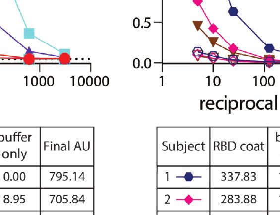
Yuen et al.                                                                                             Sensitive Detection of SARS-CoV-2 Antibodies

and found significant differences between the pre-pandemic                  detection of low levels of antigen-specific antibodies in
and acutely infected Abbott test negative groups, as well as               human specimens.
between acutely infected Abbott test negative versus positive                  The BU ELISA is straightforward, comprised of reagents that
groups (Figures 5F, G). These results indicate that multi-                 are readily available from commercial vendors and can easily be
parameter detection, comprised of multiple isotypes and                    adapted for other applications and analytes. It should be noted
antigen reactivities using a sensitive serology test could                 that nine different operators have performed the BU ELISA to
improve serologic diagnostics of SARS-CoV-2 infection.                     date and the reported signal:noise was always achieved with ease,
                                                                           even by individuals without prior experience running ELISAs.
Three-Way Comparison of the BU ELISA                                       However, a limitation of this assay is that it is currently
With the Antagen LFD and Abbott CMIA                                       considerably lower in throughput compared to other
Tests Reveal 9 of 10 Abbott Test Negative                                  serological platforms. This protocol requires an operator for
                                                                           the manual wash steps, limiting the number of plates that can be
COVID-19 Subjects Exhibit SARS-CoV-2                                       run compared to automated methods, however, there is potential
Specific Antibody Levels Above All Pre-                                     for throughput increase if automated washers/ELISA systems
Pandemic Samples for at Least One                                          can be adapted to more closely mimic this protocol. Specifically,
Readout by the BU ELISA                                                    we believe the manual wash procedure in this method minimizes
The AU values from the 10 pre-pandemic subjects with the                   cross-tip contamination, ensures thorough removal of wash
highest AU values for SARS-CoV-2 RBD and/or N-reactive IgG                 buffer from the well, and prevents cross-well contamination.
were directly compared with results from the Abbott CMIA assay             Application of these changes to an automated washer may
and a lateral flow rapid test (Antagen Pharmaceuticals). The                improve sensitivity of higher-throughput serology test
Abbott CMIA test measures IgG reactive to SARS-CoV-2 N, and                platforms. Another study limitation is the inclusion of only
the Antagen LFD test measures SARS-CoV-2 RBD-reactive IgG                  cross-sectional samples; in particular, it would be of high
and IgM. Both commercial tests detected no SARS-CoV-2                      interest to measure SARS-CoV-2 antibody levels of the ten
reactive antibodies in the pre-pandemic samples, and correctly             acute COVID subjects with negative Abbott test results both
identified the infection status of 9/10 and 10/10 subjects within           before infection and over time in the coming months to better
the convalescent group (LFD and Abbot tests, respectively)                 understand the dynamics of the antibody response, e.g. to
(Table 2). Among the acutely infected subjects, the 10 samples             determine whether or not some antibody specificities/isotypes
that scored positive for IgG the Abbott test were also positive for        are never elicited in these infected individuals or if the quantities
IgG the LFD test. Of the 10 acutely infected subjects that scored          are low (still in the pre-pandemic range) but higher than pre-
negative on the Abbott test, the LFD test successfully identified 5/        SARS-CoV-2 infection amounts. A third limitation of this work
10 subjects as SARS-COV-2 antibody positive via IgG and/or                 is the numbers of subjects included are not high enough to
IgM (Table 2). Nine of 10 acute COVID-19 subjects that scored              properly compare the specificity or sensitivity of the BU ELISA
negative on the Abbott test have AU values above all pre-                  with commercial COVID-19 serology tests. Also, we were unable
pandemics tested for at least one of the six BU ELISA readouts             to measure RBD-specific IgG for two eCoV strains due to lack of
(all but Subject A4). There are some discrepancies between the             availability (229E and OC43) which would be of interest given
different assays in detection of RBD- and N-reactive antibodies            the lack of correlation between levels of reactive IgG to SARS-
from the samples; this could be due to differences in the antigen          CoV-2 RBD with HKU1 RBD (Figure 3B).
preparations used, with varying expression vectors, impurities,                Other important features of our approach include the
tags, etc. These results underscore the importance of testing              inclusion of paired sample dilutions with buffer only coated
antigens from multiple sources and derivation methods, possible            wells to enable detection of true antigen-reactive signal and
manipulation of antigens to minimize non-specific binding, and              adjustment of the length of substrate incubation time based on
comprehensive assay validation to ensure maximal detection of              standard curve development for OD standardization to enable
true antigen specific signal.                                               direct comparison between samples on different plates.
                                                                           Quantification of relative antibody levels via Arbitrary Units
                                                                           (AU) or a similar method will be imperative for determining
DISCUSSION                                                                 which convalescent samples have antibody levels sufficient for
                                                                           effective plasma transfer as well as other applications. However,
Accurate and sensitive measurement of virus-specific antibodies             while we believe this is a preferred approach for determining
could complement diagnostic testing, provide information about             relative output values within all samples, it is critical to note that
the true prevalence of infection, provide insight into anti-viral          the unique dynamics of the panoply of antibodies of varying
immunity, and help assess vaccine responses. However, a lack of            affinities and isotypes within a given specimen causes inherent
required sensitivity and specificity of many of the SARS-CoV-2              confounding factors to serologic readouts. For example, a
antibody tests available to date have led some to conclude that            specimen with a high level of SARS-CoV-2 RBD reactive IgM
they have limited clinical utility in combating COVID-19 (36).             antibodies could have a lower detected signal for IgG and IgA
Here, we present a modified ELISA protocol with exceptional                 due to IgM’s pentameric conformation blocking many binding
sensitivity with high concentration samples that enables the               sites. Also, higher affinity antibody clones (IgG and IgA vs IgM,

Frontiers in Immunology | www.frontiersin.org                         10                                     April 2021 | Volume 12 | Article 614676
Yuen et al. Sensitive Detection of SARS-CoV-2 Antibodies
TABLE 2 | Comparison of SARS-CoV-2 reactive antibody results measured by the BU ELISA protocol, the EUA approved Abbott IgG chemiluminescent microparticle
assay, and Antagen’s lateral flow rapid test.
Assay ELISA CMIA LFD
Antigen RBD N N RBD
Isotype IgM IgG IgA IgM IgG IgA IgG IgM IgG
Pre-pandemic
P1 n.d. 811 n.d. n.d. 1165 n.d. - - -
P2 n.d. 322 n.d. n.d. 500 n.d. - - -
P3 n.d. 269 n.d. n.d. 14579 n.d. - - -
P4 n.d. 236 n.d. n.d. 54025 n.d. - - -
P5 n.d. 210 n.d. n.d. 427 n.d. - - -
P6 n.d. 201 n.d. n.d. 434 n.d. - - -
P7 n.d. -5 n.d. n.d. 18880 n.d. - - -
P8 n.d. 112 n.d. n.d. 12074 n.d. - - -
P9 n.d. 76 n.d. n.d. 9273 n.d. - - -
P10 n.d. 125 n.d. n.d. 6936 n.d. - - -
Acute
A1 54703 403 7861 933 15561 11067 - + +
A2 28388 171 2308 20037 1320 15811 - + -
A3 3091 306 383 3271 148738 221262 - + -
A4 74 158 201 158 4352 753 - + -
A5 3117 48 87 332 447 64 - - -
A6 7015 140 810 2354 462 2578 - - -
A7 127 609 1129 195 46225 1748 - - -
A8 460 4520 368 459 1255 1255 - + -
A9 0 1724 1034 193 1454 512 - - -
A10 3618 646 424 2377 5851 618 - - -
A11 1258 287496 12852 5871 2886248 178981 + - +
A12 3415 8722 3370 128747 401532 68030 + + +
A13 20404 39617 2736 15613 3805905 890383 + + +
A14 7068 1137 2290 167263 4543051 37356518 + + +
A15 1006 2243 836 63391 494137 118120 + + +
A16 20177 69285 16511 80754 28704356 392309 + + +
A17 29098 18804 6681 157527 127798 50048 + + +
A18 1560 49322 2869 118944 249225 68610 + - +
A19 678 7266 540 4922 1995094 7929190 + - +
A20 1341 268701 22853 803 17483216 218923 + - +
Convalescent
C1 66 3186 311 269 37745 1658 + - +
C2 25 2630 1054 111 8759 34 + - +
C3 1377 120984 1150 5809 557571 20315 + + +
C4 57 9893 604 79 111654 13386 + + +
C5 11 5970 956 5049 82953 286 + + +
C6 25 6455 535 68 11921 29 + - +
C7 894 8918 198 1026 786367 640 + - +
C8 55 2897 255 271 9909 290 + - -
C9 0 1121 39 133 2584 55 + - +
BU ELISA AU values (RBD): ≤ 795, 811, 806 (pre-pandemic range) : for IgM, IgG, IgA respectively ; 796, 812, 807 - 10,000 : for IgM, IgG, IgA respectively ; 10,001 -
100,000 : ; ≥100,001 :
BU ELISA AU values (N): ≤ 56230, 54025, 1544 (pre-pandemic range) : for IgM, IgG, IgA respectively ; 56231, 54026, 1545 - 100,000 : for IgM, IgG, IgA respectively ; 100,001 -
6
10 : ; ≥10 :
6
Negative as determined by Metric 1 :
n.d. = not done
Frontiers in Immunology | www.frontiersin.org 11 April 2021 | Volume 12 | Article 614676Yuen et al. Sensitive Detection of SARS-CoV-2 Antibodies
for example) may outcompete for binding sites of the coated associated with virus neutralization both in vitro and in animal
antigen and thereby be detected more readily than others. We models (18, 38–40), performing detailed functional analyses of
can account for this issue to some extent via measurement of all plasma samples from pre-pandemic samples with RBD-reactive
three major isotypes in all samples. Due to these unique antibodies is an important next step. Preliminary experiments
dynamics of competition between immunoglobulins of from our group indicated that neutralization activity was not
different isotypes within an indirect ELISA assay, we did not present in four subjects (data not shown) but future experiments
use monoclonal antibodies for all antigen reactivities as are needed to more thoroughly address this question.
standards; alternatively, we chose to employ one representative Unexposed individuals over 70 years of age possessed lower
monoclonal antibody curve of a given isotype for AU levels of both eCoV and cross-reactive SARS-CoV-2 antibodies
calculations for all antigen-reactive IgM, IgG, and IgA (Figure 4C). A study comparing pre-existing SARS-CoV-2
quantification. This approach resulted in reproducible results reactive T cell immunity in different age groups found that
enabling comparison of relative levels of antibodies specific for a levels were lower with older age (41). As individuals over 70
several different antigen between all subjects with only one are more likely to present with serious COVID-19 complications
standard curve on each plate. (42–45) future research investigating connections between age,
Here, we report links between antibody responses to endemic eCoV and SARS-CoV-2 reactive immunity, and vulnerability to
coronavirus (eCoV) strains and levels of cross-reactive severe COVID-19 is warranted.
antibodies with SARS-CoV-2 in unexposed individuals. Firstly, Direct comparison of our ELISA protocol with two
these results provide an important biological validation of the commercially available serological assays for SARS-CoV-2,
viral specificity of the OD values detected via use of the Ragon Antagen’s LFD test and Abbott’s CMIA IgG assay yielded
Institute SARS-CoV-2 RBD antigen and commercial N antigen interesting results. Both the LFD and CMIA performed well in
(Sino Biologicals). Also, these findings support recent reports of identification of the convalescent subjects via detection of SARS-
SARS-CoV-2 T cell responses correlating with eCoV T cell CoV-2 IgG to RBD (Antagen test) and N (Abbott test), and the
memory in pre-pandemic samples. Importantly, hospitalized BU ELISA detected signal from many pre-pandemic samples
COVID-19 subjects with recent eCoV infections were which all scored negative in the Antagen and Abbott tests
significantly less likely to require ICU admission or succumb (Table 2). However, these commercial tests are specifically
to the disease (37), and blood samples from virally unexposed designed to detect SARS-CoV-2 infection, unlike the BU
children were found to possess neutralization activity against ELISA which is measuring all SARS-CoV-2 reactive antibodies;
SARS-CoV-2 (11). These results collectively implicate past eCoV therefore, it is possible the antigens have been modified in
infections with protection from severe outcomes to COVID-19 the commercial assays to minimize cross-reactive antibody
due to cross-reactive immunity, including antibodies. Although detection, designated as noise in these tests. Interestingly, of
all of the pre-pandemic subjects screened for SARS-CoV-2 N- the 10 acute subjects that scored negative on the Abbott test, five
reactive IgG scored positive in our assay, we found widely scored positive for IgM and/or IgG by the LFD test; also, of the
varying quantities (> four log differences) between subjects, an six parameters measured by the BU ELISA, AU values were
agreement with another study (12) and parallel reports of N- higher than all pre-pandemic samples for at least one readout for
reactive T cell immunity in unexposed subjects (7–10). The 9/10 subjects. Taken together, these results indicate multi-
magnitude of SARS-CoV-2 N reactive IgG in our pre- parameter detection of SARS-CoV-2 reactive antibodies with
pandemic cohort did not track with HIV infection, SLE, or sensitive tests may improve use of serologic data for diagnostics.
with age among subjects under 70 years old (Figure 4) but did The BU ELISA protocol enables the measurement of
correlate strongly with eCoV N IgG levels (Figure 3). These low levels of antigen-specific antibodies within high
results strongly suggest that individuals with more recent eCoV concentration human specimens. Use of this assay could
infections and/or more robust eCoV immune responses could provide new insight into viral transmission and help elucidate
have some immune protection against COVID-19 via cross- the nature of the virus-specific antibody response. Also, this
reactive antibodies, and this may partially explain the profound protocol may aid diagnostics, measurement of vaccine responses
diversity of outcomes that occur upon SARS-COV-2 infection. and perhaps most importantly, help accurately determine a
Screening for this cross-reactive immunity may provide new history of exposure to SARS-COV-2.
insight into an individual’s risk of poor infection outcomes.
44 percent of the pre-pandemic subjects possessed antibodies
of at least one isotype that bind to SARS-CoV-2 RBD, albeit at DATA AVAILABILITY STATEMENT
very low levels. These results are in contrast with the conclusions The raw data supporting the conclusions of this article will be
of other reports, which state that RBD-reactive antibodies are not made available by the authors, without undue reservation.
detected in unexposed individuals (17, 20). However, in these
studies the assays were run at higher sample dilutions and
therefore low signal may have been missed or misinterpreted ETHICS STATEMENT
as noise. While these RBD-reactive antibody levels are low in the
blood, it is possible that they are present in higher concentrations The studies involving human participants were reviewed and
in other sites, such as the mucosa. Also, as antibodies to RBD are approved by Boston University Institutional Review Board.
Frontiers in Immunology | www.frontiersin.org 12 April 2021 | Volume 12 | Article 614676Yuen et al. Sensitive Detection of SARS-CoV-2 Antibodies
Written informed consent for participation was not required for Fund, NIH grants R01AG06505-01 and R01AG058538-01, and
this study in accordance with the national legislation and the sample collection was partially supported by the Providence/
institutional requirements. Boston Center for AIDS Research (CFAR).
AUTHOR CONTRIBUTIONS ACKNOWLEDGMENTS
JS-C conceived of the experimental plan. RY and JS-C designed We thank Dr. Aaron Schmidt/Dr. Jared Feldman and Dr. Galit
the approach. AC, RY, and JS-C designed experiments. RC, NL, Alter from the Ragon Institute of MGH, MIT, and Harvard for
KQ, PU, NB, GM, WG, IR, MS, and JS-C contributed to study the gifts of the recombinant SARS-CoV-2 RBD antigen and
subject specimen collection/use, provided reagents, and/or monoclonal SARS-CoV-2 RBD IgG antibody, respectively. We
funding. RY, ES, RP, DS, EC, JS-C, AO, LB, FK, JB, AB, and also thank Dr. Andrew Lodge at ThermoFisher Scientific for his
YK performed experiments. RY, DS, EC, JB, SG, AB, GM, YK, advice regarding initial reagent selection and protocol steps. The
and JS-C analyzed data. RY and JS-C wrote the manuscript. manuscript has been released as a pre-print at MedRxIV, (Yuen
All authors contributed to the article and approved the et al.) (46).
submitted version.
SUPPLEMENTARY MATERIAL
FUNDING
The Supplementary Material for this article can be found online at:
This work was supported by the Boston University's National https://www.frontiersin.org/articles/10.3389/fimmu.2021.614676/
Emerging Infectious Diseases Laboratories (NEIDL) Director's full#supplementary-material
11. Ng KW, Faulkner N, Cornish GH, Rosa A, Harvey R, Hussain S, et al.
REFERENCES Preexisting and de novo humoral immunity to SARS-CoV-2 in humans.
1. Zhu N, Zhang D, Wang W, Li X, Yang B, Song J, et al. A Novel Coronavirus Science (2020) 370:1339–43. doi: 10.1126/science.abe1107
from Patients with Pneumonia in China, 2019. New Engl J Med (2020) 12. Nguyen-Contant P, Embong AK, Kanagaiah P, Chaves FA, Harvey H,
382:727–33. doi: 10.1056/NEJMoa2001017 Branche AR, et al. S Protein-Reactive IgG and Memory B Cell Production
2. Wu F, Zhao S, Yu B, Chen YM, Wang W, Song ZG, et al. A new coronavirus after Human SARS-CoV-2 Infection Includes Broad Reactivity to the S2
associated with human respiratory disease in China. Nature (2020) 579:265–9. Subunit. mBio (2020) 11(5):e01991–20. doi: 10.1128/mBio.01991-20
doi: 10.1038/s41586-020-2008-3 13. Sette A, Crotty S. Pre-existing immunity to SARS-CoV-2: the knowns and
3. Huang C, Wang Y, Li X, Ren L, Zhao J, Hu Y, et al. Clinical features of patients unknowns. Nat Rev Immunol (2020) 20:457–8. doi: 10.1038/s41577-020-
infected with 2019 novel coronavirus in Wuhan, China. Lancet (2020) 0389-z
395:497–506. doi: 10.1016/S0140-6736(20)30183-5 14. Plotkin SA. Vaccines: correlates of vaccine-induced immunity. Clin Infect Dis
4. Grant MC, Geoghegan L, Arbyn M, Mohammed Z, McGuinness L, Clarke EL, (2008) 47:401–9. doi: 10.1086/589862
et al. The prevalence of symptoms in 24,410 adults infected by the novel 15. Ayouba A, Thaurignac G, Morquin D, Tuaillon E, Raulino R, Nkuba A, et al.
coronavirus (SARS-CoV-2; COVID-19): A systematic review and meta- Multiplex detection and dynamics of IgG antibodies to SARS-CoV2 and the
analysis of 148 studies from 9 countries. PloS One (2020) 15:e0234765. doi: highly pathogenic human coronaviruses SARS-CoV and MERS-CoV. J Clin
10.1371/journal.pone.0234765 Virol (2020) 129:104521. doi: 10.1016/j.jcv.2020.104521
5. Wu Z, McGoogan JM. Characteristics of and Important Lessons From 16. Marien J, Ceulemans A, Michiels J, Heyndrickx L, Kerkhof K, Foque N, et al.
the Coronavirus Disease 2019 (COVID-19) Outbreak in China: Evaluating SARS-CoV-2 spike and nucleocapsid proteins as targets for
Summary of a Report of 72314 Cases From the Chinese Center for antibody detection in severe and mild COVID-19 cases using a Luminex
Disease Control and Prevention. JAMA (2020) 323(13):1239–42. doi: bead-based assay. J Virol Methods (2020) 288:114025. doi: 10.1016/
10.1001/jama.2020.2648 j.jviromet.2020.114025
6. Gao Z, Xu Y, Sun C, Wang X, Guo Y, Qiu S, et al. A Systematic Review of 17. Amanat F, Stadlbauer D, Strohmeier S, Nguyen THO, Chromikova V,
Asymptomatic Infections with COVID-19. J Microbiol Immunol Infect (2021) McMahon M, et al. A serological assay to detect SARS-CoV-2
54(1):12–6. doi: 10.1016/j.jmii.2020.05.001 seroconversion in humans. Nat Med (2020) 26:1033–6. doi: 10.1038/
7. Braun J, Loyal L, Sun M, Wendisch D, Georg P, Kurth F, et al. SARS-CoV-2- s41591-020-0913-5
reactive T cells in healthy donors and patients with COVID-19. Nature (2020) 18. Robbiani DF, Gaebler C, Muecksch F, Lorenzi JCC, Wang Z, Cho A, et al.
587(7833):270–4. doi: 10.1038/s41586-020-2598-9 Convergent Antibody Responses to SARS-CoV-2 Infection in Convalescent
8. Grifoni A, Weiskopf D, Ramirez SI, Mateus J, Dan JM, Moderbacher CR, et al. Individuals. Nature (2020) 584(7821):437–42. doi: 10.1038/s41586-020-
Targets of T Cell Responses to SARS-CoV-2 Coronavirus in Humans with 2456-9
COVID-19 Disease and Unexposed Individuals. Cell (2020) 181:1489–501. 19. Okba NMA, Muller MA, Li W, Wang C, GeurtsvanKessel CH, Corman WM,
e1415. doi: 10.1016/j.cell.2020.05.015 et al. Severe Acute Respiratory Syndrome Coronavirus 2-Specific Antibody
9. Le Bert N, Tan AT, Kunasegaran K, Tham CYL, Hafezi M, Chia A, et al. Responses in Coronavirus Disease Patients. Emerg Infect Dis (2020) 26:1478–
SARS-CoV-2-specific T cell immunity in cases of COVID-19 and SARS, and 88. doi: 10.3201/eid2607.200841
uninfected controls. Nature (2020) 584(7821):457–62. doi: 10.1038/s41586- 20. Premkumar L, Segovia-Chumbez B, Jadi R, Martinez DR, Raut R, Markmann
020-2550-z A, et al. The receptor binding domain of the viral spike protein is an
10. Mateus J, Grifoni A, Tarke A, Sidney J, Ramirez SI, Dan JM, et al. Selective and immunodominant and highly specific target of antibodies in SARS-CoV-2
cross-reactive SARS-CoV-2 T cell epitopes in unexposed humans. Science patients. Sci Immunol (2020) 5(48):eabc8413. doi: 10.1126/sciimmunol.
(2020) 370(6512):89–94. doi: 10.1126/science.abd3871 abc8413
Frontiers in Immunology | www.frontiersin.org 13 April 2021 | Volume 12 | Article 614676Yuen et al. Sensitive Detection of SARS-CoV-2 Antibodies
21. Salvatori G, Luberto L, Maffei M, Aurisicchio L, Roscill G, Palombo F, et al. 36. Bisoffi Z, Pomari E, Deiana M, Piubelli C, Ronzoni N, Beltrame A, et al.
SARS-CoV-2 SPIKE PROTEIN: an optimal immunological target for Sensitivity, Specificity and Predictive Values of Molecular and Serological
vaccines. J Trans Med (2020) 18:222. doi: 10.1186/s12967-020-02392-y Tests for COVID-19: A Longitudinal Study in Emergency Room. Diagn
22. Ravichandran S, Coyle EM, Klenow L, Tang J, Grubbs G, Liu S, et al. Antibody (Basel) (2020) 10(9):669. doi: 10.3390/diagnostics10090669
signature induced by SARS-CoV-2 spike protein immunogens in rabbits. Sci 37. Sagar M, Reifler K, Rossi M, Miller NS, Sinha P, White L, et al. Recent endemic
Transl Med (2020) 12(550):eabc3539. doi: 10.1101/2020.05.12.091918 coronavirus infection is associated with less severe COVID-19. J Clin Invest
23. Horber S, Soldo J, Relker L, Jurgens S, Guther J, Peter S, et al. Evaluation of (2020) 131(1):e143380. doi: 10.1172/JCI143380
three fully-automated SARS-CoV-2 antibody assays. Clin Chem Lab Med 38. Deng W, Bao L, Liu J, Xiao C, Liu J, Xue J, et al. Primary exposure to SARS-
(2020) 58(12):2113–20. doi: 10.1515/cclm-2020-0975 CoV-2 protects against reinfection in rhesus macaques. Science (2020)
24. Theel ES, Harring J, Hilgart H, Granger D. Performance Characteristics of 369:818–23. doi: 10.1126/science.abc5343
Four High-Throughput Immunoassays for Detection of IgG Antibodies 39. Rogers TF, Zhao F, Huang D, Beutler N, Burns A, He WT, et al. Isolation of
against SARS-CoV-2. J Clin Microbiol (2020) 58(8):e01243–20. doi: potent SARS-CoV-2 neutralizing antibodies and protection from disease in
10.1128/JCM.01243-20 a small animal model. Science (2020) 369:956–63. doi: 10.1126/
25. Montesinos I, Gruson D, Kabamba B, Dahma H, Van den Wijngaert S, Reza S, science.abc7520
et al. Evaluation of two automated and three rapid lateral flow immunoassays 40. Tan CW, Chia WN, Qin X, Liu P, Chen MI, Tiu C, et al. A SARS-CoV-2
for the detection of anti-SARS-CoV-2 antibodies. J Clin Virol (2020) surrogate virus neutralization test based on antibody-mediated blockage of
128:104413. doi: 10.1016/j.jcv.2020.104413 ACE2-spike protein-protein interaction. Nat Biotechnol (2020) 38:1073–8.
26. Whitman JD, Hiatt J, Mowery CT, Shy BR, Yu R, Yamamoto TN, et al. Test doi: 10.1038/s41587-020-0631-z
performance evaluation of SARS-CoV-2 serological assays. medRxiv (2020) 41. Saletti G, Gerlach T, Jansen JM, Molle A, Elbahesh H, Ludlow M, et al. Older
2020.04.25.20074856. doi: 10.1101/2020.04.25.20074856 adults lack SARS CoV-2 cross-reactive T lymphocytes directed to human
27. Deeks JJ, Dinnes J, Takwoingi Y, Davenport C, Spijker R, Taylor-Phillips S, coronaviruses OC43 and NL63. Sci Rep (2020) 10:21447. doi: 10.1038/s41598-
et al. Antibody tests for identification of current and past infection with SARS- 020-78506-9
CoV-2. Cochrane Database Syst Rev (2020) 6(6):CD013652. doi: 10.1002/ 42. Garg S, Kim L, Whitaker M, O’Halloran A, Cummings C, Holstein R, et al.
14651858.CD013652 Hospitalization Rates and Characteristics of Patients Hospitalized with
28. Long QX, Tang XJ, Shi QL, Li Q, Deng HJ, Yuan J, et al. Clinical and Laboratory-Confirmed Coronavirus Disease 2019 - COVID-NET, 14 States,
immunological assessment of asymptomatic SARS-CoV-2 infections. Nat March 1-30, 2020. MMWR Morb Mortal Wkly Rep (2020) 69:458–64. doi:
Med (2020) 26:1200–4. doi: 10.1038/s41591-020-0965-6 10.15585/mmwr.mm6915e3
29. Belkina AC, Starchenko A, Drake KA, Proctor EA, Pihl RMF, Olson A, et al. 43. Stokes EK, Zambrano LD, Anderson KN, Marder EP, Raz KM, El Burai Felix
Multivariate Computational Analysis of Gamma Delta T Cell Inhibitory S, et al. Coronavirus Disease 2019 Case Surveillance - United States, January
Receptor Signatures Reveals the Divergence of Healthy and ART- 22-May 30, 2020. MMWR Morb Mortal Wkly Rep (2020) 69:759–65. doi:
Suppressed HIV+ Aging. Front Immunol (2018) 9:2783. doi: 10.3389/ 10.15585/mmwr.mm6924e2
fimmu.2018.02783 44. Onder G, Rezza G, Brusaferro S. Case-Fatality Rate and Characteristics of
30. Kaneko N, Kuo HH, Boucau J, Farmer JR, Allard-Chamard H, Mahajan VS, Patients Dying in Relation to COVID-19 in Italy. JAMA (2020) 323:1775–6.
et al. Loss of Bcl-6-Expressing T Follicular Helper Cells and Germinal Centers doi: 10.1001/jama.2020.4683
in COVID-19. Cell (2020) 143–57.e13. doi: 10.1016/j.cell.2020.08.025 45. Zhou F, Yu T, Anderson R, Fan G, Liu Y, Liu Z, et al. Clinical course and risk
31. Stadlbauer D, Amanat F, Chromikova V, Jiang K, Strohmeier S, Arunkumar factors for mortality of adult inpatients with COVID-19 in Wuhan, China: a
GA, et al. SARS-CoV-2 Seroconversion in Humans: A Detailed Protocol for a retrospective cohort study. Lancet (2020) 395:1054–62. doi: 10.1016/S0140-
Serological Assay, Antigen Production, and Test Setup. Curr Protoc Microbiol 6736(20)30566-3
(2020) 57:e100. doi: 10.1002/cpmc.100 46. Yuen REA. SARS-CoV-2 reactive antibodies in unexposed individuals
32. Guven E, Duus K, Lydolph MC, Jorgensen CS, Laursen I, Houen G. Non- revealed by a high sensitivity, low noise serologic assay. MedRxiV (2020).
specific binding in solid phase immunoassays for autoantibodies correlates doi: 10.1101/2020.09.15.20192765
with inflammation markers. J Immunol Methods (2014) 403:26–36. doi:
10.1016/j.jim.2013.11.014
Conflict of Interest: WG was employed by Antagen Pharmaceuticals.
33. Terato K, Do C, Chang J, Waritani T. Preventing further misuse of the ELISA
technique and misinterpretation of serological antibody assay data. Vaccine The remaining authors declare that the research was conducted in the absence of
(2016) 34:4643–4. doi: 10.1016/j.vaccine.2016.08.007 any commercial or financial relationships that could be construed as a potential
34. Schepp RM, de Haan CAM, Wilkins D, Layman H, Graham BS, Esser MT, conflict of interest.
et al. Development and Standardization of a High-Throughput Multiplex
Immunoassay for the Simultaneous Quantification of Specific Antibodies to Copyright © 2021 Yuen, Steiner, Pihl, Chavez, Olson, Smith, Baird, Korkmaz, Urick,
Five Respiratory Syncytial Virus Proteins. mSphere (2019) 4(2):e00236–19. Sagar, Berrigan, Gummuluru, Corley, Quillen, Belkina, Mostoslavsky, Rifkin, Kataria,
doi: 10.1128/mSphere.00236-19 Cappione, Gao, Lin, Bhadelia and Snyder-Cappione. This is an open-access article
35. Kusi KA, Faber BW, Riasat V, Thomas AW, Kocken CH, Remarque EJ, distributed under the terms of the Creative Commons Attribution License (CC BY). The
et al. Generation of humoral immune responses to multi-allele use, distribution or reproduction in other forums is permitted, provided the original
PfAMA1 vaccines; effect of adjuvant and number of component alleles author(s) and the copyright owner(s) are credited and that the original publication in this
on the breadth of response. PloS One (2010) 5:e15391. doi: 10.1371/ journal is cited, in accordance with accepted academic practice. No use, distribution or
journal.pone.0015391 reproduction is permitted which does not comply with these terms.
Frontiers in Immunology | www.frontiersin.org 14 April 2021 | Volume 12 | Article 614676You can also read