Localised increase in regional cerebral perfusion in patients with visual snow syndrome: a pseudo-continuous arterial spin labelling study
←
→
Page content transcription
If your browser does not render page correctly, please read the page content below

Migraine
J Neurol Neurosurg Psychiatry: first published as 10.1136/jnnp-2020-325881 on 14 July 2021. Downloaded from http://jnnp.bmj.com/ on August 1, 2021 by guest. Protected by copyright.
Original research
Localised increase in regional cerebral perfusion in
patients with visual snow syndrome: a pseudo-
continuous arterial spin labelling study
Francesca Puledda ,1,2 Christoph J Schankin ,3 Owen O’Daly,4 Dominic Ffytche,5
Ozan Eren,6 Nazia Karsan ,1 Steve C R Williams,4 Fernando Zelaya,4
Peter J Goadsby 1,2
►► Additional supplemental ABSTRACT such as migraine and tinnitus3; in such cases, the
material is published online Objectives We aimed to investigate changes in condition is perceived as very disabling.4
only. To view, please visit
the journal online (http://dx. regional cerebral blood flow (rCBF) using arterial spin Although the pathophysiology of VSS remains
doi.org/10.1136/jnnp-2020- labelling (ASL) in patients with visual snow syndrome largely unknown5 a growing body of literature
325881). (VSS), in order to understand more about the underlying has started offering some insight on the possible
neurobiology of the condition, which remains mostly biological mechanisms underlying the condition.
For numbered affiliations see Behavioural6 and neurophysiological studies7 8 have
end of article. unknown.
Methods We performed an MRI study in which shown patterns of changes pointing to increased
Correspondence to whole-brain maps of rCBF were obtained using pseudo- cortical excitability and visual cortex dysfunc-
Dr Francesca Puledda, Headache continuous ASL. Twenty-four patients with VSS and tion. Through neuroimaging, it has been possible
Group, Department of Basic and an equal number of gender and age-matched healthy to determine that VSS is characterised by altered
Clinical Neuroscience, Institute volunteers took part in the study. All subjects were metabolism of the extrastriate visual cortex9 10
of Psychiatry, Psychology &
examined with both a visual paradigm consisting of a as well as structural changes involving the visual
Neuroscience, King’s College
London, London SE5 9PJ, UK; visual-snow like stimulus, simulating key features of the system, and further expanding beyond it.9 11 12
francesca.puledda@kcl.ac.uk snow, and a blank screen at rest, randomly presented. Arterial spin labelling (ASL) is a quantitative,
Results Patients with VSS had higher rCBF than non-invasive functional MRI technique that has
Received 14 December 2020 evolved considerably in the last decade13; this
Accepted 9 June 2021 controls over an extensive brain network, including
the bilateral cuneus, precuneus, supplementary motor method exploits the phenomenon of neuro-vascular
cortex, premotor cortex and posterior cingulate cortex, coupling to use resting perfusion as an indirect but
as well as the left primary auditory cortex, fusiform gyrus sensitive marker of neuronal activity.
and cerebellum. These areas were largely analogous In this study we investigated intrinsic differences
comparing patients either at rest, or when looking at in brain activity in patients with VSS compared
a ’snow-like’ visual stimulus. This widespread, similar with controls, determining differences in regional
pattern of perfusion differences in either condition cerebral blood flow (rCBF) using ASL. Given that
suggests a neurophysiological signature of visual VSS is characterised by an abnormal perception,
snow. Furthermore, right insula rCBF was increased in order to differentiate changes due to altered
in VSS subjects compared with controls during visual visual processing from those of the ongoing visual
stimulation, reflecting a greater task-related change and snow effect itself, we studied subjects both at rest
suggesting a difference in interoceptive processing with and during a visual task that simulated the visual
constant perception of altered visual input. snow experience. We hypothesised those areas
Conclusion The data suggest VSS patients have marked in the visual network would be characterised by
differences in brain processing of visual stimuli, validating changes in cerebral blood flow in VSS subjects,
indirectly reflecting changes in neuronal activity. A
its neurobiological basis.
further hypothesis was that these areas would not
show particular changes in different states of brain
activity, since visual snow is a continuous phenom-
enon that does not appear to be influenced by
INTRODUCTION external conditions.
Visual snow is a neurological condition defined by
© Author(s) (or their the presence of a continuous and unremitting visual
employer(s)) 2021. Re-use disturbance in the form of uncountable tiny dots METHODS
permitted under CC BY.
covering the whole visual field.1 Patients affected by Subject population and recruitment
Published by BMJ.
visual snow syndrome (VSS) experience a complex Twenty-four patients with a diagnosis of VSS
To cite: Puledda F, Schankin array of neurological and visual symptoms in addi- according to the current criteria1 and an equal
CJ, O’Daly O, et al. J Neurol tion to the static itself, such as palinopsia, entoptic number of age and gender matched healthy volun-
Neurosurg Psychiatry Epub
ahead of print: [please phenomena, nyctalopia and photophobia.2 Visual teers were selected for the study. This number was
include Day Month Year]. snow represents a spectrum type disorder that at based on the calculation that to detect a signifi-
doi:10.1136/jnnp-2020- its worse manifests with most of these additional cant two-tailed difference with 94% power (with a
325881 symptoms, as well as with distressing comorbidities minimum CBF change of 5 mL per 100 g of tissue
Puledda F, et al. J Neurol Neurosurg Psychiatry 2021;0:1–9. doi:10.1136/jnnp-2020-325881 1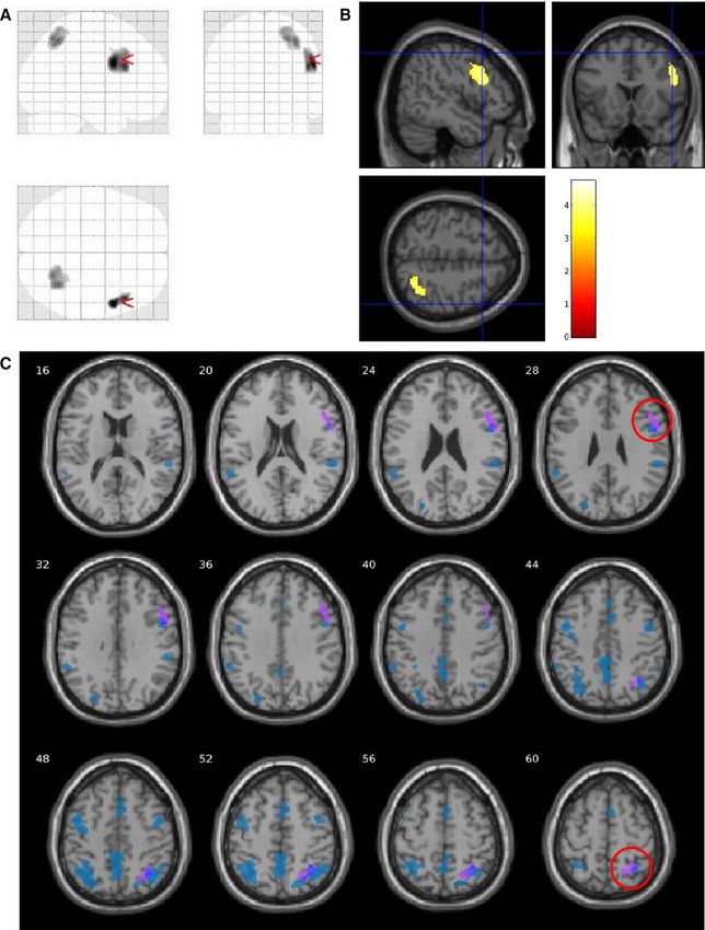
Migraine
J Neurol Neurosurg Psychiatry: first published as 10.1136/jnnp-2020-325881 on 14 July 2021. Downloaded from http://jnnp.bmj.com/ on August 1, 2021 by guest. Protected by copyright.
per min) between two independent groups with a SD of 8%, we matrix=256×256; slice thickness=1.2 mm; 196 slice partitions,
would require at least eighteen subjects per group. We recruited ASSET factor=1.75; in-plane resolution=1 mm.17
VSS patients by email, re-approaching subjects who had previ- Whole-brain CBF maps were generated by means of a three-
ously contacted our study team asking to participate in research. dimensional pseudo- continuous ASL (3D- pCASL) sequence.
The healthy controls (Ctrls) were recruited through internal Labelling of arterial blood was achieved with an 1825 ms train of
advertisement at King’s College London. Hanning shaped radiofrequency pulses of 500 µs duration in the
Recruitment was limited to individuals of 20–60 years of age presence of a net magnetic field gradient along the flow direction
with no contraindications to MRI, no serious medical condi- (the z-axis of the magnet). After a post-labelling delay of 2025
tions, consumption of no more than six cups of coffee per day ms, a whole-brain volume was read using a three-dimensional
and who were naïve to any type of recreational drugs, including inter-leaved ‘stack-of-spirals’ Fast Spin Echo readout, consisting
cannabis. Any participant taking recurrent medications with of eight interleaved spiral arms in the in-plane direction, with 512
an action on the central nervous system was excluded from points per spiral interleave. The images had 60 axial slice loca-
the study. Patients with a history of psychosis or psychological tions (3 mm thickness) and an in-plane FOV of 240×240 mm after
diseases either requiring ongoing psychoactive drugs, or that was transformation to a rectangular matrix (TE/TR=11.088/5180
thought to affect the patient’s neural pathways, were excluded ms, FA=111°). A proton density image volume with the same
from the study. Controls were selected based on matching age parameters was acquired within the same sequence in order to use
(±5 years) and gender of our patient population. All controls as a reference to compute the CBF maps in conventional physio-
were thoroughly screened to exclude any visual snow symptoms, logical units (mL blood per 100 g tissue per min). The sequence
as well a migraine history and migraine markers, by a trained used four background suppression pulses to minimise static tissue
neurologist and headache specialist. signal at the time of image acquisition. Four control-label pairs
were acquired. CBF maps were computed from the mean perfu-
sion weighted difference image derived from the four control-
Study protocol label pairs, by dividing the difference image against a proton
The study involved a telephone interview, in which eligibility density image acquired at the end of the sequence, using iden-
of the participant was assessed, followed by two visits to our tical readout parameters. This computation was done according
research facility. During the first visit patients had a general and to the formula suggested in the recent ASL consensus,18 and is
neurological examination, blood pressure and heart rate moni- described with the full preprocessing procedure of CBF maps in
toring. Each patient underwent a clinical interview, focusing online supplemental material. The entire acquisition time of the
on VSS symptoms, medical history and migraine history. VSS 3D-pCASL sequence was 6 min and 20 s.
diagnosis was based on the presence of visual static lasting for All participants in the study were subject to two separate
at least 3 months, as well as at least two additional visual symp- pCASL acquisitions, one at baseline and one during a visual
toms among: palinopsia, entoptic phenomena, photophobia and task. During the baseline sequence, participants were lying still
nyctalopia.1 with their eyes open while looking at a blank screen, which they
The PHQ-8 and GAD-7 questionnaires were used to assess viewed through a mirror system. For the visual task sequence,
respectively potential depression and anxiety, with clinical rele- participants had to view a visual task that mimicked the static of
vance defined by a score of above 9.14 15 visual snow, shown continuously through the same screen. The
Eligible patients were invited for a second visit in which the development of the visual task has been described in detail in
scanning took place; controls only came for the scanning visit. a previous publication by our group.10 Overall, this ‘snow-like’
All participants were scanned at the same time of day (between visual simulation was evaluated as very similar to the subjects’
09:00 and 12:00), as it is known that circadian rhythms can own snow (see online supplemental material).
influence rCBF.16 Subjects were instructed to consume a light
breakfast and to avoid caffeine immediately prior to the visit. Analysis of pCASL data
Participants were asked to refrain from the use of any type of Processed whole-brain CBF images were analysed using a voxel-
medication for 24 hours prior to scanning. Female patients were wise general linear model in SPM 12 ( www.fil.
ion.
ucl.
ac.
uk/
asked to keep a menstruation diary for the time of the study, spm/). A voxel- wise flexible- factorial design using two- way
in order to avoid scanning on days of active menstruation. To ANOVA allowed analysis of changes in CBF related to group and
ensure VSS patients were not scanned during an acute migraine, stimulus effect. The resulting Z statistic images were reviewed
they were instructed to inform the investigators if a migraine with an initial cluster-forming voxel threshold of p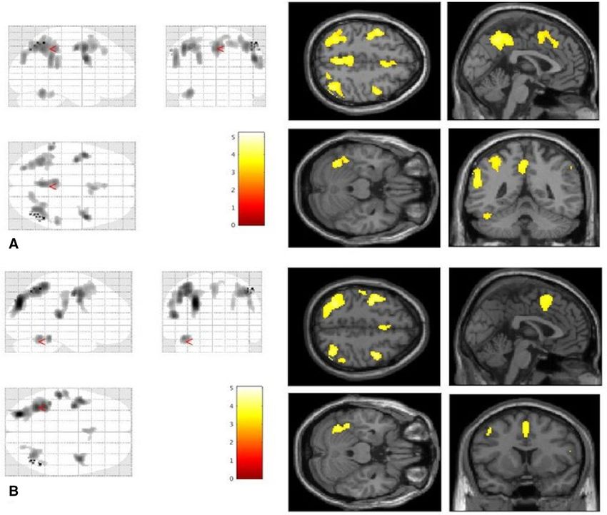
Migraine
J Neurol Neurosurg Psychiatry: first published as 10.1136/jnnp-2020-325881 on 14 July 2021. Downloaded from http://jnnp.bmj.com/ on August 1, 2021 by guest. Protected by copyright.
to select subjects with isolated VSS and to rule out spurious This analysis showed an extensive network of unilateral and
effects of migraine on investigation results. For this purpose, and bilateral brain regions with increased rCBF in patients with VSS
to understand more about VSS and migraine comorbidity, we compared with Ctrls. Specifically, bilateral clusters of significant
conducted two subanalyses in our cohort, as follows. First, we rCBF change were found in the cuneus, precuneus, inferior pari-
ran a validation analysis with a cohort of 24 episodic migraineurs, etal lobule (IPL), superior parietal lobule (SPL), supplementary
with no history of visual snow, enrolled in a previous pCASL motor area, frontal eye fields (FEF), premotor cortex, posterior
imaging study performed within our group.20 pCASL images for cingulate cortex (PCC), middle frontal gyrus, angular gyrus
this study followed the same acquisition and processing param- (AG), post central gyrus, middle and superior occipital lobule.
eters as the ones described here. A one-way ANOVA design In the left hemisphere only, areas of increased rCBF were found
was used, comparing the three groups—VSS patients, migraine in the primary auditory cortex, fusiform gyrus, area VI of the
patients, healthy controls—at baseline, when not subject to cerebellum and supramarginal gyrus. Anatomic locations and
visual stimulus. descriptions of all the significant clusters are highlighted in
We further performed a post-hoc group comparison for the table 1. There were no areas of reduced rCBF in VSS compared
baseline condition on subjects with VSS and no migraine history with Ctrls.
(n=9), compared with all Ctrls (n=24). Given the low power of We then proceeded to test the group effect (VSS vs Ctrls)
this analysis, the initial cluster-forming threshold was lowered to separately in the two conditions (ie, at baseline only and during
p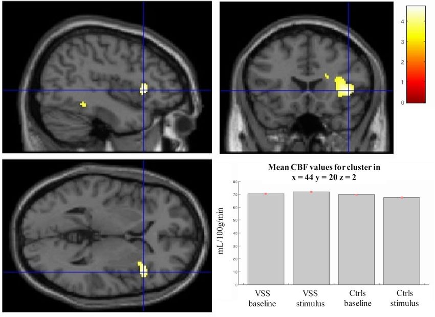
Migraine
J Neurol Neurosurg Psychiatry: first published as 10.1136/jnnp-2020-325881 on 14 July 2021. Downloaded from http://jnnp.bmj.com/ on August 1, 2021 by guest. Protected by copyright.
Figure 1 Areas of increased regional cerebral blood flow in patients with visual snow syndrome (n=24) compared to healthy controls (n=24) when
looking at a blank screen at baseline (A) and when observing a ‘snow-like’ visual stimulus (B). All areas are significant at the cluster level whole-brain
analyses and corrected for cluster extent. Bars represent T values.
following the stimulation, there is a task-related rCBF increase (ie, stimulus effects) can be found in online supplemental mate-
in this specific brain area in the VSS group, as opposed to the rial. No differences to the main effect of group analysis were
deactivation seen in Ctrls. found when comparing separately subjects who first received the
task to subjects who first were at rest, showing no carry-over
Comparative analysis between VSS and migraine effects of the visual paradigm on brain perfusion. A post-hoc
By comparing VSS patients, Ctrls (the same subjects as the analysis with tinnitus presence as a covariate showed overlap-
main analysis) and migraine patients from a previous study, we ping clusters with respect to the main analysis, but required
found that two of the nine clusters of increased rCBF in VSS lowering the significance threshold to pMigraine
J Neurol Neurosurg Psychiatry: first published as 10.1136/jnnp-2020-325881 on 14 July 2021. Downloaded from http://jnnp.bmj.com/ on August 1, 2021 by guest. Protected by copyright.
Figure 2 Comparative illustration of areas of increased regional cerebral blood flow in visual snow syndrome versus healthy controls when looking at a
blank screen (red colour areas—as seen in figure 1A) and when observing a ‘snow-like’ visual stimulus (green colour areas—as seen in figure 1B).
The parietal cortex has a fundamental role in the integra- Further, the sensorimotor region of the anterior precuneus
tion of different sensory stimuli.22 In particular with regards to has been shown to connect directly to the supplementary motor
visual stimulus processing, the dorsal visual stream represents a area,30 also exhibiting increased perfusion. These regions, as
brain pathway in which visual information is delivered from the well as the angular gyrus, are typically activated in cognitively
primary visual cortex to the posterior parietal lobe and onwards demanding tasks,31 in the control of internally generated or visu-
to other integrative areas of the brain. It is involved in the ally guided movement32 33 and in visuoproprioceptive integra-
visual location of objects and is essential in determining action- tion.34 The more medial aspects of the precuneus are also an
oriented behaviours dependent on the perception of space.23 24 element of the fronto-parietal network,35 which specialises in
Elements of the dorsal visual stream showed increased rCBF in external attention36 and visuospatial perception.37 The collec-
our analysis, as well as Brodmann area 7, a point of convergence tive involvement of these brain areas could thus potentially
between vision and proprioception, which allows to determine lead, in visual snow, to an abnormal focusing on normal sensory
where objects are in relation to the body.25 26 phenomena.
It is also relevant to note that the precuneus and PCC consti-
tute the posterior elements of the default mode network (DMN),
an organised mode of brain function active when the brain is at Visual motion function
rest and suspended during specific goal-directed behaviours.27 28 The large parietal region of increased perfusion encompasses
These areas are strongly linked to the recollection of prior expe- visual area V5, a brain region that specialises in processing and
riences, involving both the external and internal world.29 The computing visual motion, by integrating and decoding inputs it
fact that these regions showed increased blood flow bilaterally, receives from the primary visual cortex.38 39 An increased func-
could potentially signify an increased function within the DMN tion of this region justifies its larger volume detected through
in VSS patients; ultimately, this could be leading to a misattribu- structural imaging in VSS11 and is certainly consistent with
tion of brain energy, favouring internal experiences over external the misperception of constantly moving objects typical of the
attention. This finding has some correspondence with data from condition. However, in order to assess fully the role of V5 in
a functional connectivity analysis performed in this same group VSS, specific tasks related to visual motion perception would be
of patients.12 needed in the future.
Puledda F, et al. J Neurol Neurosurg Psychiatry 2021;0:1–9. doi:10.1136/jnnp-2020-325881 5Migraine
J Neurol Neurosurg Psychiatry: first published as 10.1136/jnnp-2020-325881 on 14 July 2021. Downloaded from http://jnnp.bmj.com/ on August 1, 2021 by guest. Protected by copyright.
Figure 3 Right insula activation in patients with VSS when testing for group and stimulation interaction. This area was significant for p=0.01 after SVC by
applying a mask over the right insula. Plots for mean CBF values for the cluster in each condition are shown in bottom right. k=98; Montreal Neurological
Institute coordinates: x=44, y=20, z=2. Bar represents T values. CBF, cerebral blood flow; Ctrls, healthy controls; VSS, visual snow syndrome.
The frontal eye fields also have a significant role in visual causing increased sensitivity of the visual and sensory pathways
motion and visuo-spatial attention.40 Interestingly, the FEF have and altered processing of visual stimuli.
also shown abnormal connectivity to the angular gyrus in a
previous functional brain study on visual snow patients.41 Given Cerebellum: a key player in VSS?
that both regions showed increased perfusion in our study, these The posterior and lateral cerebellum, particularly of the left
findings seem to confirm changes in attentional control, as well hemisphere, play a relevant role in complex cognitive operations
as integration of visual movement and proprioception, in VSS. linked to spatial processing, language and memory.45 46 Further-
more, activity in cerebellar lobule VI has shown high correlation
Insular involvement with activation of the salience network.47 This, taken together
Our data also showed an increased activation in response to the with the finding of increased volume of overlapping cerebellar
‘snow-like’ stimulus within the right anterior insula, in patients areas in a structural study of VSS,11 suggests that the cerebellum
with VSS compared with controls (figure 3). This heightened might represent a key structure in the biology of the condition,
activation was opposite to the stimulus- induced deactivation possibly involved in dysfunctional feed-forward mechanisms of
that was seen in healthy subjects. sensory processing.
The insular lobe of Reil has a pivotal salience function, relaying
different sensory inputs to other areas of the limbic system and Visual and auditory cortices
the brain.42 The right anterior insula, in particular, represents a Even if we did not detect specific perfusion changes in the right
hub capable of ‘switching’ brain engagement from the internally lingual gyrus, previously linked to VSS pathophysiology,9 48
oriented activity of the DMN to the externally oriented regions other parts of the extrastriate visual cortex—the left fusiform
of the executive network, which mediate attention, memory and gyrus in particular—showed increased activation, confirming the
higher order cognitive processes; this ultimately allows to deter- importance of these associative areas in the syndrome.49 This
mine appropriate behavioural responses to salient stimuli.43 44 mismatch could be attributed to differences in spatial resolution
Insular involvement in VSS pathophysiology had already been between [¹⁸F]FDG PET and MRI. Further, while several publica-
recorded by our group using functional MRI,10 being linked to tions have highlighted a degree of correlation between regional
the altered processing of an analogous visual stimulus to the one cerebral metabolic rate of glucose metabolism measured by PET
used here. Interestingly, we had previously found the insula to and regional CBF, it is not possible to assume that the same
deactivate—in contrast to controls who showed a null activa- correspondence can be detected in subjects with pre-established
tion—in response to this stimulus, rather than an increase in conditions such as visual snow.
its activity. It must be noted, however, that even if the stimuli Intuitively, it would have been expected for the visual stimu-
used in the two techniques were identical in their visual param- lation to have a bigger impact on the control group, since visual
eters, their time presentations were entirely different. Here, the snow patients were more ‘familiar’ with it. Nonetheless, as can be
MRI acquisition lasted several minutes, allowing to detect a seen from the analysis (online supplemental eFigure 1) conducted
more prolonged and sustained change in brain activity. It is thus to assess the effect of the stimulus, this produced a very similar
possible that the insula exhibits altered function in VSS, possibly cortical activation in both groups. Hence, it is possible to speculate
6 Puledda F, et al. J Neurol Neurosurg Psychiatry 2021;0:1–9. doi:10.1136/jnnp-2020-325881Migraine
J Neurol Neurosurg Psychiatry: first published as 10.1136/jnnp-2020-325881 on 14 July 2021. Downloaded from http://jnnp.bmj.com/ on August 1, 2021 by guest. Protected by copyright.
Figure 4 Analysis comparing visual snow syndrome (VSS) patients versus migraine patients versus healthy controls (Ctrls), showing two clusters of
increased regional cerebral blood flow in VSS patients imposed over a glass brain (A) and standard T1 image (B). Clusters are located in the right precentral
gyrus (x=54, y=6, z=28; T=4.40; k =420; p=0.001) and right precuneus (x=34, y=−56, z=50; T=4.04; k=404; p=0.001). A comparative image (C) with
the whole-brain analysis between VSS patients and Ctrls shows that the two clusters overlap the significant areas previously found. SVC, small volume
correction.
that the internal percept of visual snow follows different neuronal migraine. The high association between the two conditions,
pathways respect to that of incoming sensory stimuli. confirmed in our patient cohort, hinders the generalisability
Finally, the increase in blood flow in the primary auditory of results in this population. On the other hand, specifically
cortex may, at least in part, be linked to the high levels of selecting VSS patients with no concomitant migraine could
concomitant tinnitus seen in our patients.50 The comorbidity result in a selection bias, and a patient group not representative
between VSS and tinnitus seems to suggest an underlying patho- of the full condition.
physiological mechanism common to both conditions, possibly Although this was performed post-hoc, the compara-
a widespread network phenomena not limited to the involve- tive analyses of our two subject cohorts with a group of
ment of the relevant primary sensory cortices, ultimately causing migraineurs without VSS, and further of the subselection of
phantom perceptions.51 VSS patients without concomitant migraine compared with
controls, showed mostly overlapping results to our original
Limitations analysis. In the future, these issues will need to be addressed
The main limitation of this study lies in the challenges of inves- in the preliminary phases of original studies directed at inves-
tigating VSS while taking into account its main comorbidity of tigating the VSS.
Puledda F, et al. J Neurol Neurosurg Psychiatry 2021;0:1–9. doi:10.1136/jnnp-2020-325881 7Migraine
J Neurol Neurosurg Psychiatry: first published as 10.1136/jnnp-2020-325881 on 14 July 2021. Downloaded from http://jnnp.bmj.com/ on August 1, 2021 by guest. Protected by copyright.
Conclusions REFERENCES
In conclusion, patients with VSS present increased activation 1 Schankin CJ, Maniyar FH, Digre KB, et al. ’Visual snow’ - a disorder distinct from
persistent migraine aura. Brain 2014;137:1419–28.
in a wide network of intrinsic brain areas that are key in the
2 Puledda F, Schankin C, Digre K, et al. Visual snow syndrome: what we know so far.
processing of complex sensory and cognitive states. The fact that Curr Opin Neurol 2018;31:52–8.
rCBF increases were independent of the presence of an external 3 Puledda F, Schankin C, Goadsby PJ. Visual snow syndrome: a clinical and phenotypical
visual stimulus, suggests that these abnormalities could be a description of 1,100 cases. Neurology 2020;94:e564–74.
causal factor of the disorder. 4 Schankin CJ, Puledda F, Goadsby PJ. Visual snow syndrome: is it normal or a disorder -
and what to do with patients? Eur J Neurol 2020;27:2393–5.
This study expands on previous neuroimaging findings,
5 Puledda F, Ffytche D, O’Daly O, et al. Imaging the visual network in the migraine
confirms VSS to be a complex brain problem, and helps to spectrum. Front Neurol 2019;10:1325.
improve our understanding of a condition for which treatment 6 McKendrick AM, Chan YM, Tien M, et al. Behavioral measures of cortical
is still lacking. hyperexcitability assessed in people who experience visual snow. Neurology
2017;88:1243–9.
Author affiliations 7 Eren O, Rauschel V, Ruscheweyh R, et al. Evidence of dysfunction in the visual
1
Headache Group, Wolfson CARD, Institute of Psychiatry, Psychology & Neuroscience, association cortex in visual snow syndrome. Ann Neurol 2018;84:946–9.
King’s College London, King’s College London, London, UK 8 Yildiz FG, Turkyilmaz U, Unal-Cevik I. The clinical characteristics and neurophysiological
2
NIHR-Wellcome Trust King’s Clinical Research Facility, SLaM NIHR Biomedical assessments of the occipital cortex in visual snow syndrome with or without migraine.
Research Centre, King’s College Hospital, London, UK Headache 2019;59:484–94.
3
Department of Neurology, Inselspital University Hospital Bern, Bern, Switzerland 9 Schankin CJ, Maniyar FH, Chou DE, et al. Structural and functional footprint of visual
4
Centre for Neuroimaging Sciences, Department of Neuroimaging, Institute of snow syndrome. Brain 2020;143:1106–13.
Psychiatry, Psychology and Neuroscience, King’s College London, London, UK 10 Puledda F, Ffytche D, Lythgoe DJ, et al. Insular and occipital changes in visual snow
5
Old Age Psychiatry, Institute of Psychiatry, Psychology and Neuroscience, King’s syndrome: a BOLD fMRI and MRS study. Ann Clin Transl Neurol 2020;7:296–306.
College London, London, UK 11 Puledda F, Bruchhage M, O’Daly O, et al. Occipital cortex and cerebellum gray matter
6
Department of Neurology, University Hospital Munich Campus Grosshadern, changes in visual snow syndrome. Neurology 2020;95:e1792–9.
Munchen, Germany 12 Puledda F, O’Daly O, Schankin C, et al. Disrupted connectivity within visual,
attentional and salience networks in the visual snow syndrome. Hum Brain Mapp
2021;42:2032–44.
Acknowledgements The authors would like to thank the patients who have taken 13 Hernandez-Garcia L, Lahiri A, Schollenberger J. Recent progress in ASL. Neuroimage
part in the study. 2019;187:3–16.
Contributors Author FP designed the study, recruited the subjects, collected and 14 Löwe B, Decker O, Müller S, et al. Validation and standardization of the generalized
analysed the data, wrote the initial and final drafts of the manuscript. Authors CJS, anxiety disorder screener (GAD-7) in the general population. Med Care
DF, OE, SCRW contributed to the design of the study, analysis and interpretation 2008;46:266–74.
of the data. Author NK helped collect and analyse the data. Authors OGOD and FZ 15 Kroenke K, Strine TW, Spitzer RL, et al. The PHQ-8 as a measure of current depression
contributed to the design of the study, analysis and interpretation of the data and in the general population. J Affect Disord 2009;114:163–73.
revisions of the manuscript. Author PJG conceptualised the study, gave substantial 16 Hodkinson DJ, O’Daly O, Zunszain PA, et al. Circadian and homeostatic modulation
interpretation of the data and amended the final draft of the manuscript. of functional connectivity and regional cerebral blood flow in humans under normal
Funding This study represents independent research part funded by the National entrained conditions. J Cereb Blood Flow Metab 2014;34:1493–9.
Institute for Health Research (NIHR) Biomedical Research Centre at South London 17 Jack CR, Bernstein MA, Borowski BJ, et al. Update on the magnetic resonance
and Maudsley NHS Foundation Trust and King’s College London. The views expressed imaging core of the Alzheimer’s disease neuroimaging initiative. Alzheimers Dement
are those of the authors and not necessarily those of the NHS, the NIHR or the 2010;6:212–20.
Department of Health and Social Care. The study was part supported by the Visual 18 Alsop DC, Detre JA, Golay X, et al. Recommended implementation of arterial spin-
Snow Initiative and by crowdfunding from the self-help group for visual snow Eye On labeled perfusion MRI for clinical applications: a consensus of the ISMRM perfusion
Vision Foundation. Study Group and the European Consortium for ASL in dementia. Magn Reson Med
2015;73:102–16.
Competing interests None declared. 19 Mai J, Paxinos G, Voss T. Atlas of the human brain. third ed. Elsevier, 2008.
Patient consent for publication Not required. 20 Karsan N, Bose P, Zelaya F. Alterations in cerebral blood flow associated with the
premonitory phase of migraine. Cephalalgia : an international journal of headache
Ethics approval All participants gave their informed consent. The study was 2018;38:36.
approved by the London—City & East Research Ethics Committee (reference 21 Headache Classification Committee of the International Headache Society (IHS).
number: 16/LO/0964). The International classification of headache disorders, 3rd edition. Cephalalgia : an
Provenance and peer review Not commissioned; externally peer reviewed. international journal of headache 2018;38:1–211.
22 Teixeira S, Machado S, Velasques B, et al. Integrative parietal cortex processes:
Data availability statement Data are available upon reasonable request. Data
neurological and psychiatric aspects. J Neurol Sci 2014;338:12–22.
are available from the corresponding author upon reasonable request.
23 Mishkin M, Ungerleider LG, Macko KA. Object vision and spatial vision: two cortical
Supplemental material This content has been supplied by the author(s). pathways. Trends Neurosci 1983;6:414–7.
It has not been vetted by BMJ Publishing Group Limited (BMJ) and may not 24 Goodale MA, Milner AD. Separate visual pathways for perception and action. Trends
have been peer-reviewed. Any opinions or recommendations discussed are Neurosci 1992;15:20–5.
solely those of the author(s) and are not endorsed by BMJ. BMJ disclaims all 25 Scheperjans F, Hermann K, Eickhoff SB, et al. Observer-independent cytoarchitectonic
liability and responsibility arising from any reliance placed on the content. mapping of the human superior parietal cortex. Cerebral Cortex 2008;18:846–67.
Where the content includes any translated material, BMJ does not warrant the 26 Scheperjans F, Eickhoff SB, Hömke L, et al. Probabilistic maps, morphometry, and
accuracy and reliability of the translations (including but not limited to local variability of cytoarchitectonic areas in the human superior parietal cortex. Cereb
regulations, clinical guidelines, terminology, drug names and drug dosages), and Cortex 2008;18:2141–57.
is not responsible for any error and/or omissions arising from translation and 27 Raichle ME, MacLeod AM, Snyder AZ, et al. A default mode of brain function. Proc
adaptation or otherwise. Natl Acad Sci U S A 2001;98:676–82.
Open access This is an open access article distributed in accordance with the 28 Shulman GL, Fiez JA, Corbetta M, et al. Common blood flow changes across visual
Creative Commons Attribution 4.0 Unported (CC BY 4.0) license, which permits tasks: II. decreases in cerebral cortex. J Cogn Neurosci 1997;9:648–63.
others to copy, redistribute, remix, transform and build upon this work for any 29 Raichle ME. The brain’s default mode network. Annu Rev Neurosci 2015;38:433–47.
purpose, provided the original work is properly cited, a link to the licence is given, 30 Margulies DS, Vincent JL, Kelly C, et al. Precuneus shares intrinsic functional
and indication of whether changes were made. See: https://creativecommons.org/ architecture in humans and monkeys. Proc Natl Acad Sci U S A 2009;106:20069–74.
licenses/by/4.0/. 31 Cabeza R, Nyberg L. Imaging cognition II: an empirical review of 275 PET and fMRI
studies. J Cogn Neurosci 2000;12:1–47.
ORCID iDs 32 Picard N, Strick PL. Activation of the supplementary motor area (SMA) during
Francesca Puledda http://orcid.org/0000-0002-1933-4049 performance of visually guided movements. Cereb Cortex 2003;13:977–86.
Christoph J Schankin http://o rcid.org/0 000-0003-4 668-6098 33 Jenkins IH, Jahanshahi M, Jueptner M, et al. Self-Initiated versus externally triggered
Nazia Karsan http://orcid.org/0000-0002-6 946-5637 movements. II. The effect of movement predictability on regional cerebral blood flow.
Peter J Goadsby http://o rcid.org/0 000-0003-3 260-5904 Brain 2000;123 (Pt 6:1216–28.
8 Puledda F, et al. J Neurol Neurosurg Psychiatry 2021;0:1–9. doi:10.1136/jnnp-2020-325881Migraine
J Neurol Neurosurg Psychiatry: first published as 10.1136/jnnp-2020-325881 on 14 July 2021. Downloaded from http://jnnp.bmj.com/ on August 1, 2021 by guest. Protected by copyright.
34 Block H, Bastian A, Celnik P. Virtual lesion of angular gyrus disrupts the relationship 43 Menon V, Uddin LQ, Saliency ULQ. Saliency, switching, attention and control: a
between visuoproprioceptive weighting and realignment. J Cogn Neurosci network model of insula function. Brain Struct Funct 2010;214:655–67.
2013;25:636–48. 44 Menon V. Large-Scale brain networks and psychopathology: a unifying triple network
35 Fair DA, Dosenbach NUF, Church JA, et al. Development of distinct control networks model. Trends Cogn Sci 2011;15:483–506.
through segregation and integration. Proc Natl Acad Sci U S A 2007;104:13507–12. 45 Schmahmann JD. Disorders of the cerebellum: ataxia, dysmetria of thought, and
36 Dosenbach NUF, Fair DA, Miezin FM, et al. Distinct brain networks for adaptive and
the cerebellar cognitive affective syndrome. J Neuropsychiatry Clin Neurosci
stable task control in humans. Proc Natl Acad Sci U S A 2007;104:11073–8.
37 Sestieri C, Corbetta M, Spadone S, et al. Domain-General signals in the cingulo- 2004;16:367–78.
opercular network for visuospatial attention and episodic memory. J Cogn Neurosci 46 Stoodley CJ, Schmahmann JD. Functional topography in the human cerebellum: a
2014;26:551–68. meta-analysis of neuroimaging studies. Neuroimage 2009;44:489–501.
38 Zeki S, Watson JD, Lueck CJ, et al. A direct demonstration of functional specialization 47 Habas C, Kamdar N, Nguyen D, et al. Distinct cerebellar contributions to intrinsic
in human visual cortex. J Neurosci 1991;11:641–9. connectivity networks. J Neurosci 2009;29:8586–94.
39 Tootell RB, Reppas JB, Kwong KK, et al. Functional analysis of human MT and 48 Schankin CJ, Maniyar FH, Sprenger T, et al. The relation between migraine, typical
related visual cortical areas using magnetic resonance imaging. J Neurosci migraine aura and "visual snow". Headache 2014;54:957–66.
1995;15:3215–30. 49 Eren O, Schankin CJ. Insights into pathophysiology and treatment of visual snow
40 Schall JD. On the role of frontal eye field in guiding attention and saccades. Vision Res
syndrome: a systematic review. Prog Brain Res 2020;255:311–26.
2004;44:1453–67.
50 Arnold W, Bartenstein P, Oestreicher E, et al. Focal Metabolic Activation in the
41 Aldusary N, Traber GL, Freund P, et al. Abnormal connectivity and brain structure in
patients with visual snow. Front Hum Neurosci 2020;14:582031. Predominant Left Auditory Cortex in Patients Suffering from Tinnitus: A PET Study with
42 Shelley BP, Trimble MR. The insular lobe of Reil--its anatamico-functional, behavioural [18F]Deoxyglucose. ORL 1996;58:195–9.
and neuropsychiatric attributes in humans--a review. World J Biol Psychiatry 51 Sedley W, Friston KJ, Gander PE, et al. An integrative tinnitus model based on sensory
2004;5:176–200. precision. Trends Neurosci 2016;39:799–812.
Puledda F, et al. J Neurol Neurosurg Psychiatry 2021;0:1–9. doi:10.1136/jnnp-2020-325881 9You can also read