CT Image Changes of Severe Acute Pancreatitis Based on Smart Electronic Medical Augmented Reality in Nursing Practice
←
→
Page content transcription
If your browser does not render page correctly, please read the page content below
Hindawi
Journal of Healthcare Engineering
Volume 2021, Article ID 5522492, 12 pages
https://doi.org/10.1155/2021/5522492

Research Article
CT Image Changes of Severe Acute Pancreatitis Based on Smart
Electronic Medical Augmented Reality in Nursing Practice

 Defen Zhang,1 Shifang Mao,1 Siyou Lan,2 Chengli Zhou,1 and Xiaoyan Liu 1

 1
 Emergency Intensive Care Unit, The Affiliated Hospital of Southwest Medical University, Luzhou 646000, Sichuan, China
 2
 Department 1 of Respiratory and Critical Diseases, The Affiliated Hospital of Southwest Medical University, Luzhou 646000,
 Sichuan, China

 Correspondence should be addressed to Xiaoyan Liu; xiaoyanliu@m.fafu.edu.cn

 Received 20 February 2021; Revised 3 April 2021; Accepted 15 April 2021; Published 27 April 2021

 Academic Editor: Zhihan Lv

 Copyright © 2021 Defen Zhang et al. This is an open access article distributed under the Creative Commons Attribution License,
 which permits unrestricted use, distribution, and reproduction in any medium, provided the original work is properly cited.
 Severe acute pancreatitis (SAP) is traditionally treated with chemical analysis. Faced with the increasing maturity of CT imaging
 technology, it is necessary to use more advantageous CT imaging to treat SAP. In this article, 72 SAP patients admitted to the
 Affiliated Hospital of Southwest Medical University were selected for study, of which 62 were severely ill, 8 were exacerbated, and 2
 changed from severe to mild. This article combines the patient’s case records and related CT images during treatment from the
 perspective of nursing and conducts nursing research on the application of CT image changes in severe acute pancreatitis in
 nursing practice. CT image processing uses CT imaging system workstation (DICOM). The results of the study showed that, in the
 care of patients, 21 cases had recurrence after internal drainage, and the cure rate was 91.1%. Internal drainage is an effective way to
 treat SAP. The higher the incidence of pancreatitis, the more likely it is to relapse after SAP internal drainage, which may be related
 to repeated episodes of pancreatitis and repeated inflammation of the pancreas and pancreatic duct damage. 4 of the relapsed cases
 in this article are postchronic pancreatitis SAP, and the relapsed cases account for 50% of the chronic pancreatic cases. This may be
 due to chronic fibrosis of the branched and main pancreatic ducts, continuous abnormal pancreatic juice drainage. Therefore, it is
 necessary to further explore the prognosis of different causes of SAP. In terms of complication care, the overall complication rate
 was 16.6%. One patient died of postoperative hemorrhage. Analysis of the causes of cyst recurrence and complications may be
 closely related to the mechanism of the occurrence and development of SAP. The initiating factor of SAP is that the pancreatic
 tissue is damaged due to inflammation, trauma, or microcirculation disorder, and then the pancreatic juice leaks out of the
 pancreas, wrapping the pancreatic juice; it takes a certain time for the capsule of fibrous knot tissue to form and strengthen.

1. Introduction atrophy of pancreatic body and tail; pan-
 creaticocholangiography showed inhomogeneous and dif-
Severe acute pancreatitis (SAP) is a disease with a high fuse pancreatic duct stenosis, nondilation of main pancreatic
mortality rate and a rapid onset. There are still some duct, and stenosis of secondary pancreatic duct [1]. Riker
problems in nursing. For the diagnosis and nursing of SAP, et al. distinguished SAP based on CT findings, serum IgG4
there are two diagnostic models that are generally recog- level, and other organ involvement. If the diagnosis cannot
nized: one is the Japanese model and the other is the be confirmed, tissue biopsy or hormone therapy is recom-
American model. These two models carry out modeling mended [2]. Deshpande et al. differentiated pancreatic
research on the serum condition of SAP. And other related cancer and SAP according to serum IgG4 level combined
organs are also involved. with CA-199 level. The results showed that serum IgG4 level
 Siegel et al. set serum IgG4 level, CT image of pancreas, was more than 2 times higher than the normal value and
and endoscopic cholangiopancreatography as observation serum CA-199 level was lower than 85 U/ml, which were
indexes, and its CT positive manifestations were low-density important diagnostic indicators for distinguishing sap from
shadow around pancreas, delayed enhancement, and pancreatic cancer [3]. Kamisawa et al.’s study pointed out
2 Journal of Healthcare Engineering

that patients with mild elevated serum IgG4 should be the pancreas is still based on the subchondral bone. During
treated with caution, and excessive dependence on serum the operation, the articular cartilage and marginal osteo-
IgG4 as a diagnostic marker of SAP may lead to missed phytes need to be divided for the placement of the pancreatic
diagnosis of pancreatic tumor [4]. In Chari et al.’s study, guide plate [15–17]. However, the subchondral bone that
there were 22 patients with pancreatic tumor whose serum cannot be controlled when biting the articular cartilage is the
IgG4 was higher than 1.35 g/L, and the median value was same or similar to the preoperative plan, and the human
2.49 g/L. Among them, there were 8 patients whose serum error is large. In the prolonged development stage of SAP,
IgG4 was more than 2 times higher than the normal value the islet cells have not yet completely failed, and the co-
[5]. existence of impaired insulin secretion and insulin resistance
 He et al. pointed out that serum IgG4 more than 2 times is the cause of glucose metabolism disorders. At this time,
the normal value can improve the specificity of diagnosis of treatment from multiple aspects is often more effective [18].
SAP [6]. However, in Okazaki et al. study, only one patient The treatment methods are becoming more abundant,
was diagnosed as pancreatic cancer with SAP, and the serum puncture drainage is convenient and economical, but the
IgG4 value was 9.33 g/L [7]. Hamano et al. believed that if the incidence of complications is high; endoscopic technology
serum IgG4 level in patients with obstructive jaundice is has obvious advantages, but the secondary treatment rate is
increased, even if it is more than 2 times of the normal value, higher; the open drainage is classic and the curative effect is
the possibility of the malignant tumor cannot be ruled out in stable. Recently, there have also been reports of internal
the absence of other organ involvement, and tissue biopsy is drainage surgery in SAP assisted by Da Vinci robot [19].
feasible to identify malignant tumor [8]. Choi et al.’s study Regardless of the method, the basic principle of its treatment
showed that simple serum IgG4 was 2 times higher than the is mainly to deal with abnormal drainage of pancreatic juice.
normal value, which could not differentiate sap from pan- The location and size of the cyst, the general state of the
creatic cancer [9]. Okazaki et al. analyzed that 10.1% of 548 patient, and comorbidities should be considered in the
patients with pancreatic cancer had elevated serum IgG4 but clinical management of SAP. Choose the treatment plan that
did not find the significance of elevated serum IgG4 on the benefits the patient the most [20]. Therefore, the early di-
prognosis of pancreatic cancer [10]. The above studies agnosis of pancreatic cancer is a challenge for abdominal
mainly analyze the physiological pathology of pancreatitis surgeons challenge for abdominal surgeons. Early detection
from the perspective of serum, but for severe acute pan- of tumors is one of the keys to successful treatment. The
creatitis, the effect is very slow from the chemical point of identification of pancreatic cancer is also crucial to the ef-
view, so it is necessary to treat patients in a faster way, and fectiveness of treatment. Abdominal images of most patients
CT imaging technology can solve the above problems to a with pancreatic tumors suggest that the pancreas occupies a
great extent. space, but it is often difficult to distinguish between benign
 This article first constructed a DICOM 3D model of the and malignant. With the continuous development of
distal pancreas and then selected the reference point of the medicine, autoimmune pancreatitis (AP) has gradually been
CT image to analyze the image and finally analyzed the recognized. The increase in serum IgG4 level more than
application of the CT image changes of severe acute pan- twice the normal value is considered an important diag-
creatitis in nursing practice. In this article, 72 SAP patients nostic indicator for type I SAP. However, there are also a
admitted to the Affiliated Hospital of Southwest Medical small number of pancreatic cancer patients. Serum IgG4 is
University were selected as the research object. Among elevated, but the elevated level usually does not exceed 2
them, 62 were severely ill, 8 were deteriorating, and 2 had times the normal value [21]. Clinically, SAP and pancreatic
changed from severe to mild symptoms. This article com- head tumors have many common features, including
bines the patient’s case records during treatment and related painless jaundice, new-onset diabetes, and weight loss [22].
CT images to study the application of CT image changes in Therefore, distinguishing between SAP and pancreatic
severe acute pancreatitis in nursing practice. CT image cancer is still challenging for abdominal surgeons [23–25].
processing uses CT imaging system workstation (DICOM). Due to the damage of SAP to acinar cells, it may cause
 multiple organ dysfunction syndrome (MODS) in the acute
2. Severe Acute Pancreatitis and CT phase of the disease. In the middle and late stages of the
 Imaging Technology disease, the translocation of intestinal bacteria causes
 extrapancreatic tissue infection (EPI) [26]. EPI is a common
2.1. Symptoms and Conventional Treatment of Severe Acute SAP complication, including pancreatic infection, urinary
Pancreatitis (SAP). At present, there is no uniform standard system infection and bacteremia, etc., which have a serious
for the image acquisition methods and preoperative pan- adverse effect on the prognosis of patients. Especially for
creatic planning methods required for pancreatic guide plate patients with severe pancreatitis (SAP), the incidence of EPI
preparation [11, 12]. Although the accuracy of CT scan in is higher than 50%, but the change of the body’s immune
cartilage display and reconstruction model is not as good as function is the basis of the occurrence and pathological
MRI, and the articular surface cannot be used as a reference, progress of SAP [27]. In addition, SAP may cause a sig-
the model surface based on CT image reconstruction is nificant increase in the level of systemic inflammatory re-
smoother and flat, which is more accurate than MRI as a sponse markers, and whether these easily available clinical
positioning reference [13, 14]. There are also studies on the indicators can be used in the diagnosis of secondary pan-
construction of a pancreatic model based on CT images, but creatic infection in SAP patients remains to be studied [28].

Journal of Healthcare Engineering 3

2.2. CT Image Model of Augmented Reality Based on Smart classification network of severe acute pancreatitis, the re-
Electronic Medical DNN. For the contradiction between the sidual structure is used:
depth and performance degradation of CT image

 n ni�1 nj�1 wij xi − x (xj − x) n ni�1 ni≠j wij xi − x (xj − x)
 2DConv � � . (1)
 ni�1 nj�1 wij (xi − x)2 S2 ni�1 nj�1 wij

 j−1
 It consists of two parts: the first part is the identity Gt � kj�2 h�1 Gjh pj sh + ph sj Djh 1 − Djh . (8)
mapping; the second part is bottleneck structure, that is, first
 The modeling process of S (a) module in amdrc net is
through a 2dconv with 1 × 1 convolution kernel, then
 divided into five stages, and the final CT image features of
through a 2dconv with 3 × 3 convolution kernel, and then
 severe acute pancreatitis are classified into three levels.
through a 2dconv with 1 × 1 convolution kernel. At the same
 The classifier uses softmax loss, which is essentially:
time, BN operation and relu operation are performed be-
 transform the new CT image into log likelihood in
tween different convolution operations [29, 30]. For the
 probability space:
contradiction between CT image resolution and receptive
field of severe acute pancreatitis, this algorithm designs an 2k 1 1 c − c 2 2 c2 − c1
 ℘κ � + + 2 1 + , (9)
expanded residual structure, as shown in equation (2). k + 1 2 2k 3 3
 nj
 nh

 kj�1 kh�1 t�1 r�1 yij − yhr ∞ y
 SCCT � . (2) djh � dFj (y) (y − x)dFh (x), (10)
 2n2 u 0 0

 Different from the residual structure, by adding 3 ∗ 3 N
expansion convolution, the larger receptive field is suc- ln softmaxit � a0 + a1 du ∗ dt + bj Xu + εu . (11)
cessfully modeled, and the defect of single receptive field is i�1
solved to a certain extent. In order to improve the detection
 The loss value is minimized by forward propagation and
sensitivity of electrochemistry, the electrode fixed CT image
 back propagation. The softmax loss function can effectively
boundary complex can be rolled
 distinguish the differences between classifications and
 uh ≤ uj ≤ ... ≤ uk , (3) provide nonlinear expression capability for the network. At
 this time, softmax normalizes the n-dimensional vector
 G � Gw + Gnb + Gt , (4) (n � 3) output from the full connection layer (the sum of all
 dimension values is 1), and the values in the n-dimensional
 K vector represent the probability value of the prediction tag,
P di , wj � P di P wj |di ; P wj |di � P wj |zk P zk |di . respectively. The specific calculation process is shown in
 k�1 (12):
 (5)
 kt1 [i] � cos w1i , w2j , (12)
 In the channel attention module, focus on the weight j
distribution of different channels, input hh × w × C conv4_ x.
After avgpool (average pooling) and maxpool (maximum 1 x2
 LS(x) � √��� exp − , (13)
pooling); the size is converted to 1 × 1 × C. Considering the 2π 2
need to make full use of the different information obtained
by the two pooling operations, MLP (multilayer perceptron) HT � tanh wc xt + uc rt Θht−1 + BC + C, (14)
with shared parameters is added [31]. Then, the scaling
factor is obtained by activating function σ. Finally, it is
 ht � zt Θht−1 + 1 − zt Θht + W. (15)
compared with the initial CT image feature map conv4 of
severe acute pancreatitis_ spatial attention module focuses In the expression, LS represents the loss function of
on the weight distribution of spatial information gradient descent for softmax results, X represents the total
 hj
 nh
 input training data, BC represents the input data, HT rep-
 Z�1 r�1 yji − yhr
 MLPjh � , (6) resents the category of CT image of severe acute pancreatitis,
 nj nh uj + uh C represents the total category of training data, and W
 represents the parameters of network model training and
 2μx μy + C1 2σ xy + C2 learning. Using LiFePO4 as a signal probe can not only
 lssim � 1 − 2 2 2 2 , (7) expand the application of lithium-ion battery materials, but
 μx + μy + C1 σ x + σ y + C2 also expand the application of biosensors:

4 Journal of Healthcare Engineering

 �������������������
 2 “calculate 3D” function in the software; if the threshold is too
 (1/n) ni�1 FIit − FIit
 σt � , (16) small, the rougher the “Mask” edge is, the lower the rec-
 FIit ognition degree of the edge is, resulting in the rougher edge
 of the reconstructed model and the appearance of redundant
 u(j/i) � wij Ai , (17) “spike.” Secondly, when manually editing the “Mask,” it is
 often necessary to process the coronal, sagittal, and cross-
 p2 − p1 + 1 sectional images at the same time. Since the recognition of
 xH � . (18)
 2 the displayed image of the osteophyte differs from person to
 person in three stages, the shape of the reconstructed 3D
 Finally, the self-attention module network provides a model also differs. During this study, the CT scan parameter
new function for long-distance modeling of CT images of of the pancreas was 1 mm thick, which could better indicate
coronal pancreatitis. The calculation process is as follows: the condition of the pancreatic marginal osteophytes within
 FIit the structure, which would help to accurately reconstruct the
 ln � α + β ln FIit − 1 + vi + It , (19) pancreatic bone model.
 FIit − 1

 θ ϑ 3.2.2. Selection of Reference Points for CT Image Location.
 Wx
 ψ � Vx � Sx . (20) In this paper, we selected the anterior cortex of the pancreas
 x�1 x�1 n1 WI
 and the osteophytes at the edge of the medial and lateral
 Although CT can well identify ischemic lesions, it is condyles as the reference points and confirmed that the an-
difficult to show quite a few ischemic lesions on CT in the terior cortex of the pancreas has the greatest reference value
early stage of the disease due to different degrees of ischemia. through intraoperative observation. The goodness of fit of the
CTperfusion imaging is an examination method to judge the pancreatic guide plate on the CT image directly affects the
blood flow status of ischemic lesions. The blood flow status position of the entire pancreatic interface and also the force line
of pancreas can be observed by injecting a contrast agent of the pancreas. However, only anterior pancreatic reference
[32]. can provide CT images of the stability of the pancreatic guide
3. Design of CT Image Changes of Severe Acute plate located distal to the pancreas. It is necessary to increase
 the osteophyte at the edge of the medial and lateral condyles of
 Pancreatitis in Nursing Practice the pancreas as a fixed reference, and it can also be used as a
3.1. Samples. This article selects 72 SAP patients admitted to reference interface for pancreatic surgery through the condyle
the Affiliated Hospital of Southwest Medical University as axis, so as to view the rotation force line more intuitively during
the research object, 62 of which are severe, 8 are deterio- the operation. In conclusion, CT image preparation based on
rating, and 2 have changed from severe to mild symptoms. pancreatic CT and lower extremity full-length weight-bearing
This article combines the patient’s case records during x-ray images, distal pancreatic guide plate assisted TKA can
treatment and related CT images to study the application of achieve accurate pancreas, obtain accurate lower extremity
CT image changes in severe acute pancreatitis in nursing coronal and sagittal force lines, reduce blood loss, and improve
practice. CT image processing uses the CT imaging system recent pancreatic function [33, 34].
workstation (DICOM) for research. This software (Made in
China) only exports images. The other functions and safety 3.2.3. Feature Processing and Analysis of Intelligent Electronic
of this software have not been involved. Medical CT Image. For the basic convolution operation in
 DNN, it naturally has the defect of paying too much
 attention to the calculation of local area in CT image of
3.2. Sample Processing and Analysis Methods severe acute pancreatitis. In view of this defect, MS in
 amdrc net (multichannel aggregate spatial coding guided
3.2.1. Construction of Three-Dimensional Model of Distal
 by long and short attention) greatly alleviates its defi-
Pancreas in DICOM. DICOM is based on the formation of
 ciency. Compared with the general channel attention
“Mask” on two-dimensional CT images. The software has
 mechanism, the feature of long and short attention in this
several image features, but it can only segment areas where
 paper is that we use parallel channel attention and spatial
the thresholds are clearly different. First of all, the pancreatic
 attention and combine the feature graph conv4_ X is
CT image of patients with pancreatic osteoarthritis will show
 multiplied by the scaling coefficient obtained by the
edge defect by software automatic segmentation, especially
 parallel attention module, and then the new feature graph
when the scanning current parameters are not fixed, and the
 conv4 after passing through the attention module is
difference of osteophyte edge display is obvious; the larger
 processed_ X by concat operation:
the scanning current is, the clearer the osteophyte display is,
but the patient’s radiation is increased; the scanning current L(p, q + 1) − L(p, q − 1)
is too small, the worse the osteophyte display is, so manual θ(p, q) � arctan . (21)
 L(p + 1, q) − L(p − 1, q)
repair is often needed; however, when the software is used to
customize, if the threshold is too large, the incomplete the Then, the magnetic nanoparticles modified with EpCAM
“Mask” edge is, the worse the division of osteophytes is, and were used to capture sap, and then MUC1 aptamer was
the reconstructed model is incomplete after using the attached to the cell surface to form a Sandwich structure:
Journal of Healthcare Engineering 5

 FIit The self-digestion process of the pancreas during SAP
 ln � α + β ln FIit − 1 + ϕXit − 1 + vi + τ t , 8
 FIit − 1

 Taeget water pressure
 7 4.93
 (22) 6
 5
 4 3.88 2.09
 1 N Xi − x 3
 f(x) � k . (23)
 Nh i�1 h 2 2.27
 1 1.69 3.58
 0
 SAP Pancreas Inflam- Partial Immune ALEX
4. CT Image Changes of Severe Acute mation

 Pancreatitis in Nursing Practice Process item

4.1. Pancreatitis and Immune Environment. As shown in Card9 AIS
Figure 1, the self-digestion process of pancreas during sap Immune cells MRI
 PPC
has an impact on the local inflammation and immune
environment around it, resulting in the activation of a Figure 1: The self-digestion process of the pancreas during SAP.
variety of inflammation and immune cytokines and local
immune imbalance, resulting in abnormal pancreatic
perfusion and decreased surfactant. Changes in pancre- Table 1: Degree of pancreatic damage in patients with severe acute
atic microcirculation cause diffuse edema of the pan- pancreatitis.
creatic interstitium and pancreatic vesicles, damaging Item Card9 Immune cells SAP SAP MRI
pancreatic lesions. Pathogenic infections of the intestine SAP 1.09 3.5 1.69 1.12 3.9
and the production of endotoxin can activate immune Pancreas 3.88 3.77 3.88 3.39 4.04
cells by invading pancreatic tissue and damage the Inflammation 2.89 2.07 4.93 1.25 3.42
capillaries and vesicles of the pancreas. In addition, the Partial 4.8 3.14 3.58 3.29 4.14
study also showed that the pancreatic injury in SAP Immune 5.72 3.08 2.09 4.02 3.19
patients was related to the changes of complement system
and nuclear factor kB expression. The results showed that
the incidence of pancreatic infection increased with the effective way to treat sap. Combined with the literature, the
severity of SAP. more the incidence of pancreatitis, the easier the recurrence
 As shown in Table 1, the degree of pancreatic injury in of SAP after internal drainage, which may be related to
patients with severe acute pancreatitis gradually aggra- repeated attacks of pancreatitis, repeated stimulation of
vated with the extension of modeling time; drug can inflammation, and pancreatic duct injury. On the other
effectively improve the early condition of sap; with the hand, 4 of the recurrent cases were SAP after chronic
progress of the disease, the expression of CARD9 mRNA pancreatitis, and 50% of the recurrent cases were SAP after
and phosphorylated CARD9 protein in pancreatic tissue chronic pancreatitis. It may be related to chronic fibrosis of
also increased; the expression and phosphorylation of branch pancreatic duct and main pancreatic duct and
CARD9 mRNA in pancreatic tissue of patients with se- continuous abnormal pancreatic drainage. Therefore, it is
vere acute pancreatitis in cqcqd group CARD9 protein necessary to further explore the prognosis of SAP with
was significantly lower than that of SAP group at the same different etiologies. In terms of complications, the overall
time point. complication rate was 16.6%, and one patient died of
 As shown in Figure 2, patients with severe acute massive hemorrhage. Analysis of the causes of cyst recur-
pancreatitis have severe neurological deficits and poor rence and complications may be closely related to the
prognosis. The abnormal CT image in pancreas is related to mechanism of SAP development. The initiating factors of
the neurophysiological changes of pancreatic tissue during SAP are pancreatic tissue injury due to inflammation,
hypoperfusion. The decrease of neurovascular coupling trauma or microcirculation disturbance, and then pancreatic
and pancreatic blood flow in ischemic penumbra can juice extravasation around the pancreas. It takes a certain
increase slow wave activity and decrease fast wave activity. time for the formation and strengthening of fibrous hoof
The decrease of pancreatic blood flow leads to the increase tissue capsule wrapping pancreatic juice.
of low frequency wave activity, which is related to pro- As shown in Table 3, early monitoring of regional blood
gressive neuronal death. Studies have shown that the flow changes and CT imaging abnormalities in ischemic
abnormality of glutamate concentration (excitatory neu- lesions by QEEG and CT perfusion imaging can evaluate the
rotransmitter) may be related to the CBF level of 20 ∼ neurological deficit of patients, which is also confirmed by
30 ml/(100gmin), and to the δ wave produced by the the correlation between NIHSS score and various indicators
surviving neurons. in this paper. This paper analyzes the correlation between
 As shown in Table 2, there were 21 cases of recurrence QEEG characteristics and CT perfusion imaging parameters,
after internal drainage, and the cure rate was 91.1%. It is and the correlation between QEEG characteristics and
consistent with the literature reports. Internal drainage is an NIHSS score. It is found that QEEG can be used as a reliable

6 Journal of Healthcare Engineering

 Figure 2: Preparation of the patient’s pancreas CT image (from Google: http://1il65.cn/tVz3ar).

 Table 2: Number of occurrences of pancreatitis. drainage and anastomosis are weak, and complications such
 as infection and recurrence are prone to occur. The expe-
Item Cells SAP SAP MRI SAP rience of our center recommends that the time of surgical
SAP 0.33 1.44 1.35 1.41 0.44 intervention should be extended as long as the patient’s
Pancreas 3.9 2.9 3.66 2.89 2.49 condition allows, rather than limited to 4–6 weeks.
Inflammation 4.04 2.14 5.46 2.67 2.61 As shown in Table 4, patients with asymptomatic SAP
Partial 3.42 1.04 1.54 1.25 5.07 less than 6 cm were examined by abdominal color Doppler
Disorders 4.14 1.06 1.55 3.35 3.87
 ultrasound every three months. Patients with cyst diameter
Card9 3.19 1.09 5.51 2.45 5.25
 more than 6 cm or less than 6 cm but with abdominal pain,
 pancreatic portal hypertension, and other symptoms were
 examined by abdominal enhanced CT. The size of pseu-
Table 3: QEEG and CT perfusion imaging to monitor ischemic
 docyst, local symptoms, and endocrine and endocrine
lesions.
 functions of patients were closely monitored, and disease
Item SAP Pancreas Inflammation Partial Disorders progression was detected as soon as possible and appropriate
Cells 1.71 0.57 1.81 0.26 0.26 intervention was given.
SAP 2.32 1.62 1.14 1.36 1.8
SAP 5.69 5.2 4.38 4.67 3.22
MRI 3.91 1.25 5.35 3.12 2 4.2. CT Image Analysis of Severe Acute Pancreatitis. As
SAP 1.21 4.53 2.08 3.87 2.82 shown in Figure 4, if the course of disease is short and the
 cyst is small, but surgical treatment is necessary due to
 related symptoms, cyst gastric drainage is the first choice,
technical means to dynamically monitor the regional because cyst gastric drainage can take an appropriate size
changes of ischemic lesions in SAP and can also dynamically opening on the gastric wall according to the size of the cyst,
assess the neurological deficit of patients in the early stage which can fully remove the necrotic tissue in the cyst, control
and timely guide the clinical adjustment of treatment inflammation as soon as possible, and reduce postoperative
strategies. However, due to the small sample size, this inflammatory edema. Gastric acid entering the cyst may
conclusion needs to be further demonstrated by increasing have hemostatic and softening effects on the wound of the
the sample size; in addition, due to the different types of CT cyst wall. Postoperative placement of gastric tube in the cyst
imaging equipment, different parameter leads will affect the cavity and prone position can help to fully drain and pro-
spatial specificity and accuracy of QEEG, and the parameters mote the closure of the cyst cavity in a short time. However,
and algorithms of QEEG adopted by various manufacturers Roux-en-Y drainage of cyst jejunum is not conducive to
are also very different. In conclusion, there is a correlation drainage due to the limited diameter of intestinal tube and
between CT images and CTP features in SAP patients. Early the inaccessibility of gastric tube; and the increased number
dynamic monitoring of CT images can effectively evaluate of Roux-en-Y anastomotic stoma also increases the risk of
the changes of ischemic focus and neuronal damage in anastomotic leakage and stenosis. If the course of disease is
patients. long enough, the cyst tends to be mature and stable, and the
 As shown in Figure 3, early pancreatic juice is highly thickness of the cyst is uniform and dense, the use of cyst
irritant and inflammatory; the capsule of fibrous nodal tissue stomach drainage or cyst jejunum (Roux-en-Y) drainage can
wrapping pancreatic juice is immature; early internal achieve good results.

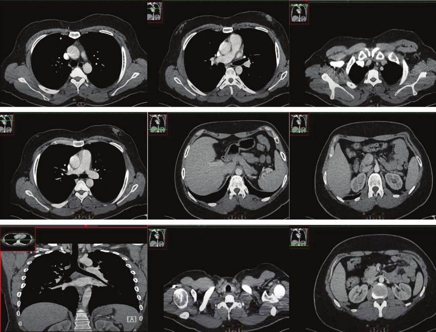
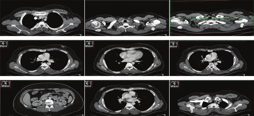
Journal of Healthcare Engineering 7

 Immature capsule of fibrous knot tissue encapsulating pancreatic juice

 5.25
 Card9
 3.19
 3.87
 Disorders
 4.14
 Mining method 5.07
 Partial
 3.42
 2.61
 Inflammation

 Pancreas
 2.49
 SAP 3.9
 0.33
 0 1 2 3 4 5 6 7
 Environmental impact

 PPC PPC
 MRI Cells
 AIS
 Figure 3: Immature capsule of fibrous knot tissue encapsulating pancreatic juice.

 Table 4: Abdominal color Doppler ultrasound examination every 3 months.
Item QEEG CT perfusion SAP CPPE mPFS PPE
SAP 3.35 1.86 1.15 3.61 1.09 3.5
Pancreas 4.98 3.79 3.64 2.46 3.88 3.77
Inflammation 2.3 3.65 2.36 4.52 2.89 2.07
Partial 2.56 3.6 4.57 3.51 4.8 3.14
Disorders 5.69 6.87 3.82 3.47 5.72 3.08
Osimertinib 3.03 5.9 1.93 6.43 2.44 6.56

 Figure 4: The effect of SAP optosis-related protein compared with the early group (from Google: http://1il65.cn/tVz3ar).

 As shown in Figure 5, clinicians should not only use As shown in Figure 6, the current CT and MRI tech-
targeted antibiotics according to the etiological results, but nology has provided great help for the diagnosis of pancreas,
also pay attention to the infection prevention of patients but studies have shown that neither CT nor MRI of cra-
during hospitalization, so as to avoid changing from non- niopancreas can achieve real-time monitoring of regional
infected patients to infected patients and increase the changes of pancreatic ischemic lesions. CT imaging exam-
probability of adverse prognosis. The pathophysiological ination is relatively simple, and it can realize and dynami-
study of SAP mechanism has proved the important value of cally observe the abnormal discharge of pancreas beside the
immune response for the disease. In the early stage of the bed. The traditional view is that QEEG has the advantage of
disease, the cascade reaction of protease does not necessarily real-time monitoring, but the spatial resolution is low, so it is
determine the severity of the disease. A variety of cytokines not valuable to locate the lesion in pancreas. It can be seen
and complex inflammatory reactions are the basis of the from the figure that the abnormal δ wave of QEEG corre-
pathogenesis and progress of SAP. sponds to the infarct center, while the θ wave and resting

8 Journal of Healthcare Engineering

 Tumor cell-centered converison to tumor growth microenvironment

 5.69 3.91
 Repair efficiency
 4.67
 3.12
 3.87
 3.22
 2.32
 2.82
 1.71

 1.36 1.21
 2
 0.26 1.8
 0.26
 Cells PPC AIS MRI PPC
 Monthly scale

 SAP Partial
 Inflammation Disorders
 Pancreas
 Figure 5: Tumor cell-centered conversion to tumor growth microenvironment.

 Figure 6: Important factors affecting DFS in patients undergoing radical surgery (from Google: http://1il65.cn/SsLUi3).
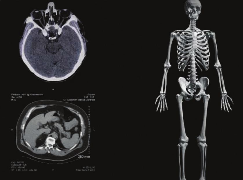
Journal of Healthcare Engineering 9

 CT perfusion imaging as an accurate assessment
 8
 6.87
 7 5.9
 5.72
 6
 4.8
 Influence level 5 3.88 2.89

 4 3.79 3.65 3.6

 3
 1.86 2.44
 2
 1.09
 1
 0
 SAP Pancreas Inflammation Partial Disorders Osimertinib
 Influence factors

 QEEG CPPE
 AIS mPFS
 CT perfusion PPE
 Figure 7: CT perfusion imaging as an accurate assessment.

 Table 5: QEEG characteristics and CT perfusion imaging parameters in SAP patients.
Item SAP Pancreas Inflammation Partial Disorders
Cells 1.71 0.57 1.81 0.26 0.26
SAP 2.32 1.62 1.14 1.36 1.8
SAP 5.69 5.2 4.38 4.67 3.22
MRI 3.91 1.25 5.35 3.12 2
SAP 1.21 4.53 2.08 3.87 2.82

electrical activity are related to ischemic penumbra, pan- monitoring effect at present. CT perfusion imaging has a
creatic edema, and separation of nerve function. These good effect in evaluating the regional blood perfusion of
findings confirm the value of QEEG in the localization of ischemic lesions, but it cannot achieve real-time
ischemic pancreas. monitoring.
 As shown in Figure 7, CT perfusion imaging has been As shown in Table 6, the study of the correlation between
used in clinical practice as a technique to accurately evaluate QEEG characteristics and CT perfusion imaging has positive
the hemodynamic changes of pancreas. In terms of pancreatic significance for the application of QEEG in dynamic
diagnosis, not only early detection of ischemic lesion areas but monitoring of ischemic pancreas and guiding clinical
also real-time monitoring of hemodynamic changes in is- treatment. The results showed that CBF was reduced, MTT
chemic lesion areas is required, so CT perfusion imaging is and TTP were delayed, delta wave index, slow wave index,
particularly important. As shown in Table 5, there is a cor- and BSI were increased, and ADR and dtabr were decreased
relation between QEEG characteristics and CT perfusion in the ischemic lesion region compared to the contralateral
imaging parameters in SAP patients. This conclusion can region, CT perfusion imaging, and qEEG diagnostic values
provide help in clinical diagnosis of SAP patients. were further confirmed.
 As shown in Figure 8, CT perfusion imaging can be As shown in Figure 10, this paper analyzed the re-
used to observe the hemodynamic changes in different lationship between different pancreatic blood flow and
pancreatic regions, which can well determine the focus qEEG and found that when the pancreatic blood flow was
area. CT images are used to record the spontaneous lower than 35 ml/(100g min), the fast beta rhythm dis-
formation of pancreatic biopotential. It is often used in the appeared, when the pancreatic blood flow was lower than
examination and diagnosis of epilepsy, pancreatitis, and 18 ML/(100g min), the delta rhythm began to appeared,
intracranial space occupying lesions. The study found that and when the pancreatic blood flow was lower than
when the blood flow of pancreatic tissue was interrupted 10 ml/(100g min), the CT image showed complete sup-
for more than 3 seconds, the CT image showed abnormal pression. In the aspect of QEEG characteristics, the main
changes in this area. indicators commonly used in the evaluation of ischemic
 As shown in Figure 9, due to the rapid progress of pancreas are relative power ratio and BSI, among which
SAP patients in the early stage, CT image as a relatively ADR and dtabr are more commonly used, which are
simple means for dynamic monitoring is easier to recognized as more accurate and stable evaluation and
achieve clinically, but there is still controversy about its prediction indicators.
10 Journal of Healthcare Engineering

 Perfusion imaging to observe hemodynamic changes in different pancreatic regions
 5 8

 7
 4
 6.13 6
 5.09

 Channel liquidity
 4.8
 Lithology
 3 5

 4
 2 3.12
 3
 4.49
 2.3 2
 1
 1

 0 0
 SAP Pancreas Inflammation Partial Disorders PPE
 Load base

 Epilepsy MRI
 Encephalitis PPC
 Intracranial Osimertinib
 Figure 8: Perfusion imaging to observe hemodynamic changes in different pancreatic regions.

 Figure 9: Cancer manifestations of DS-8201 on HER2 expression (from Google: http://1il65.cn/SsLUi3).

 Table 6: Correlation between QEEG features and CT perfusion imaging.
Item Epilepsy Encephalitis Intracranial MRI SAP
SAP 1.71 2.32 5.69 3.91 1.21
Pancreas 0.57 1.62 5.2 1.25 4.53
Inflammation 1.81 1.14 4.38 5.35 2.08
Partial 0.26 1.36 4.67 3.12 3.87
Disorders 0.26 1.8 3.22 2 2.82
Journal of Healthcare Engineering 11

 The relationship between different pancreatic blood flow and QEEG
 40
 35 5.05 2.49
 2.33
 30
 Index’s influence 1.83 6.96
 25
 20
 15 2.21
 1.98 5.21 3.51
 10
 3.95
 5 1.01

 0
 QEEG PPE MRI PPC DESTINY Mine
 Tube item

 DTABR Inflammation
 ADR Partial
 SAP Disorders
 Pancreas
 Figure 10: The relationship between different pancreatic blood flow and QEEG.

5. Conclusions Due to the limited sample size in this article, individual
 differences in patients with severe acute pancreatitis may
There are significantly more studies on CT image pan- have an impact on the experimental results; the research
creatic guides in correcting coronal force lines than other team should also prepare pathological sections of multiple
plane force lines. Studies have shown that, compared with organs to observe the protective effect of drugs on multiple
traditional TKA, CT imaging pancreas guide can better organs and at the same time continue to collect clinical trials
restore the coronal force line of the lower limbs, but there in the future cases, a large sample size, multicenter clinical
are also reports that there is no significant difference experimental research to verify the results of animal ex-
between the two. The results of this article show that the periments. In summary, this article is the first to conduct a
difference between postoperative mFTA and preoperative retrospective analysis of patients undergoing internal
measurement is statistically significant and is similar to drainage treatment of SAP to explore the risk factors that
the mFTA result obtained by preparing CT image pan- affect the recurrence and complications of pseudocysts after
creatic guide plate combined with MRI and CT images of internal drainage and provide clinical guidance.
the whole lower limb. However, there was no statistically
significant difference between FMAA and aLDFA and the
preoperative measurement, indicating that the lower limb
 Data Availability
force line returned to the preoperative physiological state. No data were used to support this study.
We believe that a distal pancreatic guide plate created
based on pancreatic CT and full-length x-ray images of the
lower limbs can also improve the coronary force lines of Conflicts of Interest
the lower limbs. For fragile and severe acute pancreatitis The authors declare that they have no conflicts of interest.
caused by SAP, treatment should also be based on spleen
deficiency.
 There are still some shortcomings in the syndrome rules References
of SAP secondary to severe acute pancreatitis discussed in [1] R. L. Siegel, K. D. Miller, and A. Jemal, “Cancer statistics,
this article. For example, under the guidance of TCM 2019,” CA: A Cancer Journal for Clinicians, vol. 69, no. 1,
treatment theory, the inflammatory state of the pancreas can pp. 7–34, 2019.
be improved in the early stage of SAP, thereby preventing or [2] A. Riker, S. K. Libutti, and D. L. Bartlett, “Advances in the
delaying the occurrence of glucose metabolism disorders. In early detection,diagnosis, and staging of pancreatic cancer,”
addition, this article is the first to introduce CT images into Surgical Oncology, vol. 6, no. 3, pp. 157–169, 2020.
the SAP model of patients with severe acute pancreatitis. [3] V. Deshpande, S. Chicano, and D. Finkelberg, “Autoimmune
Through chart analysis, it is found that the CARD9 mRNA pancreatitis: a systemic immune complex mediated disease,”
expression level of the SAP model of severe acute pancre- The American Journal of Surgical Pathology, vol. 30, no. 12,
 pp. 1537–1545, 2020.
atitis is more sensitive, specific, and AUC than TNF-α, IL-
 [4] T. Kamisawa, N. Egawa, and H. Nakajima, “Clinical diffi-
1β, pathological scores. A high value indicates high diag- culties in the differentiation of autoimmune pancreatitis and
nostic accuracy and can be used as a new target for assessing pancreatic carcinoma,” American Journal of Gastroenterology,
the state of SAP. vol. 98, no. 12, pp. 2694–2699, 2020.
12 Journal of Healthcare Engineering

 [5] S. T. Chari, N. Takahashi, and M. J. Levy, “A diagnostic [21] M. Bojková, P. Dı́tě, and J. Dvořáčková, “Immunoglobulin
 strategy to distinguish autoimmune pancreatitis from pan- G4, autoimmune pancreatitis and pancreatic cancer,” Di-
 creatic cancer,” Clinical Gastroenterology and Hepatology, gestive Diseases, vol. 33, no. 1, pp. 86–90, 2020.
 vol. 7, no. 10, pp. 1097–1103, 2020. [22] P. Dite, I. Novotny, J. Dvorackova et al., “Pancreatic solid focal
 [6] C. He, D. Rong, W. Hu et al., “A feasible CT feature to dif- lesions: differential diagnosis between autoimmune pancre-
 ferentiate focal-type autoimmune pancreatitis from pancre- atitis and pancreatic cancer,” Digestive Diseases, vol. 37, no. 5,
 atic ductal adenocarcinoma,” Cancer Medicine, vol. 8, no. 14, pp. 416–421, 2019.
 pp. 6250–6257, 2019. [23] L. D. Dickerson, A. Farooq, F. Bano et al., “Differentiation of
 [7] K. Okazaki, S. Kawa, and T. Kamisawa, “Amendment of the autoimmune pancreatitis from pancreatic cancer remains
 JSA Panese consensus guidelines for autoimmune pancrea- challenging,” World Journal of Surgery, vol. 43, no. 6,
 titis, concept and diagnosis of autoimmune pancreatitis,” pp. 1604–1611, 2019.
 Journal of Gastroenterology, vol. 49, no. 4, pp. 567–588, 2020. [24] X. F. Wang, P. Gao, and Y. F. Liu, “Predicting thermophilic
 [8] H. Hamano, S. Kawa, and A. Horiuchi, “High serum proteins by machine learning,” Current Bioinformatics,
 IgG4concentrations in patients with sclerosing pancreatitis,” vol. 15, 2020.
 Chinese Journal of Bases and Clinics in General Surgery, [25] J. Zhang and B. Liu, “A review on the recent developments of
 vol. 344, no. 10, pp. 732–738, 2020. sequence-based protein feature extraction methods,” Current
 [9] E. K. Choi, M. H. Kim, and T. Y. Lee, “The sensitivity and Bioinformatics, vol. 14, no. 3, 2019.
 specificity of serum immunoglobulin G and immunoglobulin [26] T. Ngwa, R. Law, and P. Hart, “Serum IgG4 elevation in
 G4 levels in the diagnosis of autoimmune chronic pancrea- pancreatic cancer: diagnostic and prognostic significance and
 titis: Korean experience,” Pancreas, vol. 35, no. 2, pp. 156–161, association with autoimmune pancreatitis,” Pancreas, vol. 44,
 2020. no. 4, pp. 557–560, 2020.
[10] K. Okazaki, S. Kawa, and T. Kamisawa, “Clinical diagnostic [27] Q. Liu, Z. Niu, and Y. Li, “Immunoglobulin G4 (IgG4)-
 criteria of autoimmune pancreatitis: revised proposal,” positive plasma cell infiltration is associated with the clinico
 Journal of Gastroenterology, vol. 41, no. 7, pp. 626–631, 2020. pathologic traits and prognosis of pancreatic cancer after
[11] A. Raina, A. M. Krasinskas, and J. B. Greer, “Serum immu- curative resection,” Cancer Immunology, Immunotherapy,
 noglobulin Gfraction 4 levels in pancreatic cancer: elevations vol. 65, no. 8, pp. 931–940, 2020.
 not associated with autoimmune pancreatitis,” Archives of [28] P. Karagiannis, A. E. Gilbert, and F. O. Nestle, “IgG4 anti-
 Pathology & Laboratory Medicine, vol. 132, no. 1, pp. 48–53, bodies and cancer-associated inflammation: insights into a
 2020. novel mechanism of immune escSAPe,” Oncoimmunology,
[12] A. Ghazale, S. T. Chari, and T. C. Smyrk, “Value of serum vol. 2, no. 7, pp. 248-249, 2020.
 IgG4 in the diagnosis of autoimmune pancreatitis and in [29] Z. Shuyun, W. Xin, and Z. Zhenjia, “Synchronous measuring
 distinguishing it from pancreatic cancer,” American Journal of of triptolide changes in rat brain and blood and its application
 Gastroenterology, vol. 102, no. 8, pp. 1646–1653, 2020. to a comparative pharmacokinetic study in normal and
[13] K. Yoshida, F. Toki, and T. Takeuchi, “Chronic pancreatitis Alzheimer’s disease rats,” Journal of Pharmaceutical and
 caused by an autoimmune abnormality. proposal of the Biomedical Analysis, vol. 185, Article ID 113263, 2020.
 concept of autoimmune pancreatitis,” Digestive Diseases and [30] M. M. Lerch, A. Stier, and U. Wahnschaffe, “Pancreatic
 Sciences, vol. 40, no. 7, pp. 1561–1568, 2020. pseudocysts: observation, endoscopic drainage, or resection?”
[14] H. Yong CAO, “Size effect of strength of inhomogeneous rock Deutsches Ärzteblatt International, vol. 106, no. 38, pp. 614–
 under conventional triaxial conditions,” Low Temperature 621, 2020.
 Building Technology, vol. 234, no. 12, pp. 136–139, 2017. [31] D. Jiang, F.-X. Chen, H. Zhou et al., “Bioenergetic crosstalk
[15] Q. Zhang, H. Zhu, and L. Zhang, “Study of scale effect on between mesenchymal stem cells and various ocular cells
 intact rock strength using particle flow modeling,” Interna- through the intercellular trafficking of mitochondria,”
 tional Journal of Rock Mechanics and Mining Sciences, vol. 48, Theranostics, vol. 10, no. 16, pp. 7260–7272, 2020.
 no. 8, pp. 1320–1328, 2020. [32] K. Sim, J. Yang, W. Lu, and X. Gao, “MaD-DLS: mean and
[16] M. Chao, C. Kai, and Z. Zhiwei, “Research on tobacco foreign deviation of deep and local similarity for image quality as-
 body detection device based on machine vision,” Transactions sessment,” IEEE Transactions on Multimedia, vol. 99, p. 1,
 of the Institute of Measurement and Control, vol. 42, no. 2, 2020.
 2020. [33] H. Peng, H. Wang, B. Du et al., “Spatial temporal incidence
[17] N. Gao, L. Tang, and J. Deng, “Design, fabrication and sound dynamic graph neural networks for traffic flow forecasting,”
 absorption test of composite porous metamaterial with em- Information Sciences, vol. 521, pp. 277–290, 2020.
 bedding I-plates into porous polyurethane sponge,” Applied [34] H. Qiu, T. Dong, T. Zhang, J. Lu, G. Memmi, and M. Qiu,
 Acoustics, vol. 175, Article ID 107845, 2021. “Adversarial attacks against network intrusion detection in
[18] T. Kamisawa, M. Imai, and P. Yui Chen, “Strategy for dif- IoT systems,” IEEE Internet of Things Journal, vol. 99, p. 1,
 ferentiating autoimmune pancreatitis from pancreatic can- 2020.
 cer,” Pancreas, vol. 37, no. 3, pp. 62–67, 2020.
[19] M. C. Chang, P. C. Liang, and S. Jan, “Increase diagnostic
 accuracy indifferentiating focal type autoimmune pancreatitis
 from pancreatic16cancer with combined serum IgG4 and
 CA19-9 levels,” Pancreatology, vol. 14, no. 5, pp. 366–372,
 2020.
[20] L. M. Pak, M. A. Schattner, V. Balachandran et al., “The
 clinical utility of immunoglobulin G4 in the evaluation of
 autoimmune pancreatitis and pancreatic adenocarcinoma,”
 Hpb, vol. 20, no. 2, pp. 182–187, 2018.
You can also read