New insights into degenerative mitral valve disease in dogs
←
→
Page content transcription
If your browser does not render page correctly, please read the page content below
Vet Clin Small Anim
34 (2004) 1209–1226
New insights into degenerative
mitral valve disease in dogs
Jens Häggström, DVM, PhDa,*,
Henrik Duelund Pedersen, DVM, Dr Vet Scib,
Clarence Kvart, DVM, PhDc
a
Department of Small Animal Medicine and Surgery, Faculty of Veterinary
Medicine and Animal Science, Swedish University of Agricultural Sciences,
PO Box 7045, S-75007 Uppsala, Sweden
b
Department of Anatomy and Physiology, Royal Veterinary and Agricultural University,
PO 7 Gronnegårdsvej, DK 1870 Fredriksberg C, Denmark
c
Department of Anatomy and Physiology, Faculty of Veterinary Medicine
and Animal Science, Swedish University of Agricultural Sciences,
PO Box 7011, S075007 Uppsala, Sweden
Degenerative mitral valve disease (DMVD) is, by far, the most commonly
encountered acquired cardiac disease in adult dogs, and the condition is
caused by a progressive myxomatous degeneration (MD) of the atrioven-
tricular (AV) valves. DMVD has been given many names in the veterinary
literature, including endocardiosis and chronic valvular disease [1,2]. Similar
changes of the mitral valve are also seen in human beings, horses, and pigs [3–
5]. In people, the condition with similarities to DMVD in dogs is called mitral
valve prolapse (MVP) syndrome [3,6]. Clinical signs of mitral regurgitation
(MR) caused by myxomatous lesions have long been recognized in veterinary
medicine [7], and because of the high prevalence of DMVD in the canine
population, the disease is important for the veterinary small animal
practitioner. Indeed, a recent estimation of the mortality caused by cardiac
disease in the general canine population indicates that about 7% of all dogs
die or are euthanized because of heart failure (HF) before 10 years of age (eg,
the third most common cause of death in dogs in that age group) [8]. Because
many dogs develop decompensated HF because of DMVD after 10 years of
age, we can assume that this proportion is even greater in dogs of all ages.
MR caused by DMVD has been reported to account for 75% of the cases of
heart disease in dogs [9–12] and for a considerably higher proportion in
* Corresponding author.
E-mail address: jens.haggstrom@kirmed.slu.se (J. Häggström).
0195-5616/04/$ - see front matter Ó 2004 Elsevier Inc. All rights reserved.
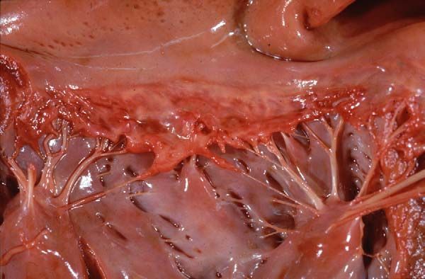
doi:10.1016/j.cvsm.2004.05.0021210 J. Häggström et al / Vet Clin Small Anim 34 (2004) 1209–1226 affected breeds [1,8,13]. Many of the affected dogs eventually need therapy for decompensated HF and die or are euthanized in the end because of refractory cardiac failure. The presence of MR and ongoing medical treatment for HF may negatively interact with other drugs, decisions for surgical procedures, or anesthesia. Because DMVD is characterized by chronic progression, the owner and veterinarian often have no other alternative than to observe how the valvular lesions and MR progress slowly, with little possibility of affecting the course of the disease, a fact that many find frustrating. Finally, there is a need for breeding measures in certain breeds with an exceptionally high prevalence of DMVD. This article does not cover all possible aspects of DMVD, because this subject is simply far too extensive to fit into this presentation. This presentation focuses on new information about some specific aspects of DMVD that may be controversial and of importance for the practicing veterinarian. Mitral valve morphology Because dogs that undergo postmortem examination are most commonly those with severe DMVD and MR, it is common to describe the macroscopic appearance of diseased leaflets as thickened and contracted with varying frequency of ruptured chordae tendineae (Fig. 1) [10,14]. The macroscopic appearance of DMVD depends on at which stage of disease the valve is examined, however. This classic description is a manifestation of severe disease that has progressed over a long time, often several years. The Fig. 1. Postmortem specimen from a dog showing classic severe degenerative mitral valve disease. The mitral valve leaflets are thickened and contracted, with nodules rolling in the free edges. Although changes are evident along the entire leaflet margin and its vicinity, they are unevenly distributed and seem to be most pronounced in sections in which chordae tendineae insert. Evidence of chordal involvement is present in the form of thickening, and some chordae are missing, presumably ruptured. This stage of degenerative mitral valve disease is to be regarded as end-stage disease. (Courtesy of Professor L. Jönsson, Uppsala, Sweden.)
J. Häggström et al / Vet Clin Small Anim 34 (2004) 1209–1226               1211

macroscopic findings in cases of mild DMVD may not be apparent and may
be overlooked, especially in dogs without clinical evidence of MR. Findings
typical for early stages of DMVD include elongated chordae tendineae and
enlarged thickened leaflets with areas showing bulging/ballooning/prolapse
toward the atrial side [11,15,16], which may be identified on a two-
dimensional (2D) echocardiogram in the living dog (Fig. 2). The changes
begin in the area of apposition of the leaflets and are usually most
pronounced in sections where chordae tendineae insert. The bulging of such
areas toward the atrial side of the leaflets has been described as rolling of the
edges. With progression, the bulging becomes worse, the free edge becomes
thickened and irregular, and the lesions spread into other parts of the leaflets
[15]. Within the same valve leaflet, one section may look relatively normal,
whereas another neighboring section is moderately or severely diseased. In
late stages, secondary fibrosis can cause marked thickening and contraction
of leaflets and chordae tendineae. The chordae tendineae may rupture [16],
leading to an unattached free edge. Microscopically, there is myxomatous
proliferation of the valve, in which the spongiosa component of the valve is
unusually prominent and the quantity of acid-staining glucosaminoglycans is
increased [11,15,16]. The valvular interstitial cells in affected areas often have
morphologic changes of the nucleus, a localized concentration of abnormally
shaped mitochondria and rough endoplasmic reticulum, a disorganized
cytoskeleton, and lack of secretory vesicles [17]. There is haphazard
arrangement, disruption, and fragmentation of the collagen fibrils surround-
ing the interstitial cells [14,17]. The endothelial cells covering affected areas
become polymorphic, and some areas completely lose the endothelium,
exposing the underlying extracellular matrix [17]. Some large-breed dogs may
present with massive MR but comparably minor mitral valve abnormalities
on a 2D echocardiogram or at postmortem examination [18,19]. It is
currently not known if the MR in some of these large-breed dogs is the
manifestation of DMVD or another cardiac disease.

Fig. 2. Right parasternal long-axis four-chamber echocardiograms in which the mitral valve in
systole is apparent. A normal mitral valve (A), mild mitral valve prolapse (B), and severe mitral
valve prolapse (C). LV, left ventricle, LA, left atrium. The arrowheads indicate the mitral valve
leaflets. (From Pedersen HD, Lorentzen KA, Kristensen B, et al. Observer variation in the two-
dimensional echocardiographic evaluation of mitral valve prolapse in dogs. Vet Radiol
Ultrasound 1996;37:65–70; with permission.)
1212         J. Häggström et al / Vet Clin Small Anim 34 (2004) 1209–1226

Etiology and pathogenesis of degenerative mitral valve disease
   Little is known with certainty about the underlying cause and pathogenesis
of the progressive thickening and degeneration of the leaflets. An old theory is
that the changes are a response to injury type lesions (ie, repeated impact to
the leaflets [especially in the areas of apposition] results in slowly progressive
changes) [20]. Because not all dogs develop DMVD, one or more primary
inciting factors probably increases the risk of disease in predisposed animals.
The nature of these primary initiating factors is not currently known,
although certain abnormalities of collagen and other extracellular matrix
components have been suggested to predispose to DMVD [1]. In people,
MVP occurs in association with a variety of connective tissue disorders [21–
25] and craniofacial skeletal deformities [26] as well as in a variety of
congenital thoracic deformities, such as straight back, pectus excavatum, or
shallow chest [27–29]. Little is known about such associations in dogs.
Recently, a relation between MVP and a narrow chest in a population of
Dachshunds was reported [30]. It was hypothesized in this report and in one
on human patients [31] that a narrow chest may lead to entrapment of the
heart within the thorax, which could predispose to MVP and DMVD.
   Regardless of the exact nature of the primary inciting factor(s), it has been
suggested that it leads to abnormal valve motion (ie, prolapse of the leaflets),
which, in turn, increases the shear stress imposed on them directly through
the abnormal leaflet apposition and indirectly through the increased
regurgitant flow [32,33]. It is likely that the endothelial damage or loss plays
an important role in the progression of the disease, because endothelial cells
are known to communicate extensively with subendothelial cells (eg, valvular
interstitial cells) [17,34]. Endothelial damage may lead to an imbalance in
local concentrations of growth-promoting and growth-inhibiting substances
produced by endothelial cells. Evidence for such imbalances in diseased
canine mitral valves includes the reported associations between disease
severity and the expression of endothelin receptors and nitric oxide synthase
[35,36]. Furthermore, collagen and other matrix components become
exposed to the blood in areas of diseased valves in which the endothelium
seems to be missing, and this exposure is expected to promote thrombosis.
Although thrombosis may develop as a complication of DMVD in dogs,
thrombus formation on the mitral valve is uncommon. Its absence in the
presence of endothelial damage in DMVD is not currently understood.
Increased knowledge and understanding of the actions of these local
mechanisms may be of great importance for future treatment of DMVD
because they may suggest ways of treating the actual valve lesions rather than
only treating the resulting circulatory disturbances.
   Because the underlying cause of DMVD remains uncertain, several
scientifically unsupported theories have been proposed. Examples of these
theories especially prevalent among breeders are that the valvular lesions
develop as a consequence of poor dental health, with hematogenic spread of
J. Häggström et al / Vet Clin Small Anim 34 (2004) 1209–1226   1213

bacteria from the oral cavity to the valves, or that DMVD may develop as an
unwanted side effect of vaccination. There is currently no scientific evidence
that any of these theories are well founded. Inflammation is not an apparent
part of DMVD [11,37], and although low-degree DMVD may macroscop-
ically or echocardiographically not always be easy to differentiate from
bacterial endocarditis, these two diseases have completely different histo-
pathologic features. In dogs, endocarditis is rare, and when it does occur, it
typically affects large-breed dogs rather than the small-breed dogs that
typically have DMVD and MR [38]. With regard to species differences of
pathologic findings between dogs and people, a major difference seems to be
that human beings are more prone than dogs to develop endocarditis as
a complication of MD. In people, endocarditis is found in approximately
10% of operatively excised and severely affected mitral valves [39,40].

Myxomatous degeneration and vascular changes
    Myxomatous degeneration is not restricted to the mitral valve, and it may
be detected in any of the four intracardiac valves. The incidence of valve
involvement in dogs was reported as follows: 62% incidence of mitral valve
alone, 32.5% incidence of mitral and tricuspid valves, and 1.3% incidence of
tricuspid valve alone [11]. The pulmonary and aortic valves are less commonly
affected. Interestingly, lesions similar to MD of the AV valves have been
described in the main pulmonary artery in Cavalier King Charles Spaniels
[41]. Other findings in dogs with advanced stages of DMVD include hyaline
or fibromuscular intramural arteriosclerosis and multiple small myocardial
infarcts [42–44]. Histologically, these vascular changes (which are common in
old dogs) resemble the changes seen in myxomatous valves, and the two
conditions often occur together [9,42]. Coexistence of these two abnormalities
is to be expected in old dogs, however, because of the high prevalence of both
conditions. Furthermore, intramural arteriosclerosis and multiple small
myocardial infarcts should predispose to sudden death. Sudden death is rare
in dogs with DMVD without decompensated HF, however, and it is
interesting that a recent retrospective study of 65 dogs with histologically
confirmed hyaline or fibromuscular arteriosclerosis of the intramural
coronary arteries showed that 16 (25%) had died suddenly [44]. Therefore,
a possible relation between DMVD and vascular changes in the intramural
coronary arteries and sudden death needs to be investigated further.

Inheritance and breeding
   Heredity has long been suspected to play a major role in the transmission
of DMVD because of the strong association of this disease with certain
small- to medium-sized breeds. Two studies of families of Cavalier King
Charles spaniels and families of Dachshunds provide evidence that genetic
1214 J. Häggström et al / Vet Clin Small Anim 34 (2004) 1209–1226 factors play a large role in the etiology (Fig. 3) [30,45]. The disease seems to have a polygenic inheritance; multiple genes influence the trait, and a certain threshold has to be reached before DMVD develops [30,45]. Male dogs have a lower threshold than female dogs, which means that male dogs develop the disease at younger age than female dogs within a family of dogs in which the offspring, on average, have the same genotype. The polygenic mode of inheritance means that a combination of a sire and a dam that both have early onset of DMVD results in offspring that have, on average, early onset of DMVD (and HF). A combination of dogs with late onset results in offspring that manifest the disease at old age or never. The major role played by genetic factors suggests that other factors (eg, level of exercise, degree of obesity, diet) play a comparably small role in the etiology. Probably because of this, little is known about the influence of such factors on the disease. Breeding measures aimed at reducing the prevalence of DMVD have been initiated in many countries in certain breeds, such as Cavalier King Charles Spaniels and Dachshunds. These breeding programs use auscultation to identify the presence of a heart murmur or echocardiography to detect and quantify MVP or regurgitation. Dogs that are younger than a specific age and have developed a heart murmur or echocardiographic findings consistent with DMVD are not allowed to breed. Likewise, offspring from parents that have developed a heart murmur or echocardiographic evidence of early DMVD younger than a certain age limit are excluded from breeding in some programs. These age limits are presumably different depending on whether auscultation or echocardiography is used as the method of diagnosing DMVD, because at a certain age, more dogs are likely to be diagnosed with DMVD when echocardiographic evidence of MVP is used as a diagnostic method than with auscultation [1,13,46]. Nevertheless, the age limits for potential breeding dogs and parents are important. Because the prevalence of DMVD is highly age dependent, the age limit should be set at an age at which dogs with early onset of DMVD are excluded from breeding but not at too high an age, because this may lead an unacceptable proportion of dogs being excluded, which may leave the breeding population at unacceptably low numbers [1]. It has been suggested that it is not advisable to exclude more than 30% of the dogs from breeding because of a single disease [1,47]. With improved DMVD status in the breed, the age limits may later be raised to push the development and manifestation of DMVD toward a higher age. Diagnosis of early degenerative mitral valve disease The diagnosis of MR caused by DMVD is often not complicated, because the clinical and echocardiographic findings are obvious and match. There are, however, situations in which the diagnosis of DMVD may be less obvious. Early stages of DMVD may be especially difficult. It may not be clinically important for managing the patient to diagnose these early stages correctly, because the effect of mild MR on the circulation is minimal and so
J. Häggström et al / Vet Clin Small Anim 34 (2004) 1209–1226              1215

            A
                         100
   Percentage dogs (%)

                          75

                                  86%                                         88%                                    Moderate intensity murmur
                                                                                                                     (Grade 3 and 4)
                          50
                                                                                                                     Low intensity murmur
                                                                                                                     (Grade 1 and 2)
                                                                                        40%
                          25                                                                                         No murmur
                                                                                                35%
                                                                                                       31%

                           0
                                                                   1          1.5       2       2.5        3
                                                                               Mean parental grading
                                         B
                                                                  2
                               MVP (mm) in litters at 4 years

                                                                1,5

                                                                  1

                                                                0,5

                                                                  0

                                                                -0,5
                                                                       -0,5         0         0,5      1       1,5        2        2,5
                                                                                        Parental MVP (mm) at 8 years
Fig. 3. Two studies have shown that genetic factors play a role in the etiology of degenerative
mitral valve disease (DMVD). (A) Relation between mean parental cardiac status and the
prevalence and intensity of cardiac murmur in offspring at 5 years of age in 30 different Cavalier
King Charles Spaniel litters. The parental cardiac status was graded 1 (late or no development
of DMVD) to 3 (DMVD present at a young age). Parents with a high mean cardiac status (ie,
developed DMVD at a young age) produced more offspring with heart murmurs than parents
with low mean parental grading (ie, late or no development of DMVD). Black, moderate-
intensity murmurs in offspring; shaded, low-intensity murmurs in offspring; white, no murmur
in offspring. (From Swenson L, Haggstrom J, Kvart C, Juneja RK. Relationship between
parental cardiac status in Cavalier King Charles Spaniels and prevalence and severity of chronic
valvular disease in offspring. J Am Vet Med Assoc 1996;208:2009–12; with permission.) (B) The
average mitral valve prolapse (MVP) severity in 18 different Dachshund litters at 4 years of age
shown as a function of mean MVP severity at 8 years of age. Parents with a high mean degree of
MVP produced offspring with a greater mean degree of MVP than parents with a low degree of
MVP. Boxes, family of long-haired Dachshunds; cross, family of short-haired Dachshunds.
(From Olsen LH, Fredholm M, Pedersen HD. Epidemiology and inheritance of mitral valve
prolapse in Dachshunds. J Vet Intern Med 1999;3:448–56; with permission.)
1216 J. Häggström et al / Vet Clin Small Anim 34 (2004) 1209–1226
is the likelihood that the disease will cause clinical signs of disease in the near
future [33,48]. Nevertheless, it is of great importance for breeding that these
dogs are correctly diagnosed, because the currently used breeding programs
are founded on the principle of excluding dogs with early onset of DMVD
and promoting the use of dogs with late or no onset. Because the age limits in
the breeding programs (especially in Cavalier King Charles Spaniels) are set
at an age at which many dogs start to develop DMVD [13], a significant
number of dogs with mild disease are screened. There is currently no ‘‘gold
standard’’ for diagnosing cases of mild DMVD.
Auscultation
The early stages of DMVD are often characterized by the presence of a soft
heart murmur with maximal intensity over the mitral area. This murmur may
occur in every heartbeat, but it may also be intermittent [14,49–51]. A systolic
click may be present in some dogs, and this click may be the only abnormal
sound, but it may also be accompanied by an early, late, or holosystolic
murmur or by no murmur at all [14, 49–51]. In the case of early systolic
murmur, potential differential diagnoses, such as physiologic flow murmurs
or low-degree aortic or pulmonic stenosis, should be ruled out. The presence
of these low-intensity murmurs is influenced by the degree of stress of the dog
at the time of examination. Stress or physical exercise may provoke murmurs
in dogs free of a murmur at rest or increase the intensity of the murmur in
dogs with a low-intensity murmur at rest [51]. Naturally, this variation may
cause confusion if the dog is examined at different times by different
auscultators and the results are in disagreement. Dogs with these ausculta-
tory findings indicative of early DMVD are not normal, even if progression
to more severe forms of DMVD does not occur in the near future.
Echocardiography often reveals changes consistent with DMVD (see below)
in the many of these dogs, but the findings may be inconclusive or normal in
others, with the latter being especially common in dogs with only a systolic
click. These early forms of DMVD may be classified as normal in some
breeding programs to ensure that only diseased dogs are classified as
diseased. This strategy has been chosen because the observer variation
among auscultators has been shown to be considerable in dogs with no or
mild DMVD but less in dogs with more progressive forms [51].
Echocardiography
Echocardiography is a valuable tool to evaluate dogs with early DMVD
because it provides information about valve morphology and valve leakage
and it helps to rule out differential diagnoses. The technique has disadvan-
tages, however, because it is comparably time-consuming and expensive
compared with auscultation and it requires trained operators, which makes it
less convenient as a screening method of large populations. Ideally, diagnosisJ. Häggström et al / Vet Clin Small Anim 34 (2004) 1209–1226 1217
of early DMVD should be founded on the findings of abnormal mitral valve
morphology typical for DMVD and valve leakage. Abnormal mitral valve
morphology may be present without leakage and vice versa, however
[46,51,52]. Although DMVD is the most common cause for mitral valve
leakage, the diagnosis of DMVD is less obvious in cases in which the only
abnormal finding is the presence of a small regurgitant jet. When examining
a mitral valve using 2D echocardiography, it is important to examine the
entire valve, because the lesions are often quite unevenly distributed
[14,17,37]. A systolic bulging of one or both leaflets to the atrial side of the
mitral annulus is an early indication of affected valves, and it may be present
in dogs with or without MR (and a murmur) (see Fig. 2) [32,46]. The presence
and degree of protrusion of the leaflets may be measured or subjectively
evaluated in the right parasternal long-axis view [2,46]. In dogs, the hinge
points of the two leaflets (imaged in the right parasternal long-axis view,
which consistently provides good images) have been used to define the
position of the mitral annulus in all recent studies assessing the presence and
severity of MVP [33,52,53]. In cases with insufficient valves, the degree of
displacement is reported to relate well to the severity of MR [2,32,46]. With
progression, the degenerative changes become more prominent and the
leaflets often have an irregular ‘‘club-like’’ appearance with greatest
thickening at the tip. The gross pathologic changes of the two leaflets
(anterior and posterior) are often equally severe at postmortem examination,
but the degenerative changes commonly appear more prominent on the
anterior leaflet in the right parasternal long-axis view on the echocardiogram.
It is rare, even in severe cases of DMVD and MR, to detect incomplete
closure of the leaflets as a means of confirming the presence of MR. Instead,
the MR may be detected and quantified by spectral or color-flow Doppler
ultrasonography [19,51,54–56]. Ideally, the regurgitant flow should be
aligned with the ultrasound beam, and this is most often achieved in the
left apical four-chamber view. Because the flow direction depends on the
orientation of the regurgitant orifice, which, in turn depends on the leaflet
morphology, other views may also give good alignment. Spectral Doppler
mapping may be used to identify the regurgitant jet when color-Doppler
mapping is not available. Furthermore, spectral Doppler mapping gives
information about the velocity of the regurgitant jet, and velocity time
tracings may help in estimating regurgitant volume (see below) [19]. Color-
flow echocardiography confirms the presence of a regurgitant jet, and the size
of the jet can be compared with the size of the left atrium. This measurement
is semiquantitative. A small jet rules out moderate to severe MR, but it is
difficult to discriminate between moderate and severe regurgitation from the
jet size. Nevertheless, the method has been reported to correlate reasonably
well with other echocardiographic measurements of regurgitant flow and
volume [56]. In case a more exact quantitative measurement of regurgitant
fraction is desired, the proximal isovelocity surface area (PISA) color-flow
method or spectral Doppler subtraction of forward aortic and regurgitant1218 J. Häggström et al / Vet Clin Small Anim 34 (2004) 1209–1226 flows may be used [54–56]. Small jets in the vicinity of the mitral valve should not be overinterpreted in dogs without any other valve abnormality, because trivial regurgitation may often be detected in normal dogs [57]. Consequences of mitral regurgitation on left ventricular function A low degree of MR caused by DMVD does not lead to an apparent change in any cardiac chamber or wall size or pump function. The forward stroke volume is maintained, and the small regurgitant volume is easily accepted by the left atrium. With progression of the valve lesions and increasing MR, however, the potential loss of forward stroke volume is compensated for by increased total stroke volume, increased force of contraction, remodeling of the left atrium and left ventricle with myocardial hypertrophy and dilatation, increased heart rate, and modulations of systemic vascular tonus and extracellular fluid volume. The exact sequence in which these compensatory mechanisms are recruited is currently not fully understood. The cardiac compensatory mechanisms are presumably re- cruited first, whereas the systemic mechanisms do not seem to be activated until the cardiac mechanisms fail to compensate the MR (ie, decompensated HF) [58]. To some extent, the MR is already compensated for by a slightly increased heart rate during compensated phases, but this increase is usually not obvious at a clinical examination because of the overall variability of heart rate in dogs [59,60]. The heart rate is usually significantly increased in advanced stages of MR, however, with evidence of decompensated HF [19,59,60]. MR creates unique hemodynamic stress by means of the development of a low-pressure form of volume overload as a result of ejection into the left atrium [61]. Myocardial systolic function is relatively well preserved, because the ejection into the left atrium at low pressure require little work by the left ventricle compared with other forms of heart disease [19,61,62]. Dogs may tolerate even severe MR for years. Nevertheless, because of chronic volume overload and the fact that the hypertrophy, although necessary, is a pathologic remodeling, myocardial contractility decreases slowly, even in clinically compensated dogs, but progressively and inevitably [12,19,59,63]. Clinical overt myocardial failure in MR is referred to as cardiomyopathy of volume overload, a condition that may also develop in other types of heart disease, such as large patent ductus arteriosus [19]. Reliable measurements of myocardial contractility are not readily obtained in MR, and it is currently not known at which stage the depressed myocardial contractility becomes of clinical significance. The reason for this is that the volume overload causes an increase in preload (increased end-diastolic filling), which, in turn, leads to an increased force of contraction according to the Frank-Starling law [64]. When the ventricles contract, the resistance to ventricular emptying is reduced in the first stages of ejection, because the regurgitant volume is ejected into the left atrium at low pressure, leading to exaggerated motion of the left ventricle (hyperkinesia) [14], which is readily
J. Häggström et al / Vet Clin Small Anim 34 (2004) 1209–1226   1219

identified on the echocardiogram of a diseased dog. In moderate to severe
MR, values of ejection phase indices obtained from the echocardiogram
(eg, left ventricular fractional shortening, ejection fraction, mean velocity of
circumferential shortening) are often greater than normal. Therefore, in the
setting of moderate or severe MR, a normal fractional shortening represents
a significant reduction of myocardial contractility. End-systolic volume
indices (eg, left ventricular end-systolic short-axis dimension, end-systolic
volume index) more accurately estimate myocardial contractility in MR
[14,62]. When decompensated HF is present and the sympathetic nervous
system is activated to increase apparent contractility, even these measure-
ments overestimate intrinsic myocardial contractility [63]. Our longitudinal
studies in Cavalier King Charles Spaniels indicate that although the end-
systolic dimension of the left ventricle does increase gradually before the
onset of signs of decompensated HF, the change is not great and may even be
within the normal reference range [58,65]. This increase in end-systolic
diameter usually becomes apparent after the onset of clinical signs of
decompensated HF [58,65]. This finding is in agreement with previously
published observations, but it does not provide information about overall
cardiac pump function [12,19,62]. Because the cardiac output is determined
by the forward stroke volume and the heart rate, evaluation of cardiac
output must take into account both heart rate and stroke volume. We
recently completed a study in which the pulmonary transit time (ie, the time it
takes for a blood cell to travel from the pulmonary trunk to the left atrium)
was studied using nuclear angiocardiography in dogs with varying severity of
MR caused by DMVD [59]. When the transit time was normalized for the
heart rate, we found that dogs with compensated MR but with evidence of
cardiomegaly had increased transit times. Dogs with signs of decompensated
HF had an even higher increase in transit times. Our interpretation of these
findings is that dogs with MR have reduced overall pump function (forward
stroke volume) even before signs of decompensated HF have developed. It is
currently not known if this finding is an indication that inotropic support is
indicated at this stage of the disease.

Diagnosis of mild decompensated heart failure
    Dogs with DMVD attributable to DMVD usually develop clinical signs of
left-sided HF (cough, dyspnea, lethargy, reduced mobility, and increased
heart rate), although evidence of right-sided HF (ascites) may develop in
advanced cases [14,19]. Diagnosing moderate to severe HF is usually not
difficult, because the clinical signs of HF are usually obvious and match the
findings on the radiographs (ie, pulmonary edema, congestion). Likewise, it
is usually not difficult to diagnose the MR because it is invariably significant
[14,19]. Mild decompensated HF may be difficult to diagnose, however,
because of the presence of vague clinical signs and the fact that the signs may
have gradually developed over a comparably long time. The stage when
1220 J. Häggström et al / Vet Clin Small Anim 34 (2004) 1209–1226 a patient starts to show clinical signs of DMVD and MR (ie, development of decompensated HF) is the end of a process that started much earlier with the onset of valve leakage. Over time, the valvular leakage was compensated through a variety of mechanisms, a condition called ‘‘asymptomatic’’ or compensated MR [14,19]. As the valve leakage increased, the valves eventually became incapable of preventing pulmonary capillary pressures from exceeding the threshold for pulmonary edema or of maintaining forward cardiac output, a condition called ‘‘symptomatic’’ or decompen- sated MR [14,19]. The distinction between these two stages is not clear, however, and it is likely that minor signs of reduced activity and mobility are present even before overt signs of decompensated HF have developed. It is difficult to evaluate the presence of slight to moderately reduced exercise capacity in most dogs with DMVD and MR objectively; many affected animals are old and small companion dogs, which if obese, have little, if any, demand on their exercise capacity. Furthermore, other concurrent diseases in the locomotor system or elsewhere are common and restrict exercise. Likewise, the hallmark of left-sided HF, coughing and dyspnea, may be caused by several conditions, such as small airway disease, tracheal in- stability, pulmonary fibrosis, neoplasia, heartworm disease, and pneumonia [14,19]. An increased heart rate and loss of respiratory sinus arrhythmia may also be indicative of decompensated HF, but heart rate is variable and is increased by many factors, such as stress and concurrent disease [14,19,60]. Many of these differential diagnoses can be excluded by different clinical tests, particularly radiography. Pulmonary findings on the radiographs may also be inconclusive, because early radiographic changes of pulmonary interstitial edema and bronchial pattern resemble the radiographic appear- ance of chronic airway disease [14,19,60]. The tendency is to overdiagnose pulmonary edema of HF [66]. Therefore, the most effective means to separate dogs with early mild decompensated HF from those with other disease is presumably to make the diagnosis based on the combined findings from the clinical examination and the radiographs, an approach that has been used in large clinical trials [48]. It is also useful to have series of radiographs and to evaluate other evidence of left-sided HF that should be present by the time pulmonary edema has developed, such as pulmonary venous distention. If the findings are still inconculsive, re-examination within a week or a 48- to 72-hour trial of diuretic therapy with repeat radiographs may help to identify the underlying cause. In the near future, ‘‘bedside’’ assays of different endogenous markers of heart disease and HF, such as natriuretic peptides (atrial natriuretic peptide [ANP] and brain natriuretic peptide [BNP]), should be available to aid in diagnosing difficult cases. When should therapy begin? Ideally, DMVD therapy should halt the progression of the valvular degeneration or improve valvular function by surgical repair or valve
J. Häggström et al / Vet Clin Small Anim 34 (2004) 1209–1226   1221

replacement. This therapy should preferably start before the onset of clinical
signs of disease. No medical therapy has been shown to change the course of
the disease by inhibiting or preventing the valvular degeneration, however,
and surgery is usually not technically, economically, or ethically possible in
dogs. Medical therapy of DMVD is therefore aimed at improving quality of
life by ameliorating the clinical signs and at improving survival. Mono-
therapy with angiotensin-converting enzyme (ACE) inhibitors has fre-
quently been prescribed for dogs with DMVD before the onset of
decompensated HF, most commonly in dogs with evidence of left atrial
and ventricular dilatation. Presumably, there are many reasons for this
strategy. Clinical trials in dogs with decompensated HF caused by DMVD
have shown that ACE inhibitor therapy improves quality of life and
increases survival when administered as adjunct therapy to other ongoing
HF therapy [67–69]. Furthermore, there is evidence from large clinical trials
in people that monotherapy with ACE inhibitors improves quality of life
and survival not only in asymptomatic patients with left ventricular
dysfunction [70] but in those without heart disease but belonging to a risk
group for developing it [71]. The local tissue renin-angiotensin-aldosterone
system (RAAS) has been suggested to be important for myocardial
remodeling in various animal models of HF [72,73]. An increased concen-
tration of plasma renin and aldosterone was reported in some asymptomatic
dogs with DMVD, indicating an early activation of the RAAS [74]. It is
therefore plausible that suppression of the RAAS could also be beneficial in
asymptomatic dogs with MR by counteracting systemic neuroendocrine
activation and left ventricular remodeling. Two large, placebo-controlled,
multicenter trials, the Scandinavian Veterinary Enalapril Prevention (SVEP)
and the VetProof trials [48,75], were undertaken to study the effect of ACE
inhibitor monotherapy on the progression of clinical signs in asymptomatic
DMVD and MR in dogs. Both failed to show a significant difference
between the placebo and treatment groups in time from onset of therapy to
confirmed decompensated HF (Fig. 4) [48,75] in dogs with or without
cardiomegaly. The two trials differed in the following features: the SVEP
trial included only dogs of one breed (Cavalier King Charles Spaniels),
whereas the VetProof trial included a variety of breeds; the dogs in the
VetProof trial more frequently had advanced DMVD than the dogs in the
SVEP trial; and the SVEP trial comprised more dogs than the VetProof trial
(229 versus 139 dogs). There are studies that may shed light on the results of
these two trials. The increased plasma concentration of renin and aldoste-
rone found in some asymptomatic dogs with MVD [74] was later found to be
associated with the presence of MVP rather than with the degree of MR per
se [76]. Furthermore, a longitudinal study involving Cavalier King Charles
Spaniels with moderate to severe MR attributable to DMVD showed no
signs of increased circulating RAAS activity during the progression from
compensated (ie, asymptomatic) to decompensated (ie, symptomatic) HF
[58]. This finding has recently been corroborated in a study involving other
1222           J. Häggström et al / Vet Clin Small Anim 34 (2004) 1209–1226

Fig. 4. The SVEP study investigated the effect of enalapril on preventing decompensated heart
failure in dogs with asymptomatic DMVD. The graph shows the percentage of dogs included in
the enalapril and placebo groups, respectively, versus time. The difference in the number of days
in the study between placebo- and enalapril-treated dogs was not significant. (From Kvart C,
Häggström J, Pedersen HD, et al. Efficacy of enalapril for prevention of congestive heart failure
in dogs with myxomatous valve disease and asymptomatic mitral regurgitation. J Vet Intern
Med 2002;16:80–8; with permission.)

breeds by Oyama and Sisson [77]. On the local tissue level, autoradiographic
studies indicate that in canine mitral valves, as opposed to rat valves,
angiotensin II receptors and ACE are scant [78]. This finding is at odds with
the theory that local RAAS systems in the valves contribute to progressive
valvular degeneration. In contrast, the canine myocardium has a comparably
high concentration of angiotensin II receptors and ACE [79]. Nevertheless,
experimental studies in dogs with MR showed no effect of an ACE inhibitor
on myocardial remodeling and progressive ventricular dilatation [79].
Because angiotensin II production may be mediated through enzymes other
than ACE, in particular through chymase in dogs and people [80], the same
authors investigated whether blocking of angiotensin II receptors could
prevent myocardial remodeling but found no effect [81]. Thus, it seems that
the remodeling process in MR may be more complicated than previously
thought; it has recently been suggested that this process is an example of
tissue activation that is difficult to stop or slow by current pharmacologic
means without changing the fundamental pathophysiology (ie, increased
heart rate and loading conditions) [82]. Finally, the large clinical trials in
asymptomatic heart disease in people have most commonly involved
patients with left ventricular dysfunction. Published clinical trials in primary
mitral valve disease and MR in people have been surprisingly few and have
reported conflicting results [82]. In conclusion, there is no evidence that any
therapy instituted before the onset of clinical signs of decompensated HF
prevents or delays the progression of DMVD.
J. Häggström et al / Vet Clin Small Anim 34 (2004) 1209–1226               1223

References
 [1] Häggström J. Chronic valvular disease in Cavalier King Charles Spaniels—epidemiology,
     inheritance and pathophysiology [thesis]. Uppsala: Swedish University of Agricultural
     Sciences; 1996.
 [2] Pedersen H. Mitral valve prolapse in the dog—pathogenesis, pathophysiology, diagnosis
     and comparative aspects of early myxomatous mitral valve disease [thesis]. Copenhagen:
     Royal Veterinary and Agricultural University; 2000.
 [3] Pedersen H, Häggström J. Mitral valve prolapse in the dog: a model of mitral valve
     prolapse in man. J Cardiovasc Res 2000;47:234–43.
 [4] Reef V, Bain F, Spencer P. Severe mitral regurgitation in horses: clinical, echocardio-
     graphic and pathological findings. Equine Vet J 1998;30:18–27.
 [5] Gagna C, Meier D, Ru G, Pospischil A, Guarda F. Pathology of mitral valve in regularly
     slaughtered pigs: an abattoir survey on the occurrence of myxoid degeneration
     (endocardiosis), fibrosis and valvulitis. J Vet Med Ser A 1998;45:383–95.
 [6] Playford D, Weyman A. Mitral valve prolapse: time for a fresh look. Rev Cardiovasc Med
     2001;2:73–81.
 [7] Ackerknecht E. Kreislauforgane (herz). In: Joest EE, editor. Spezielle pathologische
     Anatomie der Haustiere. Berlin: Richard Schoetz; 1925. p. 317–586.
 [8] Bonnett B, Egenvall A, Olson P, Hedhammar A. Mortality in insured Swedish dogs: rates
     and causes of death in various breeds. Vet Rec 1997;141:40–4.
 [9] Detweiler DK, Pattersson DF. The prevalence and types of cardiovascular disease in dogs.
     Ann NY Acad Sci 1965;127:481–516.
[10] Das KM, Tashjihan RJ. Chronic mitral valve disease in the dog. Vet Med Small Anim Clin
     1965;60:1209–15.
[11] Buchanan JW. Chronic valvular disease (endocardiosis) in dogs. Adv Vet Sci 1977;21:
     57–106.
[12] Sisson D, Kvart C, Darke P. Aquired valvular heart disease in dogs and cats. In: Fox P,
     Sisson D, Moise N, editors. Textbook of canine and feline cardiology. 2nd edition.
     Philadelphia: WB Saunders; 1999. p. 536–65.
[13] Häggström J, Hansson K, Kvart C, Swenson L. Chronic valvular disease in the Cavalier
     King Charles spaniel in Sweden. Vet Rec 1992;131:549–53.
[14] Kvart C, Häggström J. Acquired valvular heart disease. In: Ettinger S, Feldman E, editors.
     Textbook of veterinary internal medicine. Diseases of dogs and cats. 5th edition.
     Philadelphia: WB Saunders; 2000. p. 787–800.
[15] Whitney JC. Observation on the effect of age on the severity of heart valve lesions in the
     dog. J Small Anim Pract 1974;15:511–22.
[16] Kogure K. Pathology of chronic mitral valve disease in the dog. Jpn Vet Sci 1980;42:323–35.
[17] Corcoran B, Black A, Anderson H, Dukes McEvan J, French A. Investigation of mitral
     valve morphology in dogs with mitral valve endocardiosis using scanning electron
     microscopy. In: Proceedings of the 12th European College of Veterinary Internal
     Medicine/European Society of Veterinary Internal Medicine Congress. Munich, Germany;
     September 19–21, 2002, p. 178.
[18] Borgarelli M, Zini E, D’Agnolo G, Tarducci A, Santilli RA, Chiavegato D, et al.
     Comparison of primary mitral valve disease in German Shepherd dogs and dogs of small-
     sized breeds. J Vet Cardiol 2004;6, in press.
[19] Kittleson M. Myxomatous atrioventricular valvular degeneration. In: Kittleson M, Kienle
     R. Small animal cardiovascular medicine. St. Louis: Mosby; 1998. p. 297–318.
[20] Pommerance A. Pathogenesis of ‘‘senile’’ nodular sclerosis of atrioventricular valves. Br
     Heart J 1966;28:815–23.
[21] Sanyal SK, Johnson WW, Dische MR, Pitner SE, Beard C. Dystrophic degeneration of
     papillary muscle and ventricular myocardium. A basis for mitral valve prolapse in
     Duchenne’s muscular dystrophy. Circulation 1980;62:430–8.
1224 J. Häggström et al / Vet Clin Small Anim 34 (2004) 1209–1226
[22] Lebwohl M, Distefano D, Prioleau P. Pseudoxanthoma, elasticum and mitral valve
prolapse. N Engl J Med 1982;307:228–31.
[23] Cabeen WR, Reza MJ, Kovick RB, Stern MS. Mitral valve prolapse and conduction
defects in Ehlers-Danlos syndrome. Arch Intern Med 1977;137:1227–31.
[24] Wood S, Thomas J, Braimbridge M. Mitral valve disease and open heart surgery in
osteogenesis imperfecta tarda. Br Heart J 1973;35:103–6.
[25] Beardsley TL, Foulks GN. An association of keratoconus and mitral valve prolapse.
Ophthalmology 1982;89:35–7.
[26] Waite P, McCallum CA. Mitral valve prolapse in craniofacial skeletal deformities. Oral
Surg Oral Med Oral Pathol Oral Radiol Endod 1986;61:15–8.
[27] Chan FL, Chen WW, Wong PH, Chow JS. Skeletal abnormalities in mitral valve prolapse.
Clin Radiol 1983;34:207–13.
[28] Chen WW, Chan FL, Wong PH, Chow JS. Familial occurrence of mitral valve prolapse: is
this related to the straight back syndrome? Br Heart J 1983;50:97–100.
[29] Hirschfeld SS, Rudner C, Nash CL Jr, Nussbaum E, Brower EM. Incidence of mitral valve
prolapse in adolescent scoliosis and thoracic hypokyphosis. Pediatrics 1982;70:451–4.
[30] Olsen L, Fredholm M, Pedersen H. Epidemiology and inheritance of mitral valve prolapse
in Dachshunds. J Vet Intern Med 1999;13:448–56.
[31] Raggi P, Callister T, Lippolis N, Russo D. Is mitral valve prolapse due to cardiac
entrapment in the chest cavity? A CT view. Chest 2000;117:636–42.
[32] Pedersen H, Lorentzen K, Kristensen B. Echocardiographic mitral valve prolapse in
Cavalier King Charles spaniels: epidemiology and prognostic significance for regurgitation.
Vet Rec 1999;144:315–20.
[33] Olsen L, Martinussen T, Pedersen H. Early echocardiographic predictors of myxomatous
mitral valve disease in dachshunds. Vet Rec 2003;152:293–7.
[34] Stein P, Wang C, Riddle J, Sabbah H, Magilligan D, Hawkins E. Scanning electron
microscopy of operatively excised severely regurgitant floppy mitral valves. Am J Cardiol
1989;64:392–4.
[35] Mow T, Pedersen H. Increased endothelin-receptor density in myxomatous canine mitral
valve leaflets. J Cardiovasc Pharmacol 1999;34:254–60.
[36] Olsen L, Mortensen K, Martinussen T, Larsson L, Baandrup U, Pedersen H. Increased
NADPH-diaphorase activity in canine myxomatous mitral valve leaflets. J Comp Pathol
2003;129:120–30.
[37] Whitney JC. Cardiovascular pathology. J Small Anim Pract 1967;8:459–65.
[38] Calvert CA. Valvular bacterial endocarditis in the dog. J Am Vet Med Assoc 1982;180:
1080–4.
[39] Davies M, Moore B, Braimbridge M. The floppy mitral valve—study of incidence,
pathology, and complications in surgical, necropsy, and forensic material. Br Heart J 1978;
40:468–81.
[40] Agozzino L, Falco A, de Vivo F, de Vincentiis C, de Luca L, Esposito S, et al. Surgical
pathology of the mitral valve: gross and histological study of 1288 surgically excised valves.
Int J Cardiol 1992;37:79–89.
[41] Karlstam E, Haggstrom J, Kvart C, Jonsson L, Michaelsson M. Pulmonary artery lesions
in Cavalier King Charles spaniels. Vet Rec 2000;147:166–7.
[42] Jonsson L. Coronary arterial lesions and myocardial infarcts in the dog. A pathologic and
microangiographic study. Acta Vet Scand 1972;38:1–80.
[43] Tidholm A, Häggström J, Jönsson L. Prevalence of attenuated wavy fibers in myocardium
of dogs with dilated cardiomyopathy. J Am Vet Med Assoc 1998;212:1732–4.
[44] Falk T, Jönsson L. Ischaemic heart disease in the dog: a review of 65 cases. J Small Anim
Pract 2000;41:97–103.
[45] Swenson L, Häggström J, Kvart J, Juneja K. Relationship between parental cardiac status
in Cavalier King Charles Spaniels and prevalence and severity of chronic valvular disease
in offspring. J Am Vet Med Assoc 1996;208:2009–12.J. Häggström et al / Vet Clin Small Anim 34 (2004) 1209–1226 1225
[46] Pedersen HD. Mitral valve prolapse in 3-year old healthy Cavalier King Charles Spaniels.
An echocardiographic study. Can J Vet Res 1995;59:294–8.
[47] Lacy R. Loss of genetic diversity from managed populations: interacting effects on drift,
mutation, immigration, selection and population subdivision. Conserv Biol 1987;1:143–58.
[48] Kvart C, Haggstrom J, Pedersen HD, Hansson K, Eriksson A, Jarvinen AK, et al. Efficacy
of enalapril for prevention of congestive heart failure in dogs with myxomatous valve
disease and asymptomatic mitral regurgitation. J Vet Intern Med 2002;16:80–8.
[49] Kvart C, Häggström J. Cardiac auscultation and phonocardiography in dogs, horses and
cats. Uppsala: Clarence Kvart Selbstverlag; 2002. p. 61–4.
[50] Häggström J, Hansson K, Kvart C. Heart sounds and murmurs: changes related to severity
of mitral regurgitation in Cavalier King Charles Spaniels. J Vet Intern Med 1995;9:75–85.
[51] Pedersen HD, Häggström J, Falk T, Mow T, Olsen LH, Iversen L, et al. Auscultation in
mild mitral regurgitation in dogs: observer variation, effects of physical maneuvers, and
agreement with color Doppler echocardiography and phonocardiography. J Vet Intern
Med 1999;13:56–64.
[52] Pedersen HD, Kristensen B, Norby B, Lorentzen KA. Echocardiographic study of mitral
valve prolapse in dachshunds. Zentralbl Veterinarmed A 1996;43:103–10.
[53] Pedersen HD, Lorentzen K, Kristensen B. Observer variation in the two-dimensional
echocardiographic evaluation of mitral valve prolapse in dogs. Vet Radiol Ultrasound
1996;37:367–72.
[54] Doiguchi O, Takahashi T. Examination of quantitative analysis and measurement of the
regurgitation rate in mitral valve regurgitation by the ‘‘proximal isovelocity surface area’’
method. J Vet Med Sci 2000;62:109–12.
[55] Kittleson M, Brown W. Regurgitant fraction measured by using the proximal isovelocity
surface area in dogs with chronic myxomatous mitral valve disease. J Vet Intern Med 2003;
17:84–8.
[56] al Muzzi R, de Araujo R, al Muzzi L, Pena JLB, Silva EF, et al. Regurgitant jet area by
Doppler color flow mapping: quantitative assessment of mitral regurgitation severity in
dogs. J Vet Cardiol 2003;5:33–8.
[57] Nakayama T, Wakao Y, Takiguchi S, Uechi M, Tanaka K, Takahashi M. Prevalence of
valvular regurgitation in normal Beagle dogs detected by color Doppler echocardiography.
J Vet Med Sci 1994;56:973–5.
[58] Häggström J, Hansson K, Kvart C, Karlberg B, Voulteenaho O, Olsson K. Effects of
naturally acquired decompensated mitral valve regurgitation on the renin-angiotensin-
aldosterone system and atrial natriuretic peptide concentration in dogs. Am J Vet Res
1997;58:77–82.
[59] Lord P, Eriksson A, Häggström J, et al. Increased pulmonary transit times in
asymptomatic dogs with mitral regurgitation. J Vet Intern Med 2003;17:824–9.
[60] Häggström J, Hamlin RL, Hansson K, Kvart C. Heart-rate variability in relation to
severity of mitral regurgitation in the Cavalier King Charles Spaniel. J Small Anim Pract
1996;37:69–75.
[61] Lord PF. Left ventricular volumes of diseased canine heart: congestive cardiomyopathy
and volume overload (patent ductus arteriosus and primary mitral insufficiency). Am J Vet
Res 1973;35:493–501.
[62] Kittleson MD, Eyster GE, Knowlen GG, Bari Olivier N, Anderson LK. Myocardial
function in small dogs with chronic mitral regurgitation and severe congestive heart failure.
J Am Vet Med Assoc 1984;184:455–9.
[63] Urabe Y, Mann DL, Kent RL, Nakano K, Tomanek RJ, Carabello BA, et al. Cellular and
ventricular contractile dysfunction in experimental canine mitral regurgitation. Circ Res
1992;70:131–47.
[64] Komamura K, Shannon RP, Ihara T, Shen YT, Mirsky I, Bishop SP, et al. Exhaustion of
Frank-Starling mechanism in conscious dogs with heart failure. Am J Physiol 1993;265:
H1119–31.1226 J. Häggström et al / Vet Clin Small Anim 34 (2004) 1209–1226
[65] Häggström J, Hansson K, Kvart C, Vuolteenaho O, Olsson K. Secretion patterns of the
natriuretic peptides in naturally acquired mitral regurgitation attributable to chronic
valvular disease in dogs. J Vet Cardiol 2000;2:7–16.
[66] Hansson K. Diagnostic imaging of cardiopulmonary structures in normal dogs and dogs
with mitral regurgitation [thesis]. Uppsala: Swedish University of Agricultural Sciences;
2004.
[67] The COVE Study Group. Controlled clinical evaluation of enalapril in dogs with heart
failure: results of the Cooperative Veterinary Enalapril Study Group. J Vet Intern Med
1995;9:243–52.
[68] Ettinger SJ, Benitz AM, Ericsson GF, Cifelli S, Jernigan AD, Longhofer SL, et al. Effects
of enalapril maleate on survival of dogs with naturally acquired heart failure. The Long-
Term Investigation of Veterinary Enalapril (LIVE) Study Group. J Am Vet Med Assoc
1998;213:1573–7.
[69] The BENCH Study Group. The effect of benazepril on survival times and clinical signs of
dogs with congestive heart failure: results of a multicenter, prospective, randomized,
double-blinded, placebo-controlled, long-term clinical trial. J Vet Cardiol 1999;1:7–18.
[70] The SOLVD Investigators. Effect of enalapril on mortality and the development of heart
failure in asymptomatic patients with reduced left ventricular ejection fractions. N Engl J
Med 1992;327:725–7.
[71] Arnold J, Yusuf S, Young J, Mathew J, Johnstone D, Avezum A, et al. Prevention of
Heart Failure in Patients in the Heart Outcomes Prevention Evaluation (HOPE) Study.
Circulation 2003;107:1234–6.
[72] Brilla C, Maisch B. Regulation of the structural remodelling of the myocardium: from
hypertrophy to heart failure. Eur Heart J 1994;15:45–52.
[73] Lee M, Bohm M, Paul M, Ganten D, et al. Tissue renin-angiotensin systems: their role in
cardiovascular disease? Circulation 1993;87(5 Suppl):IV7–13.
[74] Pedersen HD, Koch J, Poulsen K, Jensen AL, Flagstad A. Activation of the renin-
angiotensin system in dogs with asymptomatic and mildly symptomatic mitral valvular
insufficiency. J Vet Intern Med 1995;9:328–31.
[75] Atkins CE. Enalapril monotherapy in asymptomatic mitral regurgitation: results of the
VetProof trial. In: Proceedings of the 20th Annual American College of Veterinary Internal
Medicine Forum. Dallas, TX, May 29–June 1, 2002. p. 75–6.
[76] Pedersen H, Olsen LH, Mow T, Christensen N. Neuroendocrine changes in Dachshunds
with mitral valve prolapse examined under different study conditions. Res Vet Sci 1999;66:
11–7.
[77] Oyama M, Sisson D. Blood based detection of occult heart disease. In: Proceedings of the
21st Annual American College of Veterinary Internal Medicine Forum. Charlotte, NC,
June 4–8, 2003. p. 88–9.
[78] Mow T, Pedersen H. No expression of angiotensin II receptors and angiotensin-converting
enzyme in myxomatous canine mitral valve leaflets. An autoradiographic study. J Vet Med
A 1999;46:465–72.
[79] Dell’italia LJ, Balcells E, Meng QC, Su X, Schultz D, Bishop SP, et al. Volume-overload
cardiac hypertrophy is unaffected by ACE inhibitor treatment in dogs. Am J Physiol 1997;
273:H961–70.
[80] Stewart JA Jr, Wei CC, Brower GL, Rynders PE, Hankes GH, Dillon AR, et al. Cardiac
mast cell- and chymase-mediated matrix metalloproteinase activity and left ventricular
remodeling in mitral regurgitation in the dog. J Mol Cell Cardiol 2003;35:311–9.
[81] Perry GJ, Wei CC, Hankes GH, Dillon SR, Rynders P, Mukherjee R, et al. Angiotensin II
receptor blockade does not improve left ventricular function and remodeling in subacute
mitral regurgitation in the dog. J Am Coll Cardiol 2002;39:1374–9.
[82] Dell’Italia L. The renin-angiotensin system in mitral regurgitation: a typical example of
tissue activation. Curr Cardiol Rep 2002;4:97–103.You can also read