Disruption of Posterior Brain Systems for Reading in Children with Developmental Dyslexia
←
→
Page content transcription
If your browser does not render page correctly, please read the page content below
Disruption of Posterior Brain Systems for Reading in
Children with Developmental Dyslexia
Bennett A. Shaywitz, Sally E. Shaywitz, Kenneth R. Pugh, W. Einar Mencl,
Robert K. Fulbright, Pawel Skudlarski, R. Todd Constable, Karen E. Marchione,
Jack M. Fletcher, G. Reid Lyon, and John C. Gore

Background: Converging evidence indicates a functional                            Introduction
disruption in the neural systems for reading in adults with
dyslexia. We examined brain activation patterns in dys-
lexic and nonimpaired children during pseudoword and
real-word reading tasks that required phonologic analysis
                                                                                  D     yslexia is characterized by an unexpected difficulty in
                                                                                        reading in children and adults who otherwise possess
                                                                                  the intelligence, motivation, and schooling considered
(i.e., tapped the problems experienced by dyslexic children                       necessary for accurate and fluent reading (Shaywitz 1998).
in sounding out words).                                                           It represents one of the most common problems affecting
Methods: We used functional magnetic resonance imag-                              children and adults with prevalence rates ranging from 5 to
ing (fMRI) to study 144 right-handed children, 70 dyslexic                        17.5% (Shaywitz 1998). Such data have led “the National
readers, and 74 nonimpaired readers as they read                                  Institute of Child Health and Human Development
pseudowords and real words.                                                       (NICHD) within the National Institutes of Health (NIH)
Results: Children with dyslexia demonstrated a disruption                         [to] consider reading failure to reflect not only an educa-
in neural systems for reading involving posterior brain                           tional problem, but a significant public health problem as
regions, including parietotemporal sites and sites in the                         well” (Lyon 1998).
occipitotemporal area. Reading skill was positively corre-                           There is now a strong consensus that the central
lated with the magnitude of activation in the left occipito-                      difficulty in dyslexia reflects a deficit within the language
temporal region. Activation in the left and right inferior                        system and, more particularly, in a lower level component,
frontal gyri was greater in older compared with younger                           phonology, which has to do with the ability to access the
dyslexic children.                                                                underlying sound structure of words (Liberman and
Conclusions: These findings provide neurobiological ev-                           Shankweiler 1991; Shaywitz 1996, 1998; Wagner and
idence of an underlying disruption in the neural systems                          Torgesen 1987). Results from large and well-studied
for reading in children with dyslexia and indicate that it is                     populations with reading disability confirm that in young
evident at a young age. The locus of the disruption places
                                                                                  school-age children, a deficit in phonologic analysis rep-
childhood dyslexia within the same neurobiological
                                                                                  resents the most reliable (Fletcher et al 1994; Stanovich
framework as dyslexia, and acquired alexia, occurring in
adults. Biol Psychiatry 2002;52:101–110 © 2002 Soci-                              and Siegel 1994) and specific (Morris et al 1998) correlate
ety of Biological Psychiatry                                                      of dyslexia. Such findings form the basis for the most
                                                                                  successful and evidence-based interventions designed to
Key Words: Dyslexia, reading, fMRI, children, brain                               improve reading (Report of the National Reading Panel
                                                                                  2000). A range of neurobiological investigations using
                                                                                  postmortem brain specimens (Galaburda et al 1985), brain
                                                                                  morphometry (Filipek 1996), and diffusion tensor mag-
                                                                                  netic resonance imaging (MRI; Klingberg et al 2000)
                                                                                  suggests that there are differences in the left temporo-
                                                                                  parieto-occipital brain regions between dyslexic and non-
From the Departments of Pediatrics (BAS, SES, KRP, WEM, KEM), Neurology           impaired readers. Converging evidence using functional
   (BAS), Diagnostic Radiology (RKF, PS, RTC, JCG), Applied Physics (JCG),        brain imaging in adult dyslexic readers also shows a
   Yale University School of Medicine, New Haven, Connecticut; Haskins
   Laboratories (KRP, WEM), New Haven, Connecticut; Department of Pediat-         failure of left hemisphere posterior brain systems to
   rics (JMF), University of Texas Medical School, Houston, Texas; Child
   Development and Behavior Branch (GRL), National Institute of Child Health
                                                                                  function properly during reading (Brunswick et al 1999;
   and Development, National Institutes of Health, Washington, DC (JCG).          Helenius et al 1999; Horwitz et al 1998; Paulesu et al
Address reprint requests to Bennett A. Shaywitz, Department of Pediatrics, Yale
   University School of Medicine, P.O. Box 3333, New Haven CT 06510-8064.
                                                                                  2001; Pugh et al 2000; Rumsey et al 1992, 1997; Salmelin
Received November 2, 2001; revised January 23, 2002; accepted February 5, 2002.   et al 1996; Shaywitz et al 1998; Simos et al 2000). In

© 2002 Society of Biological Psychiatry                                                                                         0006-3223/02/$22.00
                                                                                                                          PII S0006-3223(02)01365-3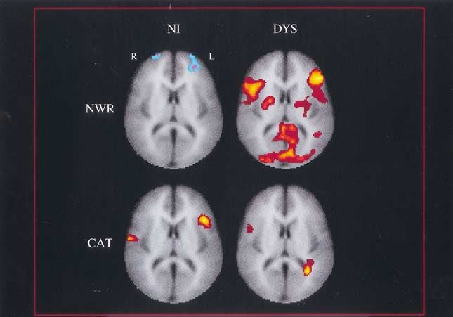
102 BIOL PSYCHIATRY B.A. Shaywitz et al
2002;52:101–110
Table 1. Characteristics of the Subjects
Group
NI (n ⫽ 74) DYS (n ⫽ 70)
Mean SD Mean SD
b
Age (years) 10.9 2.4 13.3 2.7
Sexa
Male 43 49
Female 31 21
Racea
Caucasian 74b 66
African American 0 4
SES
High (1 ⫹ 2) 70 41
Average (3) 4 20
Low (4) 0 6b
Family history of reading problema
No 61 34
Yes 13 36b
WISC-III Full-Scale IQ 116 12.2 99.5b 15.1
Woodcock–Johnson Psycho-Educational Battery
Letter-word identification SS 122 13.7 84.2b 10.7
Word Attack SS 120 17.1 85.1b 11.0
NI, nonimpaired readers; DYS, dyslexic readers; SES, social class, based on the Hollingshead index (unpublished data); SS,
standard score (mean of 100, SD 15); WISC-III, Wechsler Intelligence Scale for Children.
a
Statistical significance determined by Fisher’s exact test.
b
p ⬍ .001.
addition, some functional brain imaging studies show sources, including referrals from pediatricians, nurses, psychol-
differences in brain activation in frontal regions in dys- ogists, educators, and family physicians, as well as through
lexic compared with nonimpaired readers; in some studies notices in parent–teacher association bulletins, public libraries,
dyslexic readers are more active in frontal regions (Bruns- scouting groups, children’s toy stores, and community organiza-
wick et al 1999; Rumsey et al 1997; Shaywitz et al 1998), tions. Children were first screened with IQ and achievement
measures and, if eligible on the basis of these tests, entered the
and in others nonimpaired readers are more active in
study and were evaluated with fMRI. All children had intelli-
frontal regions (Corina et al 2001; Georgiewa et al 1999;
gence in the average range. Criteria for DYS were met if the
Gross-Glenn et al 1991; Paulesu et al 1996). average of the two decoding subtests (Word Identification and
These previous functional imaging studies of dyslexia Word Attack) from the Woodcock–Johnson Psycho-Educational
were in adults, and the findings in adults were used to infer Test Battery (Woodcock and Johnson 1989) were below a
what might be found in children with dyslexia, without Standard Score of 90 (below the 25th percentile) or 1.5 standard
actually studying them. To determine whether these find- errors of prediction lower than the expected reading achievement
ings are the result of a lifetime of poor reading or whether score using the WISC-III (Wechsler 1991) Full-Scale IQ score.
they are there during the period of literacy acquisition, we Both of these definitions validly identify children as poor
used functional magnetic resonance imaging (fMRI) to readers, with little evidence for differences among subgroups of
compare dyslexic and nonimpaired children during tasks children formed with these definitions (Fletcher et al 1994;
that required phonologic analysis, that is, tapped the Shaywitz et al 1992a). To ensure good reading skills and that
problems experienced by dyslexic children in sounding there was no overlap between groups, criteria for NI were
reading scores above the 39th percentile. We excluded from the
out words.
study children with sensory disorders, brain injury, and where the
cause of the reading problem was likely attributable to emotional
Methods and Materials disturbance; clinically apparent neurogenetic disorders; or social,
cultural, or economic disadvantage.
Subjects This study was approved by the Institutional Review Board
We studied 144 right-handed children, 70 dyslexic (DYS) and written informed consent was obtained from all subjects.
readers (21 girls, 49 boys, aged 7–18 years, mean age 13.3 years) The subjects’ demographic characteristics are shown in Table
and 74 nonimpaired (NI) readers (31 girls, 43 boys, aged 7–17 1. There were no differences in gender (2, [1; n ⫽ 144] ⫽ 2.205,
years, mean age 10.9 years) after informed consent had been p ⫽ .14) or race (Fisher’s exact p ⫽ .053); the groups did differ
obtained. Subjects for this study were recruited from a number of on age (t [142] ⫽ 5.62, p ⬍ .0001) and family history (first-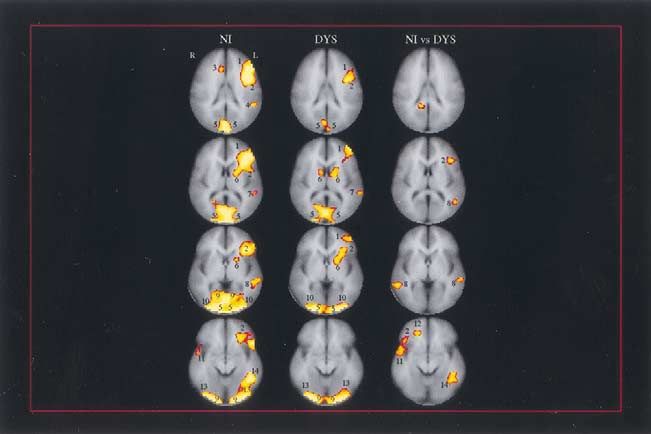
fMRI in Children with Dyslexia BIOL PSYCHIATRY 103
2002;52:101–110
degree relative) of reading problems (2, [1; n ⫽ 144] ⫽ 18.37 [TE (echo time), 11 msec; TR (repetition time), 500 msec; FOV
p ⬍ .001). Full-Scale IQ and Woodcock–Johnson reading mea- (field of view), 20 ⫻ 20 cm; 8-mm-thick contiguous slices;
sures were higher in NI than DYS (all p ⬍ .001). By history, 20% 256 ⫻ 192 ⫻ 2 NEX (number of excitations)] were prescribed
of the sample had been previously treated for reading difficulties. parallel to the intercommissural line based on sagittal localizer
images (TE, 11; TR, 600 msec; FOV, 24 cm; 5-mm contiguous
slices; 256 ⫻ 192 ⫻ 1 NEX). Ten axial-oblique functional
Preparation of Subjects activation images were obtained at the same relative slice
Our general approach to maintaining optimum compliance with location in each subject, extending from the inferior aspect of the
the fMRI procedure focused on decreasing anticipatory anxiety temporal lobes to the parietal convexity, effectively covering the
and desensitizing the children to the components of the proce- entire brain. Activation images were collected using single shot,
dure. This was accomplished by first using a coloring book to gradient echo, echo planar acquisitions (flip angle, 60°; TE, 60
explain the process and then showing a film illustrating a child msec; TR, 2000 msec; FOV, 20 ⫻ 40 cm; 8-mm contiguous
going through the entire procedure. Following this introduction slices; 64 ⫻ 64 ⫻ 1 NEX) in the same slice locations used for
the child practiced in a mock-scanner. For this, we used a room anatomic images.
that was set up with a table made to mimic the imaging gantry. In each of the eight total imaging runs, 100 images per slice
The sounds of the fMRI were recorded and played on a tape location were collected while the subject performed one of the
recorder, thus acclimating the child to the sound of the scanner. four activation tasks (C, SLR, NWR, or CAT) and the line
A mock helmet was used as well. In addition, the child practiced baseline task. The activation tasks and the baseline line task were
the computer tasks that he or she would be performing during the presented in a block design, with five epochs of line task and four
fMRI. Using this procedure, we were able to obtain a high epochs of each activation task within each run. Trials were 4500
imaging success rate. We imaged 155 children; in 11 of the msec in duration; on each trial, stimuli were presented simulta-
children, one or more tasks were not successfully completed, neously for 2500 msec followed by a blank screen for 2000 msec.
resulting in the 144 subjects reported here. Blocks of the baseline task of 22.5-sec duration were inter-
leaved with blocks of the activation task; task order was
randomized across subjects. Two imaging runs with each acti-
Imaging
vation task were acquired, resulting in a total of 100 images per
Subjects were imaged in a 1.5 Tesla SignaLX imaging system slice per activation task and 400 images per slice for the line
from General Electric Medical Systems (Waukesha, WI). Chil- baseline task across the experiment.
dren lay supine in the imaging system, looking up through a
prism at a screen that was attached to the gantry; stimuli were
projected on the screen using a Macintosh laptop computer Data Analysis
programmed in Psyscope. The tasks were designed to differen- Data analysis was performed using software written in MAT-
tially tap the component processes in reading: identifying letters, LAB (MathWorks, Natick, MA). Motion criteria for rejection of
sounding out letters, sounding out pseudowords (pseudowords a study were motion exceeding 2 mm translation or 3° rotation.
are used so that the child cannot have memorized the word and All studies that did not exceed these criteria were included in the
actually has to sound out the never-before-seen pseudoword), final analyses, and all were motion corrected. Before statistical
and sounding out and getting to the meaning of a real word. analysis, the images from each run were motion corrected for
Specifically, the tasks were as follows: identifying letters (i.e., three translation directions and for the three possible rotations.
letter case [C] judgment; e.g., Are [t] and [V] both in the same (Friston et al 1996) Images acquired at the beginning of exper-
upper/lowercase?); sounding out letters (i.e., single letter rhyme imental blocks, corresponding to the period of transient hemo-
[SLR]; e.g., Do the letters [T] and [V] rhyme?); sounding out dynamic change that occurs initially in response to a task, were
pseudowords (i.e., nonword rhyme [NWR]; e.g., Do [LEAT] and discarded, leaving 84 images per activation task for analysis. The
[JETE] rhyme?); and getting to the meaning of words (i.e., remaining images were thresholded (the signal outside of the
Semantic Category [CAT] judgment; e.g., Are [CORN] and brain was set to zero) and Gaussian filtered (FWHM 2.6 mm).
[RICE] in the same category?). A common baseline, the line For generation of single-subject activation maps, activation of
orientation (L) judgment task (e.g., Do [/] and [/] match?) pixels was measured by comparing the images for each task to
was used in analysis; each individual task, C, SLR, NWR, and the line task using a split Student’s t test with correction for
CAT, was contrasted with the (L) baseline condition. The line linear drift. This definition of activation provides a conservative
task was employed as a control because it makes no demands on criterion for identifying task-related activity in the presence of
the major components of reading (orthographic, phonological, or other sources of signal variation (Skudlarski et al 1999). Ana-
semantic processing) but does engage the same sensory modality tomic images and activation maps from individual subjects were
(i.e., visual) used in reading. Children responded to the task with transformed into a proportional three-dimensional grid (Ta-
a button press, for example, pressing one button for “yes, the two lairach and Tournoux 1988). This was performed first by
nonwords rhyme” versus pressing another button for “no, the two in-plane transformation and then by slice interpolation into the
nonwords do not rhyme.” In-magnet proportion correct responses 10 most superior slices of Talairach space, centered at z ⫽ ⫹69,
on the L, C, SLR, NWR, and CAT tasks were, respectively, for ⫹60, ⫹51, ⫹42, ⫹33, ⫹23, ⫹14, ⫹5, ⫺5, and ⫺16,
NI: .86, .89, .87, .79, .91; for DYS: .83, .82, .75, .59, .75. respectively.
Before functional imaging 10 axial-oblique anatomic images The activation maps from individual subjects were used as a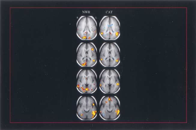
104       BIOL PSYCHIATRY                                                                                           B.A. Shaywitz et al
          2002;52:101–110

                                                                       voxel (p ⬍ .05) was then overlaid on the mean anatomic image
                                                                       for display (Figure 1, column 3).

                                                                       Skill-Correlation Analysis
                                                                       To examine the relationship between reading performance and
                                                                       brain activation in posterior brain regions, we correlated the
                                                                       activations observed for NWR and CAT during fMRI and out of
                                                                       magnet performance on the Word Attack (pseudoword) reading
                                                                       test (Woodcock and Johnson 1989). For each subject, we
                                                                       correlated the mean change in t values between NWR and L (and
                                                                       CAT and L) in each voxel with the child’s reading score on
                                                                       performance on Woodcock–Johnson pseudoword reading
                                                                       (Woodcock and Johnson 1989). In these analyses, age was
                                                                       included as a covariate, effectively removing the effects of age.
Figure 1. Composite maps (columns 1 and 2) demonstrating
brain activation in nonimpaired (NI) and dyslexic (DYS) readers
during the nonword rhyme task and composite contrast maps              Age-Correlation Analysis
(column 3) comparing directly the brain activation of the two          To examine the relationship between age and brain activation, we
groups. In columns 1 and 2, red-yellow indicates areas that had
                                                                       correlated the activations observed for NWR and CAT during
significantly greater activation (p ⫽ .05) in the NWR task
compared with the line task, and in column 3, red-yellow               fMRI and age. For each subject, we correlated the mean change
indicates brain regions that were more active in NI compared           in t values between NWR and L (and CAT and L) in each voxel
with DYS during the NWR task. The four rows of images from             with the child’s age in months.
top to bottom correspond to z ⫽ ⫹23, ⫹14, ⫹5, and –5 in
Talairach space (Talairach and Tournoux 1988). The legend for
brain activation is as follows: 1) middle frontal gyrus, 2) inferior
frontal gyrus, 3) anterior cingulate gyrus, 4) supramarginal gyrus,
5) cuneus, 6) basal ganglia, 7) superior temporal gyrus, 8)
superior temporal sulcus and posterior aspect of the superior and
middle temporal gyri, 9) lingual gyrus, 10) middle occipital
gyrus, 11) anterior aspect of superior temporal gyrus. 12) medial
orbital gyrus, 13) inferior occipital gyrus, and 14) posterior
aspect of middle temporal gyrus and anterior aspect of middle
occipital gyrus. NWR, non-word rhyme.

derived measure of task-related activity and were combined to
obtain a group composite activation map comparing, for exam-
ple, NWR with line (Figure 1 columns 1 and 2) and CAT with
line (Figure 2 columns 1 and 2). A randomization procedure was
used to generate the distribution of the task-related activation
measure to estimate p values (Manly 1997). To randomize, the           Figure 2. Composite maps (columns 1 and 2) demonstrating
sign of the mean t value (the activation measure) for each voxel       brain activation in nonimpaired (NI) and in dyslexic (DYS)
was reversed in half of the subjects. The mean value of the            readers during the category task and composite contrast maps
                                                                       (column 3) comparing directly the brain activation of the two
activation measure was then recalculated. This procedure was
                                                                       groups. In columns 1 and 2, red-yellow indicates areas that had
repeated 1000 times, generating a distribution of the mean             significantly greater activation (p ⫽ .05) in the category task
activation measure. The observed measure, calculated without           compared with the line task, and in column 3, red-yellow
sign reversal, was assigned a p value based on its position in this    indicates brain regions that were more active in NI compared
distribution. The proportion of times that the observed measure        with DYS during the category task. The four rows of images
was more extreme than a randomized value represents a p value,         from top to bottom correspond to z ⫽ ⫹23, ⫹14, ⫹5, and –5 in
that is, it is the proportion of times we would expect to obtain a     Talairach space. The legend for brain activation is as follows: 1)
mean activation as large or larger than the one obtained if the null   middle frontal gyrus, 2) inferior frontal gyrus, 3) anterior
hypothesis (no effect) were true. The p value for each voxel           cingulate gyrus, 4) supramarginal gyrus, 5) cuneus, 6) basal
exhibiting a positive activation above threshold (p ⬍ .05) was         ganglia, 7) superior temporal gyrus, 8) posterior aspect of middle
                                                                       temporal gyrus and anterior aspect middle occipital gyrus, 9)
overlaid on the mean anatomic image for display. To compare
                                                                       lingual gyrus, 10) middle occipital gyrus, 11) anterior aspect of
directly the NI and DYS readers, the activation measure com-           superior temporal gyrus, 12) medial orbital gyrus, 13) inferior
puted at each voxel comparing NWR and line tasks for the NI            occipital gyrus, 14) posterior aspect of middle temporal gyrus
readers was compared with the same measure for DYS readers.            and anterior aspect of middle occipital gyrus, 15) postcentral
Significance levels for this contrast were assessed by the ran-        gyrus, 16) precuneus, 17) angular gyrus, and 18) middle temporal
domization procedure as described above. The p value at each           gyrus.
fMRI in Children with Dyslexia BIOL PSYCHIATRY 105
2002;52:101–110
Results rior temporal gyrus, posterior aspect of the middle tempo-
ral gyrus and anterior aspect of the middle occipital gyrus,
Reading performance in the dyslexic children was signif-
lingual gyrus, middle occipital gyrus, inferior occipital
icantly impaired: the mean standard score on a measure of
gyrus and posterior aspect of middle temporal gyrus, and
pseudoword reading (Woodcock and Johnson 1989;
anterior aspect of middle occipital gyrus and precuneus)
mean ⫾ SD) was 85.1 ⫾ 11.0 in DYS compared with
and right hemisphere sites in inferior frontal gyrus, cu-
120 ⫾ 17.1 in NI (p ⬍ .001). During fMRI, significant
neus, basal ganglia, posterior aspect of middle temporal
differences between NI and DYS children were observed
gyrus and anterior aspect middle occipital gyrus, lingual
while the children were engaged in the tasks requiring
gyrus, middle occipital gyrus, anterior aspect of superior
phonologic analysis (SLR, NWR, and CAT) and not temporal gyrus, inferior occipital gyrus, posterior aspect of
during the case task, which relies on visual perception and middle temporal gyrus, and anterior aspect of middle
not phonology. Because the results for SLR and NWR occipital gyrus and precuneus. The DYS readers (Figure 2,
were very similar and because SLR did not add any column 2) also activated left hemisphere sites (including
additional explanatory power, in the interest of parsimony middle frontal gyrus, inferior frontal gyrus, cuneus, basal
we have chosen to focus on the results for NWR and CAT. ganglia, superior temporal gyrus, posterior aspect of mid-
During NWR, the NI readers (Figure 1, column 1) acti- dle temporal gyrus and anterior aspect middle occipital
vated primarily left hemisphere regions (including middle gyrus, lingual gyrus, middle occipital gyrus, inferior oc-
frontal gyrus, inferior frontal gyrus, supramarginal gyrus, cipital gyrus, posterior aspect of middle temporal gyrus,
cuneus, basal ganglia, superior temporal gyrus, superior and anterior aspect of middle occipital gyrus and precu-
temporal sulcus and posterior aspect of the superior and neus) and right hemisphere sites in middle frontal gyrus,
middle temporal gyri, lingual gyrus, middle occipital inferior frontal gyrus, cuneus, basal ganglia, superior
gyrus, inferior occipital gyrus, posterior aspect of the temporal gyrus, posterior aspect of middle temporal gyrus
middle temporal gyrus, and anterior aspect of the middle and anterior aspect middle occipital gyrus, lingual gyrus,
occipital gyrus) and right hemisphere regions in the middle occipital gyrus, inferior occipital gyrus, postcentral
anterior cingulate gyrus, cuneus, lingual gyrus, middle gyrus, precuneus, and middle temporal gyrus. In Figure 2,
occipital gyrus, anterior aspect of superior temporal gyrus, column 3, the groups are contrasted directly. The NI
and inferior occipital gyrus. The DYS readers (Figure 1, readers demonstrated significantly greater activation than
column 2) also activated left hemisphere sites (including the DYS children in left hemisphere sites (including the
middle frontal gyrus, inferior frontal gyrus, cuneus, basal angular gyrus, posterior aspect of middle temporal gyrus
ganglia, superior temporal gyrus, lingual gyrus, middle and anterior aspect middle occipital gyrus and posterior
occipital gyrus, and inferior occipital gyrus) and right aspect of middle temporal gyrus, and anterior aspect of
hemisphere sites in cuneus, basal ganglia, lingual gyrus, middle occipital gyrus) and in right hemisphere sites in the
middle occipital gyrus, and inferior occipital gyrus. In posterior aspect of middle temporal gyrus and anterior
Figure 1, column 3, the groups are contrasted directly. The aspect of the middle occipital gyrus.
NI readers demonstrated significantly greater activation To address the issue of the difference in age between
than DYS children in left hemisphere sites (including dyslexic and nonimpaired children, we examined a subset
inferior frontal gyrus, superior temporal sulcus and poste- that was carefully matched for age: 102 of the 144 children
rior aspect of the superior and middle temporal gyri, and with 53 NI (age [mean ⫾ SD, range] ⫽ 11.8 ⫾ 2.2,
posterior aspect of middle temporal gyrus and anterior 7.8 –17.8) and 49 DYS (age [mean ⫾ SD, range[ ⫽ 12.0 ⫾
aspect of middle occipital gyrus) and right hemisphere 2.4, 7.9 –17.4). The group contrasts on NWR and CAT
sites in inferior frontal gyrus, superior temporal sulcus and were essentially identical with the results shown for the
posterior aspect of the superior and middle temporal gyri, entire group in Figures 1 and 2.
anterior aspect of superior temporal gyrus, and medial
orbital gyrus. We did not find differences in the insula, as
some investigators have reported (Corina et al 2001; Skill Correlation
Paulesu et al 1996), although in the NWR task the region Of interest is the correlation between individual differ-
of activation in NI readers (Figure 1, column 1) did ences in reading performance on standard measures of
include the insula. On the contrast image between NI and reading skill out of magnet and individual differences in
DYS (Figure 1, column 3) this region is not significantly brain activation patterns in left hemisphere posterior re-
different between groups, however. gions. As shown in Figure 3 performance on Woodcock–
During CAT, the NI readers (Figure 2, column 1) Johnson Word Attack test of pseudoword reading (Wood-
activated primarily left hemisphere regions (including cock and Johnson 1989) was positively correlated with
middle frontal gyrus, inferior frontal gyrus, cuneus, supe- activation in posterior regions, particularly in the left106 BIOL PSYCHIATRY B.A. Shaywitz et al
2002;52:101–110
activation is often equated with attentional demands and
effort, and it is reasonable to interpret this finding as
indicating that the poorest readers are putting forth a great
deal of effort as they attempt to read words.
Age Correlation
We calculated a Pearson correlation coefficient (r) at each
voxel between age and activation for each subject group
individually for both NWR and CAT tasks (Figure 4).
During NWR in the DYS readers, increasing age was
positively correlated with bilateral activation primarily in
the inferior frontal gyri as well as basal ganglia, posterior
cingulate gyri, cuneus, and middle occipital gyri and in the
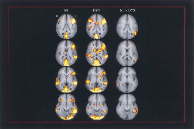
Figure 3. Correlation map between reading skill as measured by posterior aspect of the left superior temporal gyrus (row 1,
the Word Attack reading test (Woodcock and Johnson 1989) column 2). In contrast, during NWR in the NI readers, few
performed out of magnet and nonword rhyme (NWR) and
semantic category CAT tasks performed during functional mag- correlations are apparent with increasing age, and here age
netic resonance imaging for the group of 144 children. At each was negatively correlated with activation in the superior
voxel, a Pearson correlation coefficient (r) was calculated with frontal sulcus and middle frontal gyri regions bilaterally
age included as a covariate; a normal distribution test was used (row 1, column 1). To further examine this issue, we
(Hays 1988). Areas in yellow-red show a positive correlation of
in-magnet tasks with the out-of-magnet reading test (threshold,
p ⬍ .01). The four rows of images from top to bottom correspond
to Z ⫽ ⫹23, ⫹14, ⫹5 and –5 of Talairach atlas. Strong
correlation was found in the inferior aspect of the temporal
occipital region (fourth row), in the more superior aspect of the
temporal occipital regions (second and third rows), and in the
parietal regions (top row). CAT, semantic category.
occipitotemporal area in both the NWR and CAT and
bilateral parietotemporal regions in CAT. The more accu-
rate the performance both on word and on pseudoword
reading tasks, the greater the magnitude of the fMRI signal
in these left hemisphere regions during in-magnet reading.
These findings across the full cohort of children reveal a
continuum from very poor to skilled readers (Shaywitz et
al 1992b). To explore this brain– behavior relation further,
Figure 4. Correlation maps between age and activation for
we isolated the average center of mass of activation in the nonimpaired (NI) and dyslexic (DYS) readers. For each group of
left occipitotemporal area (Talairach coordinates x: ⫺42; readers, a Pearson correlation coefficient (r) was calculated at
y: ⫺42; z: ⫺5) and performed multiple regression analy- each voxel between age and activation for both nonword rhyme
ses, adjusting for the effects of age by covariance. The (NWR) and semantic category (CAT tasks). Areas in yellow-red
indicate a positive correlation between age and activation
correlation between left occipitotemporal activation dur-
(threshold, p ⬍ .05). Brain regions in blue-purple indicate a
ing NWR and reading performance on the Woodcock– negative correlation between age and activation (threshold, p ⬍
Johnson Word Attack was .33 (p ⬍ .001). For CAT .05). The slice location is at z ⫽ ⫹12 in Talairach space. During
(Talairach coordinates x: ⫺53; y: ⫺38; z: ⫺5), the NWR in DYS readers, increasing age was positively correlated
correlation was .26 (p ⬍ .002). with bilateral activation in the inferior frontal gyri, basal ganglia,
posterior cingulate gyri, cuneus, and middle occipital gyri, and in
In addition to these positive correlations of CAT acti-
the posterior aspect of the left superior temporal gyrus (row 1,
vation with reading performance, we also noted a signif- column 2). In contrast, during NWR in NI readers, increasing age
icant negative correlation with performance in the right was negatively correlated with activation in the superior frontal
occipitotemporal region (shown in blue, z ⫽ ⫺5). This sulcus and middle frontal gyri regions bilaterally (row 1, column
suggests that as the poorest readers attempted to read the 1). During the CAT task in DYS readers, increasing age was
positively correlated with activation in the right inferior frontal
real words in the CAT task, they were engaging an
gyrus (row 2, column 2). During the CAT task in NI readers,
ancillary system in the right hemisphere. Similarly, a increasing age was positively correlated with activation in the left
negative correlation with performance was evident in the inferior frontal gyrus and the right central sulcus region (row 2,
anterior cingulate region (blue, z ⫽ 23). Anterior cingulate column 1).fMRI in Children with Dyslexia BIOL PSYCHIATRY 107
2002;52:101–110
isolated the average center of mass of activation in the
contrast map (Figure 1, column 3) in the inferior frontal
gyrus in the left hemisphere and its homologue in the right
hemisphere, regions comprising a radius of 9 mm with
coordinates (x, y, z) ⫾ 38, ⫹23, and ⫹12. For each
subject, we determined the amount of activation in this
region of interest (ROI) by averaging the mean change in
t values between NWR and L in each voxel of the ROI.
The amount of activation in each ROI was then correlated
with age. Significant Pearson r values were observed in
the DYS children in both the left (r ⫽ .34, p ⬍ .01) and
Figure 5. Neural systems for reading. Converging evidence
right (r ⫽ .30, p ⬍ .05) inferior frontal gyri; in contrast, in
indicates three important systems in reading, all primarily in the
the NI readers, no significant correlations between age and left hemisphere. These include an anterior system and two
brain activations were observed in these frontal regions. posterior systems: 1) anterior system in the left inferior frontal
During CAT, significant positive correlations with age region; 2) dorsal parietotemporal system involving angular gy-
were noted in NI, but not in DYS, in the left inferior rus, supramarginal gyrus and posterior portions of the superior
temporal gyrus; 3) ventral occipitotemporal system involving
frontal gyrus and right precentral sulcus (Figure 4).
portions of the middle temporal gyrus and middle occipital gyrus.
For details, please see text.
Discussion differences. Specifically, we found that during the most
These results, acquired on an exceptionally large sample difficult and specific phonologic task (nonword rhyming)
representing a broad age range across childhood, indicate older dyslexic readers engaged the left and right inferior
significant differences in brain activation patterns during frontal gyrus, a finding consistent with results in adult
phonologic analysis in nonimpaired compared with dys- dyslexic readers which indicate an increase in activation in
lexic children. Specifically, nonimpaired children demon- frontal regions (Brunswick et al 1999; Shaywitz et al
strate significantly greater activation than do dyslexic 1998). It is reasonable to suggest that older dyslexic
children in left hemisphere sites including the inferior readers engage neural systems in frontal regions to com-
frontal, superior temporal, parietotemporal, and middle pensate for the disruption in posterior regions. During the
temporal–middle occipital gyri and right hemisphere sites CAT task, older dyslexic readers engage the right inferior
including the inferior frontal, superior temporal, cingulate, frontal gyrus, whereas older nonimpaired readers engage
and medial orbital gyri. These data converge with reports the left inferior frontal gyrus and right central sulcus
from many investigators using functional brain imaging region. The category task is considerably more complex
that show a failure of left hemisphere posterior brain than nonword rhyming, engaging not only phonology but
systems to function properly during reading (Brunswick et lexical and semantic processes as well. The older nonim-
al 1999; Helenius et al 1999; Horwitz et al 1998; Paulesu paired readers begin to engage the left frontal systems to
et al 2001; Pugh et al 2000; Rumsey et al 1992, 1997; perform this task; in contrast, older dyslexic readers fail to
Salmelin et al 1996; Shaywitz et al 1998; Simos et al engage left frontal systems but rather begin using an
2000) as well as during nonreading visual processing tasks ancillary system, the right inferior frontal gyrus.
(Demb et al 1998; Eden et al 1996). Our data indicate that Finally, the significant correlations between perfor-
dysfunction in left hemisphere posterior reading circuits is mance on a reading measure out of the magnet and brain
already present in dyslexic children and cannot be ascribed activations during fMRI tasks suggest that the left occipi-
simply to a lifetime of poor reading. totemporal region may be a critical component of a neural
In anterior regions the NI children demonstrated greater system for skilled reading. Accumulating evidence from
activation during NWR (Figure 1, column 3) than the DYS laboratories around the world indicates that there are a
children; this finding is consonant with two other reports number of interrelated neural systems used in reading, at
in children (Corina et al 2001; Georgiewa et al 1999) as least two in posterior brain regions, as well as distinct and
well as reports in adults (Gross-Glenn et al 1991; Paulesu related systems in anterior regions (Figure 5). As early as
et al 1996). At the same time, this finding contrasts with 1891, the French neurologist Dejerine (1891) suggested
what we (Shaywitz et al 1998) and others (Brunswick et al that a portion of the left posterior brain region is critical
1999) have reported in adults, where dyslexic readers for reading. Beginning with Dejerine, a large literature on
showed greater activation in the inferior frontal gyrus. acquired inability to read (alexia) describes neuroanatomic
Consideration of the correlation between age and brain lesions most prominently centered in the parietotemporal
activation provided an explanation that could resolve these area (including the angular gyrus, supramarginal gyrus and108 BIOL PSYCHIATRY B.A. Shaywitz et al
2002;52:101–110
posterior portions of the superior temporal gyrus) as a words, they remain slow, nonautomatic readers (Bruck
region pivotal in mapping the visual percept of the print 1992; Felton et al 1990). These data now suggest an
onto the phonologic structures of the language system explanation for these observed clinical findings. In dys-
(Damasio and Damasio 1983; Friedman et al 1993; Ge- lexic readers disruption of both dorsal and ventral left
schwind 1965). Another posterior brain region, this more hemisphere posterior reading systems underlies the failure
ventral in the occipitotemporal area, was also described by of skilled reading to develop, whereas a shift to ancillary
Dejerine (1892) as critical in reading. systems in left and right anterior regions and right poste-
More recently, Logan (Logan 1988, 1997) proposed two rior regions supports accurate, but not automatic, word
systems critical in the development of skilled, automatic reading.
processing, one involving word analysis (operating on This study was designed to minimize some of the
individual units of words such as phonemes, requiring problems encountered in previous studies, and thus we
attentional resources and processing relatively slowly) and examined a large sample, particularly for a functional
the second system operating on the whole word (word imaging study; we included a broad age range and studied
form; an obligatory system that does not require attention both boys and girls. We also recognize that there are
and processes very rapidly, on the order of 150 msec after limitations of our study, notably that inferences about
a word is read; Price et al 1996). Converging evidence development are based on the cross-sectional features of
from a number of lines of investigation indicate that the study design. A longitudinal study of the development
Logan’s word analysis system is localized within the of reading in children with dyslexia would be of particular
parietotemporal region, whereas the automatic, rapidly interest. Knowledge that dyslexic children and adults
responding system is localized within the occipitotemporal demonstrate a disruption within the neural systems en-
area, functioning as a visual word form area (Cohen et al gaged in accessing the sound structure of words under-
2000, in press; Dehaene et al 2001; Moore and Price scores the importance of evaluating phonologic skills in
1999). The visual word form area appears to respond the diagnosis of dyslexia and also of focusing on these
preferentially to rapidly presented stimuli (Price et al skills and their underlying neural systems as targets for
1996) and is engaged even when the word has not been informed phonologically based interventions for children
consciously perceived (Dehaene et al 2001). Still another and for adults.
reading-related neural circuit involves an anterior system Finally, we emphasize that fMRI studies of reading are
in the inferior frontal gyrus (Broca’s area), a region that very much investigational, and the data presented here
has long been associated with articulation and also serves represent group data. At the present time, fMRI has not
an important function in silent reading and naming (Fiez progressed to a point where it can be, nor should be, used
and Peterson 1998; Frackowiak et al 1997). in the diagnosis of individuals with dyslexia.
Recognition of these systems allows us to suggest an
explanation for the brain activation patterns observed in The authors thank Carmel Lepore, Hedy Sarofin, and Terry Hickey for
dyslexic children. We suppose that rather than the their invaluable help in imaging subjects. The authors thank also John
smoothly functioning and integrated reading systems ob- Holahan and Cheryl Lacadie for their help with data analysis. This work
was supported by grants from the National Institute of Child Health and
served in nonimpaired children, disruption of the posterior
Human Development (Grant Nos. PO1 HD 21888 and P50 HD25802).
reading systems results in dyslexic children attempting to
compensate by shifting to other, ancillary systems, for
example, anterior sites such as the inferior frontal gyrus References
and right hemisphere sites. The anterior sites, critical in
Bruck M (1992): Persistence of dyslexics’ phonological aware-
articulation (Brunswick et al 1999; Fiez and Peterson ness deficits. Dev Psychol 28:874 – 886.
1998; Frackowiak et al 1997; Pugh et al 1997), may help Brunswick N, McCrory E, Price CJ, Frith CD, Frith U (1999):
the child with dyslexia develop an awareness of the sound Explicit and implicit processing of words and pseudowords
structure of the word by forming the word with his lips, by adult developmental dyslexics: A search for Wernicke’s
tongue, and vocal apparatus and thus allow the child to Wortschatz. Brain 122:1901–1917.
read, albeit more slowly and less efficiently than if the fast Cohen L, Dehaene S, Naccache L, et al (2000): The visual word
occipitotemporal word identification system were func- form area: Spatial and temporal characterization of an initial
stage of reading in normal subjects and posterior split-brain
tioning. The right hemisphere sites may represent the patients. Brain 123:291–307.
engagement of brain regions that allow the poor reader to
Cohen L, Lehéricy S, Chochon F, Lemer C, Rivaud S, Dehaene
use other perceptual processes to compensate for his or her S (in press): Language-specific tuning of visual cortex?
poor phonologic skills. A number of studies of young Functional properties of the Visual Word Form Area. Brain.
adults with childhood histories of dyslexia indicate that Corina D, Richards T, Serafini S, Richards AL, Steury K, Abbott
although they may develop some accuracy in reading RD, et al (2001): fMRI auditory language differences be-fMRI in Children with Dyslexia BIOL PSYCHIATRY 109
2002;52:101–110
tween dyslexic and able reading children. Neuroreport 12: Horwitz B, Rumsey JM, Donohue BC (1998): Functional con-
1195–1201. nectivity of the angular gyrus in normal reading and dyslexia.
Damasio AR, Damasio H (1983): The anatomic basis of pure Proc Natl Acad Sci USA 95:8939 – 8944.
alexia. Neurology 33:1573–1583. Klingberg T, Hedehus M, Temple E, Salz T, Gabrieli JD,
Dehaene S, Naccache L, Cohen L, et al (2001): Cerebral Moseley ME, Poldrack RA (2000): Microstructure of tem-
mechanisms of word masking and unconscious repetition poro-parietal white matter as a basis for reading ability:
priming. Nat Neurosci 4:752–758. Evidence from diffusion tensor magnetic resonance imaging.
Neuron 25:493–500.
Dejerine J (1891): Sur un cas de cécité verbale avec agraphie,
suivi d’autopsie. C R Société du Biologie 43:197–201. Liberman IY, Shankweiler D (1991): Phonology and beginning
Dejerine J (1892): Contribution a l’etude anatomo-pathologique to read: A tutorial. In: Rieben L, Perfetti CA, editors.
et clinique des differentes varietes de cecite verbale. Memoi- Learning to Read: Basic Research and Its Implications.
res de la Société de Biologie 4:61–90. Hillsdale, NJ: Lawrence Erlbaum Associates.
Demb J, Boynton G, Heeger D (1998): Functional magnetic Logan G (1988): Toward an instance theory of automatization.
resonance imaging of early visual pathways in dyslexia. Psychol Rev 95:492–527.
J Neuroscience 18:6939 – 6951. Logan G (1997): Automaticity and reading: Perspectives from
Eden GF, VanMeter JW, Rumsey JM, Maisog JM, Woods RP, the instance theory of automatization. Read Writing Q Over-
Zeffiro TA (1996): Abnormal processing of visual motion in coming Learning Disabilities 13:123–146.
dyslexia revealed by functional brain imaging. Nature 382: Lyon G (1998): Statement of Dr. G. Reid Lyon, Committee on
66 – 69. Labor and Human Resources. US Congress. Washington, DC.
Felton RH, Naylor CE, Wood FB (1990): Neuropsychological Manly B (1997): Randomization, Bootstrap and Monte Carlo
profile of adult dyslexics. Brain Lang 39:485– 497. Methods in Biology. London: Chapman and Hall.
Fiez JA, Peterson SE (1998): Neuroimaging studies of word Moore C, Price C (1999): Three distinct ventral occipitotemporal
reading. Proc Natl Acad Sci USA 95:914 –921. regions for reading and object naming. Neuroimage 10:181–
Filipek P (1996): Structural variations in measures in the devel- 192.
opmental disorders. In: Thatcher R, Lyon G, Rumsey J, Morris RD, Stuebing KK, Fletcher JM, Shaywitz SE, Lyon GR,
Krasnegor N, editors. Developmental Neuroimaging: Map- Shankweiler DP (1998): Subtypes of reading disability: Vari-
ping the Development of Brain and Behavior. San Diego, CA: ability around a phonological core. J Educ Psychol 90:347–
Academic Press, pp 169 –186. 373.
Fletcher JM, Shaywitz SE, Shankweiler DP, Katz L, Liberman Paulesu E, Demonet J-F, Fazio F, McCrory E, Chanoine V,
IY, Stuebing KK (1994): Cognitive profiles of reading Brunswick N (2001): Dyslexia-cultural diversity and biolog-
disability: Comparisons of discrepancy and low achievement ical unity. Science 291:2165–2167.
definitions. J Educ Psychol 86:6 –23.
Paulesu E, Frith U, Snowling M, Gallagher A, Morton J,
Frackowiak R, Friston K, Frith C, Dolan R, Mazziotta (1997): Frackowiak RS, Frith CD(1996): Is developmental dyslexia a
Human Brain Function. New York: Academic Press. disconnection syndrome? Evidence from PET scanning.
Friedman RF, Ween JE, Albert ML (1993): Alexia. In: Heilman Brain 119:143–157.
KM, Valenstein E, editors. Clinical Neuropsychology, 3rd ed. Price C, Moore C, Frackowiak RSJ (1996): The effect of varying
New York: Oxford University Press, pp 37– 62. stimulus rate and duration on brain activity during reading.
Friston K, Frith C, Poline JB, Heather J, Frackowiak R (1996): Neuroimage 3:40 –52.
Spatial registration and moralization of images. Hum Brain Pugh K, Mencl EW, Shaywitz BA, et al (2000): The angular
Map 2:165–189. gyrus in developmental dyslexia: Task-specific differences in
Galaburda AM, Sherman GF, Rosen GD, Aboitiz F, Geschwind functional connectivity in posterior cortex. Psychol Sci 11:
N (1985): Developmental dyslexia: Four consecutive patients 51–56.
with cortical anomalies. Ann Neurol 18:222–233. Pugh KR, Shaywitz BA, Shaywitz SE, et al (1997): Predicting
Georgiewa P, Rzanny R, Hopf J, Knab R, Glauche V, Kaiser reading performance from neuroimaging profiles: The cere-
WA, Blanz B (1999): FMRI during word processing in bral basis of phonological effects in printed word identifica-
dyslexic and normal reading children. Neuroreport 10:3459 – tion. J Exp Psychol Hum Perception Performance 23:299 –
3465. 318.
Geschwind N (1965): Disconnection syndromes in animals and Report of the National Reading Panel (2000): Teaching Children
man. Brain 88:237–294. to Read: An Evidence Based Assessment of the Scientific
Gross-Glenn K, Duara R, Barker WW, Loewenstein D, Chang Research Literature on Reading and Its Implications for
JY, Yoshii F, et al (1991): Positron emission tomographic Reading Instruction, NIH Pub. No. 00 – 4754. Washington,
studies during serial word-reading by normal and dyslexic DC: U.S. Department of Health and Human Services, Public
adults. J Clin Exp Neuropsychol 13:531–544. Health Service, National Institutes of Health, National Insti-
Hays WL (1988): Statistics. Orlando, FL: Holt, Rinehart and tute of Child Health and Human Development.
Winston. Rumsey JM, Andreason P, Zametkin AJ, Aquino T, King AC,
Helenius P, Tarkiainen A, Cornelissen P, Hansen PC, Salmelin R Hamburger SD, (1992): Failure to activate the left temporopa-
(1999): Dissociation of normal feature analysis and deficient rietal cortex in dyslexia. Arch Neurol 49:527–534.
processing of letter-strings in dyslexic adults. Cereb Cortex Rumsey JM, Nace K, Donohue B, Wise D, Maisog JM, Andrea-
4:476 – 483. son P (1997): A positron emission tomographic study of110 BIOL PSYCHIATRY B.A. Shaywitz et al
2002;52:101–110
impaired word recognition and phonological processing in dyslexic children: A magnetic source imaging approach.
dyslexic men. Arch Neurol 54:562–573. Cereb Cortex 10:809 – 816.
Salmelin R, Service E, Kiesila P, Uutela K, Salonen O (1996): Skudlarski P, Constable R, Gore JC (1999): ROC analysis of
Impaired visual word processing in dyslexia revealed with statistical methods used in functional MRI: Individual sub-
magnetoencephalography. Ann Neurol 40:157–162. jects. Neuroimage 9:311–329.
Shaywitz BA, Fletcher JM, Holahan JM, Shaywitz SE (1992a): Stanovich KE, Siegel LS (1994): Phenotypic performance profile
Discrepancy compared with low achievement definitions of of children with reading disabilities: A regression-based test
reading disability: Results from the Connecticut Longitudinal of the phonological-core variable-difference model. J Educ
Study. J Learning Disabilities 25:639 – 648. Psychol 86:24 –53.
Shaywitz S (1998): Current concepts: Dyslexia. N Engl J Med
338:307–312. Talairach J, Tournoux P (1988): Coplanar Stereotaxic Atlas of
the Human Brain. Three-Dimensional Proportional System:
Shaywitz SE (1996): Dyslexia. Sci Am 275:98 –104. An Approach to Cerebral Imaging. New York: Thieme
Shaywitz SE, Escobar MD, Shaywitz BA, Fletcher JM, Makuch Medical.
R (1992b): Evidence that dyslexia may represent the lower
Wagner R, Torgesen J (1987): The nature of phonological
tail of a normal distribution of reading ability. N Engl J Med
326:145–150. processes and its causal role in the acquisition of reading
skills. Psychol Bull 101:192–212.
Shaywitz SE, Shaywitz BA, Pugh KR, Fulbright RK, Constable
RT, Mencl WE, et al (1998): Functional disruption in the Wechsler D (1991): Wechsler Intelligence Scale for Children,
organization of the brain for reading in dyslexia. Proc Natl 3rd ed. San Antonio, TX: Psychological Corporation.
Acad Sci USA 95:2636 –2641. Woodcock RW, Johnson MB (1989): Woodcock–Johnson Psy-
Simos P, Breier J, Fletcher J, Bergman E, Papanicolaou A cho-Educational Battery–Revised. Allen, TX: Developmental
(2000): Cerebral mechanisms involved in word reading in Learning Materials.You can also read