RNAi: Double-Stranded RNA Directs the ATP-Dependent Cleavage of mRNA at 21 to 23 Nucleotide Intervals
←
→
Page content transcription
If your browser does not render page correctly, please read the page content below
Cell, Vol. 101, 25–33, March 31, 2000, Copyright 2000 by Cell Press
RNAi: Double-Stranded RNA Directs the
ATP-Dependent Cleavage of mRNA
at 21 to 23 Nucleotide Intervals
Phillip D. Zamore,*# Thomas Tuschl,†# 1999), hydra (Lohmann et al., 1999), zebrafish (Wargelius
Phillip A. Sharp,‡§ and David P. Bartel§k et al., 1999), and mice (Wianny and Zernicka-Goetz,
* Department of Biochemistry and Molecular Biology 2000), and appears to be related to gene silencing phe-
University of Massachusetts Medical School nomena in plants (“cosuppression”; Vaucheret et al.,
Worcester, Massachusetts 01655 1998; Waterhouse et al., 1998, 1999; Baulcombe, 1999)
† Department of Cellular Biochemistry and the fungus Neurospora (“quelling”; Cogoni et al.,
Max-Planck-Institute for Biophysical Chemistry 1996; Cogoni and Macino, 1999a, 1999b).
Am Faßberg 11 RNAi occurs posttranscriptionally and involves mRNA
D-37077 Göttingen degradation (Montgomery et al., 1998; Ngo et al., 1998).
Germany In addition to providing a powerful tool for creating gene-
‡ Center for Cancer Research and specific phenocopies of loss-of-function mutations,
§ Department of Biology RNAi may also play an important biological role in pro-
Massachusetts Institute of Technology tecting the genome against instability caused by the
Cambridge, Massachusetts 02139 accumulation of transposons and repetitive sequences
k The Whitehead Institute for Biomedical Research (Ketting et al., 1999; Tabara et al., 1999). In C. elegans,
9 Cambridge Center dsRNA blocks specific gene expression even when ex-
Cambridge, Massachusetts 02142 pressed by bacteria fed to the worms (Timmons and Fire,
1998). RNAi in animals may also represent an ancient
antiviral response, just as posttranscriptional gene si-
Summary lencing appears to protect plants from viral infection
(Baulcombe, 1999; Grant, 1999; Ratcliff et al., 1999). The
Double-stranded RNA (dsRNA) directs the sequence- breadth of RNAi-like processes suggests that RNAi may
specific degradation of mRNA through a process encompass gene silencing phenomena, including cellu-
known as RNA interference (RNAi). Using a recently lar strategies for gene regulation, well beyond the initial
developed Drosophila in vitro system, we examined observation that dsRNA can produce RNAi.
the molecular mechanism underlying RNAi. We find Genetic screens in both C. elegans and Neurospora
that RNAi is ATP dependent yet uncoupled from mRNA have identified genes required for RNAi (Cogoni and
translation. During the RNAi reaction, both strands of Macino, 1997; Tabara et al., 1999). Mutations in a subset
the dsRNA are processed to RNA segments 21–23 of these genes, including rde-2, rde-3, mut-2, and mut-7,
nucleotides in length. Processing of the dsRNA to the permit the mobilization of transposons in the worm
small RNA fragments does not require the targeted germline (Ketting et al., 1999; Tabara et al., 1999; Grishok
mRNA. The mRNA is cleaved only within the region of et al., 2000). A second class of mutants, including the
identity with the dsRNA. Cleavage occurs at sites rde-1 and rde-4 loci, are defective for RNAi but show
21–23 nucleotides apart, the same interval observed for no other phenotypic abnormalities (Tabara et al., 1999).
the dsRNA itself, suggesting that the 21–23 nucleotide The rde-1 and rde-4 genes are required for the initiation
fragments from the dsRNA are guiding mRNA cleavage. of heritable RNAi, a phenomenon in which RNAi estab-
lished by injection of dsRNA in a worm leads to heritable
gene silencing in the F2 generation and beyond (Grishok
et al., 2000). In contrast, rde-2 and mut-7 are not required
Introduction for the initiation of heritable interference but are required
downstream in the tissue where the interference occurs.
The term RNA interference, or “RNAi,” was initially Mello and colleagues have proposed that rde-1 and
coined by Fire and coworkers (Fire et al., 1998) to de- rde-4 respond to dsRNA by producing a secondary ex-
scribe the observation that double-stranded RNA (dsRNA) tragenic agent that is used by the downstream genes
can block gene expression when it is introduced into rde-2 and mut-7 to target specific mRNAs for posttran-
worms (for reviews see Fire, 1999; Hunter, 2000; Hunter, scriptional gene silencing (Grishok et al., 2000). In this
1999; Montgomery and Fire, 1998; Sharp, 1999; Wagner view, rde-1 and rde-4 act as initiators of RNAi, whereas
and Sun, 1998). Their discovery built upon the previous, rde-2 and mut-7 are effectors. These authors propose
puzzling observation that sense and antisense RNA that other stimuli that lead to gene silencing, such as
(asRNA) were equally effective in suppressing specific the accumulation of transposons or repetitive DNA in
gene expression (Guo and Kemphues, 1995), a paradox the genome or the introduction of a transgene, are inter-
resolved by the finding that small amounts of dsRNA preted by a separate set of initiator genes that produce
contaminate sense and antisense preparations (Fire et the same secondary extragenic agent.
al., 1998). RNAi has since been discovered in a wide In Neurospora, the qde-3 gene, which is required for
variety of animals, including flies (Kennerdell and Carthew, quelling (a form of posttranscriptional silencing in which
1998; Misquitta and Paterson, 1999), trypanosomes (Ngo an endogenous gene is silenced by the introduction of
et al., 1998), planaria (Sánchez-Alvarado and Newmark, a transgenic copy of the gene), may be an example of
an initiator gene that responds to the presence of a
# To whom correspondence should be addressed (e-mail: phillip. transgene (Cogoni and Macino, 1999b). qde-3 is a mem-
zamore@umassmed.edu [P. D. Z.], ttuschl@mpibpc.gwdg.de [T. T.]). ber of the RecQ DNA helicase family, which includes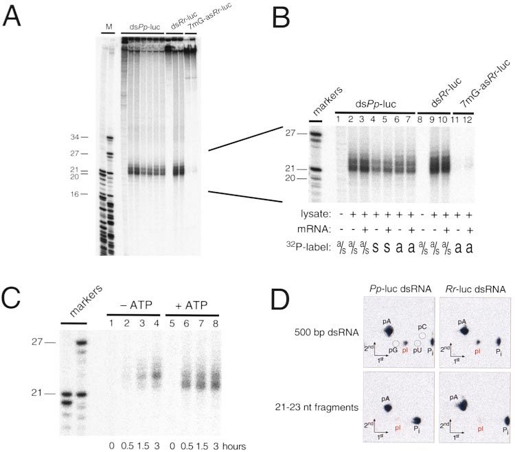
Cell
26
the human genes for Bloom’s syndrome and Werner’s
syndrome.
One candidate for the secondary extragenic agent
itself is the 25 nucleotide–long RNAs associated with
posttranscriptional gene silencing in plants (Hamilton
and Baulcombe, 1999). These RNAs, which correspond
to both the sense and antisense strands of the silenced
gene, are only detected in plants undergoing silencing.
The level of expression of these short RNAs also corre-
lates with the extent of gene silencing. It remains to be
shown if the 25 nt RNAs are the actual agents or merely
the products of gene silencing.
Two other genes implicated in posttranscriptional
gene silencing, qde-1 in Neurospora (Cogoni and Ma-
cino, 1999a) and ego-1 in C. elegans (Smardon et al.,
2000), are homologous to a tomato protein that displays
RNA-directed RNA-polymerase activity in vitro (Schiebel
et al., 1993a, 1993b, 1998). RNA-directed RNA polymer-
ases have been implicated in the initial formation of
the silencing agent or in the amplification of dsRNA.
Amplification of injected dsRNA by an endogenous
RNA-directed RNA polymerase would help explain how
a very small number of dsRNA molecules can inactivate
a much larger population of mRNAs and how the dsRNA
can apparently persist in the animal for many days and
even into subsequent generations. ego-1 mutants are
Figure 1. RNAi Requires ATP
defective for RNAi for maternally, but not zygotically,
expressed mRNAs. Interestingly, ego-1 is also required (A) Denaturing agarose-gel analysis of 5⬘-32P-radiolabeled Rr-luc
mRNA incubated for the times indicated in an in vitro RNAi reaction
for germline development in C. elegans (Qiao et al., with or without ATP, creatine phosphate (CP), or creatine kinase
1995). (CK), as indicated below each panel.
Biochemical analysis of RNAi has become possible (B) Quantitation of the data in (A). Circles, ⫹ATP, ⫹CP, ⫹CK;
with the development of an in vitro Drosophila embryo squares, -ATP, ⫹CP, ⫹CK; triangles, -ATP, -CP, ⫹CK; inverted trian-
lysate that recapitulates dsRNA-dependent silencing of gles, -ATP, ⫹CP, -CK.
gene expression (Tuschl et al., 1999). In the in vitro
system, dsRNA—but not sense or asRNA—targets a luciferase protein from the targeted mRNA. Thus, these
corresponding mRNA for degradation yet does not af- RNAi reactions contained an ATP-regenerating system,
fect the stability of an unrelated control mRNA. Further- needed for the efficient translation of the mRNA. To test
more, preincubation of the dsRNA in the lysate potenti- if ATP was, in fact, required for RNAi, the lysates were
ates its activity for target mRNA degradation, suggesting depleted for ATP by treatment with hexokinase and glu-
that the dsRNA must be converted to an active form by cose, which converts ATP to ADP, and RNAi was moni-
binding proteins in the extract or by covalent modifica- tored directly by following the fate of 32P-radiolabeled
tion (Tuschl et al., 1999). Renilla reniformis luciferase (Rr-luc) mRNA (Figure 1).
Here, we use the in vitro system to analyze the require- Treatment with hexokinase and glucose reduced the
ments of RNAi and to determine the fate of the dsRNA endogenous ATP level in the lysate from 250 M to
and the mRNA. RNAi in vitro requires ATP but does not below 10 M (data not shown). ATP regeneration re-
require either mRNA translation or recognition of the quired both exogenous creatine phosphate and creatine
7-methyl-guanosine cap of the targeted mRNA. The kinase, which acts to transfer a high-energy phosphate
dsRNA but not single-stranded RNA is processed in from creatine phosphate to ADP. When ATP-depleted
vitro to a population of 21–23 nt species. Deamination extracts were supplemented with either creatine phos-
of adenosines within the dsRNA does not appear to be phate or creatine kinase separately, no RNAi was ob-
required for formation of the 21–23 nt RNAs. Further- served. Therefore, RNAi requires ATP in vitro. When ATP,
more, we find that the mRNA is cleaved only in the region creatine phosphate, and creatine kinase were all added
corresponding to the sequence of the dsRNA and that together to reactions containing the ATP-depleted ly-
the mRNA is cleaved at 21–23 nt intervals, strongly sug- sate, dsRNA-dependent degradation of the Rr-luc mRNA
gesting that the 21–23 nt fragments from the dsRNA are was restored (Figure 1). The addition of exogenous ATP
targeting the cleavage of the mRNA. was not required for efficient RNAi in the depleted lysate,
provided that both creatine phosphate and creatine ki-
Results and Discussion nase were present, demonstrating that the endogenous
concentration (250 M) of adenosine nucleotide is suffi-
RNAi Requires ATP cient to support RNAi. RNAi with a Photinus pyralis lucif-
Drosophila embryo lysates faithfully recapitulate RNAi erase (Pp-luc) mRNA was also ATP dependent (data not
(Tuschl et al., 1999). Previously, dsRNA-mediated gene shown).
silencing was monitored by measuring the synthesis of The stability of the Rr-luc mRNA in the absence of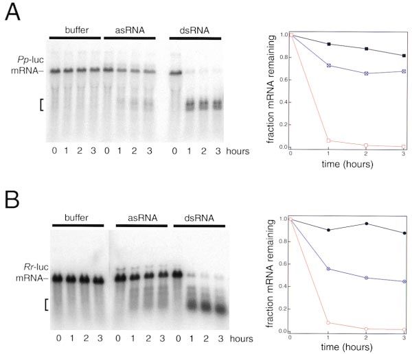
dsRNA Directs Cleavage of mRNA at 21–23 nt Intervals
27

                                                                             Figure 2. RNAi Does Not Require mRNA
                                                                             Translation
                                                                             (A) Protein synthesis, as reflected by lucifer-
                                                                             ase activity produced after incubation of Rr-
                                                                             luc mRNA in the in vitro RNAi reaction for 1 hr, in
                                                                             the presence of the protein synthesis inhibitors
                                                                             anisomycin, cycloheximide, or chlorampheni-
                                                                             col, relative to a reaction without any inhibitor.
                                                                             (B) Denaturing agarose-gel analysis of 5⬘-32P-
                                                                             radiolabeled Pp-luc mRNA after incubation
                                                                             for the indicated times in a standard RNAi
                                                                             reaction with and without protein synthesis
                                                                             inhibitors. The arrowhead indicates the posi-
                                                                             tion of full-length mRNA in the gel, and the
                                                                             bracket marks the position of stable, 5⬘ cleav-
                                                                             age products.
                                                                             (C) Translation of 7-methyl-guanosine- and
                                                                             adenosine-capped Pp-luc mRNAs (circles
                                                                             and squares, respectively) in the RNAi reac-
                                                                             tion in the absence of dsRNA, as measured
                                                                             by luciferase activity produced in a 1 hr incu-
                                                                             bation.
                                                                             (D) Incubation in an RNAi reaction of uni-
                                                                             formly 32P-radiolabeled 7-methyl-guanosine-
                                                                             capped Pp-luc mRNA (circles) and adeno-
                                                                             sine-capped Pp-luc mRNA (squares), in the
                                                                             presence (open symbols) and absence (filled
                                                                             symbols) of 505 bp Pp-luc dsRNA.

Rr-dsRNA was reduced in ATP-depleted lysates relative            Translational initiation is an ATP-dependent process
to that observed when the energy regenerating system          that involves recognition of the 7-methyl guanosine cap
was included, but decay of the mRNA under these condi-        of the mRNA (Merrick and Hershey, 1996; Kozak, 1999).
tions did not display the rapid decay kinetics character-     The Drosophila lysate used to support RNAi in vitro also
istic of RNAi in vitro, nor did it generate the stable mRNA   recapitulates the cap dependence of translation: Pp-luc
cleavage products characteristic of dsRNA-directed            mRNA with a 7-methyl-guanosine cap was translated
RNAi (data not shown). These experiments do not estab-        greater than 10-fold more efficiently than was the same
lish if the ATP requirement for RNAi is direct, implicating   mRNA with an A(5⬘)ppp(5⬘)G cap (Figure 2C). Both RNAs
ATP in one or more steps in the RNAi mechanism, or            were equally stable in the Drosophila lysate, showing
indirect, reflecting a role for ATP in maintaining high       that this difference in efficiency cannot be merely ex-
concentrations of another nucleoside triphosphate in          plained by more rapid decay of the mRNA with an adeno-
the lysate.                                                   sine cap (also see Gebauer et al., 1999). Although the
                                                              translational machinery can discriminate between Pp-
Translation Is Not Required for RNAi In Vitro                 luc mRNAs with 7-methyl-guanosine and adenosine
The requirement for ATP suggested that RNAi might be          caps, the two mRNAs were equally susceptible to RNAi
coupled to mRNA translation, a highly energy-depen-           in the presence of Pp-dsRNA (Figure 2D). These results
dent process. To test this possibility, various inhibitors    suggest that steps in cap recognition are not involved
of protein synthesis were added to the reaction. We           in RNAi.
tested the eukaryotic translation inhibitors anisomycin,
an inhibitor of initial peptide bond formation, cyclohexi-    dsRNA Is Processed to 21–23 Nucleotide Species
mide, an inhibitor of peptide chain elongation, and puro-     RNAs 25 nt in length are generated from both the sense
mycin, a tRNA mimic that causes premature termination         and antisense strands of genes undergoing posttran-
of translation (Cundliffe, 1981). Each of these inhibitors    scriptional gene silencing in plants (Hamilton and Baul-
reduced protein synthesis in the Drosophila lysate by         combe, 1999). We find that dsRNA is also processed to
more than 1,900-fold (Figure 2A; data not shown). In          small RNA fragments (Figures 3A and 3B). When incu-
contrast, chloramphenicol, an inhibitor of Drosophila mi-     bated in lysate, approximately 15% of the input radioac-
tochondrial protein synthesis (Page and Orr-Weaver,           tivity of both the 501 bp Rr-dsRNA and the 505 bp Pp-
1997), had no effect on translation in the lysates (Figure    dsRNA appeared in 21 to 23 nt RNA fragments. Because
2A). Despite the presence of anisomycin, cycloheximide,       the dsRNAs are more than 500 bp in length, the 15%
or chloramphenicol, RNAi proceeded at normal effi-            yield of fragments implies that multiple 21–23 nt RNAs
ciency (Figure 2B). Puromycin also did not perturb effi-      are produced from each full-length dsRNA molecule. No
cient RNAi (data not shown). Thus, protein synthesis is       other stable products were detected. The small RNA
not required for RNAi in vitro.                               species were produced from dsRNAs in which both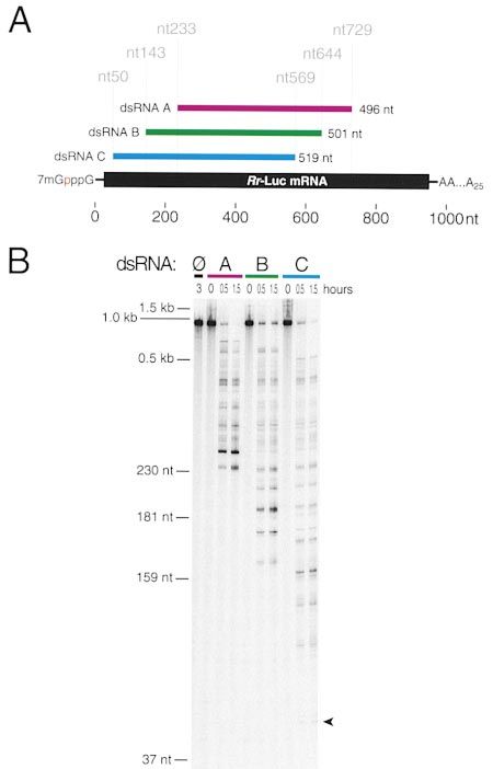
Cell
28

Figure 3. 21–23 nt RNA Fragments Are Produced upon Incubation of dsRNA in Drosophila Embryo Lysate
(A) Denaturing acrylamide-gel analysis of the products formed in a 2 hr incubation of uniformly 32P-radiolabeled dsRNAs or capped asRNA
in lysate under standard RNAi conditions, in the presence or absence of target mRNAs.
(B) An enlargement of the portion of the gel in (A) corresponding to 17 to 27 nt. For Pp-dsRNA, the sense (lanes 4 and 5) or the antisense
(lanes 6 and 7) or both strands (lanes 1, 2, and 3) were labeled. For Rr-luc dsRNA, both strands were radioactive (lanes 8, 9, and 10).
(C) An enlargement of the 17 to 27 nt region of a gel showing the products formed upon incubation of uniformly 32P-radiolabeled dsRNAs in
lysate without and with ATP.
(D) Adenosine deamination in full-length dsRNA and the 21–23 nt RNA species assessed by two-dimensional thin-layer chromatography.
Circles correspond to positions of unlabeled 5⬘-nucleotide monophosphate standards visualized under UV light. Inorganic phosphate (Pi) was
produced by the degradation of mononucleotides by phosphatases that contaminate commercially available nuclease P1 (Auxilien et al.,
1996).

strands were uniformly 32P-radiolabeled (Figure 3B,                     converted to the 21–23 nt product when it was incubated
lanes 2, 3, 9, and 10). Formation of the 21–23 nt RNAs                  with 5 nM nonradioactive 505 bp Pp-dsRNA (data not
from the dsRNA did not require the presence of the                      shown). Second, in the absence of mRNA, a 501 nt
corresponding mRNA (Figure 3B, compare lane 2 with                      7-methyl-guanosine-capped Rr-asRNA produced only
lane 3 and lane 9 with lane 10), demonstrating that the                 a barely detectable amount of 21–23 nt RNA (Figure 3B,
small RNA species is generated by processing of the                     lane 11; capped single-stranded RNAs are as stable in
dsRNA, rather than as a product of dsRNA-targeted                       the lysate as dsRNA [Tuschl et al., 1999]), probably due
mRNA degradation. We note that 22 nucleotides corre-                    to a small amount of dsRNA contaminating the antisense
sponds to two turns of an A-form RNA–RNA helix.                         preparation. However, when Rr-luc mRNA was included
   When dsRNAs radiolabeled within either the sense or                  in the reaction with the 32P-radiolabeled, capped Rr-
the antisense strand were incubated with lysate in a                    asRNA, a small amount of 21–23 nt product was gener-
standard RNAi reaction, 21–23 nt RNAs were generated                    ated, corresponding to 4% of the amount of 21–23 nt
with comparable efficiency (Figure 3B, compare lanes                    RNA produced from an equimolar amount of Rr-dsRNA.
4 and 6). These data support the idea that the 21–23 nt                 This result is unlikely to reflect the presence of contami-
RNAs are generated by symmetric processing of the                       nating dsRNA in the Rr-asRNA preparation, since signifi-
dsRNA. A variety of data support the idea that the 21–23                cantly more product was generated from the asRNA in
nt RNA is efficiently generated only from dsRNA and is                  the presence of the Rr-luc mRNA than in the absence
not the consequence of an interaction between single-                   (compare lanes 12 and 11). Instead, the data suggest
stranded RNA and the dsRNA. First, a 32P-radiolabeled                   that asRNA can interact with the complementary mRNA
505 nt Pp-luc sense RNA or asRNA was not efficiently                    sequences to form dsRNA in the reaction and that the
dsRNA Directs Cleavage of mRNA at 21–23 nt Intervals
29

                                                                                Figure 4. asRNA Causes a Small Amount of
                                                                                RNAi In Vitro
                                                                                (A) Denaturing agarose-gel analysis of Pp-luc
                                                                                mRNA incubated in a standard RNAi reaction
                                                                                with buffer, 505 nt Pp-asRNA, or 505 bp Pp-
                                                                                dsRNA for the times indicated.
                                                                                (B) The same analysis for the Rr-luc mRNA.
                                                                                Quantitation of the gel data in both (A) and
                                                                                (B) is given to the right of each panel. Buffer,
                                                                                black symbols; asRNA, blue symbols; dsRNA,
                                                                                red symbols.

resulting dsRNA is subsequently processed to the small        cleaved to mononucleotides with nuclease P1, and ana-
RNA species. Rr-asRNA can support a low level of bona         lyzed by two-dimensional thin-layer chromatography.
fide RNAi in vitro (see below), consistent with this expla-      A significant fraction of the adenosines in the full-
nation.                                                       length dsRNA were converted to inosine after 2 hr (3.1%
   We next asked if production of the 21–23 nt RNAs           and 5.6% conversion for Pp-luc and Rr-luc dsRNAs,
from dsRNA required ATP (Figure 3C). When the 505 bp          respectively). In contrast, only 0.4% (Pp-dsRNA) or 0.7%
Pp-dsRNA was incubated in a lysate depleted for ATP           (Rr-dsRNA) of the adenosines in the 21–23 nt species
by treatment with hexokinase and glucose, 21–23 nt            were deaminated. These data imply that fewer than 1
RNA was produced (lanes 1–4, “⫺ATP”), albeit six times        in 27 molecules of the 21–23 nt RNA species contain an
slower than when ATP was regenerated in the depleted          inosine. Therefore, it is unlikely that dsRNA-dependent
lysate by the inclusion of creatine kinase and creatine       adenosine deamination within the 21–23 nt species is
phosphate (lanes 5–8, “⫹ ATP”). Therefore, ATP may            required for its production.
not be required for production of the 21–23 nt RNA
species but may instead simply enhance its formation.         asRNA Generates a Small Amount of RNAi In Vitro
Alternatively, ATP may be required for processing of the      When mRNA was 32P-radiolabeled within the 5⬘-7-
dsRNA, but at a concentration less than that remaining        methyl-guanosine cap, stable 5⬘ decay products accu-
after hexokinase treatment. We do not yet understand          mulated during the RNAi reaction (see, for example,
the molecular basis for the slower mobility of the small      Figures 1A and 2B). Such stable 5⬘ decay products were
RNA fragments generated in the ATP-depleted lysate.           observed for both the Pp-luc and Rr-luc mRNAs when
   Wagner and Sun (1998) and Sharp (1999) have specu-         they were incubated with their cognate dsRNAs (indi-
lated that the requirement for dsRNA in gene silencing        cated by the brackets in Figures 4A and 4B). Previously,
by RNAi reflects the involvement of a dsRNA-specific          we reported that efficient RNAi does not occur when
adenosine deaminase in the process. dsRNA adenosine           asRNA is used in place of dsRNA (Tuschl et al., 1999).
deaminases unwind dsRNA by converting adenosine to            Nevertheless, mRNA was measurably less stable when
inosine, which does not base pair with uracil. dsRNA          incubated with asRNA than with buffer (Figures 4A and
adenosine deaminases function in the posttranscrip-           4B). This was particularly evident for the Rr-luc mRNA:
tional editing of mRNA (reviewed by Bass, 1997). To test      approximately 90% of the RNA remained intact after a
for the involvement of dsRNA adenosine deaminase in           3 hr incubation in lysate, but only 50% when asRNA was
RNAi, we examined the degree of conversion of adeno-          added. Less than 5% remained when dsRNA was added.
sine to inosine in the 501 bp Rr-luc and 505 bp Pp-luc        Interestingly, the decrease in mRNA stability caused by
dsRNAs after incubation with Drosophila embryo lysate         asRNA was accompanied by the formation of a small
in a standard in vitro RNAi reaction (Figure 3D). We also     amount of the stable 5⬘ decay products characteristic
determined the degree of adenosine deamination in the         of the RNAi reaction with dsRNA. This finding parallels
21–23 nt species. The full-length dsRNA radiolabeled          the observation that a small amount of 21–23 nt product
with [32P]-adenosine was incubated in the lysate, and         formed from the asRNA when it was incubated with the
both the full-length dsRNA and the 21–23 nt RNA prod-         mRNA (see above) and lends strength to the idea that
ucts were purified from a denaturing acrylamide gel,          asRNA can enter the RNAi pathway, albeit inefficiently.
Cell
30
ⵑ750 nt; dsRNA A spans nucleotides 233 to 729 of the
Rr-luc mRNA. Incubation of the mRNA with dsRNA B
produced mRNA 5⬘ cleavage products ranging in length
from 150 to ⵑ600 nt; dsRNA B spans nucleotides 143
to 644 of the mRNA. Finally, dsRNA C produced mRNA
cleavage products from 66 to ⵑ500 nt in length. This
dsRNA spans nucleotides 50 to 569 of the Rr-luc mRNA.
Therefore, the dsRNA not only provides specificity for
the RNAi reaction, selecting which mRNA from the total
cellular mRNA pool will be degraded, but also deter-
mines the boundaries of cleavage along the mRNA se-
quence.
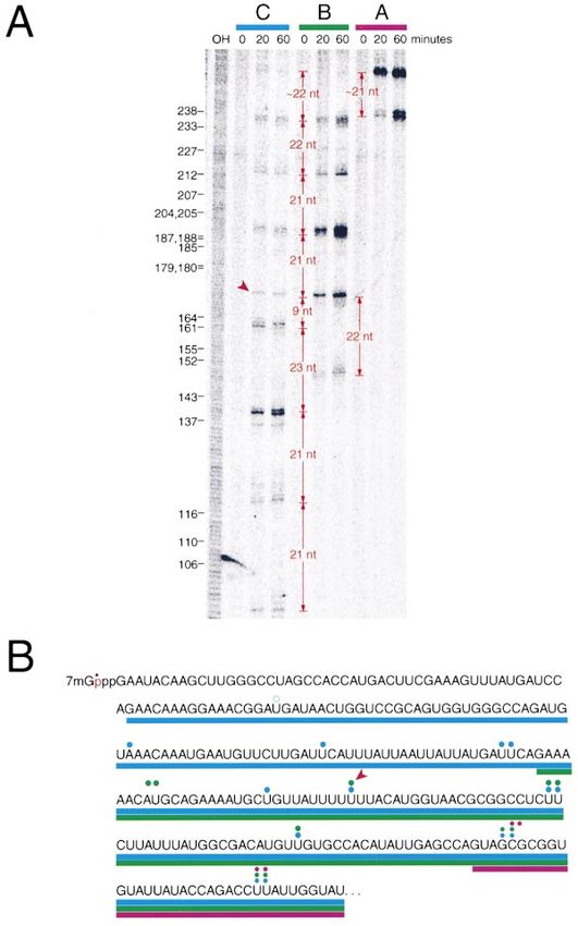
The mRNA Is Cleaved at 21–23 Nucleotide Intervals
To gain further insight into the mechanism of RNAi, we
mapped the positions of several mRNA cleavage sites
for each of the three dsRNAs (Figure 6). Remarkably,
most of the cleavages occurred at 21–23 nt intervals
(Figure 6A). This spacing is especially striking in light of
our observation that the dsRNA is processed to a 21–23
nt RNA species and the finding of Hamilton and Baul-
combe that a 25 nt RNA correlates with posttranscrip-
tional gene silencing in plants (Hamilton and Baulcombe,
1999). Of the 16 cleavage sites we mapped (two for
dsRNA A, five for dsRNA B, and nine for dsRNA C), all
but two reflect the 21–23 nt interval. One of the two
exceptional cleavages was a weak cleavage site pro-
duced by dsRNA C (indicated by an arrowhead in Figure
5B and an open blue circle in Figure 6B). This cleavage
occurred 32 nt 5⬘ to the next cleavage site. The other
exception is particularly intriguing. After four cleavages
spaced 21–23 nt apart, dsRNA C caused cleavage of
the mRNA just 9 nt 3⬘ to the previous cleavage site
Figure 5. The dsRNA Determines the Boundaries of the Cleavage (Figures 6A and 6B, red arrowhead). This cleavage oc-
Products curred in a run of seven uracil residues and appears to
(A) Schematic of the positions of the three dsRNAs, A, B, and C, “reset” the ruler for cleavage; the next cleavage site was
relative to the Rr-luc mRNA. 21–23 nt 3⬘ to the exceptional site. The three subsequent
(B) Denaturing acrylamide-gel analysis of the stable, 5⬘ cleavage cleavage sites that we mapped were also spaced 21–23
products produced after incubation of the Rr-luc mRNA for the
nt apart. Curiously, of the sixteen cleavage sites mapped
indicated times with each of the three dsRNAs, A, B, and C, or
with buffer (zero with strikethrough). The positions of RNA markers for the three different dsRNAs, fourteen occur at uracil
radiolabeled within their 5⬘ cap is shown at left. The arrowhead residues. We do not yet understand the significance
denotes a faint cleavage site that is indicated with an open blue of this finding, but it suggests that mRNA cleavage is
circle in Figure 6B. determined by a process that measures 21–23 nt inter-
vals and that has a sequence preference for cleavage
at uracil. In preliminary experiments, the 21–23 nt RNA
mRNA Cleavage Sites Are Determined species produced by incubation of ⵑ500 bp dsRNA in
by the Sequence of the dsRNA the lysate caused sequence-specific interference in vitro
The sites of mRNA cleavage were examined using three when isolated from an acrylamide gel and added to a
different dsRNAs, “A,” “B,” and “C,” displaced along new RNAi reaction in place of the full-length dsRNA (our
the Rr-luc sequence by approximately 100 nt. The posi- unpublished data).
tions of these relative to the Rr-luc mRNA sequence
are shown (Figure 5A). Each of the three dsRNAs was A Model for dsRNA-Directed mRNA Cleavage
incubated in a standard RNAi reaction with Rr-luc mRNA Our biochemical data, together with recent genetic ex-
32
P-radiolabeled within the 5⬘ cap (Figure 5B). In the periments in C. elegans and Neurospora (Cogoni and
absence of dsRNA, no stable 5⬘ cleavage products were Macino, 1999a; Ketting et al., 1999; Tabara et al., 1999;
detected for the mRNA, even after 3 hr of incubation in Grishok et al., 2000), suggest a model for how dsRNA
lysate. In contrast, after a 20 min incubation, each of targets mRNA for destruction (Figure 7). In this model,
the three dsRNAs produced a ladder of bands corre- the dsRNA is first cleaved to 21 to 23 nt long fragments
sponding to a set of mRNA cleavage products charac- in a process likely to involve genes such as the C. ele-
teristic for that particular dsRNA. For each dsRNA, the gans loci rde-1 and rde-4. The resulting fragments, prob-
stable, 5⬘ mRNA cleavage products were restricted to ably as short asRNAs bound by RNAi-specific proteins,
the region of the Rr-luc mRNA that corresponded to the would then pair with the mRNA and recruit a nuclease
dsRNA (Figures 5B and 6). For dsRNA A, the lengths of that cleaves the mRNA. Alternatively, strand exchange
the 5⬘ cleavage products ranged from 236 to just under could occur in a protein–RNA complex that transientlydsRNA Directs Cleavage of mRNA at 21–23 nt Intervals
31
Figure 6. The mRNA Is cleaved in 21–23 nt
Intervals
(A) High-resolution denaturing acrylamide-
gel analysis of a subset of the 5⬘ cleavage
products described in Figure 5B. The posi-
tions of some of the partial T1 digestion prod-
ucts of Rr-luc mRNA are indicated at left.
“OH” marks the lane in which a partial base-
hydrolysis ladder was loaded.
(B) The cleavage sites in (A) mapped onto the
first 267 nt of the Rr-luc mRNA. The blue bar
below the sequence indicates the position of
dsRNA C, and blue circles indicate the posi-
tion of cleavage sites caused by this dsRNA.
The green bar denotes the position of dsRNA
B, and green circles, the cleavage sites. The
magenta bar indicates the position of dsRNA
A, and magenta circles, the cleavages. An
exceptional cleavage within a run of seven
uracils is marked with a red arrowhead in both
(A) and (B).
holds a 21–23 nt dsRNA fragment close to the mRNA. not required for initiation of heritable RNAi (Grishok et
Separation of the two strands of the dsRNA following al., 2000). These “effector” genes (Grishok et al., 2000)
fragmentation might be assisted by an ATP-dependent are likely to encode proteins functioning in the actual
RNA helicase, explaining the ATP enhancement of 21–23 selection of mRNA targets and in their subsequent
nt RNA production we observed. cleavage. ATP may be required at any of a number of
We envision that each small RNA fragment produces steps during RNAi, including complex formation on the
one, or at most two, cleavages in the mRNA, perhaps dsRNA, strand dissociation during or after dsRNA cleav-
at the 5⬘ or 3⬘ ends of the 21–23 nt fragment. The small age, pairing of the 21–23 nt RNAs with the target mRNA,
RNAs may be amplified by an RNA-directed RNA poly- mRNA cleavage, and recycling of the targeting complex.
merase such as that encoded by the ego-1 gene in C. Testing these ideas with the in vitro RNAi system will
elegans (Smardon et al., 2000) or the qde-1 gene in be an important challenge for the future.
Neurospora (Cogoni and Macino, 1999a), producing
long-lasting posttranscriptional gene silencing in the ab- Experimental Procedures
sence of the dsRNA that initiated the RNAi effect. Herita-
In Vitro RNAi
ble RNAi in C. elegans requires the rde-1 and rde-4
In vitro RNAi reactions and lysate preparation were as described
genes to initiate but not to persist in subsequent genera- previously (Tuschl et al., 1999) except that the reaction contained
tions. The rde-2, rde-3, and mut-7 genes in C. elegans 0.03 g/ml creatine kinase, 25 mM creatine phosphate (Fluka), and
are required in the tissue where RNAi occurs but are 1 mM ATP. Creatine phosphate was freshly dissolved at 500 mM inCell
32
synthesis was determined by measuring the activity of Rr luciferase
protein produced by translation of the Rr-luc mRNA in the RNAi
reaction after 1 hr as described previously (Tuschl et al., 1999).
Analysis of dsRNA Processing
Internally ␣-32P-ATP-labeled dsRNAs (505 bp Pp-luc or 501 Rr-luc)
or 7-methyl-guanosine-capped Rr-luc antisense RNA (501 nt) were
incubated at 5 nM final concentration in the presence or absence
of unlabeled mRNAs in Drosophila lysate for 2 hr in standard condi-
tions. Reactions were stopped by the addition of 2⫻ proteinase K
buffer and deproteinized as described previously (Tuschl et al.,
1999). Products were analyzed by electrophoresis in 15% or 18%
polyacrylamide sequencing gels. Length standards were generated
by complete RNase T1 digestion of ␣-32P-ATP-labeled 501 nt Rr-
luc sense RNA and asRNA.
For analysis of mRNA cleavage, 5⬘-32P-radiolabeled mRNA (de-
scribed above) was incubated with dsRNA as described previously
(Tuschl et al., 1999) and analyzed by electrophoresis in 5% (Figure
5B) and 6% (Figure 6C) polyacrylamide sequencing gels. Length
standards included commercially available RNA size standards
(FMC Bioproducts) radiolabeled with guanylyl transferase as de-
scribed above and partial base hydrolysis and RNase T1 ladders
generated from the 5⬘-radiolabeled mRNA.
Deamination Assay
Internally ␣-32P-ATP-labeled dsRNAs (5 nM) were incubated in Dro-
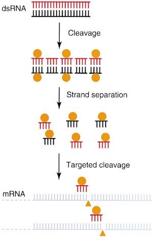
Figure 7. Proposed Model for RNAi sophila lysate for 2 hr at standard conditions. After deproteinization,
RNAi is envisioned to begin with cleavage of the dsRNA to 21–23 samples were run on 12% sequencing gels to separate full-length
nt products by a dsRNA-specific nuclease, perhaps in a multiprotein dsRNAs from the 21–23 nt products. RNAs were eluted from the gel
complex. These short dsRNAs might then be dissociated by an ATP- slices in 0.3 M NaCl overnight, ethanol precipitated, collected by
dependent helicase, possibly a component of the initial complex, centrifugation, and redissolved in 20 l water. The RNA was hy-
to 21–23 nt asRNAs that could then target the mRNA for cleavage. drolyzed into nucleoside 5⬘ phosphates with nuclease P1 (10 l
The short asRNAs are imagined to remain associated with the RNAi- reaction containing 8 l RNA in water, 30 mM KOAc [pH 5.3], 10
specific proteins (ochre circles) that were originally bound by the mM ZnSO4, and 10 g or 3 units nuclease P1, for 3 hr at 50⬚C).
full-length dsRNA, thus explaining the inefficiency of asRNA to trig- Samples (1 l) were cospotted with nonradioactive 5⬘ mononucleo-
ger RNAi in vivo and in vitro. Finally, a nuclease (triangles) would tides (0.05 O. D. units [A260] of pA, pC, pG, pI, and pU) on cellulose
cleave the mRNA. HPTLC plates (EM Merck) and separated in the first dimension in
isobutyric acid/25% ammonia/water (66/1/33, v/v/v) and in the sec-
ond dimension in 0.1 M sodium phosphate, pH 6.8/ammonium sul-
water for each experiment. GTP was omitted from the reactions, fate/1-propanol (100/60/2, v/w/v; Silberklang et al., 1979). Migration
except in Figures 2 and 3. of the nonradioactive internal standards was determined by UV
shadowing.
RNA Synthesis
Pp-luc and Rr-luc mRNAs and Pp- and Rr-dsRNAs (including dsRNA Acknowledgments
B in Figure 6) were synthesized by in vitro transcription as described
previously (Tuschl et al., 1999). To generate transcription templates We acknowledge Heike Taubner, Sayda M. Elbashir, and Winfried
for dsRNA C, the 5⬘ sense RNA primer was gcgtaatacgactcactata Lendeckel for assistance, and Herbert Jäckle and his research group
GAACAAAGGAAACGGATGAT and the 3⬘ sense RNA primer was and members of the Bartel laboratory for their support and advice.
GAAGAAGTTATTCTCCAAAA; the 5⬘ asRNA primer was gcgtaatac The authors thank Terri Orr-Weaver for providing fly resources and
gactcactataGAAGAAGTTATTCTCCAAAA and the 3⬘ asRNA primer Marlene Castle for maintaining population cages. This work was
was GAACAAAGGAAACGGATGAT. For dsRNA A, the 5⬘ sense RNA supported by a German BMBF Biofuture grant number 0311856
primer was gcgtaatacgactcactataGTAGCGCGGTGTATTATACC and (T. T.), by the National Institutes of Health through a United States
the 3⬘ sense RNA primer was GTACAACGTCAGGTTTACCA; the 5⬘ Public Health Service MERIT award, the National Science Founda-
asRNA primer was gcgtaatacgactcactataGTACAACGTCAGGTTT tion, and partially by the National Cancer Institute through a Cancer
ACCA and the 3⬘ asRNA primer was GTAGCGCGGTGTATTATACC Center Support core grant (P. A. S.), and by the Searle Scholars
(lowercase, T7 promoter sequence). Program/The Chicago Community Trust (D. P. B.).
mRNAs were 5⬘ end labeled using guanylyl transferase (Gibco/
BRL), S-adenosyl methionine (Sigma), and ␣-32P-GTP (3000 Ci/ Received March 2, 2000; revised March 10, 2000.
mmol; New England Nuclear) according to the manufacturer’s direc-
tions. Radiolabeled RNAs were purified by poly(A) selection using References
the Poly(A) Tract III kit (Promega). Nonradioactive 7-methyl-guano-
sine- and adenosine-capped RNAs were synthesized in in vitro tran- Auxilien, S., Crain, P.F., Trewyn, R.W., and Grosjean, H. (1996).
scription reactions with a 5-fold excess of 7-methyl-G(5⬘)ppp(5⬘)G Mechanism, specificity and general properties of the yeast enzyme
or A(5⬘)ppp(5⬘)G relative to GTP. Cap analogs were purchased from catalysing the formation of inosine 34 in the anticodon of transfer
New England Biolabs. RNA. J. Mol. Biol. 262, 437–458.
Bass, B.L. (1997). RNA editing and hypermutation by adenosine
ATP Depletion and Protein Synthesis Inhibition
deamination [published erratum appears in Trends Biochem Sci.
ATP was depleted by incubating the lysate for 10 min at 25⬚C with
1997 Jul;22(7):278]. Trends Biochem. Sci. 22, 157–162.
2 mM glucose and 0.1 U/l hexokinase (Sigma). Protein synthesis
inhibitors were purchased from Sigma and dissolved in absolute Baulcombe, D.C. (1999). Fast forward genetics based on virus-
ethanol as 250-fold concentrated stocks. The final concentrations induced gene silencing. Curr. Opin. Plant Biol. 2, 109–113.
of inhibitors in the reaction were anisomycin, 53 g/ml; cyclohexi- Cogoni, C., and Macino, G. (1997). Isolation of quelling-defective
mide, 100 g/ml; and chloramphenicol, 100 mg/ml. Relative protein (qde) mutants impaired in posttranscriptional transgene-induceddsRNA Directs Cleavage of mRNA at 21–23 nt Intervals 33 gene silencing in Neurospora crassa. Proc. Natl. Acad. Sci. USA 94, Maine, E.M. (1995). Enhancers of glp-1, a gene required for cell- 10233–10238. signaling in Caenorhabditis elegans, define a set of genes required Cogoni, C., and Macino, G. (1999a). Gene silencing in Neurospora for germline development. Genetics 141, 551–569. crassa requires a protein homologous to RNA-dependent RNA poly- Ratcliff, F.G., MacFarlane, S.A., and Baulcombe, D.C. (1999). Gene merase. Nature 399, 166–169. silencing without DNA. RNA-mediated cross-protection between Cogoni, C., and Macino, G. (1999b). Posttranscriptional gene silenc- viruses. Plant Cell 11, 1207–1216. ing in Neurospora by a RecQ DNA helicase. Science 286, 2342–2344. Sánchez-Alvarado, A., and Newmark, P.A. (1999). Double-stranded Cogoni, C., Irelan, J.T., Schumacher, M., Schmidhauser, T.J., Selker, RNA specifically disrupts gene expression during planarian regener- E.U., and Macino, G. (1996). Transgene silencing of the al-1 gene ation. Proc. Natl. Acad. Sci. USA 96, 5049–5054. in vegetative cells of Neurospora is mediated by a cytoplasmic Schiebel, W., Haas, B., Marinkovic, S., Klanner, A., and Sanger, effector and does not depend on DNA-DNA interactions or DNA H.L. (1993a). RNA-directed RNA polymerase from tomato leaves. I. methylation. EMBO J. 15, 3153–3163. Purification and physical properties. J. Biol. Chem. 268, 11851– Cundliffe, E. (1981). Antibiotic inhibitors of ribosome function. In The 11857. Molecular Basis of Antibiotic Action, E. Gale et al., eds. (New York: Schiebel, W., Haas, B., Marinkovic, S., Klanner, A., and Sanger, Wiley), pp. 402–547. H.L. (1993b). RNA-directed RNA polymerase from tomato leaves. II. Fire, A. (1999). RNA-triggered gene silencing. Trends Genet. 15, Catalytic in vitro properties. J. Biol. Chem. 268, 11858–11867. 358–363. Schiebel, W., Pelissier, T., Riedel, L., Thalmeir, S., Schiebel, R., Fire, A., Xu, S., Montgomery, M.K., Kostas, S.A., Driver, S.E., and Kempe, D., Lottspeich, F., Sanger, H.L., and Wassenegger, M. Mello, C.C. (1998). Potent and specific genetic interference by dou- (1998). Isolation of an RNA-directed RNA polymerase-specific cDNA ble-stranded RNA in Caenorhabditis elegans. Nature 391, 806–811. clone from tomato. Plant Cell 10, 2087–2101. Gebauer, F., Corona, D.F., Preiss, T., Becker, P.B., and Hentze, M.W. Sharp, P.A. (1999). RNAi and double-strand RNA. Genes Dev. 13, (1999). Translational control of dosage compensation in Drosophila 139–141. by Sex-lethal: cooperative silencing via the 5⬘ and 3⬘ UTRs of msl-2 Silberklang, M., Gillum, A.M., and RajBhandary, U.L. (1979). Use of in mRNA is independent of the poly(A) tail. EMBO J. 18, 6146–6154. vitro 32P labeling in the sequence analysis of nonradioactive tRNAs. Grant, S.R. (1999). Dissecting the mechanisms of posttranscriptional Methods Enzymol. 59, 58–109. gene silencing: divide and conquer. Cell 96, 303–306. Smardon, A., Spoerke, J., Stacey, S., Klein, M., Mackin, N., and Grishok, A., Tabara, H., and Mello, C. (2000). Genetic requirements Maine, E. (2000). EGO-1 is related to RNA-directed RNA polymerase for inheritance of RNAi in C. elegans. Science, in press. and functions in germ-line development and RNA interference in C. Guo, S., and Kemphues, K.J. (1995). par-1, a gene required for elegans. Curr. Biol. 10, 169–178. establishing polarity in C. elegans embryos, encodes a putative Ser/ Tabara, H., Sarkissian, M., Kelly, W.G., Fleenor, J., Grishok, A., Tim- Thr kinase that is asymmetrically distributed. Cell 81, 611–620. mons, L., Fire, A., and Mello, C.C. (1999). The rde-1 gene, RNA Hamilton, A.J., and Baulcombe, D.C. (1999). A species of small anti- interference, and transposon silencing in C. elegans. Cell 99, sense RNA in posttranscriptional gene silencing in plants. Science 123–132. 286, 950–952. Timmons, L., and Fire, A. (1998). Specific interference by ingested Hunter, C.P. (1999). A touch of elegance with RNAi. Curr. Biol. 9, dsRNA. Nature 395, 854. R440–R442. Tuschl, T., Zamore, P.D., Lehmann, R., Bartel, D.P., and Sharp, P.A. Hunter, C. (2000). Gene silencing: shrinking the black box of RNAi. (1999). Targeted mRNA degradation by double-stranded RNA in Curr. Biol. 10, R137–R140. vitro. Genes Dev. 13, 3191–3197. Kennerdell, J.R., and Carthew, R.W. (1998). Use of dsRNA-mediated Vaucheret, H., Beclin, C., Elmayan, T., Feuerbach, F., Godon, C., genetic interference to demonstrate that frizzled and frizzled 2 act Morel, J.B., Mourrain, P., Palauqui, J.C., and Vernhettes, S. (1998). in the wingless pathway. Cell 95, 1017–1026. Transgene-induced gene silencing in plants. Plant J. 16, 651–659. Ketting, R.F., Haverkamp, T.H., van Luenen, H.G., and Plasterk, R.H. Wagner, R., and Sun, L. (1998). Functional genomics: double- (1999). Mut-7 of C. elegans, required for transposon silencing and stranded RNA poses puzzle. Nature 391, 744–745. RNA interference, is a homolog of Werner syndrome helicase and Wargelius, A., Ellingsen, S., and Fjose, A. (1999). Double-stranded RNaseD. Cell 99, 133–141. RNA induces specific developmental defects in zebrafish embryos. Kozak, M. (1999). Initiation of translation in prokaryotes and eukary- Biochem. Biophys. Res. Commun. 263, 156–161. otes. Gene 234, 187–208. Waterhouse, P.M., Graham, M.W., and Wang, M.B. (1998). Virus Lohmann, J.U., Endl, I., and Bosch, T.C. (1999). Silencing of develop- resistance and gene silencing in plants can be induced by simultane- mental genes in Hydra. Dev. Biol. 214, 211–214. ous expression of sense and antisense RNA. Proc. Natl. Acad. Sci. Merrick, W., and Hershey, J. (1996). The pathway and mechanism USA 95, 13959–13964. of eukaryotic protein synthesis. In Translational Control, J. Hershey Waterhouse, P.M., Smith, N.A., and Wang, M.-B. (1999). Virus resis- et al., eds. (Cold Spring Harbor, NY: Cold Spring Harbor Laboratory tance and gene silencing: killing the messenger. Trends Plant Sci. Press), pp. 31–69. 4, 452–457. Misquitta, L., and Paterson, B.M. (1999). Targeted disruption of gene Wianny, F., and Zernicka-Goetz, M. (2000). Specific interference with function in Drosophila by RNA interference (RNA-i): a role for nautilus gene function by double-stranded RNA in early mouse development. in embryonic somatic muscle formation. Proc. Natl. Acad. Sci. USA Nat. Cell Biol. 2, 70–75. 96, 1451–1456. Montgomery, M.K., and Fire, A. (1998). Double-stranded RNA as a Note Added in Proof mediator in sequence-specific genetic silencing and co-suppres- sion. Trends Genet. 14, 255–258. Recently, Hammond et al. have shown that ⵑ25 nt RNAs are gener- Montgomery, M.K., Xu, S., and Fire, A. (1998). RNA as a target of ated in cultured Drosophila S2 cells transfected with cyclin E dsRNA double-stranded RNA-mediated genetic interference in Caenorhab- (Hammond, S.M., Bernstein, E., Beach, D., and Hannon, G.J. [2000]. ditis elegans. Proc. Natl. Acad. Sci. USA 95, 15502–15507. Nature 404, 293–296. Ngo, H., Tschudi, C., Gull, K., and Ullu, E. (1998). Double-stranded RNA induces mRNA degradation in Trypanosoma brucei. Proc. Natl. Acad. Sci. USA 95, 14687–14692. Page, A.W., and Orr-Weaver, T.L. (1997). Activation of the meiotic divisions in Drosophila oocytes. Dev. Biol. 183, 195–207. Qiao, L., Lissemore, J.L., Shu, P., Smardon, A., Gelber, M.B., and
You can also read