Chaperone -usher pathways: diversity and pilus assembly mechanism
←
→
Page content transcription
If your browser does not render page correctly, please read the page content below
Downloaded from http://rstb.royalsocietypublishing.org/ on July 23, 2015

                                                                                      Phil. Trans. R. Soc. B (2012) 367, 1112–1122
                                                                                                         doi:10.1098/rstb.2011.0206

                                                                 Review

           Chaperone –usher pathways: diversity and
                 pilus assembly mechanism
                                  Andreas Busch and Gabriel Waksman*
        Institute of Structural and Molecular Biology (ISMB), University College London and Birkbeck College,
                                        Malet Street, WC1E 7HX London, UK
        Up to eight different types of secretion systems, and several more subtypes, have been described in
        Gram-negative bacteria. Here, we focus on the diversity and assembly mechanism of one of the best-
        studied secretion systems, the widespread chaperone– usher pathway known to assemble and secrete
        adhesive surface structures, called pili or fimbriae, which play essential roles in targeting bacterial
        pathogens to the host.
                       Keywords: chaperone– usher; pilus biogenesis; host– pathogen interactions

1. INTRODUCTION                                                        fimbrial/pilus subunit-encoding gene [8]. The chaper-
Non-flagellar surface filaments were initially described               one and usher proteins are the accessory proteins
in Gram-negative bacteria in the 1950s in Escherichia                  needed to assemble pilus subunits into a pilus and
coli [1] and 5 years later the term ‘fimbriae’ was                     secrete the assembled pilus. These are relatively con-
coined when their role in cell adhesion processes                      served. However, classification schemes for CU
became evident [2]. The term ‘pilus’ [3] was later                     pathways based only on sequence homology between
introduced referring to the same proteinaceous non-                    fimbrial subunits and/or between chaperones have a
flagellar surface appendages, and therefore the terms                  significant shortcoming: the CU pathway-encoding
fimbriae and pili can be used as synonyms. Before                      gene clusters or operons may vary in the number of
whole genomes became available, fimbriae or pili                       chaperones, fimbrial subunits as well as of additional
were classified in terms of their morphology as seen                   adhesin-encoding genes that group to distant branches
under the microscope and, if known, their function                     in a phylogenetic tree and would therefore make any
[4– 6]. Yet this did not account for the phylogenetic                  assignment ambiguous. However, there is always only
relatedness or the genomic variability with respect to                 one outer membrane (OM) usher present. As a conse-
the number of components involved in secreting and                     quence, Nuccio & Bäumler [8] proposed a classification
building these fimbriae. Nowadays, the classification                  scheme based on the usher protein. The fimbrial usher
of fimbriae or pili is the result of a combination of gen-             protein (FUP) family is distributed among the Proteobac-
etics, biochemistry and structure that has led to a                    teria, Cyanobacteria and Deinococcus–Thermus phyla [9].
classification on the basis of the membrane-embedded                   The FUP is divided into six clades (table 1), designated
assembly and secretion systems involved in their bio-                  a-, b-, g-, k-, p- and s-fimbriae, each stemming from a
genesis (reviewed in Fronzes et al. [7]). This has led                 common ancestor. The g-fimbrial clade is further sub-
to the identification of four types of non-flagellar sur-              divided into four subclades, termed g1-, g2-, g3- and
face filaments produced by Gram-negative bacteria                      g4-fimbriae. The a-, k-, p- and s-fimbrial clade names
(reviewed in Fronzes et al. [7]), among which the                      were assigned arbitrarily to recall a particular character-
so-called chaperone– usher (CU) pathway of pilus                       istic of the clade or a prominent member as follows:
biogenesis is the most ubiquitous. We review here the                  a-fimbriae, for alternate CU family; k-fimbriae, for
mechanism of pilus assembly and secretion by these                     K88 (F4) fimbriae; p-fimbriae, for pyelonephritis-
CU systems, highlighting recent mechanistic insights                   associated fimbriae (P fimbriae); and s-fimbriae, for
and also their diversity.                                              spore coat protein U from Myxococcus xanthus. The
                                                                       b- and g-fimbriae were assigned names alphabetically.
                                                                           Another mode of classification of CU pathways is
2. CLASSIFICATION OF CHAPERONE – USHER                                 based on the chaperone structure, particularly on the
PATHWAY                                                                length of a loop that connects their F1 and G1 strands
The CU secretion systems are mostly grouped into                       [11]: the FGL (long F1 – G1 loop) chaperone system
gene clusters, some of them identified as operons,                     subfamily assemble non-fimbrial surface structures,
with a minimum of an usher-, a chaperone- and a                        while the FGS (short F1 –G1 loop) chaperone system
                                                                       subfamily assemble fimbrial filaments [11,12]. Within
                                                                       the FUP clades classification, the FGL chaperones
* Author for correspondence (g.waksman@ucl.ac.uk).                     CU systems fall into just one clade, the g3-clade;
One contribution of 11 to a Theme Issue ‘Bacterial protein secretion   however, the FGS chaperones CU systems fall into
comes of age’.                                                         the b-, g1-, g2-, g4-, k- and p-clades.

                                                                   1112                         This journal is q 2012 The Royal Society
Downloaded from http://rstb.royalsocietypublishing.org/ on July 23, 2015

                                                      Review. Pilus biogenesis and diversity A. Busch & G. Waksman         1113

Table 1. Classification of chaperone –usher systems according to Nuccio & Bäumler [8] into fimbrial usher protein
(FUP) clusters, clades and subclades. One best representative of the core gene organization of each clade/subclade
has been chosen and the typical host target tissues are listed, if known. For more details on adhesins and host
receptor molecules, please refer to Nicastro et al. [10].

3. GENETIC DIVERSITY OF CHAPERONE –USHER                                diversity in gene organization found within each clade
PATHWAYS                                                                never involved lateral exchange of subunit genes
The classification of the fimbrial gene clusters into                   among these four major clusters, instead coevolving
FUP clades based on the usher sequences was shown                       as complete clusters. In cases where there are diver-
to correlate with a core gene arrangement. The six                      gences from the core gene structure, for example,
established clades can be subdivided into four major clus-              when there is more than one chaperone within one
ters based on their gene organization and evolutionary                  fimbrial gene cluster, this could be explained by a prob-
relationship of their pilus subunit sequences. The first                able gene duplication event, as on a tree based on
cluster is the gkp cluster, containing the g, k and p                   chaperone sequence, when the chaperones encoded
clades. This cluster is characterized by a common pilus                 within the same gene cluster formed sister groups [8].
subunit homology domain (PFAM00419). Additionally,                         In addition to the variability in gene organization,
the p- and k-clades both share a core structure composed                sequencing of whole genomes has identified CU path-
of genes encoding first a major subunit, then an usher,                 ways which can be considered as hybrid: their gene
followed by a chaperone (MUC; table 1). The g-clade                     clusters include sequences which code for components
does not share the same gene organization and this struc-               that are normally part of unrelated clusters encoding
ture is different for each subclade: MCUT for g1,                       unrelated types of secretion systems. This is the case
MCCU for g2, MCU or CUM for g3 and MCUT                                 for the g4-clade, which in some cases includes genes
for g4 (T standing for the tip adhesin). The a, b or                    of the type Vb or two-partner secretion (TPS) systems
the s-clades form their own individual clusters, with dis-              either within the coding sequence for the CU pathway
tinct gene organization: CMUT, MCU and MCUT,                            or flanking the CU gene cluster (table 1): in both
respectively. They also contain distinct fimbrial subunit               Bordetella pertussis and Bordetella avium, the fim gene
homology domains: PFAM04449 for a, PFAM06551                            clusters are flanked upstream by FhaB (TpsA protein
for b and COG5430 for s. This would suggest that the                    and adhesin) and downstream by a FhaC (TpsB
Phil. Trans. R. Soc. B (2012)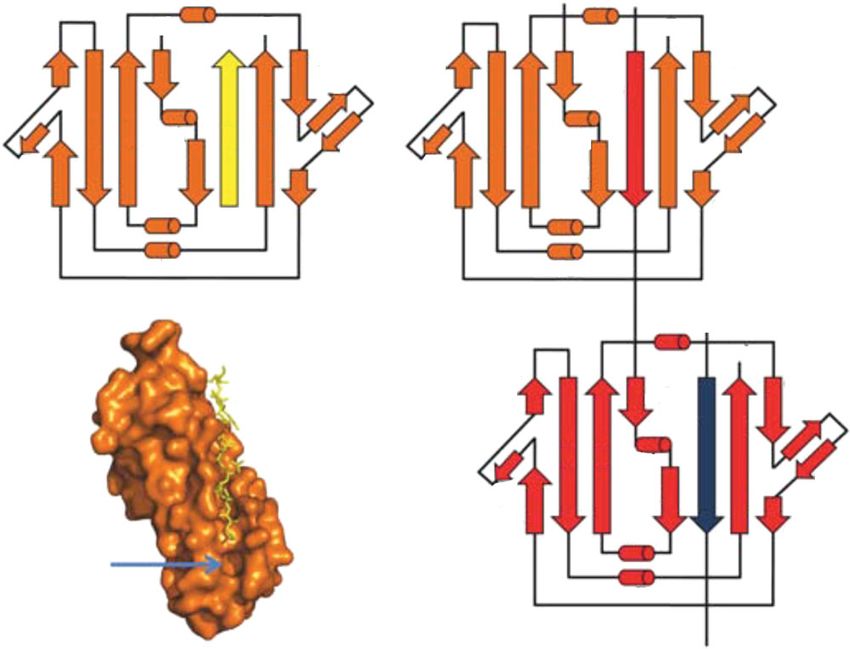
Downloaded from http://rstb.royalsocietypublishing.org/ on July 23, 2015

1114     A. Busch & G. Waksman            Review. Pilus biogenesis and diversity

                            (a)                     PapG                  (b)

                                                    PapF
                                                                                             FimH
                                                    PapE (5–10)   tip fibrillum
                                                                                         FimG

                                                     PapK                                    FimF

                          PapA                                                                    FimA
                          >1000 copies                                                       ~1000 copies
                                                                      Pilus rod

                                                PapC                                    FimD             OM

                                               H       C1                                     C1
                                         N                                        N                 periplasm
                                                      C2                                     C2
                                             PapD                                     FimC

Figure 1. A schematic of (a) P and (b) type 1 pili assembled by the Pap and Fim systems, respectively. The chaperones attached
to the last subunit to be incorporated into each pilus are shown in yellow. Periplasmic chaperones assist in folding pilus
subunits and targeting them to the OM usher. P pili are terminated at the OM by the termination subunit, PapH. No such
subunit is known in the Fim system. The periplasmic NTD and CTDs, respectively, of the usher are indicated with the
letters N and C.

and OM pore) protein. In Pseudomonas fluorescens, a                     pilus, through its PapG tip adhesion, binds to Gala-
complete TPS cluster is inserted within the CU gene                     1,4 Galb moieties present in the globoseries of glyco-
cluster and in Pseudomonas aeruginosa, an orphan                        lipids (GbO3, GbO4 and GbO5) on the surface of
gene coding for an FhaC-like adhesin is found in the                    kidney epithelial cells and erythrocytes. The tip adhe-
same location [8]. It is likely that whole-genome                       sins of the a-clade (mainly enterotoxigenic E. coli
sequencing will eventually unveil more such hybrid                      (ETEC)) are known, but their receptor partners on
pathways, involving other clades than the g4-clade                      epithelial intestinal cells remain elusive [15].
and other secretion systems/pathways than TPS.                              Most bacteria carry more than one CU system.
                                                                        Whole-genome sequencing of many strains of entero-
                                                                        bacteria has indicated that the presence of multiple
                                                                        fimbrial gene clusters is the norm. In pathogenic
4. ADHESINS AND PILUS – RECEPTOR                                        bacteria, such as P. aeruginosa, five different so-called
INTERACTION                                                             Cup (CU pathway) secretion systems (CupA–CupE)
The first pilus protein to be identified as responsible                 are described [10,19,20]. The apparent redundancy in
for binding to host epithelial cells was FimH [13,14].                  such secretion machineries might reflect a high diversity
FimH forms part of the tip fibrillum in type I fimbriae                 in lectin domains, thereby ensuring attachment to a
and is the adhesive structure responsible for interaction               broader set of hosts.
with D-mannosylated proteins such as epithelial bladder                     While each adhesin expressed independently can
and kidney cells [15] or uroplakins [16,17] (table 1).                  promote adhesion of bacteria to a specific tissue, a
FimH was also the first structure to be determined, con-                sequential or synergistic expression of adhesins with
sisting of two subdomains: an N-terminal lectin domain                  diverse specificities could eventually determine the
containing the mannose-binding site, connected via a                    final host tissue destination of the bacteria. This was
linker chain to a C-terminal pilin domain responsible                   first proposed for uropathogenic E. coli cells
for incorporating FimH into the fimbrial structure                      (UPEC), with expression of first type 1 fimbriae and
[18]. Type 1C and S fimbriae, which, like the type I fim-               then P fimbriae [21], progressively targeting the
briae, belong to the g1-clade, use as receptors the                     bacterium from the bladder (type 1) to the kidney
GalNAc-b-1,4-Galb- and the NeuAca-2,3-Galb-con-                         (P pilus). Determining how sequential expression
taining surface proteins, respectively. g2-Fimbriae                     of different lectins affects tissue tropism is a key
bind mainly to human intestinal epithelial cells and                    aspect in understanding bacterial colonization. Simi-
g3-fimbriae are the blood group fimbriae binding                        larly, the expression of the Cup and other pathways
either to neutrophils and erythrocytes or more specifi-                 known to be involved in host colonization in P. aerugin-
cally to decay accelerating factor (DAF or CD55) or                     osa seems to be dependent on the stage of biofilm
a5b1 integrin, among others. The receptors for the g4-                  formation [22,23], a process itself dependent on the
fimbriae lectin remain unknown. In the p-clade, the P                   production of fimbriae/pili.
Phil. Trans. R. Soc. B (2012)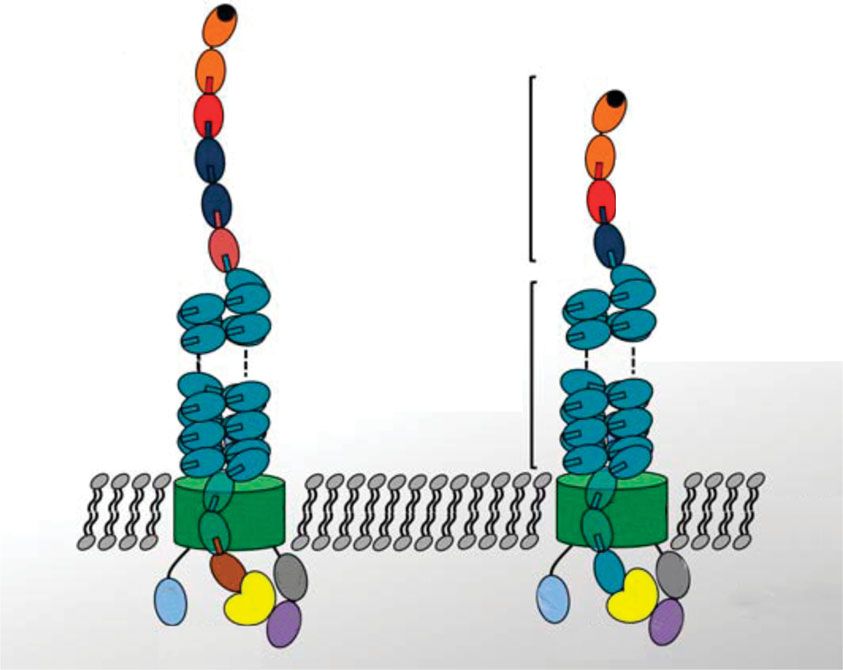
Downloaded from http://rstb.royalsocietypublishing.org/ on July 23, 2015

                                                        Review. Pilus biogenesis and diversity A. Busch & G. Waksman                1115

                                          310C                                                310C N
                 (a)                  N            C                (c)                                 C
                                          A1             C1                                                     C1
                    D2                                                    D2               A1
                                                           C′                               Nte                   C′

                            E B 310A             G1 F                              E B 310A
                                                              C′′                                                  C′′
                    D′                 A2                                 D′             A2
                    D1                                                    D1
                                      aB                 C2                              aB                   C2

                                     aD                                                 aD

                                                                                                       310C N
                  (b)                                                                                            C
                                                                                                       A1                C1
                                                                                   D2
                                                                                                        Nte                C′

                                                                                          E B3 A
                                                                                              10
                                                                                                                              C′′
                                                                                   D′             A2
                                                                                   D1
                                                                                                  aB                     C2
                            P5
                                                                                                  aD

Figure 2. Donor-strand complementation (DSC) and exchange. (a) A topology diagram of the FimH pilin domain (FimHp;
orange) complemented via DSC with the G1 strand of the chaperone FimC (yellow). Arrows and cylinders represent b-strands
and a-helices, respectively. The C- and the N-terminus are indicated. (b) A topology diagram of donor-strand exchange (DSE)
between FimHp (orange) and FimG (red). The red arrow in the upper diagram represents the N-terminal extension (Nte) of
the incoming subunit FimG, complementing in trans the hydrophobic groove of FimHp. The blue arrow in the lower panel
represents the Nte of the incoming FimF subunit. (c) A surface and stick representation of the FimH pilin domain
(orange) in complex with chaperone FimC (yellow) during DSC. The empty P5 pocket where the incoming subunit starts
the zip-in, zip-out mechanism is shown. For clarity, only the G1 strand of FimC is shown.

5. PILUS MORPHOLOGY AND ASSEMBLY                                               rod adopts a right-handed, one-start superhelical
MECHANISMS                                                                     structure with a 25 Å pitch, a 7.54 Å rise per subunit
The CU pathway pili are assembled into linear                                  and 3.3 subunits per turn. The filament has a max-
unbranched polymers consisting of several hundreds to                          imum diameter of 82 Å and an axial channel 25 Å in
thousands of pilus subunits (also known as pilins) that                        diameter runs straight up the centre of the helical axis
range in size from approximately 12 kDa to approxima-                          [27]. Type 1 pili are composed of four different subunit
tely 20 kDa. CU organelles differ widely in complexity                         types (FimH, FimG, FimF and FimA). One copy of the
and morphology, ranging from non-fimbrial 2–5 nm in                            distal adhesin FimH, followed by one copy each of
diameter, flexible fibrillae (g3-clade: Dr; k-clade F4),                       FimG and FimF [28], forms a flexible tip fibrillum
to rigid helical fimbrial shafts of up to 10 mm in diameter                    shorter than the Pap tip [29]. The tip fibrillum is
(a-clade: CS1; g1-clade: type1; g2-clade: F6; g4-clade:                        attached to an extended rigid and helically wound rod
Mrk; p-clade: P) [24].                                                         of circa 1000 FimA subunits. No termination subunit
   This review focuses on the type I and P pili of uro-                        has yet been characterized for the type 1 pilus.
pathogenic E. coli, or rod-like fimbrial organelles,                              Pilus subunits are translocated from the cytoplasm
which are members of the g1- and p-fimbrial clades,                            to the periplasm via the general secretory pathway
respectively, from which most of our current know-                             SecYEG. Pilus subunits are unable to fold and
ledge of the pilus assembly process has been derived                           unable to self-assemble at the cell surface on their
(figure 1).                                                                    own [30]. Two accessory proteins are needed: (i) a
   The P pilus is formed by six different subunits                             periplasmic chaperone essential in stabilization/
arranged into two distinct subassemblies: the tip fibril-                      folding of subunits, in avoiding premature subunit
lum and the pilus rod (figure 1). The distal tip                               polymerization in the periplasm, and in targeting
fibrillum is approximately 2 nm in diameter, is flexible                       chaperone–subunit complexes to the other accessory
and composed of one PapG adhesin at the distal end,                            protein, (ii) an OM assembly platform termed usher.
followed by one adaptor subunit PapF and 5 –10
copies of the PapE subunit (figure 1). More than                               (a) Donor-strand complementation
1000 copies of the PapA subunit form the long, rigid                           The mechanism of stabilization of pilus subunits by
and 6.8 nm wide pilus rod. Both subassemblies are                              periplasmic chaperones was first described for the
connected by the adaptor subunit PapK and the rod                              PapD – PapK [31] and FimC – FimH [18] chaper-
is terminated at the proximal end (in the cell wall)                           one – subunit complexes. All pilus subunits exhibit an
by the termination subunit PapH [25,26]. The PapA                              incomplete Ig-like fold, where the seventh C-terminal
Phil. Trans. R. Soc. B (2012)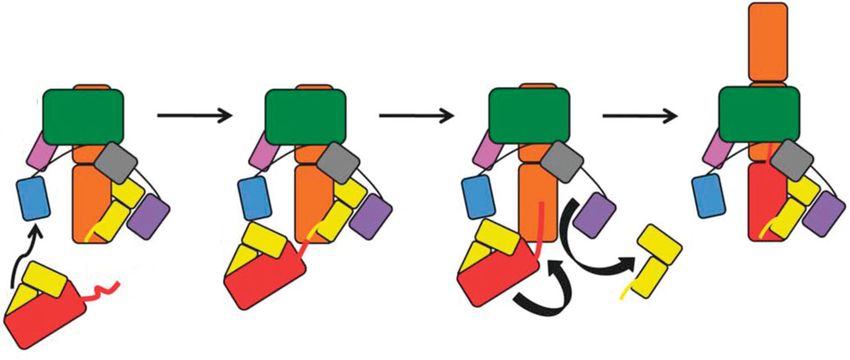
Downloaded from http://rstb.royalsocietypublishing.org/ on July 23, 2015

1116     A. Busch & G. Waksman          Review. Pilus biogenesis and diversity

b-strand is missing. As a result, a deep hydrophobic                 occupied by the G1 strand of the chaperone [36,37].
groove is clearly visible on the subunit’s surface                   Thus, the P5 pocket is the primary site of interaction for
where the missing strand should be. Because of the                   the incoming Nte. The identification (using non-denatur-
missing secondary structure, pilus subunits are stable               ing mass spectrometry) of a transient ternary complex
only when either bound to the chaperone or to an                     between the chaperone–subunit complex and the Nte
adjacent subunit within the nascent pilus. The chaper-               of the incoming subunit during in vitro DSE reactions
one provides in trans the missing secondary structure                was crucial to the discovery of this mechanism [36].
by inserting one of its own strands, strand G1, into                 Single-site mutagenesis of the P5, P4 and P3 residues to
the subunit’s groove in a process called donor-strand                Ala in the Nte revealed a gradient of decreasing DSE effi-
complementation (DSC; figure 2a). Periplasmic chap-                  ciency moving away from the P5 initiation site, suggesting
erones (approx. 25 kDa in mass) consist of two Ig-like               a ‘zip-in–zip-out’ mechanism, with the incoming Nte
domains forming a boomerang-like structure and it is                 gradually displacing the chaperone G1 donor strand in
the G strand of the N-terminal domain (NTD; or                       a stepwise process from P5 to P1 [36]. Molecular
domain 1, hence G1) that provides the missing sec-                   dynamics simulations provided the first evidence for a
ondary structure [32,33]. By inserting into the                      zipper mechanism [38]. In the simulations, the chaperone
subunit’s groove, the chaperone’s G1 strand reconsti-                donor strand was seen to unbind from the pilus subunit,
tutes the incomplete Ig-fold of the subunit, but,                    residue by residue, in support of the ‘zip-in–zip-out’
because it runs parallel to strand F, it does so                     hypothesis. Because the insertion and the subsequent
atypically: in a regular Ig fold, strand G would run                 zip-in of the incoming Nte occur in parallel to the accep-
antiparallel to strand F. Strand G1 contains a motif of              tor subunit’s strand F, the resulting Nte-complemented
four alternating hydrophobic residues (termed P1– P4                 Ig-fold of the acceptor subunit is now ‘canonical’. The
residues), which, in chaperone– subunit complexes,                   topological transition occurring upon DSE (from non-
interact with four sites/pockets (termed P1 – P4 sites/              canonical before DSE to canonical after DSE) is
pockets) in the subunit’s hydrophobic groove. All                    linked to conformational changes that result in the clos-
subunits’ hydrophobic grooves, except the pilus biogen-              ing of the groove around the incoming Nte, creating a
esis terminator PapH [26], have an additional P5 site/               groove– Nte interaction that is one of the strongest
pocket which is either never occupied by the chaper-                 ever documented [39]. Such a topological transition is
one’s strand or only partially occupied, depending on                thought to provide the energy driving the DSE reaction
the length of the chaperone’s strand. This empty P5                  in one direction, that of pilus assembly.
pocket is crucial in pilus subunit polymerization, as
will be detailed below (figure 2b).
                                                                     (c) Termination of pilus biogenesis
                                                                     Verger et al. [26] showed that PapH was the terminator
                                                                     subunit regulating the pilus length. The authors ruled
(b) Donor-strand exchange: ‘zip-in – zip-out’
                                                                     out as explanation a stronger PapD – PapH interaction
mechanism
                                                                     preventing a displacement of the G1 strand of the chap-
The polymerization of pilus subunits at the usher
                                                                     erone via ‘zip-in –zip-out’ mechanism by a further
occurs via a mechanism termed ‘donor-strand
                                                                     subunit. Instead, the structure of the PapD – PapH
exchange’ (DSE) [34– 36] (figure 2c). All CU pilus
                                                                     complex revealed the absence of a P5 pocket that a
subunits contain a 10–20 residue-long N-terminal exten-
                                                                     previous study identified as the initiator site for DSE
sion (Nte) peptide that is disordered in the chaperone–
                                                                     [36]. PapH functions not only as a terminator of
subunit complex and is not part of the subunit fold.
                                                                     pilus biogenesis, but also it anchors the pilus to the
Subunit polymerization occurs when the G1 b-strand
                                                                     OM [25]. This is because the usher barrel can only
of the chaperone complementing the subunit’s groove
                                                                     accommodate the passage of subunits and not chaper-
(termed ‘previously-assembled’ or ‘receiving’ or ‘accep-
                                                                     one – subunit complexes (see §5e). No homologue of
tor’ subunit) is replaced by the Nte of the subunit next
                                                                     PapH has yet been found for the type I pilus system,
in assembly (termed ‘donating’ and ‘incoming’ subunit).
                                                                     and thus the mechanism for controlling type I pilus
As described before, the P1–P4 pockets in the acceptor
                                                                     length is unclear.
subunit’s groove are occupied by the P1–P4 residues of
the chaperone’s G1 strand in the chaperone–subunit
complex (figure 2b), but after DSE, it is the P2–P5 pock-            (d) Subunit ordering
ets of the acceptor subunit’s groove that are occupied by            Immuno-electronmicroscopy (EM) studies resolved
the hydrophobic residues (termed P2–P5 residues) of                  the order by which the P pilus subunits are arranged,
the incoming subunit’s Nte. Residue P4 of the Nte of                 the PapG adhesin being at the tip followed by PapF,
the acceptor pilus subunit is a Gly and is conserved                 PapE and PapK [40,41]. These four subunits form
among all pilus subunits. The P4 pocket in the subunit’s             the flexible tip fibrillum [42], followed by the helical
groove contains a bulk formed by an aromatic residue,                rod formed by a multimer of PapA subunits. PapF is
and the small residue Glycine is the only residue able to            required for the correct linkage of the adhesin at the
adapt to this bulk: this ensures the proper registering of           distal end of the tip fibrillum, and PapK regulates the
the Nte within the acceptor subunit’s groove. How is                 length of the tip fibrillum and joins it to the pilus rod
the G1 strand of the chaperone displaced by the Nte of               [42]. Only PapE, present in several copies in the tip
the incoming subunit? The first step in the DSE reaction             fibrillum, and PapA, present in more than 1000
is the insertion of the P5 residue of the incoming Nte into          copies in the rod, were shown to have self-associating
the empty P5 pocket of the previously assembled or                   properties in the periplasm [43]. PapF is an adaptor
acceptor subunit, while the P1–P4 pockets remain                     subunit essential in the display of PapG at the tip of
Phil. Trans. R. Soc. B (2012)
Downloaded from http://rstb.royalsocietypublishing.org/ on July 23, 2015
Review. Pilus biogenesis and diversity A. Busch & G. Waksman 1117
the fibrillum, and PapK has a dual role as a terminator The b-barrel closes in an end-to-end fashion, posi-
of the tip fibrillum and an initiator of rod assembly tioning the N- and C-termini on the periplasmic
[42]. After elucidation of the DSC and DSE processes, side of the membrane. The N- and C-terminal glob-
awareness of the essential role of the Ntes in them led to ular domains are thus juxtaposed and reside in the
a study of their role in the specific order of assembly. periplasm, consistent with their role in chaperone–
Deletion of the entire Nte of the adaptor subunit subunit recruitment and adhesin-induced pore acti-
PapF impeded the incorporation of PapG into the vation [30,47,49–52]. The plug domain (residues
pilus, while swapping of the Ntes of PapE and PapF 257–332) is positioned laterally inside the b-barrel
led to PapF being unable to play its adaptor role pore, occluding the luminal volume of the trans-
[44]. These results suggest both that the N-terminal location pore, preventing passage of solutes or
extension of PapE does not fit into the hydrophobic periplasmic proteins across the channel in its non-
groove of the PapG pilin body and that the pilin body activated or apo form (figure 3). Very recently, the
of PapF is not required in interactions between PapF structure of full-length FimD usher was solved in
and PapG [44]. Similar Nte swapping experiments complex with its cognate FimC–FimH substrate
were performed with PapE and PapK, where the Nte [50] (figure 4a). Like PapC, FimD contains a 24-
of PapF was fused to PapE (NteFPapE) and the Nte stranded b-barrel (residues 139–665) translocation
of PapE to PapK (NteEPapK): the sole presence of pore domain. However, in contrast to the PapC
the Nte allowed an interaction of NteFPapE with structure in its non-activated state, the plug domain
PapG and the incoming PapK was still able to undergo (residues 241–324) in the FimD–FimC–FimH com-
DSE, being able to assemble a pilus in a PapF deletion plex now resides in the periplasm, underneath the
mutant (PapF2). In the same fashion, NteEPapK was translocation domain and next to the NTD, and
able to complement the groove of PapF and pilus the shape of the translocation domain has dramati-
assembly was observed in a PapE2 strain [42,44]. cally changed. Compared with the apo-PapC or
Another study examining the specificity of DSE in apo-FimD pore domain structure, the 24-strand b-
the Pap system confirmed the Nte – groove interaction barrel rearranges from an oval-shaped pore with a
as a determining factor in subunit ordering [45]. In 52 28 Å diameter to a near circular pore 32 Å in
an in vitro DSE assay, all six chaperone– subunit com- diameter (figure 3). With the plug domain displaced
plexes were incubated individually in the presence of into the periplasm, the open and circular 32 Å chan-
the Ntes of each of the five Pap subunits (except nel is occupied instead by the FimH lectin domain,
PapG, the adhesin located at the distal end of the providing the first view of a transporter caught in
pilus which acts only as an Nte acceptor). The dis- the act of substrate transport.
sociation rate of the chaperone– subunit complex Previous work has established that the FimC –
owing to DSE was followed for each of the 30 combi- FimH complex displays the highest affinity for FimD
nations by electrospray mass spectrometry. The [47,52] and is the only complex capable of inducing
fastest DSE rate occurred uniformly with the cognate the conformational changes in FimD required for
partners, suggesting a complementarity of Ntes and usher activation [53]. The FimC chaperone alone
cognate hydrophobic grooves. The same study [45] was unable to bind to FimD. FimC – FimA, FimC –
and a subsequent one [46] ascribe a decisive role in FimG and FimC– FimF bound to the FimD usher
subunit ordering to the P5 site and immediately with KD values of 176 nM (FimC – FimA), 670 nM
adjacent residues. (FimC – FimG) and 1.37 mM (FimC – FimF), 20- to
Apart from interaction specificities between sub- 150-fold higher than FimC – FimH (KD 9.1 nM).
units, the usher also plays an important role in subunit The relatively high association constant of the
ordering as will be discussed in §5e. FimC – FimH complex is probably critical for it to
associate first to the usher ensuring the localization
of FimH at the tip of the pilus, and the FimH-induced
(e) The usher assembly platform: structure conformational change constitutes a crucial activation
and function step that is required for subunit polymerization and
OM ushers are approximately 800-residue proteins translocation at the usher assembly platform [53,54].
comprising four functional domains: an N-terminal, The differences in KD between the other subunits
approximately 125-residue periplasmic domain; a might not affect their order of incorporation into the
C-terminal, approximately 170-residue periplasmic pilus, as these differences are relatively small. More
domain and a large, central, approximately 500- important is the specificity of interaction between the
residue translocation pore domain that is interrupted receiving subunits’ hydrophobic grooves and the
by a conserved plug domain of approximately 110 incoming subunits’ Ntes.
residues [30,47,48] (figure 4b). The first structure The structure of FimD in the FimD – FimC – FimH
of a membrane-integral usher part to be solved complex reveals four periplasmic domains: the NTD,
was the full translocation pore domain of PapC the plug and two C-terminal domains (CTDs),
[49], a 55 kDa fragment comprising residues 130– CTD1 and CTD2. Which roles do these periplasmic
640 corresponding to the predicted OM translocation domains play? Earlier co-expression studies of FimD
domain and the middle or plug domain [48,49]. The with FimC – FimH and subsequent trypsin digestion
structure of PapC130 – 640 has a kidney shaped, 24- and pull-down assays isolated a C-terminal region of
stranded b-barrel (residues 146–635), 45 Å in FimD bound to FimC – FimH [52]. The authors con-
height and with outer and inner dimensions of cluded that the CTDs are the probable site of
65 45 Å and 45 25 Å, respectively (figure 3). interaction. Later studies, however, showed that the
Phil. Trans. R. Soc. B (2012)Downloaded from http://rstb.royalsocietypublishing.org/ on July 23, 2015
1118 A. Busch & G. Waksman Review. Pilus biogenesis and diversity
(a) (b)
(c) (d)
Figure 3. Structures of the apo and activated (FimC–FimH-engaged) FimD usher pore. (a) Top and (b) side view ribbon rep-
resentations of the superimposed apo-FimD (slate) and activated FimD (green) b-barrel. The plug domain in the channel
lumen in apo FimD (magenta) rotates into the periplasm following FimD activation (pink). (c) Top view surface representation
of the apo-FimD. (d) Activated FimD with FimH lectin domain (FimHL, orange) in the translocation pore. The plug and
FimHL are coloured magenta and orange, respectively.
(a) (b) (c)
FimH N FimHL FimHP C
1 157 158 279
FimC N FimCN FimCC C
1 120 121 205
FimD N NTD Pore Plug Pore CTD1 CTD2 C
26 138 139 241 324 665 666 750 751834
Figure 4. Usher-mediated pilus biogenesis. (a) Schematic of the domains of the chaperone FimC, the usher FimD and the
adhesin FimH. Numbers indicate residues where the respective domains start and end. (b) FimD –FimC– FimH complex.
Side view ribbon representation of FimD (green), FimC (yellow) and surface representation of FimH (orange). The NTD,
CTD1 and CTD2 are coloured light blue, grey and purple, respectively; the plug is coloured magenta. The FimC G1
strand complementing the groove (P1–P4 pockets) of FimHp is coloured cyan. (c) Superposition of FimD –FimC– FimH
complex with the FimDN –FimC–FimF structure (PDB 3BWU) leading to the proposed chaperone-subunit incorporation
cycle at the FimD usher (see text and figure 5). The Nte of the incoming modelled subunit FimG occupying the P5
pocket is coloured purple.
usher’s NTD is also a binding site for chaperone –sub- FimD on its own displayed the highest affinity towards
unit complexes, including the chaperone– adhesin the FimC – FimH complex [47] and showed that resi-
complex FimC– FimH [47,51,55]. The NTD of dues 1– 24 of the NTD specifically interact with
Phil. Trans. R. Soc. B (2012)Downloaded from http://rstb.royalsocietypublishing.org/ on July 23, 2015
Review. Pilus biogenesis and diversity A. Busch & G. Waksman 1119
recruitment DSE transfer to CTDs secretion
to NTD ‘zip-in–zip-out’
FimH
FimD
Plug
CTD1
NTD
CTD2
FimC
FimC′ Nte
FimG
Figure 5. Schematic of the proposed chaperone –subunit incorporation cycle. Initial targeting of the periplasmic chaperone –
subunit complex (FimC0 –FimG) to the NTD of the outer membrane usher (FimD), followed by DSE between the previously
assembled subunit (FimH) and the incoming subunit FimG via a ‘zip-in –zip-out’ mechanism, which releases the chaperone
FimC from FimH. Subsequently, the incoming FimG– FimC0 complex is handed over from the NTD to the CTDs, and the
nascent pilus is secreted.
both FimC and the subunit [55], FimH’s lectin incorporation cycle [47,51,55]. In all, the FimD –
domain being essential in the activation of the usher FimC – FimH structure together with previous
[53]. In the FimD – FimC – FimH complex structure, structural and biochemical evidence on the function
however, the NTD lies idle, making no interactions of the NTD of FimD allowed a pilus subunit incorpor-
with FimC; the FimC– FimH complex instead is ation cycle to be proposed where the usher is fully
bound to two Ig-like domains formed at the usher C functional for pilus biogenesis as a monomer [50]
terminus, CTD1 and CTD2 (residues 666 – 750 and (figure 5): the chaperone –subunit complex at the
751 – 834, respectively) [50]. The most extensive inter- base of the growing pilus fibre resides at the usher’s
action with the FimC – FimH complex is with the CTDs; new subunits are recruited to the NTD and
usher’s CTD1 (figure 4a,b), which contacts the brought into ideal orientation to undergo DSE with
FimH lectin domain and FimC. CTD2 contacts pri- the subunit bound at the CTDs (now the penultimate
marily FimC. Removal of both CTDs, or CTD2 subunit, figure 5); upon DSE, the chaperone is dis-
alone or point mutations in CTD1 abrogate pilus bio- placed from the penultimate subunit and dissociates
genesis [50,56]. Using electron paramagnetic from the CTDs; to reset the assembly machinery for
resonance (EPR) spectroscopy, it was also demon- a new incorporation cycle, the chaperone– subunit
strated that subsequent subunits localize to the complex dissociates from the NTD site and transfers
CTDs’ binding sites after undergoing DSE. to the CTDs’ site, concomitantly pushing the penul-
As there was now clear evidence that the usher’s timate subunit into the translocation channel.
NTD and the CTDs are both bona fide chaperone– According to this model, a rotational translation of
subunit binding sites, the question arises whether the incoming chaperone – subunit complex must prob-
these sites work in a parallel and coordinated or in a ably take place. However, how this ‘hand-over’ of the
sequential manner. As the structure of the NTD of chaperone– subunit complex from the usher’s NTD
FimD (FimDN) bound to the FimC – FimF complex to the CTDs occurs remains elusive.
was available [57], both the structures of FimDN –
FimC – FimF and FimD– FimC – FimH were super-
imposed (using FimDN) to investigate whether, in 6. HYBRID CHAPERONE –USHER PATHWAYS
the FimD – FimC – FimH complex, both the NTD So far, the only hybrid CU pathway reasonably well
and the CTD binding sites would be able to accom- characterized is the CupB pathway in P. aeruginosa
modate two chaperone – subunit complexes at the PAO1 [22,23,58,59]. The cupB gene cluster codes for
same time, with the knowledge that the last incorpor- six proteins: a pilin subunit (CupB1), an usher
ated chaperone– subunit complex remains bound to (CupB3), two chaperones (CupB2 and CupB4),
the usher CTDs [51,52]. This superimposition an adhesin (CupB6) and, remarkably, a protein
(figure 4c) demonstrated that, in fact, the NTD in (CupB5) sharing 44 per cent similarity with the
the FimD – FimC – FimH complex is available for the TpsA4 protein identified in the PAO1 genome. TpsA
recruitment of a chaperone– subunit complex without proteins are adhesins normally transported to the cell
steric clashes with the FimC – FimH complex bound at surface by a cognate TpsB OM pore protein in what
the CTDs. Inactivation of the NTD by a bulky is called the type Vb or TPS system. However, no
molecule severely disrupted further subunit incorpor- gene encoding a TpsB-like protein was found within
ation in vitro, confirming the NTD of the usher as the cupB operon and CupB5 can therefore be classified
the recruitment site operating first in the pilus subunit as an orphan TpsA-like protein within a CU cluster. In
Phil. Trans. R. Soc. B (2012)Downloaded from http://rstb.royalsocietypublishing.org/ on July 23, 2015
1120 A. Busch & G. Waksman Review. Pilus biogenesis and diversity
silico analyses comparing CupB5 with other TpsA pro- Unravelling the mechanisms of pilus biogenesis has
teins showed sequences at the N-terminus that are already helped in efforts to inhibit biofilm formation
characteristic of TpsA-like molecules and are known and bacterial adhesion in uropathogenic E. coli [61].
to interact with specific sequences in TpsB-like The recent FimD – FimC – FimH structure provides a
transporters called polypeptide transport-associated whole new cadre of targets for the next generation of
(POTRA) domains. The usher CupB3 transports and pilus biogenesis inhibitors. This structure exemplifies
assembles CupB1, the pilin subunit, to the surface, as the need for structural and mechanistic studies of
would be expected from an usher. However, transport bacterial secretion in order to design a new anti-
of the TpsA-like adhesin CupB5 to the cell surface microbial arsenal specifically targeting virulence
was also shown to be CupB3-dependent [58]. Sure factors and tame the resurgence of hospital-acquired
enough, a POTRA domain was identified at the N-ter- antibiotic-resistant pathogens.
minus of CupB3. Interestingly, this POTRA domain
This work was funded by Long-Term Fellowships from the
seems to coordinate transport of the CupB1 pilus sub- European Molecular Biology Organization (EMBO, ALTF
unit and the non-fimbrial adhesin CupB5. In a 745-2009) and Marie Curie FP7-PEOPLE-2009-IEF
truncated version of cupB3 lacking the POTRA (252616) to Andreas Busch, and by grant 85602 from the
domain, CupB1 transport was impeded and CupB5 MRC and grant 082227 from the Wellcome Trust to
transport apparently not. However, when in addition Gabriel Waksman.
to the POTRA-truncated version of cupB3 the cupB5
gene was interrupted, CupB1 transport was restored.
This indicated that the POTRA domain is not essential REFERENCES
for CupB1 transport, but might be a prerequisite for 1 Houwink, A. L. & van Iterson, W. 1950 Electron micro-
translocation of CupB5, which otherwise impedes scopical observations on bacterial cytology; a study on
correct CupB1 assembly. Moreover, the whole secretion flagellation. Biochim. Biophys. Acta 5, 10–44. (doi:10.
process of both CupB1 and CupB5 is dependent—either 1016/0006-3002(50)90144-2)
directly or indirectly, this remains to be established—on 2 Duguid, J. P., Smith, I. W., Dempster, G. & Edmunds,
the chaperone CupB4. So far, only the structure of P. N. 1955 Non-flagellar filamentous appendages (fim-
briae) and haemagglutinating activity in Bacterium coli.
CupB2 has been resolved and it is most likely the
J. Pathol. Bacteriol. 70, 335–348. (doi:10.1002/path.
chaperone targeting CupB1 to the usher [60]. In con- 1700700210)
clusion, the most interesting feature of this system is 3 Brinton, C. C. 1959 Non-flagellar appendages of bac-
that the usher is able to transport to the cell surface teria. Nature 183, 782–786. (doi:10.1038/183782a0)
both fimbrial (CU pathway components) and non-fim- 4 Brinton, C. C. 1965 The structure, function, synthesis
brial adhesins (TpsA-like CupB5). Mechanistic details and genetic control of bacterial pili and a molecular
as to how this is achieved remain to be described. model for DNA and RNA transport in Gram negative
bacteria. Trans. N. Y. Acad. Sci. 27, 1003– 1054.
5 Duguid, J. P., Anderson, E. S. & Campbell, I. 1966 Fim-
briae and adhesive properties in Salmonellae. J. Pathol.
Bacteriol. 92, 107 –138. (doi:10.1002/path.1700920113)
7. CONCLUSION
6 Desvaux, M., Hébraud, M., Talon, R. & Henderson, I. R.
There has been much progress in recent years in the 2009 Secretion and subcellular localizations of bacterial
understanding of key aspects of secretion through the proteins: a semantic awareness issue. Trends Microbiol.
CU pathway: first, how the subunits are stabilized 17, 139 –145. (doi:10.1016/j.tim.2009.01.004)
when secreted to the periplasm (DSC) and targeted 7 Fronzes, R., Remaut, H. & Waksman, G. 2008 Architec-
to the assembly platform at the OM, the usher; tures and biogenesis of non-flagellar protein appendages
second, how the subunits polymerize (DSE) and in in Gram-negative bacteria. EMBO J. 27, 2271 –2280.
what order; and third, how the usher is activated to a (doi:10.1038/emboj.2008.155)
secretion competent form, catalyses DSE and how its 8 Nuccio, S.-P. & Bäumler, A. J. 2007 Evolution of the
various domains are involved in chaperone – subunit chaperone/usher assembly pathway: fimbrial classification
complex recruitment, subunit assembly and pilus goes Greek. Microbiol. Mol. Biol. Rev. 71, 551 –575.
(doi:10.1128/MMBR.00014-07)
secretion. Probably the most intriguing aspect to be
9 Yen, M.-R., Peabody, C. R., Partovi, S. M., Zhai, Y.,
resolved is how a single usher manages the hand-over Tseng, Y. H. & Saier, M. H. 2002 Protein-translocating
of the chaperone – subunit complex from NTD to outer membrane porins of Gram-negative bacteria.
CTD, which has to involve a major relocation of its Biochim. Biophys. Acta 1562, 6 –31. (doi:10.1016/
periplasmic domains at each assembly step. Thus, S0005-2736(02)00359-0)
acquiring a dynamic view of the usher as it incorpor- 10 Nicastro, G. G., Boechat, A. L., Abe, C. M., Kaihami,
ates subunits to the nascent pilus will be a major G. H. & Baldini, R. L. 2009 Pseudomonas aeruginosa
research challenge for the next few years. PA14 cupD transcription is activated by the RcsB
Hybrid secretion systems are fascinating, but their response regulator, but repressed by its putative cognate
evolutionary significance remains to be elucidated. sensor RcsC. FEMS Microbiol. Lett. 301, 115–123.
(doi:10.1111/j.1574-6968.2009.01803.x)
Do they confer a selective advantage to the bacterium?
11 Zav’yalov, V. P., Zav’yalova, G. A., Denesyuk, A. I. &
Do they borrow the best from both the CU and TPS Korpela, T. 1995 Modelling of steric structure of a peri-
‘worlds’? Why would an usher be more advantageous plasmic molecular chaperone Caf1M of Yersinia pestis, a
as a means of secretion than a TpsB transporter? prototype member of a subfamily with characteristic
Why use the same transporter to secrete adhesins structural and functional features. FEMS Immunol.
that are so different in structure? All these questions Med. Microbiol. 11, 19–24. (doi:10.1111/j.1574-695X.
will no doubt drive future research in the field. 1995.tb00074.x)
Phil. Trans. R. Soc. B (2012)Downloaded from http://rstb.royalsocietypublishing.org/ on July 23, 2015
Review. Pilus biogenesis and diversity A. Busch & G. Waksman 1121
12 Hung, D. L., Knight, S. D., Woods, R. M., Pinkner, J. S. & 26 Verger, D., Miller, E., Remaut, H., Waksman, G. &
Hultgren, S. J. 1996 Molecular basis of two subfamilies Hultgren, S. 2006 Molecular mechanism of P pilus ter-
of immunoglobulin-like chaperones. EMBO J. 15, mination in uropathogenic Escherichia coli. EMBO Rep.
3792–3805. 7, 1228– 1232. (doi:10.1038/sj.embor.7400833)
13 Barnhart, M. M., Sauer, F. G., Pinkner, J. S. & 27 Mu, X.-Q. & Bullitt, E. 2006 Structure and assembly of
Hultgren, S. J. 2003 Chaperone–subunit – usher inter- P-pili: a protruding hinge region used for assembly of a
actions required for donor strand exchange during bacterial adhesion filament. Proc. Natl Acad. Sci. USA
bacterial pilus assembly. J. Bacteriol. 185, 2723 –2730. 103, 9861 –9866. (doi:10.1073/pnas.0509620103)
(doi:10.1128/JB.185.9.2723-2730.2003) 28 Le Trong, I., Aprikian, P., Kidd, B. A., Thomas, W. E.,
14 Bäumler, A. J., Gilde, A. J., Tsolis, R. M., van der Sokurenko, E. V. & Stenkamp, R. E. 2010 Donor
Velden, A. W., Ahmer, B. M. & Heffron, F. 1997 Contri- strand exchange and conformational changes during E.
bution of horizontal gene transfer and deletion events to coli fimbrial formation. J. Struct. Biol. 172, 380 –388.
development of distinctive patterns of fimbrial operons (doi:10.1016/j.jsb.2010.06.002)
during evolution of Salmonella serotypes. J. Bacteriol. 29 Hahn, E. et al. 2002 Exploring the 3D molecular archi-
179, 317 –322. tecture of Escherichia coli type 1 pili. J. Mol. Biol. 323,
15 Korea, C.-G., Ghigo, J.-M. & Beloin, C. 2011 The sweet 845 –857. (doi:10.1016/S0022-2836(02)01005-7)
connection: solving the riddle of multiple sugar-binding 30 Thanassi, D. G., Stathopoulos, C., Dodson, K., Geiger,
fimbrial adhesins in Escherichia coli: multiple E. coli fim- D. & Hultgren, S. J. 2002 Bacterial outer membrane
briae form a versatile arsenal of sugar-binding lectins ushers contain distinct targeting and assembly domains
potentially involved in surface-colonisation and tissue for pilus biogenesis. J. Bacteriol. 184, 6260–6269.
tropism. BioEssays 33, 300 –311. (doi:10.1002/bies. (doi:10.1128/JB.184.22.6260-6269.2002)
201000121) 31 Sauer, F. G., Fütterer, K., Pinkner, J. S., Dodson, K. W.,
16 Bäumler, A. J. & Heffron, F. 1995 Identification and Hultgren, S. J. & Waksman, G. 1999 Structural basis of
sequence analysis of lpfABCDE, a putative fimbrial chaperone function and pilus biogenesis. Science 285,
operon of Salmonella typhimurium. J. Bacteriol. 177, 1058 –61. (doi:10.1126/science.285.5430.1058)
2087–2097. 32 Holmgren, A. & Bränden, C. I. 1989 Crystal structure of
17 Bäumler, A. J., Tsolis, R. M. & Heffron, F. 1996 The lpf chaperone protein PapD reveals an immunoglobulin
fimbrial operon mediates adhesion of Salmonella typhi- fold. Nature 342, 248 –51. (doi:10.1038/342248a0)
murium to murine Peyer’s patches. Proc. Natl Acad. Sci. 33 Kuehn, M. J., Ogg, D. J., Kihlberg, J., Slonim, L. N.,
USA 93, 279 –283. (doi:10.1073/pnas.93.1.279) Flemmer, K., Bergfors, T. & Hultgren, S. J. 1993 Struc-
18 Choudhury, D., Thompson, A., Stojanoff, V., tural basis of pilus subunit recognition by the PapD
Langermann, S., Pinkner, J., Hultgren, S. J. & Knight, chaperone. Science 262, 1234–1241. (doi:10.1126/
S. D. 1999 X-ray structure of the FimC–FimH chaper- science.7901913)
one-adhesin complex from uropathogenic Escherichia 34 Sauer, F. G., Pinkner, J. S., Waksman, G. & Hultgren,
coli. Science 285, 1061–1066. (doi:10.1126/science.285. S. J. 2002 Chaperone priming of pilus subunits facilitates
5430.1061) a topological transition that drives fiber formation. Cell
19 He, J. et al. 2004 The broad host range pathogen Pseudo- 111, 543 –551. (doi:10.1016/S0092-8674(02)01050-4)
monas aeruginosa strain PA14 carries two pathogenicity 35 Zavialov, A. V., Berglund, J., Pudney, A. F., Fooks, L. J.,
islands harboring plant and animal virulence genes. Ibrahim, T. M., MacIntyre, S. & Knight, S. D. 2003
Proc. Natl Acad. Sci. USA 101, 2530–2535. (doi:10. Structure and biogenesis of the capsular F1 antigen
1073/pnas.0304622101) from Yersinia pestis: preserved folding energy drives fiber
20 Mikkelsen, H., Ball, G., Giraud, C. & Filloux, A. 2009 formation. Cell 113, 587– 596. (doi:10.1016/S0092-
Expression of Pseudomonas aeruginosa CupD fimbrial 8674(03)00351-9)
genes is antagonistically controlled by RcsB and the 36 Remaut, H., Rose, R. J., Hannan, T. J., Hultgren, S. J.,
EAL-containing PvrR response regulators. PLoS ONE Radford, S. E., Ashcroft, A. E. & Waksman, G. 2006
4, e6018. (doi:10.1371/journal.pone.0006018) Donor-strand exchange in chaperone-assisted pilus
21 Snyder, J. A., Haugen, B. J., Lockatell, C. V., Maroncle, assembly proceeds through a concerted beta strand dis-
N., Hagan, E. C., Johnson, D. E., Welch, R. A. & placement mechanism. Mol. Cell 22, 831 –842. (doi:10.
Mobley, H. L. T. 2005 Coordinate expression of fimbriae 1016/j.molcel.2006.05.033)
in uropathogenic Escherichia coli. Infect. Immun. 73, 37 Vetsch, M., Erilov, D., Molière, N., Nishiyama, M.,
7588–7596. (doi:10.1128/IAI.73.11.7588-7596.2005) Ignatov, O. & Glockshuber, R. 2006 Mechanism of
22 Vallet, I., Diggle, S., Stacey, R., Camara, M., Ventre, I., fibre assembly through the chaperone –usher pathway.
Lory, S., Lazdunski, A., Williams, P. & Filloux, A. 2004 EMBO Rep. 7, 734 –738. (doi:10.1038/sj.embor.
Biofilm formation in Pseudomonas aeruginosa: fimbrial 7400722)
cup gene clusters are controlled by the transcriptional 38 Rose, R. J., Welsh, T. S., Waksman, G., Ashcroft, A. E.,
regulator MvaT. J. Bacteriol. 186, 2880 –2890. (doi:10. Radford, S. E. & Paci, E. 2008 Donor-strand exchange
1128/JB.186.9.2880-2890.2004) in chaperone-assisted pilus assembly revealed in atomic
23 Vallet, I., Olson, J., Lory, S., Lazdunski, A. & Filloux, A. detail by molecular dynamics. J. Mol. Biol. 375, 908–
2001 The chaperone/usher pathways of Pseudomonas 919. (doi:10.1016/j.jmb.2007.10.077)
aeruginosa: identification of fimbrial gene clusters (cup) 39 Zavialov, A. V., Tischenko, V. M., Fooks, L. J., Brandsdal,
and their involvement in biofilm formation. Proc. Natl B. O., Aqvist, J., Zav’yalov, V. P., MacIntyre, S. & Knight,
Acad. Sci. USA 98, 6911–6916. (doi:10.1073/pnas. S. D. 2005 Resolving the energy paradox of chaperone/
111551898) usher-mediated fibre assembly. Biochem. J. 389,
24 Waksman, G. & Hultgren, S. J. 2009 Structural biology of 685 –694. (doi:10.1042/BJ20050426)
the chaperone –usher pathway of pilus biogenesis. Nat. 40 Kuehn, M. J., Heuser, J., Normark, S. & Hultgren, S. J.
Rev. Microbiol. 7, 765 –774. (doi:10.1038/nrmicro2220) 1992 P pili in uropathogenic E. coli are composite fibres
25 Båga, M., Norgren, M. & Normark, S. 1987 Biogenesis with distinct fibrillar adhesive tips. Nature 356, 252 –255.
of E. coli Pap pili: papH, a minor pilin subunit involved in (doi:10.1038/356252a0)
cell anchoring and length modulation. Cell 49, 241– 251. 41 Lindberg, F., Lund, B., Johansson, L. & Normark, S.
(doi:10.1016/0092-8674(87)90565-4) 1987 Localization of the receptor-binding protein
Phil. Trans. R. Soc. B (2012)Downloaded from http://rstb.royalsocietypublishing.org/ on July 23, 2015
1122 A. Busch & G. Waksman Review. Pilus biogenesis and diversity
adhesin at the tip of the bacterial pilus. Nature 328, 84– 52 Saulino, E. T., Thanassi, D. G., Pinkner, J. S. &
87. (doi:10.1038/328084a0) Hultgren, S. J. 1998 Ramifications of kinetic partitioning
42 Jacob-Dubuisson, F., Heuser, J., Dodson, K., Normark, S. on usher-mediated pilus biogenesis. EMBO J. 17,
& Hultgren, S. 1993 Initiation of assembly and association 2177–2185. (doi:10.1093/emboj/17.8.2177)
of the structural elements of a bacterial pilus depend on two 53 Nishiyama, M., Ishikawa, T., Rechsteiner, H. &
specialized tip proteins. EMBO J. 12, 837–847. Glockshuber, R. 2008 Reconstitution of pilus assembly
43 Striker, R., Jacob-Dubuisson, F., Freiden, C. & reveals a bacterial outer membrane catalyst. Science
Hultgren, S. J. 1994 Stable fiber-forming and nonfiber- 320, 376 –379. (doi:10.1126/science.1154994)
forming chaperone-subunit complexes in pilus biogenesis. 54 Saulino, E. T., Bullitt, E. & Hultgren, S. J. 2000
J. Biol. Chem. 269, 12 233 –12 239. Snapshots of usher-mediated protein secretion and
44 Lee, Y. M., Dodson, K. W. & Hultgren, S. J. 2007 ordered pilus assembly. Proc. Natl Acad. Sci. USA 97,
Adaptor function of PapF depends on donor strand 9240–9245. (doi:10.1073/pnas.160070497)
exchange in P-pilus biogenesis of Escherichia coli. 55 Nishiyama, M. et al. 2005 Structural basis of chaperone-
J. Bacteriol. 189, 5276–5283. (doi:10.1128/JB.01648-06) subunit complex recognition by the type 1 pilus assembly
45 Rose, R. J., Verger, D., Daviter, T., Remaut, H., Paci, E., platform FimD. EMBO J. 24, 2075–2086. (doi:10.1038/
Waksman, G., Ashcroft, A. E. & Radford, S. E. 2008 sj.emboj.7600693)
Unraveling the molecular basis of subunit specificity in 56 Li, Q., Ng, T. W., Dodson, K. W., So, S. S. K., Bayle,
P pilus assembly by mass spectrometry. Proc. Natl K.-M., Pinkner, J. S., Scarlata, S., Hultgren, S. J. &
Acad. Sci. USA 105, 12 873– 12 878. Thanassi, D. G. 2010 The differential affinity of the
46 Leney, A. C., Phan, G., Allen, W., Verger, D., Waksman, usher for chaperone-subunit complexes is required for
G., Radford, S. E. & Ashcroft, A. E. 2011 Second order assembly of complete pili. Mol. Microbiol. 76, 159 –172.
rate constants of donor-strand exchange reveal individual (doi:10.1111/j.1365-2958.2010.07089.x)
amino acid residues important in determining the subunit 57 Eidam, O., Dworkowski, F. S. N., Glockshuber, R.,
specificity of pilus biogenesis. J. Am. Soc. Mass Spectrom. Grütter, M. G. & Capitani, G. 2008 Crystal structure
22, 1214–1223. (doi:10.1007/s13361-011-0146-4) of the ternary FimC-FimF(t)-FimD(N) complex indicates
47 Nishiyama, M., Vetsch, M., Puorger, C., Jelesarov, I. & conserved pilus chaperone-subunit complex recognition by
Glockshuber, R. 2003 Identification and characterization the usher FimD. FEBS Lett. 582, 651–655. (doi:10.1016/j.
of the chaperone-subunit complex-binding domain from febslet.2008.01.030)
the type 1 pilus assembly platform FimD. J. Mol. Biol. 58 Ruer, S., Ball, G., Filloux, A. & de Bentzmann, S. 2008
330, 513– 525. (doi:10.1016/S0022-2836(03)00591-6) The ‘P-usher’, a novel protein transporter involved in
48 Capitani, G., Eidam, O. & Grütter, M. G. 2006 Evi- fimbrial assembly and TpsA secretion. EMBO J. 27,
dence for a novel domain of bacterial outer membrane 2669–2680. (doi:10.1038/emboj.2008.197)
ushers. Proteins 65, 816–823. (doi:10.1002/prot.21147) 59 Ruer, S., Filloux, A. & de Bentzmann, S. 2007 Assem-
49 Remaut, H., Tang, C., Henderson, N. S., Pinkner, J. S., bly of fimbrial structures in Pseudomonas aeruginosa:
Wang, T., Hultgren, S. J., Thanassi, D. G., Waksman, G. & functionality and specificity of chaperone – usher machin-
Li, H. 2008 Fiber formation across the bacterial outer mem- eries. J. Bacteriol. 189, 3547– 3555. (doi:10.1128/JB.
brane by the chaperone/usher pathway. Cell 133, 640–652. 00093-07)
(doi:10.1016/j.cell.2008.03.033) 60 Cai, X., Wang, R., Filloux, A., Waksman, G. &
50 Phan, G. et al. 2011 Crystal structure of the FimD usher Meng, G. 2011 Structural and functional characteriz-
bound to its cognate FimC–FimH substrate. Nature 474, ation of Pseudomonas aeruginosa CupB chaperones.
49–53. (doi:10.1038/nature10109) PLoS ONE 6, e16583. (doi:10.1371/journal.pone.
51 Ng, T. W., Akman, L., Osisami, M. & Thanassi, D. G. 0016583)
2004 The usher N terminus is the initial targeting site 61 Pinkner, J. S. et al. 2006 Rationally designed small com-
for chaperone-subunit complexes and participates in pounds inhibit pilus biogenesis in uropathogenic bacteria.
subsequent pilus biogenesis events. J. Bacteriol. 186, Proc. Natl Acad. Sci. USA 103, 17 897–17 902.
5321–5331. (doi:10.1128/JB.186.16.5321-5331.2004) (doi:10.1073/pnas.0606795103)
Phil. Trans. R. Soc. B (2012)You can also read