The C-terminal protoxin domain of Bacillus thuringiensis Cry1Ab toxin has a functional role in binding to GPI-anchored receptors in the insect midgut
←
→
Page content transcription
If your browser does not render page correctly, please read the page content below
JBC Papers in Press. Published on November 1, 2018 as Manuscript RA118.005101
          The latest version is at http://www.jbc.org/cgi/doi/10.1074/jbc.RA118.005101

   The C-terminal protoxin domain of Bacillus thuringiensis Cry1Ab toxin
   has a functional role in binding to GPI-anchored receptors in the insect
   midgut
   Arlen Peña-Cardeña1, Ricardo Grande2, Jorge Sánchez1, Bruce E. Tabashnik3, Alejandra Bravo1,
   Mario Soberón1 and Isabel Gómez1*
   1
     Departamento de Microbiología Molecular. Instituto de Biotecnología, Universidad Nacional
   Autónoma de México, Avenida Universidad 2001, Colonia Chamilpa, Cuernavaca, Morelos 62210,
   México.
   2
      Unidad de Secuenciación Masiva y Bioinformática. Instituto de Biotecnología, Universidad
   Nacional Autónoma de México, Avenida Universidad 2001, Colonia Chamilpa, Cuernavaca,
   Morelos 62210, México.
   3
     Department of Entomology, University of Arizona, Tucson, AZ, 85721, USA.

                                                                                                                 Downloaded from http://www.jbc.org/ by guest on December 17, 2018
   Running title: C-terminal region of Cry1Ab protoxin binds to APN and ALP

   * Corresponding author.
   Phone-Fax: (52-777) 329-1624.
   E-mail: isabelg@ibt.unam.mx

   Keywords: Bacillus thuringiensis, Cry toxin, Aminopeptidase, Alkaline phosphatase, C-terminal
   region, receptor binding, insecticide, insecticidal protein, cadherin.

Abstract                                                   CAD but not to ALP or APN, supporting the
Bacillus thuringiensis (Bt) Cry toxins are used            notion that protoxins have additional binding
worldwide for controlling insects. Cry1Ab is               sites. These results imply that two different
produced as a 130 kDa protoxin that is activated           regions mediate the binding of Cry1Ab protoxin
by proteolytic removal of an inert 500 amino-              to membrane receptors, one located in domain
acids-long C-terminal region, enabling the                 II-III of the toxin and another in its C-terminal
activated toxin to bind to insect midgut receptor          region, suggesting an active role of the C-
proteins and leading to its membrane insertion             terminal protoxin fragment in the mode of action
and pore formation. It has been proposed that the          of Cry toxins. These results suggest that future
C-terminal region is only involved in toxin                manipulations of the C-terminal protoxin region
crystallization, but its role in receptor binding is       could alter specificity and increase toxicity of Bt
undefined. Here we show that the C-terminal                proteins.
region of Cry1Ab protoxin provides additional
binding sites to alkaline phosphatase (ALP) and            Introduction
aminopeptidase N (APN) insect receptors.                   Insecticidal proteins from the soil bacterium
ELISA, ligand blot, SPR and pull-down assays               Bacillus thuringiensis (Bt) are used extensively
revealed that the Cry1Ab C-terminal region                 in transgenic plants and sprays to control insect
binds to both ALP and APN, but not to cadherin             pests (1,2). These Bt proteins are especially
(CAD). Thus, the C-terminal region provides                valuable because they kill some of the world’s
both higher binding affinity of the protoxin to            most harmful pests, yet are not toxic to people
the gut membrane that correlated with higher               and other organisms (3-5). Cultivation of crops
toxicity of protoxin than activated toxin.                 genetically engineered to produce Bt proteins
Moreover, Cry1Ab domain II loop 2 or 3                     increased to 98 million ha in 2016 (1). Although
mutations reduced binding of the protoxin to               Bt proteins have provided substantial economic

                                                       1
and environmental benefits (1,2,6-11) rapid               (21,22). Furthermore, both forms promoted
evolution of pest resistance is reducing these            different post-binding events in the toxic
advantages (12,13).                                       pathway, since two different pathways of
Better understanding of the mode of action of Bt          oligomerization and pore formation, were
proteins is needed to improve and sustain their           described which are based in the interaction of
efficacy. Many studies have investigated the              protoxin or the activated toxin with the CAD
mode of action of the crystalline (Cry) Bt                receptor (18,21). One oligomer is formed by
proteins in the Cry1A family, which kill                  protease activation of the protoxin after binding
caterpillar pests and are produced by widely              to CAD and a different oligomer is formed by
adopted transgenic Bt corn, cotton, and soybean           the activated toxin after binding to CAD (21).
(1,2,14,15). Cry1A proteins bind to insect                These oligomers have different sensitivity to
midgut receptors such as glycosy-phosphatidyl-            temperature, and differ in their open probability
inositol    (GPI)     anchored     proteins    like       and conductance (21). In bioassays performed in
aminopeptidase N (APN), alkaline phosphatase              a cell line CF203 from Choristoneura
(ALP) or to a transmembrane cadherin (CAD) to             fumiferana, showed that the intact Cry1Ac
exert toxicity (14,16). In particular, loops 2 and        protoxin, without activation, or the activated
3 of domain II of Cry1A toxins are important for          Cry1Ac toxin were toxic to these cells, but the

                                                                                                                Downloaded from http://www.jbc.org/ by guest on December 17, 2018
binding to midgut receptors (14,16). The                  cytological damage to treated cells differed
different models of Bt mode of action described           between Cry1Ac protoxin and activated toxin
so far include the conversion of the full-length          (23). The aforementioned in vitro experiments
Cry1A protoxins by insect midgut proteases to             performed in the cell line directly tested the
yield activated toxins that bind to insect midgut         effects of intact protoxins because they excluded
receptors (14-17). This activation entails                the proteolytic activation that occurs in insect
removal of ≈40 amino acids from the N-terminus            midguts.
and more than 500 amino acids from the C-                 The C-terminal protoxin region of Cry1A toxins
terminus, converting the protoxins (≈130 kDa)             has been proposed to be an inert region of the
into activated toxins (≈65 kDa) (14,15).                  protein that is only involved in crystallization of
The “classical model” of Bt mode of action                Cry proteins during the sporulation phase of Bt,
asserts that protoxins must be converted to               and no other role in the mechanism of action of
activated toxins before receptor binding, toxin           Cry proteins has been attributed to this region
oligomerization and pore formation. Thus, this            (15,24). However, this C-terminal protoxin
model does not take in account any role that the          region that is removed during activation is
C-terminal fragment may have in the mode of               organized into distinct structural domains (24),
action of Cry toxins (14,15). Contrary to this            where domains V and VII resemble
paradigm, bioassays performed against at least            carbohydrate-binding       modules     and      are
10 resistant strains selected with activated              structurally similar to domains II and III of the
Cry1Ac toxin of four major lepidopteran pests,            activated toxin (24), which mediate binding to
showed that the Cry1Ac protoxin was still able            midgut receptors (15).
to kill these populations resistant to activated          The objective of this work was to define if the C-
toxin, showing 5 to 50 fold higher potency than           terminal region of the protein has a role in Cry
the activated toxin (18-20). These results with           toxicity. Our hypothesis was that the C-terminal
whole insects imply that the intact protoxin or           portion of the protoxin has an active role in Cry
some part of the protoxin other than the activated        toxicity by binding to insect midgut receptors.
toxin contributes to toxicity.                            Here we report the first tests of that hypothesis
In vitro experiments with different fragments of          performed by different binding assays. Also, we
CAD receptor from Pectinophora gossypiella or             analyzed different single point Cry1Ab mutants
Manduca sexta demonstrated that both the                  with alterations in domain II, that were
protoxin and activated toxin forms of Cry1Ac or           previously described to be affected in binding
Cry1Ab bind to CAD receptor, specifically to              interaction with Cry-receptors, but which were
the CAD repeats 8-11 (CR8-11) from P.                     analyzed only as activated toxins (25-31).
gossypiella-CAD and to CR7-12 from M. sexta               Overall, our results indicate that the C-terminal

                                                      2
fragment of Cry toxins is directly involved in           cells (Fig. S3). We used the CAD fragment CR7-
protoxin mode of action by binding to Cry toxin          12 (residues M810-A1485 of M. sexta CAD)
receptors. This region of protoxin contributes           since this fragment contains all three epitopes of
with additional binding sites to ALP and APN,            CAD protein involved in Cry1Ab binding (32-
while domain II contributes to CAD binding.              34). Our data showed that C-terminal fragment
                                                         bound to ALP and APN with high affinity (Kd
Results                                                  values of 55 ± 9 nM and 24 ± 3 nM,
Binding of the C-terminal fragment of                    respectively) (Fig. 3A), but showed extremely
Cry1Ab-protoxin to M. sexta brush border                 low absorbance values at 490 nm of the ELISA
membrane vesicles (BBMV) and to Cry1Ab-                  binding assay in the interaction with CAD (CR7-
receptors. To determine if C-terminal portion of         12). These data were confirmed by kinetic
Cry1Ab protoxin binds to insect midgut                   binding studies performed with immobilized C-
receptors, the C-terminal region of Cry1Ab               terminal fragment in Surface Plasmon
protoxin was cloned, expressed in Escherichia            Resonance (SPR) assays, that showed that the C-
coli    cells    and    purified    by    affinity       terminal region was able to bind to APN and
chromatography (Fig. 1). The Cry1Ab protoxin             ALP with high affinity (Kd values of 185 ± 3
was obtained from solubilized purified crystal           nM and 88 ± 5 nM, respectively) but not to CAD

                                                                                                              Downloaded from http://www.jbc.org/ by guest on December 17, 2018
inclusions and the Cry1Ab activated toxin was            (CR7-12) (Fig. 3B). We also performed ligand
purified by anion exchange chromatography                blot assays of the protoxin, activated toxin and
after protoxin activation with trypsin (Fig. S1).        C-terminal fragment to different concentrations
Figure S2 shows the western blot analysis of             of each receptor molecule (CR7-12, ALP and
these samples using anti-Cry1Ab-toxin or anti-           APN). These data confirmed that C-terminal
C-terminal antibodies, showing that anti-                fragment did not bind to CAD (CR7-12), but
Cry1Ab-toxin antibody cross react with                   was able to bind to ALP and APN (Fig. 4).
activated toxin and protoxin but not with C-             Finally, to demonstrate that C-terminal fragment
terminal fragment and the anti-C-terminal                of Cry1Ab was able to interact with APN and
antibody recognized both protoxin and C-                 ALP but not with the complete CAD receptor
terminal fragment, but did not recognize the             that is present in the insect BBMV, pull-down
activated toxin.                                         assays with BBMV proteins from M. sexta were
The toxicity of these samples was tested against         performed. Figure 5 shows that CAD was pulled
M. sexta neonate larvae. The C-terminal                  down by the Cry1Ab protoxin and by the
fragment was not toxic to the larvae since no            activated toxin, but not by the C-terminal
toxicity was observed with 5 µg/cm2 of this              fragment, supporting that C-terminal region of
protein. However, the protoxin showed a LC50             Cry1Ab was unable to bind to CAD receptor. As
value of 3.4 ng/cm2 (confidence limits 2.5-4.3)          expected, we found that APN was pulled down
in contrast to the activated toxin that showed a         by the Cry1Ab protoxin, activated toxin and by
LC50 value of 10.3 ng/cm2 (confidence limits             the C-terminal fragment (Fig. 5). However, the
7.8-13), respectively, showing that the protoxin         ALP was pulled down strongly by the C-
was 3-fold more toxic than the activated toxin.          terminal fragment, but not by protoxin and only
The binding analysis of purified protoxin,               weakly by activated toxin (Fig. 5).
activated toxin or C-terminal fragment to BBMV
from M. sexta showed that C-terminal fragment            Binding of wild type and mutant Cry1Ab
also interacts with BBMV with a high apparent            protoxins and activated toxins to recombinant
binding affinity (Kd = 25 nM), similar to the            CAD, ALP and APN receptors.
apparent binding affinity of the protoxin (Kd =          We compared the binding interaction of Cry1Ab
16 nM) or the activated toxin (Kd =18 nM) (Fig.          protoxin and activated toxin with the
2). To determine if the C-terminal fragment was          recombinant purified receptors. Figure 6A shows
able to bind to Cry-protein receptors such as            the results of ELISA binding assays of wild type
CAD, APN or ALP, we performed ELISA                      Cry1Ab protoxin and activated toxin to CAD
binding assays using purified ALP, APN and               (CR12), where the apparent binding affinity was
CAD (CR7-12 fragment) expressed in E. coli               2.4-fold higher for the activated toxin than the

                                                     3
protoxin (Fig. 6A and Table 1). Conversely, for         these mutants showed that Cry1Ab-F371A did
APN and ALP receptors the apparent binding              not reduce binding to ALP or APN; Cry1Ab-
affinity was higher for protoxin than activated         RR368-369AA reduced binding 1.3-fold to ALP
toxin (11 and 3.7-fold higher for protoxin              and 1.9-fold to APN, and Cry1Ab-G439D
binding to for ALP and APN, respectively, than          reduced binding 2.7-fold to ALP and 2.9-fold to
the activated toxin) (Fig 6B, 6C and Table 1).          APN (Table 1, Fig. 6B, 6C). These results
We chose to work with some Cry1Ab mutant                suggest that loops 2 and 3 of domain II,
proteins (Cry1Ab-RR368-369AA, Cry1Ab-                   particularly loop 3, contribute to binding of
F371A and Cry1Ab-G439D), that are located in            Cry1Ab activated toxin to ALP and APN. The
domain II of Cry1Ab toxin and were previously           new data showed that these mutations were not
characterized, showing reduced binding to               affected in the binding interaction of their
BBMV and lower toxicity to M. sexta (25-31). It         protoxin molecules to ALP and APN, supporting
is important to mention that the binding data of        that additional regions present in the protoxin are
these mutants to the BBMV or to the purified            involved in ALP and APN binding.
receptors that were reported before were
performed only with activated toxin. Here we            Discussion
compared the binding of the protoxin and the            We discovered that ALP and APN from M. sexta

                                                                                                              Downloaded from http://www.jbc.org/ by guest on December 17, 2018
activated toxin from these mutants to purified          interact with high affinity with the C-terminal
APN, ALP and CAD (CR12) receptors. The                  region of Cry1Ab protoxin (Fig. 3-5). The C-
Cry1Ab-RR368-369AA and Cry1Ab-F371A                     terminal fragment was produced in E. coli since
have mutations in domain II loop 2 (16,25-28).          after treatment of protoxins with trypsin, the C-
Previous work showed that the activated                 terminal fragment is further cleaved making
Cry1Ab-RR368-369AA mutant toxin had                     impossible to purify the C-terminal fragment
reduced binding to APN in SPR assays and the            after protoxin activation with trypsin (36).
activated form of Cry1Ab-F371A mutant toxin             The binding affinity of Cry1Ab protoxin to both
showed reduced binding to M. sexta BBMV                 receptors was higher than activated toxin (Table
(16,25-28). Finally, previous work showed that          1) supporting that C-terminal fragment have
the activated form of Cry1Ab-G439D mutant               additional binding sites for these receptors.
toxin with a mutation in domain II loop 3               These results do not support the notion that C-
showed reduced binding with the CAD (CR12)              terminal region of protoxins is solely involved in
fragment (29-31). Figure S1 show the purified           crystal formation and has no role in the toxicity
protoxin and the activated toxin molecules from         of Cry proteins (14,15,35,37). Conversely, they
the different mutant proteins. We used the CAD          are consistent with the proposition that the C-
fragment CR12 (residues G1370-A1485 of M.               terminal region of protoxins contribute to
sexta CAD) since it is an important Cry1Ab-             binding and toxicity of Cry proteins (18,21). The
binding region of CAD (33-35) and the binding           notion that protoxins participate in toxicity by
phenotype of the Cry1Ab mutants was                     binding to Cry toxins receptors is also supported
previously reported with this CAD binding site          by the in vitro binding experiments of Cry1Ab
CR12 (30). For the three Cry1Ab mutants the             and Cry1Ac protoxin performed with CAD
apparent binding affinity of protoxins and              protein from P. gossypiella and M. sexta larvae
activated toxins to CAD was reduced >11-fold            (21,22), with bioassays performed in the cell line
relative to the wild type Cry1Ab and a final Kd         CF203 and 10 resistant strains of Diatraea
value could not be determined (Table 1 and Fig.         saccharalis, Helicoverpa armigera, Helicoverpa
6A). These results imply that both of these             zea, and O. nubilalis, against which protoxin was
domain II exposed loops participate in the              more potent than activated toxin (18-20).
binding of protoxin and activated toxin to CAD.         The new results revealing binding of C-terminal
In contrast, the apparent binding affinity to ALP       region of Cry1Ab protoxin to ALP and APN
and APN was similar for the protoxins of these          provide evidence of an additional mechanism by
three mutants relative to the wild-type Cry1Ab          which protoxin could exert toxicity by binding to
protoxin (Table 1 and Fig. 6B and 6C). The              receptors leading also to toxin oligomerization
binding analysis of the activated toxin form of         and pore formation. For protoxins to exert

                                                    4
toxicity it is required that the full protoxin           In other pore forming toxins, it was reported that
reaches CAD receptor located in brush border             they bind to their receptors as protoxins and the
membranes before activation by midgut                    pro-peptides, once they are proteolyzed, may
proteases. Thus, high affinity binding sites of          display additional functions such as chaperones
protoxins to the abundant GPI-anchored proteins          that help to keep the protein in question soluble
could provide means for the binding of full              under certain conditions; assist in its secretion or
protoxin to brush border membranes before its            its oligomerization (42). For example, the C-
activation. Figure 7 shows a model of the                terminal pro-peptide of aerolysin, from
mechanism of action of Cry1A protoxin,                   Aeromonas hydrophila, assists oligomerization
showing that interaction with APN and ALP                and pore formation (43). Also the Clostridium
helps protoxin to reach CAD receptor before              septicum pore forming α-toxin is secreted as a
activation by midgut proteases, interaction with         protoxin and its propeptide function as
CAD, by means of domain II exposed loops, in             chaperone (44). Here we found that the C-
the presence of midgut proteases would induce            terminal region of Cry1Ab protoxin binds ALP
the formation of a robust oligomer that display          and APN. An additional important implication of
pore formation activity with a single                    this finding is that this region may influence
conductance and high open probability as                 specificity and toxicity. If so, then similar to

                                                                                                                Downloaded from http://www.jbc.org/ by guest on December 17, 2018
previously demonstrated (21).                            achievements previously observed with activated
The new results presented here show that                 toxins, where it was shown that domain
mutations Cry1Ab-RR368-369AA in loop 2 and               swapping and site directed mutagenesis as well
Cry1Ab-G439D in loop 3 of domain II reduced              as evolution of toxins resulted in improved
binding to ALP and APN of the activated toxin,           toxins (45-47), the engineering of the C-terminal
but not of the protoxin indicating differences in        region could potentially generate novel Cry
binding sites to ALP and APN between Cry1Ab              proteins that could be more potent against
protoxin and activated toxin. The specific sites         specific target insects or may help to counter
of protoxin involved in interaction with ALP and         insect resistance.
APN remain to be elucidated. In particular, it
will be useful to test the hypothesis that binding       Materials and Methods
occurs via domains V and VII, which resemble             Purification of Cry1Ab wild type and mutant
carbohydrate-binding       modules     and     are       proteins.
structurally similar to domains II and III of the        The Bt 407-strain (48) transformed with
activated toxin (24). It also remains to be              pHT315-cry1Ab (49), pHT315-cry1AbRR368-
analyzed if the C-terminal region binds to ATP           369AA, pHT315-cry1AbF371A, or pHT315-
binding cassette (ABC) transporter proteins that         cry1AbG439D (16,30) plasmids were grown for
are important proteins implicated in the toxicity        three days at 30 °C until complete sporulation in
of Cry1A proteins (38-40). Recent data have              SP medium (0.8 % nutrient broth, 1 mM
showed that domain II loops are the regions of           MgSO4·7H2O, 13 mM KCl, 10 mM
Cry1A activated toxins involved in the                   MnCl2·4H2O, pH 7.0 supplemented with 2 ml/L
interaction with ABCC2 transporter (41).                 of sterile solution of 131 mM FeSO4·7H2O in 1N
In contrast to the results with ALP and APN, the         H2SO4 and 1 ml/L of sterile 0.5 M CaCl2)
binding to CAD (CR12) by protoxin and                    supplemented with erythromycin at 10 µg/ml.
activated toxin was greatly reduced for all three        Spores/crystals were washed three times in 300
of the Cry1Ab mutants tested (Cry1Ab-RR368-              mM NaCl, 10 mM EDTA, then three times with
369AA and Cry1Ab-F371A in loop 2 and                     1 mM phenylmethylsulfonyl fluoride (PMSF)
Cry1Ab-G439D in loop 3). These findings                  and stored at 4 ºC. The crystal inclusions were
suggest that loops 2 and 3 of domain II are              purified by discontinuous sucrose gradients as
important for binding of both Cry1Ab protoxin            described (50). Protoxins were solubilized in
and activated toxin to CAD in M. sexta. Thus,            alkaline buffer: 50 mM Na2CO3, 0.02 % β-
similar sites of domain II of both molecules             mercaptoethanol, pH 10.5 for 1 h at 37 ºC and
participate in CAD interaction.                          centrifuged 20 min at 12,857 xg. For activation,
                                                         the pH of protoxin solution was lowered to pH

                                                     5
8.5 by adding 1: 4 (w/w) of 1 M Tris buffer pH 8         were prepared as described (51) and stored at -70
and 1: 50 trypsin (trypsin: toxin) (TPCK treated         °C. The BBMV protein concentrations were
trypsin from bovine pancreas, SIGMA Aldrich)             determined by the Lowry DC protein assay
was added for 1 h at 37 °C, after this incubation        (BioRad, Hercules, CA) using BSA as standard.
PMSF (1 mM final concentration) was added.               The enrichment of APN (five fold) and ALP
The trypsin-activated toxins were loaded into a          (four fold) activities in the BBMV in relation to
HiTrap Q HP column connected to the fast                 the homogenate was analyzed as reported (16).
protein liquid chromatography system (ÄKTA,
GE Healthcare Life Sciences) washed with                 Expression and purification of recombinant
Buffer A (50 mM NaCl, 50 mM Tris buffer, pH              Cry receptors and C-terminal fragment. CAD
8). Eluted with a 0-100 % gradient of Buffer B           (CR12 and CR7-12), ALP and APN from M.
(1 M NaCl, 50 mM Tris buffer, pH 8.5). The               sexta larvae were cloned in pET-22b and
protein concentration was determined by using            expressed in E. coli cells (52-54). The CAD
the Bradford assay (BioRad, Hercules, CA), with          fragments CR12 (G1370-A1485) and CR7-12
bovine serum albumin (BSA) as standard. The              (M810-A1485) were expressed in E. coli
quality of the samples was analyzed by SDS-              ER2566. APN, ALP and C-terminal fragment
PAGE (10% acrylamide) stained with                       were expressed in E. coli BL21 (DE3)

                                                                                                              Downloaded from http://www.jbc.org/ by guest on December 17, 2018
Coomassie blue and by western blot using                 (Invitrogen, Carlsbad CA). Expression was
specific anti-Cry1Ab antibody (Fig S1).                  induced with 1 mM isopropyl β -D-1-
                                                         thiogalactopyranoside (IPTG) and inclusion
Cloning of C-terminal fragment. The C-                   bodies solubilized with 8 M urea as previously
terminal fragment was amplified with specific            reported (52-54). The recombinant proteins were
primers: ForC-ter: 5’-TAT CTG GGA TCC                    purified through a nickel affinity column that
TCG AAT TGA ATT TGT TCC GGC AG-3’;                       bind the recombinant proteins through the His-
RevC-ter: 5’-GAG CTC GAA TTC AAT TCC                     tag peptide added in the pET-22b cloning vector.
TCC ATA AGA AGT AAT TCC-3’. The PCR                      Proteins were eluted with 250 mM imidazole in
product was digested with BamHI and EcoRI                PBS as described previously (29,52-54). The
and cloned into pET-22b (Novagen, Madison,               quality of the samples was analyzed by SDS-
WI). Plasmids were DNA sequenced in the                  PAGE (10% acrylamide) stained with
facilities of Instituto de Biotecnología,                Coomassie blue and by western blot using a
Universidad Nacional Autónoma de México.                 specific anti-C terminal-antibody (Fig. S1 and
                                                         S2).
Toxicity Assays against M. sexta larvae.
Bioassays were performed with neonate larvae             ELISA binding assays. ELISA plates were
using five concentrations of Cry1Ab protoxin,            coated with 0.5 µg of M. sexta CAD (CR12 or
activated toxin or C-terminal fragment solutions         CR7-12), APN, or ALP in 100 µl of PBS per
that were poured on the surface of the diet. We          well over night at 4 °C or with 1 µg of BBMV.
used 24 well polystyrene plates and one plate per        Plates were washed three times with PBS,
dose in triplicate. Mortality was analyzed after 7       blocked with 200 µl/well of PBS-M (PBS, 2%
days and the 50 % lethal concentration (LC50)            skim milk) 2 h at 37 °C and washed three times
was calculated with Probit LeOra software.               with PBS. Different concentrations of protoxin
Negative controls without protein addition were          or activated toxins of wild type or mutants or the
included.                                                C-terminal fragment from Cry1Ab were added in
                                                         a total 100 µl volume of PBST (PBS + 0.1 %
Preparation of brush border membrane                     Tween 20) for 1 h at 37 ºC. The unbound
vesicles (BBMV) from M. sexta larvae. The M.             proteins were removed by three washes with
sexta colony is maintained on an artificial diet         PBST and three washes with PBS. The bound
under laboratory conditions at 28 ± 2 °C and 65          proteins were detected using 100 µl PBST buffer
% ± 5 % relative humidity, under a 12: 12 (light-        containing anti-Cry1Ab (1: 20,000) polyclonal
dark) photoperiod. The midgut tissue was                 antibody for 1 h at 37 °C or anti-C-terminal (1:
dissected from 3rd instar larvae. The BBMV               30,000) polyclonal antibody. After three washes

                                                     6
with PBST and three washes with PBS, we                  1 min injection of 20 mM NaOH. Injections
added 100 µl of PBST containing the anti-rabbit          were performed three times for each receptor-
HRP conjugated antibody (1: 20,000) (Santa               protein concentration. The data were analyzed
Cruz Biotechnology) for 1 h at 37 °C. Finally,           using the SensiQ Software Qdat version B.02.
three washes with PBST were done and                     This software employs non-linear regression and
incubated with 100 µl per well of substrate              the Levenberg-Marquadt algorithm to fit
mixture (2 mM o-phenylenediamine (Sigma) and             experimental data to a binding interaction model
0.05 % H2O2 in 0.1 M phosphate buffer pH 5.0).           that defines the interaction.
Reaction was stopped with 60 µl of 5 M HCl and
measured at OD 490 nm using an ELISA plate               Ligand blot assays. Serial dilutions of the
reader. Each experiment was performed in                 recombinant CAD (CR7-12), and ALP (5, 2.5,
duplicate with three repetitions. Data were              1.25 and 0.625 µg) and APN (10, 5 and 2.5 µg)
analyzed with SigmaPlot program (version 12.0)           proteins were separated by 10% SDS-PAGE and
and adjusted with Scatchard plot analysis.               transferred to PVDF membrane. The PVDF was
The anti-C-terminal polyclonal antibody was              blocked with PBS supplemented with 5 %
raised in a New Zealand white rabbit after               skimmed milk and blots were incubated for 1 h
subcutaneous immunization with purified C-               in at room temperature with the Cry1Ab

                                                                                                            Downloaded from http://www.jbc.org/ by guest on December 17, 2018
terminal fragment. The rabbit was boosted three          protoxin, the activated toxin or the C-terminal
times with 1 mg of the C-terminal fragment               fragment. We used 5 nM of these proteins in
mixed with incomplete Freund´s adjuvant, at 15-          washing buffer (0.1 % Tween 20 in PBS) for the
day intervals. Blood serum was obtained.                 ligand blot analysis with CR7-12, and 200 nM of
Specificity and sensitivity was determined in a          these proteins for the ligand blot analysis with
ligand-blot assays of the C-terminal fragment            ALP and APN. Unbound proteins were removed
spotted on nitrocellulose strips analyzed with           by washing three times in washing buffer for 15
different concentrations of the polyclonal anti-C-       min. Bound toxin and protoxin were identified
terminal antibody (from 1: 10,000 to 1: 50,000           with anti-Cry1Ab polyclonal antibody (1: 20,000
dilutions) and the secondary goat anti-rabbit            dilution) and bound C-terminal fragment with
HRP antibody (1: 30,000 dilution). All                   anti-C-terminal polyclonal antibody (1: 20,000
procedures involving animals were conducted              dilution) for 1 h at room temperature. After
according to the ethical guidelines of the               washing the membrane was incubated with the
Instituto de Biotecnología, Universidad Nacional         secondary goat anti-rabbit HRP antibody (1:
Autónoma de México.                                      20,000 dilution) 1 h at room temperature. After
                                                         washing the excess of unbound secondary
Biosensor (SPR) analysis of C-terminal                   antibody, proteins were visualized with Super
binding to Cry1Ab-receptors. SENSI-Q                     Signal chemiluminescence substrate (Pierce).
instrument (Oklahoma City, OK) was used for
performing Surface Plasmon Resonance (SPR)               Pull-down assays. BBMVs from M. sexta were
measurements. Running buffer (HBS buffer, pH             solubilized 2 h at 4 °C in 20 mM Tris-HCl, pH
7.4, containing 0.1 M HEPES, 1.5 M NaCl, and             7.4, 150 mM NaCl, 5 mM EDTA, 1 mM PMSF
0.005 % (v/v) Tween 20) was freshly prepared,            containing 1 % CHAPS (v/v). Undissolved
filtered (pore size of 0.22 µm) and degassed. The        material was removed by centrifugation at
C-terminal      fragment     of   Cry1Ab      was        100,000 xg for 1 h at 4 °C. We used 100 µg of
immobilized onto a COOH functionalized sensor            purified Cry1Ab activated toxin, protoxin, or C-
chip (ICX nomadics) by conventional amine                terminal that were incubated with 200 µl CNBr
coupling at densities less than 1500 RU. Then 1          agarose (GE Healthcare), in 0.1 M sodium
M ethanolamine at a flow rate of 10 µl/min for 5         phosphate buffer (pH 7.5) at 4 °C overnight.
min was injected to block flow cells. The                The non-coupled proteins were removed by
analytes (CAD (CR7-12), APN or ALP) were                 centrifugation. The unreacted CNBr groups were
injected at a flow rate of 25 µl/min. Serial             blocked with 0.1 M Tris-HCl pH 8 at room
doubling dilutions of Cry1Ab-receptors were              temperature for 2 h. The resin was washed 5
analyzed and the surface was regenerated with a          times with 500 µl PBS. The coupled CNBr bead

                                                     7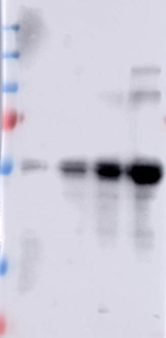
to -protoxin -activated toxin or -C-terminal            laboratory. The anti-C-terminal antibody was
fragment were incubated with 200 µg solubilized         raised in this work as reported above. For
BBMV proteins in 500 µl for 1 h at 4 °C, the            western blot assays the PVDF membranes were
unbound BBMV proteins were removed by                   blocked with 5 % skimmed milk in PBS buffer
centrifugation at 18,400 xg for 10 min at 4 °C.         pH 7.4 plus 0.1 % Tween 20, for 1 h at room
The coupled CNBr-agarose beads containing the           temperature. The membranes were rinsed once
bound proteins from BBMV were washed five               with same buffer. The different proteins were
times with 500 µl PBS supplemented with 1 M             detected after 1 h incubation with the
NaCl, followed by five washes with 500 µl PBS           corresponding polyclonal antibody (anti-CAD
to remove unbound proteins. The proteins that           1/20,000; anti-ALP and anti-APN 1/10,000; anti-
remained bound to the CNBr-protoxin, -                  Cry1Ab 1/20,000 and anti-C-terminal 1/20,000
activated toxin or -C-terminal agarose beads            dilutions) and 1 h with goat anti-rabbit HPR
were dissociated after 5 min at 100 ºC in 50 µl         secondary antibody (Santa Cruz) (1/20,000
loading buffer (100 mM TrisCl, 200 mM DTT, 4            dilution). Finally visualized by incubation with
% SDS w/v, 0.2 % bromophenol blue w/v, 20 %             Super Signal chemiluminescence substrate
glycerol v/v, pH 6.8). As negative control, the         (Pierce), according to the manufacturer
activated CNBr agarose was incubated without            instructions.

                                                                                                              Downloaded from http://www.jbc.org/ by guest on December 17, 2018
Cry1Ab protein, blocked as described above, and
incubated with solubilized BBMV. The pulled             ACKNOWLEDGMENTS
down proteins were separated in 10 % SDS-               We thank Lizbeth Cabrera for technical
PAGE and transferred to PVDF membrane that              assistance. This work was supported in part by
were revealed in western blot assays using anti-        CONACyT Fronteras 008, and PAPIIT
CAD, anti-ALP and anti-APN polyclonal                   IN202718 and IN213514.
antibodies as described below.
                                                        Conflict of interest: The authors declare that they
Western blot assays. The anti-Cry1Ab, anti-             have no conflicts of interest with the contents of
CAD, anti-ALP and anti-APN polyclonal                   this article.
antibodies were previously raised in our

References

1.     James, C. (2010) Global Status of Commercialized Biotech/GM Crops: 2010. (ISAAA ed.,
       Ithaca, NY
2.     Sanahuja, G., Banakar, R., Twyman, R. M., Capell, T., and Christou, P. (2011) Bacillus
       thuringiensis: a century of research, development and commercial applications. Plant
       biotechnology journal 9, 283-300
3.     Mendelsohn, M., Kough, J., Vaituzis, Z., and Matthews, K. (2003) Are Bt crops safe? Nature
       biotechnology 21, 1003-1009
4.     Nicolia, A., Manzo, A., Veronesi, F., and Rosellini, D. (2014) An overview of the last 10 years of
       genetically engineered crop safety research. Critical reviews in biotechnology 34, 77-88
5.     Comas, C., Lumbierres, B., Pons, X., and Albajes, R. (2014) No effects of Bacillus thuringiensis
       maize on nontarget organisms in the field in southern Europe: a meta-analysis of 26 arthropod
       taxa. Transgenic research 23, 135-143
6.     Downes, S., Mahon, R. J., Rossiter, L., Kauter, G., Leven, T., Fitt, G., and Baker, G. (2010)
       Adaptive management of pest resistance by Helicoverpa species (Noctuidae) in Australia to the
       Cry2Ab Bt toxin in Bollgard II(R) cotton. Evolutionary applications 3, 574-584
7.     Hutchison, W. D., Burkness, E. C., Mitchell, P. D., Moon, R. D., Leslie, T. W., Fleischer, S. J.,
       Abrahamson, M., Hamilton, K. L., Steffey, K. L., Gray, M. E., Hellmich, R. L., Kaster, L. V.,
       Hunt, T. E., Wright, R. J., Pecinovsky, K., Rabaey, T. L., Flood, B. R., and Raun, E. S. (2010)

                                                   8
Areawide suppression of European corn borer with Bt maize reaps savings to non-Bt maize
      growers. Science 330, 222-225
8.    Tabashnik, B. E., Sisterson, M. S., Ellsworth, P. C., Dennehy, T. J., Antilla, L., Liesner, L.,
      Whitlow, M., Staten, R. T., Fabrick, J. A., Unnithan, G. C., Yelich, A. J., Ellers-Kirk, C., Harpold,
      V. S., Li, X., and Carriere, Y. (2010) Suppressing resistance to Bt cotton with sterile insect
      releases. Nature biotechnology 28, 1304-1307
9.    Lu, Y., Wu, K., Jiang, Y., Guo, Y., and Desneux, N. (2012) Widespread adoption of Bt cotton and
      insecticide decrease promotes biocontrol services. Nature 487, 362-365
10.   Wan, P., Wu, K., Huang, M., Yu, D., and Wu, J. (2008) Population dynamics of Spodoptera litura
      (Lepidoptera: Noctuidae) on Bt cotton in the Yangtze River Valley of China. Environmental
      entomology 37, 1043-1048
11.   Dively, G. P., Venugopal, P. D., Bean, D., Whalen, J., Holmstrom, K., Kuhar, T. P., Doughty, H.
      B., Patton, T., Cissel, W., and Hutchison, W. D. (2018) Regional pest suppression associated with
      widespread Bt maize adoption benefits vegetable growers. Proceedings of the National Academy
      of Sciences of the United States of America 115, 3320-3325
12.   Wu, Y. D. (2014) Detection and Mechanisms of Resistance Evolved in Insects to Cry Toxins from
      Bacillus thuringiensis. Adv Insect Physiol 47, 297-342

                                                                                                              Downloaded from http://www.jbc.org/ by guest on December 17, 2018
13.   Tabashnik, B. E., and Carriere, Y. (2017) Surge in insect resistance to transgenic crops and
      prospects for sustainability. Nature biotechnology 35, 926-935
14.   Pardo-Lopez, L., Soberón, M., and Bravo, A. (2013) Bacillus thuringiensis insecticidal three-
      domain Cry toxins: mode of action, insect resistance and consequences for crop protection. FEMS
      microbiology reviews 37, 3-22
15.   Adang, M. J., Crickmore, N., and Jurat-Fuentes, J. L. (2014) Diversity of Bacillus thuringiensis
      Crystal Toxins and Mechanism of Action. Adv Insect Physiol 47, 39-87
16.   Arenas, I., Bravo, A., Soberón, M., and Gómez, I. (2010) Role of alkaline phosphatase from
      Manduca sexta in the mechanism of action of Bacillus thuringiensis Cry1Ab toxin. The Journal of
      biological chemistry 285, 12497-12503
17.   Zhang, X., Candas, M., Griko, N. B., Taussig, R., and Bulla, L. A., Jr. (2006) A mechanism of
      cell death involving an adenylyl cyclase/PKA signaling pathway is induced by the Cry1Ab toxin
      of Bacillus thuringiensis. Proceedings of the National Academy of Sciences of the United States of
      America 103, 9897-9902
18.   Tabashnik, B. E., Zhang, M., Fabrick, J. A., Wu, Y., Gao, M., Huang, F., Wei, J., Zhang, J.,
      Yelich, A., Unnithan, G. C., Bravo, A., Soberón, M., Carriere, Y., and Li, X. (2015) Dual mode of
      action of Bt proteins: protoxin efficacy against resistant insects. Scientific reports 5, 15107
19.   Siqueira, H. A., Nickerson, K. W., Moellenbeck, D., and Siegfried, B. D. (2004) Activity of gut
      proteinases from Cry1Ab-selected colonies of the European corn borer, Ostrinia nubilalis
      (Lepidoptera: Crambidae). Pest management science 60, 1189-1196
20.   Anilkumar, K. J., Rodrigo-Simon, A., Ferre, J., Pusztai-Carey, M., Sivasupramaniam, S., and
      Moar, W. J. (2008) Production and characterization of Bacillus thuringiensis Cry1Ac-resistant
      cotton bollworm Helicoverpa zea (Boddie). Applied and environmental microbiology 74, 462-469
21.   Gómez, I., Sanchez, J., Munoz-Garay, C., Matus, V., Gill, S. S., Soberón, M., and Bravo, A.
      (2014) Bacillus thuringiensis Cry1A toxins are versatile proteins with multiple modes of action:
      two distinct pre-pores are involved in toxicity. The Biochemical journal 459, 383-396
22.   Fabrick, J. A., and Tabashnik, B. E. (2007) Binding of Bacillus thuringiensis toxin Cry1Ac to
      multiple sites of cadherin in pink bollworm. Insect biochemistry and molecular biology 37, 97-
      106
23.   Li, X. D., Zhao, F., Qiu, X. F., Ren, X. M., Mo, X. T., Ding, X. Z., Xia, L. Q., and Sun, Y. J.
      (2018) The full-length Cry1Ac protoxin without proteolytic activation exhibits toxicity against
      insect cell line CF-203. Journal of invertebrate pathology 152, 25-29
24.   Evdokimov, A. G., Moshiri, F., Sturman, E. J., Rydel, T. J., Zheng, M., Seale, J. W., and Franklin,
      S. (2014) Structure of the full-length insecticidal protein Cry1Ac reveals intriguing details of

                                                   9
toxin packaging into in vivo formed crystals. Protein science : a publication of the Protein Society
      23, 1491-1497
25.   Rajamohan, F., Alcantara, E., Lee, M. K., Chen, X. J., Curtiss, A., and Dean, D. H. (1995) Single
      amino acid changes in domain II of Bacillus thuringiensis CryIAb delta-endotoxin affect
      irreversible binding to Manduca sexta midgut membrane vesicles. Journal of bacteriology 177,
      2276-2282
26.   Rajamohan, F., Cotrill, J. A., Gould, F., and Dean, D. H. (1996) Role of domain II, loop 2
      residues of Bacillus thuringiensis CryIAb delta-endotoxin in reversible and irreversible binding to
      Manduca sexta and Heliothis virescens. The Journal of biological chemistry 271, 2390-2396
27.   Jenkins, J. L., and Dean, D. H. (2000) Exploring the mechanism of action of insecticidal proteins
      by genetic engineering methods. Genetic engineering 22, 33-54
28.   Lee, M. K., Rajamohan, F., Jenkins, J. L., Curtiss, A. S., and Dean, D. H. (2000) Role of two
      arginine residues in domain II, loop 2 of Cry1Ab and Cry1Ac Bacillus thuringiensis delta-
      endotoxin in toxicity and binding to Manduca sexta and Lymantria dispar aminopeptidase N.
      Molecular microbiology 38, 289-298
29.   Pacheco, S., Gómez, I., Gill, S. S., Bravo, A., and Soberón, M. (2009) Enhancement of
      insecticidal activity of Bacillus thuringiensis Cry1A toxins by fragments of a toxin-binding

                                                                                                             Downloaded from http://www.jbc.org/ by guest on December 17, 2018
      cadherin correlates with oligomer formation. Peptides 30, 583-588
30.   Rodriguez-Almazan, C., Zavala, L. E., Munoz-Garay, C., Jimenez-Juarez, N., Pacheco, S.,
      Masson, L., Soberón, M., and Bravo, A. (2009) Dominant negative mutants of Bacillus
      thuringiensis Cry1Ab toxin function as anti-toxins: demonstration of the role of oligomerization
      in toxicity. PloS one 4, e5545
31.   Smedley, D. P., and Ellar, D. J. (1996) Mutagenesis of three surface-exposed loops of a Bacillus
      thuringiensis insecticidal toxin reveals residues important for toxicity, receptor recognition and
      possibly membrane insertion. Microbiology 142 ( Pt 7), 1617-1624
32.   Chen, J., Hua, G., Jurat-Fuentes, J. L., Abdullah, M. A., and Adang, M. J. (2007) Synergism of
      Bacillus thuringiensis toxins by a fragment of a toxin-binding cadherin. Proceedings of the
      National Academy of Sciences of the United States of America 104, 13901-13906
33.   Gómez, I., Dean, D. H., Bravo, A., and Soberón, M. (2003) Molecular basis for Bacillus
      thuringiensis Cry1Ab toxin specificity: two structural determinants in the Manduca sexta Bt-R1
      receptor interact with loops alpha-8 and 2 in domain II of Cy1Ab toxin. Biochemistry 42, 10482-
      10489
34.   Xie, R., Zhuang, M., Ross, L. S., Gómez, I., Oltean, D. I., Bravo, A., Soberón, M., and Gill, S. S.
      (2005) Single amino acid mutations in the cadherin receptor from Heliothis virescens affect its
      toxin binding ability to Cry1A toxins. The Journal of biological chemistry 280, 8416-8425
35.   de Maagd, R. A., Bravo, A., Berry, C., Crickmore, N., and Schnepf, H. E. (2003) Structure,
      diversity, and evolution of protein toxins from spore-forming entomopathogenic bacteria. Annual
      review of genetics 37, 409-433
36.   Choma, C. T., Surewicz, W. K., Carey, P. R., Pozsgay, M., Raynor, T., and Kaplan, H. (1990)
      Unusual proteolysis of the protoxin and toxin from Bacillus thuringiensis. Structural implications.
      Eur J Biochem 189, 523-527
37.   Schnepf, E., Crickmore, N., Van Rie, J., Lereclus, D., Baum, J., Feitelson, J., Zeigler, D. R., and
      Dean, D. H. (1998) Bacillus thuringiensis and its pesticidal crystal proteins. Microbiology and
      molecular biology reviews : MMBR 62, 775-806
38.   Park, Y., Gonzalez-Martinez, R. M., Navarro-Cerrillo, G., Chakroun, M., Kim, Y., Ziarsolo, P.,
      Blanca, J., Canizares, J., Ferre, J., and Herrero, S. (2014) ABCC transporters mediate insect
      resistance to multiple Bt toxins revealed by bulk segregant analysis. BMC biology 12, 46
39.   Gahan, L. J., Pauchet, Y., Vogel, H., and Heckel, D. G. (2010) An ABC transporter mutation is
      correlated with insect resistance to Bacillus thuringiensis Cry1Ac toxin. PLoS genetics 6,
      e1001248
40.   Tabashnik, B. E. (2015) ABCs of Insect Resistance to Bt. PLoS genetics 11, e1005646

                                                  10
41. Adegawa, S., Nakama, Y., Endo, H., Shinkawa, N., Kikuta, S., and Sato, R. (2017) The domain II
loops of Bacillus thuringiensis Cry1Aa form an overlapping interaction site for two Bombyx mori
larvae functional receptors, ABC transporter C2 and cadherin-like receptor. Biochimica et
biophysica acta 1865, 220-231
42. Eder, J., and Fersht, A. R. (1995) Pro-sequence-assisted protein folding. Molecular microbiology
16, 609-614
43. Iacovache, I., Degiacomi, M. T., Pernot, L., Ho, S., Schiltz, M., Dal Peraro, M., and van der Goot,
F. G. (2011) Dual chaperone role of the C-terminal propeptide in folding and oligomerization of
the pore-forming toxin aerolysin. PLoS Pathog 7, e1002135
44. Sellman, B. R., and Tweten, R. K. (1997) The propeptide of Clostridium septicum alpha toxin
functions as an intramolecular chaperone and is a potent inhibitor of alpha toxin-dependent
cytolysis. Molecular microbiology 25, 429-440
45. de Maagd, R. A., Kwa, M. S., van der Klei, H., Yamamoto, T., Schipper, B., Vlak, J. M.,
Stiekema, W. J., and Bosch, D. (1996) Domain III substitution in Bacillus thuringiensis delta-
endotoxin CryIA(b) results in superior toxicity for Spodoptera exigua and altered membrane
protein recognition. Applied and environmental microbiology 62, 1537-1543
46. Bravo, A., Gómez, I., Porta, H., Garcia-Gomez, B. I., Rodriguez-Almazan, C., Pardo, L., and
Downloaded from http://www.jbc.org/ by guest on December 17, 2018
Soberón, M. (2013) Evolution of Bacillus thuringiensis Cry toxins insecticidal activity. Microbial
biotechnology 6, 17-26
47. Badran, A. H., Guzov, V. M., Huai, Q., Kemp, M. M., Vishwanath, P., Kain, W., Nance, A. M.,
Evdokimov, A., Moshiri, F., Turner, K. H., Wang, P., Malvar, T., and Liu, D. R. (2016)
Continuous evolution of Bacillus thuringiensis toxins overcomes insect resistance. Nature 533,
58-63
48. Lereclus, D., Agaisse, H., Gominet, M., and Chaufaux, J. (1995) Overproduction of encapsulated
insecticidal crystal proteins in a Bacillus thuringiensis spo0A mutant. Bio/technology 13, 67-71
49. Meza, R., Nunez-Valdez, M. E., Sanchez, J., and Bravo, A. (1996) Isolation of Cry1Ab protein
mutants of Bacillus thuringiensis by a highly efficient PCR site-directed mutagenesis system.
FEMS microbiology letters 145, 333-339
50. Thomas, W. E., and Ellar, D. J. (1983) Bacillus thuringiensis var israelensis crystal delta-
endotoxin: effects on insect and mammalian cells in vitro and in vivo. Journal of cell science 60,
181-197
51. Wolfersberger, M. G. (1993) Preparation and partial characterization of amino acid transporting
brush border membrane vesicles from the larval midgut of the gypsy moth (Lymantria dispar).
Archives of insect biochemistry and physiology 24, 139-147
52. Flores-Escobar, B., Rodriguez-Magadan, H., Bravo, A., Soberón, M., and Gómez, I. (2013)
Differential role of Manduca sexta aminopeptidase-N and alkaline phosphatase in the mode of
action of Cry1Aa, Cry1Ab, and Cry1Ac toxins from Bacillus thuringiensis. Applied and
environmental microbiology 79, 4543-4550
53. Torres-Quintero, M. C., Gómez, I., Pacheco, S., Sanchez, J., Flores, H., Osuna, J., Mendoza, G.,
Soberón, M., and Bravo, A. (2018) Engineering Bacillus thuringiensis Cyt1Aa toxin specificity
from dipteran to lepidopteran toxicity. Scientific reports 8, 4989
54. Jimenez-Juarez, N., Munoz-Garay, C., Gómez, I., Saab-Rincon, G., Damian-Almazo, J. Y., Gill,
S. S., Soberón, M., and Bravo, A. (2007) Bacillus thuringiensis Cry1Ab mutants affecting
oligomer formation are non-toxic to Manduca sexta larvae. The Journal of biological chemistry
282, 21222-21229
Table 1. Equilibrium dissociation constant (Kd in nM) for interactions between wild type and
mutant Cry1Ab protoxins and activated toxins with recombinant CR12, ALP, and APN
11CR12 ALP APN
Type of Cry1Ab Protoxin Toxin Protoxin Toxin Protoxin Toxin
Wild type 85±14 35±4 28±2 296±3 79±8 296±5
RR368-9AA NB NB 22±3 381±36 55±6 572±53
F371A NB NB 16±2 242±48 40±5 214±8
G439D NB NB 14±2 806±88 30±4 870±32
NB, No binding
Downloaded from http://www.jbc.org/ by guest on December 17, 2018
Figures
Figure 1. Expression and purification of recombinant C-terminal region of the Cry1Ab protoxin
in E. coli cells.
The C-terminal fragment was purified by affinity chromatography through a nickel affinity
column by using the His-tag provided in the pET22b cloning vector and eluted with 250 mM
imidazole. The samples were analyzed by SDS-PAGE (10 % acrylamide) stained with
Coomassie blue. Molecular masses are indicated in kDa.
12A B
1.4 0.7
1.2 0.6
1.0 0.5
Absorbance 490 nm
Absorbance 490 nm
0.8 0.4
0.6 0.3
0.4 Cry1Ab Protoxin 0.2 Cry1Ab Protoxin
Cry1Ab Toxin Cry1Ab Toxin
Cry1Ab C-Terminal Cry1Ab C-Terminal
0.2 0.1
0.0 0.0
0 50 100 150 200 250 0 50 100 150 200 250
Concentration (nM) Concentration (nM)
Downloaded from http://www.jbc.org/ by guest on December 17, 2018
Figure 2. ELISA binding analysis of Cry1Ab-protoxin, Cry1Ab-activated toxin and C-terminal
fragment from Cry1Ab to BBMV purified from M. sexta midgut tissue.
Panel A, shows the binding of these proteins revealed with anti-Cry1Ab antibody that was raised
against the activated Cry1Ab-toxin. This antibody does not cross-react with C-terminal fragment
(Fig. S1). Panel B, shows the binding of these proteins revealed with anti-C-terminal antibody
that was raised against the purified C-terminal fragment. This antibody does not cross-react with
Cry1Ab-activated toxin (Fig. S1). A total of 1 µg protein of BBMV was bound to each of the
wells of the ELISA plate. Each experiment was performed in duplicate with a total of three
repetitions. Standard deviations are shown.
13A B 800 800 800
APN ALP CR7-12
1.2 APN 700 700 700
600 600 600
Response (RU)
1.0
ALP
Absorbance 490 nm
500 500 500
0.8
400 400
400
0.6
300 300 300
0.4
200 200
CR7-12 200
0.2 100 100
100
0.0 0 0 0
0 20 40 60 80 100 0 20 40 60 80 100 0 20 40 60 80 100
0 100 200 300 400 500
Time (sec) Time (sec) Time (sec)
Concentration (nM)
Figure 3. Binding analysis of C-terminal fragment to CAD, APN and ALP.
Panel A, ELISA binding assays of C-terminal fragment of Cry1Ab to the purified recombinant
CR7-12 fragment, ALP or APN expressed in E. coli cells. Each experiment was performed in
Downloaded from http://www.jbc.org/ by guest on December 17, 2018
duplicate with a total of three repetitions. Standard deviations are shown in the figure. Panel B,
SPR binding analyses of C-terminal fragment to Cry1Ab-receptors performed by immobilizing
the C-terminal fragment by conventional amine coupling. Sensograms of serial doubling dilutions
of each purified receptor (CAD (CR7-12), APN and ALP) are shown.
14A Protoxin (5 nM) Toxin (5 nM) C-terminal (5 nM)
kDa M 0.6 1.2 2.5 5 CAD (µg) kDa M 0.6 1.2 2.5 5 CAD (µg) kDa M 0.6 1.2 2.5 5 CAD (µg)
250 250 250
130 130 130
95 95 95
72 72 191 337 477 536 72
100 215 347 464 0 0 0 0
55 55 55
B Protoxin (200 nM) Toxin (200 nM) C-terminal (200 nM)
kDaM 0.6 1.2 2.5 5 ALP (µg) kDa 0.6 1.2 2.5 5 ALP (µg) kDa 0.6 1.2 2.5 5 ALP (µg)
250 250
130 250
130
Downloaded from http://www.jbc.org/ by guest on December 17, 2018
95 95 130
72 72 95
55 72
55
100 366 767 1095 67 365 706 891 55
18 192 649 1101
C Protoxin (200 nM) Toxin (200 nM) C-terminal (200 nM)
kDa M 2.5 5 10 APN (µg) kDa 2.5 5 10 APN kDa 2.5 5 10 APN (µg)
250 250 (µg) 250
130 130 130
95 100 205 876 95 171 185 438 95
152 258 953
72 72 72
55 55 55
Figure 4. Ligand blot assays of Cry1Ab-protoxin, Cry1Ab-activated toxin and C-terminal
fragment from Cry1Ab to the purified recombinant CAD (CR7-12) fragment, ALP or APN
proteins expressed in E. coli cells.
Serial dilutions of the recombinant receptor proteins were loaded in the SDS-PAGE and
transferred to the PVDF membranes. These blots were used for ligand blot binding assays.
15Numbers under the bands represent the percentage of each band on the blot calculated after
densitometry analysis of the bands, using ImageJ software and selecting one band of the protoxin
bound to the lower concentration of receptor as 100% reference. Molecular masses are indicated
in kDa.
Panel A, ligand blot assays of 5 nM of Cry1Ab-protoxin, Cry1Ab-activated toxin and C-terminal
fragment from Cry1Ab to CAD (CR7-12). Panel B, ligand blot assays of 200 nM of Cry1Ab-
protoxin, Cry1Ab-activated toxin and C-terminal fragment from Cry1Ab to ALP. Panel C, ligand
blot assays of 200 nM of Cry1Ab-protoxin, Cry1Ab-activated toxin and C-terminal fragment
Downloaded from http://www.jbc.org/ by guest on December 17, 2018
from Cry1Ab to APN.
anti-CAD Anti-APN Anti-ALP
Protoxin
Protoxi
Protoxi
C-term
C-term
C-term
Toxin
Toxin
Toxi
n
n
n
kDa kDa kD
a
250 250 250
130 130 130
95 95 95
72 72 72
55 55 55
36 36 36
Figure 5. Pull down assays of BBMV proteins using Cry1Ab-protoxin, Cry1Ab-activated toxin
and C-terminal fragment from Cry1Ab bound to CNBr agarose.
The precipitated BBMV proteins with Cry1Ab-protoxin, Cry1Ab-activated toxin and C-terminal
16fragment from Cry1Ab were reveled with specific antibodies that recognize CAD, APN or ALP
proteins. Molecular masses are indicated in kDa.
Downloaded from http://www.jbc.org/ by guest on December 17, 2018
17A Binding to CAD receptor
1.0 1.0
Cry1Ab
0.8 0.8
Absorbance 490 nm
Absorbance 490 nm
Cry1Ab
0.6 0.6 F371A
F371A RR368-369AA
0.4 0.4
RR368-369AA G439D
0.2 0.2
G439D
0.0 0.0
0 200 400 600 800 0 200 400 600 800
Cry1Ab Protoxin (nM) Cry1Ab Toxin (nM)
B Binding to ALP receptor
F371A Cry1Ab
Cry1Ab 1.2
Downloaded from http://www.jbc.org/ by guest on December 17, 2018
1.2
RR368-369AA
1.0
1.0
G439
F371A
Absorbance 490 nm
Absorbance 490 nm
0.8
D 0.8
RR368-369AA
0.6 0.6
0.4 0.4
G439D
0.2 0.2
0.0 0.0
0 100 200 300 400 500 0 100 200 300 400 500
Cry1Ab Protoxin (nM) Cry1Ab Toxin (nM)
C Binding to APN receptor
1.4
Cry1Ab
1.2 Cry1Ab
1.2
1.0 F371A
1.0 F371A
Absorbance 490 nm
Absorbance 490 nm
0.8
RR368-369AA
0.8 RR368-369AA
G439D
0.6
0.6 G439D
0.4 0.4
0.2 0.2
0.0 0.0
0 20 40 60 80 100 0 50 100 150 200 250
Cry1Ab Protoxin (nM) Cry1Ab Toxin (nM)
Figure
6. ELISA binding assays of Cry1Ab and Cry1Ab-mutant proteins to the purified recombinant
CAD (CR12), ALP or APN expressed in E. coli cells.
The three Cry1Ab mutants (Cry1Ab-RR368-369AA, Cry1Ab-F371A and Cry1Ab-G439D) were
affected in toxicity against M. sexta larvae.
18Panel A, Binding of protoxin or activated toxin molecules to purified CAD (CR12) receptor
fragment. Panel B, Binding of protoxin or activated toxin molecules to purified ALP. Panel C,
Binding of protoxin or activated toxin molecules to purified APN. Each experiment was
performed in duplicate with a total of three repetitions. Standard deviations are shown in the
figure.
Downloaded from http://www.jbc.org/ by guest on December 17, 2018
Figure 7. Proposed mechanism of action of Cry proteins from Bt.
1, the parasporal crystals are solubilized into protoxin. 2, the C-terminal region of protoxin binds
with highly abundant APN and ALP receptors. 3, the protoxin binds to CAD receptor by the
loops of domain II. 4, proteases activate the protoxin inducing oligomer formation. 5, the
oligomer inserts into the membrane, forming a pore that kills the cell.
The interaction with APN and ALP by C-terminal region, helps protoxin to reach CAD receptor
before proteolytical activation, and the interaction with CAD in the presence of midgut proteases
19induce the formation of a robust oligomer that display high pore formation activity with single
conductance and high open probability as previously demonstrated (18).
Downloaded from http://www.jbc.org/ by guest on December 17, 2018
20The C-terminal protoxin domain of Bacillus thuringiensis Cry1Ab toxin has a
functional role in binding to GPI-anchored receptors in the insect midgut
Arlen Peña-Cardeña, Ricardo Grande, Jorge Sánchez, Bruce E. Tabashnik, Alejandra
Bravo, Mario Soberón and Isabel Gómez
J. Biol. Chem. published online November 1, 2018
Access the most updated version of this article at doi: 10.1074/jbc.RA118.005101
Alerts:
• When this article is cited
• When a correction for this article is posted
Click here to choose from all of JBC's e-mail alerts
Downloaded from http://www.jbc.org/ by guest on December 17, 2018You can also read