Distribution of Tetrodotoxin in the New Zealand Clam, Paphies australis, Established Using Immunohistochemistry and Liquid Chromatography-Tandem ...
←
→
Page content transcription
If your browser does not render page correctly, please read the page content below
toxins
Article
Distribution of Tetrodotoxin in the New Zealand
Clam, Paphies australis, Established Using
Immunohistochemistry and Liquid
Chromatography-Tandem Quadrupole
Mass Spectrometry
Laura Biessy 1,2, *, Kirsty F. Smith 1 , Michael J. Boundy 1 , Stephen C. Webb 1      ID
                                                                                           , Ian Hawes 2
and Susanna A. Wood 1 ID
 1    Cawthron Institute, Private Bag 2, Nelson 7010, New Zealand; kirsty.smith@cawthron.org.nz (K.F.S.);
      michael.boundy@cawthron.org.nz (M.J.B.); steve.webb@cawthron.org.nz (S.C.W.);
      susie.wood@cawthron.org.nz (S.A.W.)
 2    Department of Biological Sciences, University of Waikato, Private Bag 3105, Hamilton 3240, New Zealand;
      ian.hawes@waikato.ac.nz
 *    Correspondence: laura.biessy@cawthron.org.nz; Tel.: +64-035-482-319

 Received: 13 June 2018; Accepted: 2 July 2018; Published: 6 July 2018

 Abstract: Tetrodotoxin (TTX) is one of the most potent neurotoxins known. It was originally thought
 to only occur in puffer fish but has now been identified in twelve different classes of freshwater
 and marine organisms, including bivalves. Despite being one of the world’s most studied biotoxins,
 its origin remains uncertain. There is contradictory evidence regarding the source of TTX and its
 pathway through food webs. To date, the distribution of TTX has not been examined in bivalves.
 In the present study, 48 Paphies australis, a TTX-containing clam species endemic to New Zealand,
 were collected. Thirty clams were dissected, and organs and tissues pooled into five categories
 (siphons, digestive gland, adductor muscles, and the ‘rest’) and analyzed for TTX using liquid
 chromatography-mass spectrometry (LC-MS). The micro-distribution of TTX was visualized in
 the remaining 18 individuals using an immunohistological technique incorporating a TTX-specific
 monoclonal antibody. The LC-MS analysis revealed that siphons contained the highest concentrations
 of TTX (mean 403.8 µg/kg). Immunohistochemistry analysis showed TTX in the outer cells of the
 siphons, but also in the digestive system, foot, and gill tissue. Observing TTX in organs involved in
 feeding provides initial evidence to support the hypothesis of an exogenous source in P. australis.

 Keywords: biotoxin; localization; marine bivalves; monoclonal antibody; neurotoxin

 Key Contribution: Using immunohistochemistry and liquid chromatography-mass spectrometry,
 we demonstrated that the highest concentrations of tetrodotoxin in Paphies australis (the New Zealand
 surf clam) occurred in the siphons.

1. Introduction
      Tetrodotoxin (TTX) is a lethal neurotoxin that selectively binds and blocks voltage-gated sodium
channels [1]. It is one of the most potent natural substances known, with ingestion of 1 to 2 mg enough
to kill a 50-kg human [2]. Tetrodotoxin was named after the puffer fish family Tetraodontidae and was
first isolated in 1910 [3], although characterization of the chemical structure was not achieved until
1964 [4–6].

Toxins 2018, 10, 282; doi:10.3390/toxins10070282                                 www.mdpi.com/journal/toxins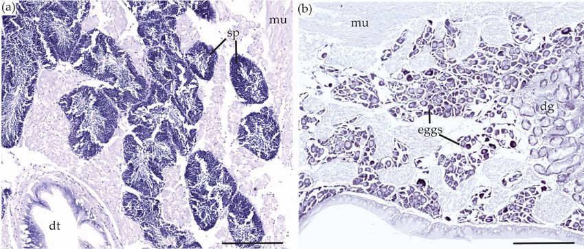
Toxins 2018, 10, 282                                                                                2 of 14

       Tetrodotoxin has long been known as the causative agent in puffer fish poisoning [7,8] and was
first recorded in 1774 by Captain James Cook who detailed the effects on his crew from the consumption
of a fish from New Caledonia, now thought to be a puffer fish [9]. It was originally thought that TTX
only occurred in puffer fish, but has since been identified in 12 different classes of marine, freshwater,
and terrestrial vertebrate and invertebrate organisms [10].
       Despite TTX being one of the most studied biotoxins in the world, its origin remains uncertain.
There is contradictory evidence regarding whether the source of TTX is exogenous or endogenous,
and the pathways and mechanisms through which TTX is incorporated in the food web are
unknown [11]. The wide distribution of TTX in many genetically unrelated species and its high
spatiotemporal variation among TTX-containing species suggest that the toxin comes from an
exogenous source such as accumulation through diet or symbiotic bacteria [12–14]. However, there is
also evidence for an endogenous source in terrestrial species. Research suggesting this began in the late
1980s when TTX-containing tree frogs (Atelopus oxyrhynchus) were shown to retain high levels of toxicity
in controlled environment for three years [15]. Hatched frogs (Atelopus varius) raised in captivity were
also found to contain TTX [16]. Similar experiments undertaken in newts (Taricha granulosa) have
shown that individuals regenerated their levels of TTX in their skin when kept in captivity and fed a
TTX-free diet [17].
       Tetrodotoxin has been identified in nine different bivalve species from six countries. The first
report of TTX in marine bivalves was in Japan in 1993 [18] when the toxin was found in the digestive
gland of scallop (Patinopecten yessoensis) after a bloom of the dinoflagellate Alexandrium tamarense.
Two decades after the first detection in marine bivalves, McNabb et al. [19] reported high levels
(800 µg/kg) of TTX in the surf clam Paphies australis in New Zealand. That research triggered
further investigations on TTX in bivalves globally. Turner et al. [20] detected TTX in Pacific oysters
(Crassostrea gigas) and common blue mussels (Mytilus edulis) in England. Shortly after, Vlamis et al. [21]
reported moderate levels (up to 223 µg/kg) of TTX in M. edulis harvested in 2012 from the Greek
islands. Oysters and mussels from production areas in the Netherlands also tested positive for TTX [22]
and trace detections (2.22 µg/kg) have also been reported in a clam species (Ruditapes philippinarum) in
China [23].
       Recent advances in chemical detection and quantification methods (i.e., Liquid Chromatography-
Tandem Quadrupole Mass Spectrometry; LC-MS/MS) have allowed the development of very specific,
sensitive, and accurate methods to measure TTX concentrations [24,25]. The use of these methods
makes it possible to investigate and quantify the distribution of TTX in specific organs within bivalves.
To date, the micro-distribution of TTX has been studied in a wide range of organisms including:
three species of pufferfish [26,27]; a sea-slug and a flatworm [28]; a nemertean and a turbellarian [29];
and a ribbon worm [30] but has never been investigated in bivalves. Improving knowledge on the
location of TTX in marine bivalves may provide new insights on the ecological functions and sources
of the neurotoxin in these organisms. For example, finding the toxin in the outside epithelium or in
the reproductive system could indicate a defense mechanism, whereas the identification of TTX in the
digestive tract could indicate a possible exogenous source.
       In the present study, LC-MS/MS was used to determine which P. australis organs contained
TTX. These data were then used to guide immunohistological experiments using a TTX-specific
monoclonal antibody [31,32] to determine the micro-distribution of TTX within these organs. Based on
similar studies on the neurotoxin saxitoxin (produced by planktonic marine dinoflagellates and
freshwater cyanobacterial species) in clams Saxidomus gigantea [33] and in other Paphies species [34],
we hypothesized that TTX would be located in the siphons where it would act as a defense mechanism,
and in the reproductive system of P. australis where it would be transferred to larvae to aid in their
protection post-spawning.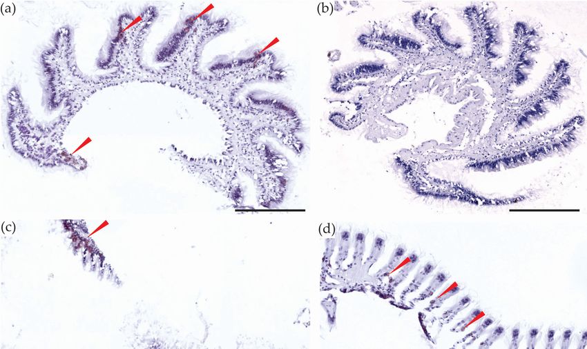
Toxins 2018, 10, 282                                                                                                   3 of 14

   2. Results and Discussion
Toxins 2018, 10, x FOR PEER REVIEW                                                                                           3 of 14
       Tetrodotoxin was the most abundant analogue in the matrices analyzed by LC-MS/MS,
  accounting
two deoxy-TTXfor >99% of the total putative
                    analogues,     TTX analogues present. Traces
                                             5-deoxy-TTX      andof 4/6-epi-TTX, 4,9-anhydro-TTX,
                                                                     11-deoxy-TTX,                and two
                                                                                       were detected    but
  deoxy-TTX analogues,
concentrations  were tooputative   5-deoxy-TTX
                          low to allow         and
                                         accurate  11-deoxy-TTX,(Figure
                                                  quantification    were detected
                                                                           S1). but concentrations were
  too low to allow accurate quantification (Figure S1).
2.1. Siphons
   2.1. Siphons
      Quantification of TTX in P. australis organs by LC-MS/MS identified the highest concentration
        Quantification of TTX in P. australis organs by LC-MS/MS identified the highest concentration
of toxin in the siphons (mean 403.8 ± 32.8 µg/kg, Figure 1). One-way ANOVA showed a significant
   of toxin in the siphons (mean 403.8 ± 32.8 µg/kg, Figure 1). One-way ANOVA showed a significant
difference
   differencebetween
                 between  thethemean
                                   mean concentrations
                                           concentrations in in
                                                              organ
                                                                 organ groups
                                                                          groups (p (p
                                                                                     < 0.001), with
                                                                                        < 0.001),  withTukey’s
                                                                                                          Tukey’s  post-hoc
                                                                                                                       post-hoc test
identifying    the difference
   test identifying                 between
                        the difference           the siphons
                                             between             and and
                                                       the siphons      all other    groups
                                                                              all other        (p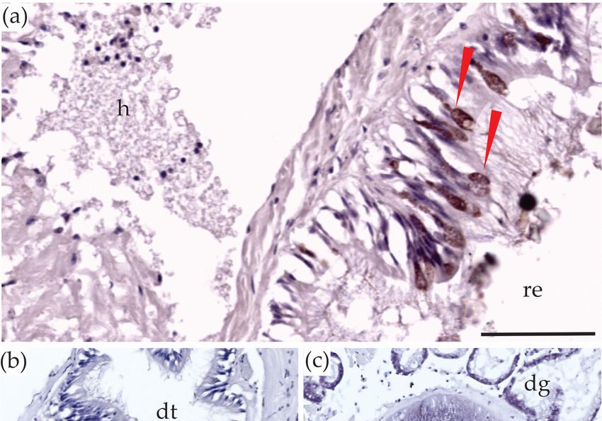
Toxins 2018, 10, 282 4 of 14
Toxins 2018, 10, x FOR PEER REVIEW 4 of 14
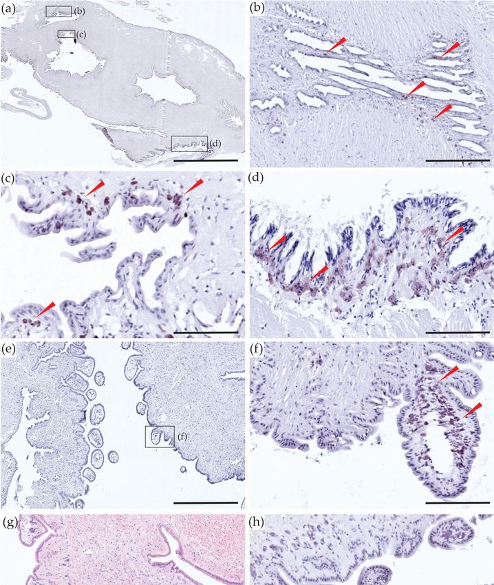
Figure 2. Longitudinal sections of the inhalant and exhalant siphons of Paphies australis. (a)
Figure 2. Longitudinal
Tetrodotoxin sections
(TTX)-specific of the inhalant
monoclonal and exhalant
antibody siphons of Paphies australis.
(mAB) immunohistological (a)longitudinal
staining in Tetrodotoxin
(TTX)-specific
section of siphons; (b–d) enlargements of different boxes on (a) to show micro-location ofsection
monoclonal antibody (mAB) immunohistological staining in longitudinal TTX
ofstaining,
siphons;identified
(b–d) enlargements
by the brown of different
color depositsboxes
shown onby(a)
redtoarrows;
show (e)micro-location
another view of TTX staining,
of TTX-specific
identified by the brown color
mAB immunohistological deposits
staining in theshown
outerby redofarrows;
layer (e) another
the siphon; view of TTX-specific
(f) enlargement of the box onmAB(e)
immunohistological
to show detailed viewstaining
of the in the outer
staining layer
in the of the
siphons siphon;
loops; (f) enlargement
(g) Hematoxylin and of the staining;
Eosin box on (e)
(h)to
show
mABdetailed
negativeview of the
control. staining
Scale bars =in200
theµmsiphons
(a), 50loops; (g) Hematoxylin
µm (b–d,f,h), 150 µm (e),and Eosin
and staining;
100 µm (g). (h) mAB
negative control. Scale bars = 200 µm (a), 50 µm (b–d,f,h), 150 µm (e), and 100 µm (g).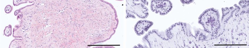
Toxins 2018, 10, 282                                                                                  5 of 14

      Bivalve siphons were an evolutionary development which enabled them to diversify into many
new ecological niches after the Paleozoic Era [37]. In bivalves, the two siphons are paired: the inhalant
siphon carries the water into the mantle cavity, and the exhalant siphon ejects the water from the
internal cavity [38]. The water flow from the siphons is variously used for feeding, respiration,
reproduction, and in locomotion for some bivalves [38]. Siphons also play a critical role in survival by
allowing water flow while buried [39]. Paphies australis and many other bivalves, bury themselves in the
sand and keep their siphons above the sediment surface to filter feed. It is well known that siphons are a
targeted food for predators including siphon-nipping fish such as the flatfish Pleuronectes platessa [40] or
shrimp [41]. Bivalves are negatively impacted by siphon-nipping predators because they require extra
energy for siphon regeneration and it reduces feeding opportunities [41]. Researchers have suggested
previously that biotoxins are accumulated as a chemical defense against predation. For example,
Kvitek [35] suggested that the clam S. gigantea retains saxitoxin in their siphons as a defense mechanism
against siphon-nipping predators and Kvitek & Bretz [42] demonstrated that sea otters stop grazing
on shellfish with high saxitoxin concentrations. We speculate that TTX accumulation in siphon tissues
could provide a similar chemical defense in P. australis. An elevated level of TTX in P. australis siphons
could also suggest that TTX is present in the water or within organisms in the water that they are
filtering while feeding, possibly indicating that the toxin comes from an exogenous source.

2.2. Foot
      After the Palaeozoic era, certain bivalve species developed an extensive foot, opening up new
life history strategies [37]. The development of a foot gave bivalves a distinct competitive advantage
by allowing them to rapidly dig down into the sand [43]. Some are also able to swim short distances
using their foot [38]. The LC-MS/MS analysis identified lower concentrations of TTX in foot tissue
(mean 44.4 ± 12.8 µg/kg, Figure 1) but no TTX was vizualized after immunostaining. In P. australis,
the foot is directly attached to the gonads. The toxic P. australis collected for this experiment were
relatively small (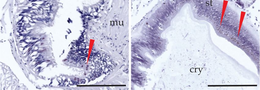
Toxins2018,
Toxins 2018,10,
             10,282
                 x FOR PEER REVIEW                                                                             66 of
                                                                                                                  of 14
                                                                                                                     14

in the digestive tracts of bivalve species, for example, the highest concentrations of saxitoxin was
biotoxins  in the
found in the      digestive
               viscera of M.tracts
                             edulisof bivalve
                                    [50]; domoicspecies,  for example,
                                                    acid was              the highest
                                                               mostly contained        concentrations
                                                                                    (>80%)             of saxitoxin
                                                                                            in mussel and   oyster’s
was found in the viscera of M. edulis [50]; domoic acid was mostly contained (>80%) in mussel and
digestive systems [51] and greater than 50% of microcystins were present in the digestive glands of
oyster’s digestive systems [51] and greater than 50% of microcystins were present in the digestive
freshwater
glands        mussels [52].
        of freshwater       This result
                         mussels          could
                                   [52]. This     indicate
                                               result  could that  TTX isthat
                                                                 indicate   sourced
                                                                                 TTX from   the diet
                                                                                      is sourced     of P.
                                                                                                  from  theaustralis.
                                                                                                             diet of
Tetrodotoxin
P.              may be initially
   australis. Tetrodotoxin  may stored     in the
                                  be initially     organs
                                                stored   in of
                                                             thethe digestive
                                                                  organs        system
                                                                           of the        and system
                                                                                  digestive  could be  transported
                                                                                                     and   could be
transported   to other
to other tissues       tissues and
                  and organs,  to useorgans,  to use asdefense.
                                        as a chemical     a chemical defense.

     Figure 3. Sections of the digestive system of Paphies australis. Tetrodotoxin-specific monoclonal
     Figure 3. Sections of the digestive system of Paphies australis. Tetrodotoxin-specific monoclonal
     antibody immunohistological staining in: (a) the rectum; (b) the intestinal epithelium; (c) the inside
     antibody immunohistological staining in: (a) the rectum; (b) the intestinal epithelium; (c) the inside
     layer of the stomach wall. The red arrows are showing the brown color deposits, indicating the
     layer of the stomach wall. The red arrows are showing the brown color deposits, indicating the presence
     presence of tetrodotoxin staining. cry = crystalline style, dg = tubules in the digestive gland, dt = lumen
     of tetrodotoxin staining. cry = crystalline style, dg = tubules in the digestive gland, dt = lumen of the
     of the digestive tract, h = haemocyte debris, mu = muscle tissue, re = rectum. Scale bars = 50 µm (a)
     digestive tract, h = haemocyte debris, mu = muscle tissue, re = rectum. Scale bars = 50 µm (a) and
     and 100 µm (b,c).
     100 µm (b,c).

2.5. The ‘rest’
2.5. The ‘rest’
      The ‘rest’ group contained all the organs and tissues that were not described above. It mainly
      The ‘rest’ group contained all the organs and tissues that were not described above. It mainly
included the
included   the gonads,
                gonads, the
                        the mantle,
                            mantle, the
                                     the labial
                                         labial palps,
                                                 palps, and
                                                         and the
                                                              the gills.
                                                                  gills. The
                                                                         The separation
                                                                               separation of
                                                                                          of these
                                                                                             these tissues
                                                                                                    tissues was
                                                                                                            was
not possible
not  possible due to
                   to the
                      the fragility
                          fragility and
                                    and small
                                        small size
                                                size of
                                                     of the
                                                        the P.
                                                            P.australis
                                                                australis collected.
                                                                           collected. The
                                                                                      The ‘rest’
                                                                                          ‘rest’ contained
                                                                                                 contained low
                                                                                                            low
concentrations of TTX (mean 46.1 ± 1.8 µg/kg; Figure 1). Although these sections could not be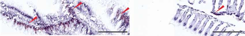
Toxins 2018, 10, 282                                                                                                   7 of 14

Toxins 2018, 10, x FOR PEER REVIEW                                                                                    7 of 14
concentrations of TTX (mean 46.1 ± 1.8 µg/kg; Figure 1). Although these sections could not be
dissected for LC-MS/MS
dissected for LC-MS/MS analysis,
                          analysis,they
                                    theycould
                                         couldbebevisualised
                                                    visualised using
                                                             using thethe immunohistological
                                                                       immunohistological     staining
                                                                                          staining and
and
key key findings
    findings      are described
              are described     below.
                            below.

2.5.1. Gonads
2.5.1. Gonads
       Like most
      Like  most bivalves,
                   bivalves, P.P. australis
                                  australis are
                                            are dioecious
                                                 dioecious andand fertilization
                                                                    fertilization usually
                                                                                   usually occurs
                                                                                            occurs inin the
                                                                                                        the water
                                                                                                             water column
                                                                                                                     column
after spawning
after   spawning[38].
                    [38].In In  immunostained
                             immunostained            sections
                                                  sections           P. australis,
                                                             of P.ofaustralis,      the gonads
                                                                               the gonads   did notdidcontain
                                                                                                         not contain     TTX.
                                                                                                                TTX. After
After   hematoxylin    staining,    the  eggs   were    dark  purple    (Figure   4), making
hematoxylin staining, the eggs were dark purple (Figure 4), making the vizualisation of TTX    the  vizualisation     of TTX
challenging. Based
challenging.    Basedon onthethe immunostained
                                  immunostainedsections,sections,wewedo donot
                                                                            notbelieve
                                                                                 believeTTX
                                                                                          TTXwaswaspresent
                                                                                                      presentin inthe
                                                                                                                   theeggs.
                                                                                                                         eggs.
The  majority   of the P.  australis  collected   for  this study   were   not  sexually  mature   (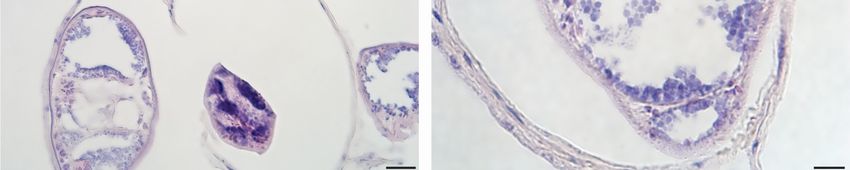
Toxins 2018, 10, 282 8 of 14
was detected in low concentrations in the ‘rest’ tissue group with LC-MS/MS. Other biotoxins have
previouslyToxins
been detected
2018, in REVIEW
10, x FOR PEER the gills of bivalves: low levels of microcystins [52] and saxitoxins 8 of 14 [50]
have been detected in gills of mussels and three species of bivalves in Palau. This result provides
[50] have been detected in gills of mussels and three species of bivalves in Palau. This result provides
further evidence
Toxins 2018,to
10, support
x FOR PEER the hypothesis of a dietary source of TTX in P. australis.
REVIEW 8 of 14
further evidence to support the hypothesis of a dietary source of TTX in P. australis.
[50] have been detected in gills of mussels and three species of bivalves in Palau. This result provides
further evidence to support the hypothesis of a dietary source of TTX in P. australis.
Figure 5. Paphies australis tissues sectioned at 7 µm. Tetrodotoxin (TTX)-specific monoclonal
Figure 5. Paphies australis
antibody (mAB) tissues sectioned
immunohistological at 7of:
staining µm. Tetrodotoxin
(a) the (TTX)-specific
labial palps with the TTX-mAB;monoclonal
(b) the labial antibody
(mAB) immunohistological staining
palps with mAB negative control; of:
(c,d)(a)
the the
gills labial
with thepalps withTTX
TTX-mAB. thewasTTX-mAB;
identified by(b)
thethe labial palps
brown
Figure
color 5. Paphies
deposits australis
shown by red tissues Scale
arrows. sectioned
bars =at100
7 µm
µm.(a,b)
Tetrodotoxin
and 50 µm (TTX)-specific monoclonal
(c,d).
with mAB negative control; (c,d) the gills with the TTX-mAB. TTX was identified by the brown color
antibody (mAB) immunohistological staining of: (a) the labial palps with the TTX-mAB; (b) the labial
deposits shown by red arrows. Scale bars = 100 µm (a,b) and 50 µm (c,d).
2.6. Parasites
palps with mAB negative control; (c,d) the gills with the TTX-mAB. TTX was identified by the brown
color deposits shown by red arrows. Scale bars = 100 µm (a,b) and 50 µm (c,d).
Digenean trematode metacercariae (sub-adult stages) were seen (Figure 6) encysted in Paphies
2.6. Parasites
australis mantle tissues (55% of P. australis observed were infected). Further identification of these
2.6. Parasites
Gymnophallid-like worms was prevented by their poor internal condition. Most of their internal
Digenean trematode
Digenean trematode
metacercariae (sub-adult
(sub-adult stages)seen were seen encysted
(Figurein 6) encysted in
organs were missing andmetacercariae stages) were
the body cavity of each contained (Figure 6) spheres
many grey/purple Paphies
resembling
Paphies australis
australis mantle
coloniesmantle tissues
tissues
of rickettsia (55%(55%
of P.of P.No
australis
australis
hyperparasites. observed
observed
TTX was were were
in theinfected).
infected).
observed Further inFurther
identification
parasites identification
of these
immunostained of
these Gymnophallid-like
Gymnophallid-like
sections. worms
worms waswasprevented
prevented by by their
their poorpoor internal
internal condition.
condition. Most of Most of their internal
their internal
organs
organs were were missing
missing and the andbody
the body cavity
cavity ofofeach
each contained
contained many
many grey/purple spheresspheres
grey/purple resemblingresembling
colonies of rickettsia hyperparasites. No TTX was observed in the parasites in immunostained
colonies of rickettsia hyperparasites. No TTX was observed in the parasites in immunostained sections.
sections.
Figure 6. Metacercariae encysted in mantle tissues of Paphies australis. (a) View of a group of encysted
metacercariae (red arrows) and cyst walls (blue arrows); (b) one metacercaria (red arrow) inside cyst
Figure 6. Metacercariae encysted in mantle tissues of Paphies australis. (a) View of a group of encysted
Figure 6. metacercariae
Metacercariae (redencysted
arrows) and
in cyst wallstissues
mantle (blue arrows); (b) oneaustralis.
of Paphies metacercaria
(a)(red
Viewarrow)
of ainside
group cyst
of encysted
metacercariae (red arrows) and cyst walls (blue arrows); (b) one metacercaria (red arrow) inside cyst wall
(blue arrow); rickettsial hyperparasite colonies inside metacercaria (green arrows). Scale bars = 20 µm (a)
and 10 µm (b).Toxins 2018, 10, x FOR PEER REVIEW 9 of 14
wall (blue arrow); rickettsial hyperparasite colonies inside metacercaria (green arrows). Scale bars =
Toxins 2018, 10, 282 9 of 14
20 µm (a) and 10 µm (b).
3. Materials 3.
and Methods
Materials and Methods
3.1. Paphies australis Collection
3.1. Paphies australis Collection
Paphies australis (n =australis
Paphies 48) were(ncollected
= 48) werefrom the Hokianga
collected from the Harbour
Hokianga (Northland, New Zealand;
Harbour (Northland, New Zealand;
◦
35°28′ S, 173°24′ 0
35 28E) S,on 173 ◦ 0
28 September
24 E) on 2017 and placed2017
28 September in a metal shellfish
and placed in collection basket. Paphies
a metal shellfish collection basket.
australis from this location
Paphies australishadfrompreviously beenhad
this location shown to contain
previously beenTTX (Harwood
shown and TTX
to contain Biessy unpub. and Biessy
(Harwood
data). Individuals
unpub.were rinsed
data). in seawater
Individuals were and placed
rinsed in in a plasticand
seawater bagplaced
insideinana insulated
plastic bag container
inside an insulated
(9–12 °C). Within
container hours◦ C).
two(9–12 of Within
collection,
twotwelve
hours of large P. australis
collection, twelvewere sectioned
large andwere
P. australis six small
sectioned and six
individuals (Toxins 2018, 10, 282 10 of 14
counterstained with Gill’s II Hematoxylin (Sigma-Aldrich, Darmstadt, Germany) and were dehydrated
using ascending grades of ethanol (60–100%), followed by two rinses in xylene for 10 min each.
The slides were mounted with DPX mountant (Sigma-Aldrich), and observed using an inverted
microscope (CKX41, Olympus) and photographs were taken using a slide scanner (VENTANA iScan
Coreo, Tucson, AZ, USA).
Table 1. Immunohistological incubation scheme. Steps were undertaken at room temperature unless
otherwise specified.
Step Solution Time (min)
1. 3% H2 O2 /10% methanol 10
2. 1 × PBS 10 × 3
3. Normal Goat Serum 20
4. 1 × PBS 10 × 3
5. mAB T20G10 * Overnight at 4 ◦ C
6. 1 × PBS 10 × 3
7. Biotinylated secondary antibody (anti-rabbit IgG) * 60
8. 1 × PBS 10 × 3
9. VECTASTAIN® ABC reagent * 60
10. 1 × PBS 10 × 3
11. DAB * 5–10
12. Deionized H2 O 5
13. Counterstain (Gill’s II Hematoxylin) 5
PBS—phosphate buffered saline, mAB—monoclonal antibody, DAB—3,30 -diaminobenzidine. * Reagents were
diluted in 1 × PBS, pH 7.2.
Antigen–antibody complexes were visualized as brown colour deposit in positive sections and
the same protocol was followed for the negative controls with the slides being incubated overnight
with 1 × PBS instead of the TTX-specific monoclonal antibody.
3.2.4. Tetrodotoxin Analysis
Each sample (ca. 0.3–3.0 g) was weighed, cut into small pieces with a sterile blade and placed
in a sterile tube (50 mL) with a corresponding volume (ca. 300–3,000 µL) of Milli-Q water containing
1% acetic acid. Samples were homogenized (Ultra-Turrax® , IKA® , Wilmington, NC, USA) for 45 s
to ensure complete disruption of tissues. The tubes were placed in boiling water (5 min) and then
cooled in an ice bath (5 min) before briefly vortexing. Samples were centrifuged (3200× g, 10 min)
and 0.3–1 mL of the supernatant transferred to a centrifuge tube (1.7 mL) containing 2.5–5 µL of 25%
ammonia (Honeywell). Samples were then centrifuged (17,000× g, 1 min) and the supernatant cleaned
with the GPC Solid Phase Extraction (SPE) method as described by Boundy et al. [24] using Supelclean
ENVI-Carb 250 mg/3 mL SPE cartridges (Sigma-Aldrich). Tetrodotoxin was analyzed and quantified
by liquid chromatography tandem-mass spectrometry analysis as described by Turner et al. [57].
3.2.5. Statistical Analysis
Tetrodotoxin data from the LC-MS/MS was analyzed using R [58]. Data from the three time
points (3, 6, and 9 days) were combined to enhance replicate number (after showing no significant
difference) and an ANOVA with a Tukey’s post-hoc test were undertaken to determine if there are
statistically significant differences in TTX concentrations among tissues and organs.
4. Conclusions
Tetrodotoxin analysis of P. australis organs and tissues using LC-MS/MS showed that the siphons
contained the highest concentrations of the biotoxin. Lower concentrations of TTX were found in the
foot, digestive system, mantle, and the combined group of organs/tissues (‘rest’) which could not be
individually dissected. The immunohistochemistry experiment demonstrated the micro-localizationToxins 2018, 10, 282 11 of 14
of TTX inside these organs/tissues. The toxin was primarily present in the outer layers of the siphons,
rectum, digestive tubes, gills, and labial palps. Observing TTX in organs involved in feeding provides
initial evidence to support the hypothesis that the source of the neurotoxin is exogenous in P. australis.
The presence of TTX in the siphon suggests that one of its ecological roles in this species may be to
reduce predation. Further studies are needed to determine if TTX migrates to other organs over time,
such as those involved in reproduction, and whether the toxin is transferred to subsequent generations.
Supplementary Materials: The following are available online at http://www.mdpi.com/2072-6651/10/7/282/s1,
Figure S1: Observed TTX analogues from the siphon of Paphies australis.
Author Contributions: L.B., I.H. and S.A.W. conceived and designed the experiments; L.B. performed the
experiments; K.F.S. and S.C.W. helped with the data interpretation and experiments, and M.J.B. and L.B. undertook
the LC-MS/MS analysis and interpretation. All authors contributed to data analysis and the editing and writing
of the manuscript.
Funding: This research was supported by an inaugural PhD scholarship from the New Zealand Food Safety
Science & Research Center to Laura Biessy, the MBIE-funded Safe New Zealand Seafood research Programme
(contract No.: CAW X1317) and the Cawthron Institute Internal Investment Fund.
Acknowledgments: The authors thank Lauren Salvitti for her advice and for providing the TTX antibody;
Joel Bowater (Cawthron) for his help in the field and in the laboratory; Eric Goodwin (Cawthron) for statistical
help and Tim Harwood and Lesley Rhodes (Cawthron) for their advice and assistance. Taranaki Medlab, 56 Vivian
Street New Plymouth NZ, is acknowledged for providing histological services.
Conflicts of Interest: The authors declare no conflict of interest.
References
1. Cestèle, S.; Catterall, W.A. Molecular mechanisms of neurotoxin action on voltage-gated sodium channels.
Biochimie 2000, 82, 883–892. [CrossRef]
2. Noguchi, T.; Ebesu, J.S. Puffer poisoning: epidemiology and treatment. J. Toxicol. Toxin Rev. 2001, 20, 1–10.
[CrossRef]
3. Tahara, Y. Uber das tetrodongift. Biochemistry 1910, 30, 255–275.
4. Goto, T.; Kishi, Y.; Takahashi, S.; Hirata, Y. Tetrodotoxin. Tetrahedron 1965, 21, 2059–2088. [CrossRef]
5. Tsuda, K.; Ikuma, S.; Kawamura, M.; Tachikawa, R.; Sakai, K.; Tamura, C.; Amakasu, O. Tetrodotoxin. VII.
On the structures of tetrodotoxin and its derivatives. Chem. Pharm. Bull. 1964, 12, 1357–1374. [CrossRef]
[PubMed]
6. Fuhrman, F.A. Tetrodotoxin. Sci. Am. 1967, 217, 60–71. [CrossRef] [PubMed]
7. Kodama, M.; Noguchi, T.; Maruyama, J.; Ogata, T.; Hashimoto, K. Release of tetrodotoxin and paralytic
shellfish poison from puffer liver by RNase. J. Biochem. 1983, 93, 243–247. [CrossRef] [PubMed]
8. Noguchi, T.; Arakawa, O.; Takatani, T. TTX accumulation in pufferfish. Comp. Biochem. Physiol. Part D
Genom. Proteom. 2006, 1, 145–152. [CrossRef] [PubMed]
9. Isbister, G.K.; Son, J.; Wang, F.; Maclean, C.J.; Lin, C.S.; Ujma, J.; Balit, C.R.; Smith, B.; Milder, D.G.;
Kiernan, M.C. Puffer fish poisoning: A potentially life-threatening condition. Med. J. Aust. 2002, 177, 650–653.
[PubMed]
10. Chau, R.; Kalaitzis, J.A.; Neilan, B.A. On the origins and biosynthesis of tetrodotoxin. Aquat. Toxicol. 2011, 104,
61–72. [CrossRef] [PubMed]
11. Bane, V.; Lehane, M.; Dikshit, M.; O‘Riordan, A.; Furey, A. Tetrodotoxin: Chemistry, toxicity, source,
distribution and detection. Toxins 2014, 6, 693–755. [CrossRef] [PubMed]
12. Noguchi, T.; Arakawa, O. Tetrodotoxin–distribution and accumulation in aquatic organisms, and cases of
human intoxication. Mar. Drugs 2008, 6, 220–242. [CrossRef] [PubMed]
13. Yasumoto, T.; Yasumura, D.; Yotsu, M.; Michishita, T.; Endo, A.; Kotaki, Y. Bacterial production of tetrodotoxin
and anhydrotetrodotoxin. Agric. Biol. Chem. 1986, 50, 793–795.
14. Wu, Z.; Yang, Y.; Xie, L.; Xia, G.; Hu, J.; Wang, S.; Zhang, R. Toxicity and distribution of tetrodotoxin-
producing bacteria in puffer fish Fugu rubripes collected from the Bohai Sea of China. Toxicon 2005, 46,
471–476. [CrossRef] [PubMed]Toxins 2018, 10, 282 12 of 14
15. Yotsu-Yamashita, M.; Mebs, D.; Yasumoto, T. Tetrodotoxin and its analogues in extracts from the toad
Atelopus oxyrhynchus (family: Bufonidae). Toxicon 1992, 30, 1489–1492. [CrossRef]
16. Daly, J.W.; Padgett, W.L.; Saunders, R.L.; Cover, J.F., Jr. Absence of tetrodotoxins in a captive-raised riparian
frog, Atelopus varius. Toxicon 1997, 35, 705–709. [CrossRef]
17. Cardall, B.L.; Brodie, E.D.; Hanifin, C.T. Secretion and regeneration of tetrodotoxin in the rough-skin newt
(Taricha granulosa). Toxicon 2004, 44, 933–938. [CrossRef] [PubMed]
18. Kodama, M.; Sato, S.; Sakamoto, S.; Ogata, T. Occurrence of tetrodotoxin in Alexandrium tamarense, a causative
dinoflagellate of paralytic shellfish poisoning. Toxicon 1996, 34, 1101–1105. [CrossRef]
19. McNabb, P.S.; Taylor, D.I.; Ogilvie, S.C.; Wilkinson, L.; Anderson, A.; Hamon, D.; Wood, S.A.; Peake, B.M.
First detection of tetrodotoxin in the bivalve Paphies australis by liquid chromatography coupled to triple
quadrupole mass spectrometry with and without precolumn reaction. J. AOAC Int. 2014, 97, 325–333.
[CrossRef] [PubMed]
20. Turner, A.; Powell, A.; Schofield, A.; Lees, D.; Baker-Austin, C. Detection of the pufferfish toxin tetrodotoxin
in European bivalves, England, 2013 to 2014. Eur. Surveill. 2015, 20, 2–8. [CrossRef]
21. Vlamis, A.; Katikou, P.; Rodriguez, I.; Rey, V.; Alfonso, A.; Papazachariou, A.; Zacharaki, T.; Botana, A.M.;
Botana, L.M. First detection of tetrodotoxin in Greek shellfish by UPLC-MS/MS potentially linked to the
presence of the dinoflagellate Prorocentrum minimum. Toxins 2015, 7, 1779–1807. [CrossRef] [PubMed]
22. Knutsen, H.K.; Alexander, J.; BarregAard, L.; Bignami, M.; Brüschweiler, B.; Ceccatelli, S.; Cottrill, B.;
Dinovi, M.; Edler, L.; Grasl-Kraupp, B. Risks for public health related to the presence of tetrodotoxin (TTX)
and TTX analogues in marine bivalves and gastropods. Eur. Food Saf. Auth. J. 2017, 15, 4752.
23. Zhang, X.; Yan, Z.; Wang, Y.; Jiang, T.; Wang, J.; Sun, X.; Guo, Y. Immunoaffinity chromatography purification
and ultrahigh performance liquid chromatography tandem mass spectrometry determination of tetrodotoxin
in marine organisms. J. Agric. Food Chem. 2015, 63, 3129–3134. [CrossRef] [PubMed]
24. Boundy, M.J.; Selwood, A.I.; Harwood, D.T.; McNabb, P.S.; Turner, A.D. Development of a sensitive and
selective liquid chromatography–mass spectrometry method for high throughput analysis of paralytic
shellfish toxins using graphitised carbon solid phase extraction. J. Chromatogr. A 2015, 1387, 1–12. [CrossRef]
[PubMed]
25. McNabb, P.; Selwood, A.I.; Holland, P.T. Multiresidue method for determination of algal toxins in shellfish:
Single-laboratory validation and interlaboratory study. J. AOAC Int. 2005, 88, 761–772. [PubMed]
26. Tanu, M.; Mahmud, Y.; Takatani, T.; Kawatsu, K.; Hamano, Y.; Arakawa, O.; Noguchi, T. Localization of
tetrodotoxin in the skin of a brackishwater puffer Tetraodon steindachneri on the basis of immunohistological
study. Toxicon 2002, 40, 103–106. [CrossRef]
27. Mahmud, Y.; Okada, K.; Takatani, T.; Kawatsu, K.; Hamano, Y.; Arakawa, O.; Noguchi, T. Intra-tissue distribution
of tetrodotoxin in two marine puffers Takifugu vermicularis and Chelonodon patoca. Toxicon 2003, 41, 13–18.
[CrossRef]
28. Salvitti, L.R.; Wood, S.A.; Winsor, L.; Cary, S.C. Intracellular immunohistochemical detection of tetrodotoxin
in Pleurobranchaea maculata (Gastropoda) and Stylochoplana sp. (Turbellaria). Mar. Drugs 2015, 13, 756–769.
[CrossRef] [PubMed]
29. Tanu, M.B.; Mahmud, Y.; Arakawa, O.; Takatani, T.; Kajihara, H.; Kawatsu, K.; Hamano, Y.; Asakawa, M.;
Miyazawa, K.; Noguchi, T. Immunoenzymatic visualization of tetrodotoxin (TTX) in Cephalothrix species
(Nemertea: Anopla: Palaeonemertea: Cephalotrichidae) and Planocera reticulata (Platyhelminthes: Turbellaria:
Polycladida: Planoceridae). Toxicon 2004, 44, 515–520. [CrossRef] [PubMed]
30. Magarlamov, T.Y.; Shokur, O.A.; Chernyshev, A.V. Distribution of tetrodotoxin in the ribbon worm
Lineus alborostratus (Takakura, 1898)(nemertea): Immunoelectron and immunofluorescence studies. Toxicon
2016, 112, 29–34. [CrossRef] [PubMed]
31. Rivera, V.R.; Poli, M.A.; Bignami, G.S. Prophylaxis and treatment with a monoclonal antibody of tetrodotoxin
poisoning in mice. Toxicon 1995, 33, 1231–1237. [CrossRef]
32. Kawatsu, K.; Hamano, Y.; Yoda, T.; Terano, Y.; Shibata, T. Rapid and highly sensitive enzyme immunoassay
for quantitative determination of tetrodotoxin. Jpn. J. Med. Sci. Biol. 1997, 50, 133–150. [CrossRef] [PubMed]
33. Smolowitz, R.; Doucette, G. Immunohistochemical localization of saxitoxin in the siphon epithelium of the
butter clam, Saxidomus giganteus. Biol. Bull. 1995, 189, 229–230. [CrossRef] [PubMed]Toxins 2018, 10, 282 13 of 14
34. MacKenzie, L.; White, D.; Adamson, J. Temporal variation and tissue localization of paralytic shellfish toxins
in the New Zealand Tuatua (surf-clam), Paphies subtriangulata. J. Shellfish Res. 1996, 15, 735–740.
35. Kvitek, R.G. Paralytic shellfish toxins sequestered by bivalves as a defense against siphon-nipping fish.
Mar. Biol. 1991, 111, 369–374. [CrossRef]
36. Hine, P.M. The inter-relationships of bivalve haemocytes. Fish Shellfish Immunol. 1999, 9, 367–385. [CrossRef]
37. Stanley, S.M. Post-Paleozoic adaptive radiation of infaunal bivalve molluscs: A consequence of mantle fusion
and siphon formation. J. Paleontol. 1968, 42, 214–229.
38. Gosling, E. Bivalve Molluscs: Biology, Ecology and Culture; Wiley-Blackwell: Oxford, UK, 2008; Volume 1, p. 448.
39. Zwarts, L.; Wanink, J. Siphon size and burying depth in deposit-and suspension-feeding benthic bivalves.
Mar. Biol. 1989, 100, 227–240. [CrossRef]
40. De Goeij, P.; Luttikhuizen, P.C.; van der Meer, J.; Piersma, T. Facilitation on an intertidal mudflat: The effect
of siphon nipping by flatfish on burying depth of the bivalve Macoma balthica. Oecologia 2001, 126, 500–506.
[CrossRef] [PubMed]
41. Kamermans, P.; Huitema, H.J. Shrimp (Crangon crangon L.) browsing upon siphon tips inhibits feeding and
growth in the bivalve Macoma balthica (L.). J. Exp. Mar. Biol. Ecol. 1994, 175, 59–75. [CrossRef]
42. Kvitek, R.; Bretz, C. Harmful algal bloom toxins protect bivalve populations from sea otter predation.
Mar. Ecol. Prog. Ser. 2004, 271, 233–243. [CrossRef]
43. Kondo, Y.; Stace, G. Burrowing ability and life position of Toheroa (Paphies ventricosa: Mesodesmatidae),
an unusually large, deep-burrowing ocean beach bivalve endemic to New Zealand. Jpn. J. Malacol. 1995, 54,
67–76.
44. Bricelj, V.M.; Shumway, S.E. Paralytic shellfish toxins in bivalve molluscs: Occurrence, transfer kinetics,
and biotransformation. Rev. Fish. Sci. 1998, 6, 315–383. [CrossRef]
45. Hwang, D.F.; Tsai, Y.H.; Cheng, C.A.; Jeng, S.S. Comparison of paralytic toxins in aquaculture of purple clam
in Taiwan. Toxicon 1992, 30, 669–672. [CrossRef]
46. Pereira, P.; Dias, E.; Franca, S.; Pereira, E.; Carolino, M.; Vasconcelos, V. Accumulation and depuration of
cyanobacterial paralytic shellfish toxins by the freshwater mussel Anodonta cygnea. Aquat. Toxicol. 2004, 68,
339–350. [CrossRef] [PubMed]
47. Baldwin, J.; Opie, A.M. On the role of octopine dehydrogenase in the adductor muscles of bivalve molluscs.
Comp. Biochem. Physiol. Part B Biochem. Mol. Bol. 1978, 61, 85–92. [CrossRef]
48. Shumway, S.E.; Cembella, A.D. The impact of toxic algae on scallop culture and fisheries. Rev. Fish. Sci. 1993, 1,
121–150. [CrossRef]
49. Helm, M.M.; Bourne, N.; Lovatelli, A. Hatchery Culture of Bivalves: A Practical Manual, 2004. Food and
Agriculture Organization of the United Nations. Available online: http://www.fao.org/docrep/007/y5720e/
y5720e00.htm (accessed on 11 June 2018).
50. Harada, T.; Oshima, Y.; Kamiya, H.; Yasumoto, T. Confirmation of paralytic shellfish toxins in the dinoflagellate
Pyrodinium bahamense var. compressa and bivalves in Palau. Nippon Suisan Gakkaishi 1982, 48, 821–825. [CrossRef]
51. Mafra, L.L., Jr.; Bricelj, V.M.; Fennel, K. Domoic acid uptake and elimination kinetics in oysters and mussels
in relation to body size and anatomical distribution of toxin. Aquat. Toxicol. 2010, 100, 17–29. [CrossRef]
[PubMed]
52. Chen, J.; Xie, P. Seasonal dynamics of the hepatotoxic microcystins in various organs of four freshwater
bivalves from the large eutrophic lake Taihu of subtropical China and the risk to human consumption.
Environ. Toxicol. 2005, 20, 572–584. [CrossRef] [PubMed]
53. Itoi, S.; Suzuki, M.; Asahina, K.; Sawayama, E.; Nishikubo, J.; Oyama, H.; Takei, M.; Shiibashi, N.; Takatani, T.;
Arakawa, O. Role of maternal tetrodotoxin in survival of larval pufferfish. Toxicon 2018, 148, 95–100.
[CrossRef] [PubMed]
54. Purchon, R.D. The Biology of The Mollusca; Pergamon Press: Oxford, UK, 1968; Volume 1, p. 560.
55. Howard, D.W.; Lewis, E.J.; Keller, B.J.; Smith, C.S. Histological Techniques for Marine Bivalve Mollusks and
Crustaceans, 2nd ed.; NOAA: Oxford, MD, USA, 2004; Volume 5, p. 218.
56. Salvitti, L.; Wood, S.A.; Taylor, D.I.; McNabb, P.; Cary, S.C. First identification of tetrodotoxin (TTX) in the
flatworm Stylochoplana sp.; a source of TTX for the sea slug Pleurobranchaea maculata. Toxicon 2015, 95, 23–29.
[CrossRef] [PubMed]Toxins 2018, 10, 282 14 of 14
57. Turner, A.D.; Boundy, M.J.; Rapkova, M.D. Development and single-laboratory validation of a liquid
chromatography tandem mass spectrometry method for quantitation of Tetrodotoxin in mussels and oysters.
J. AOAC Int. 2017, 100, 1469–1482. [CrossRef] [PubMed]
58. R Core Team. R: A Language and Environment for Statistical Computing; R Foundation for Statistical Computing:
Vienna, Austria, 2013. Available online: www.r-project.org (accessed on 22 May 2018).
© 2018 by the authors. Licensee MDPI, Basel, Switzerland. This article is an open access
article distributed under the terms and conditions of the Creative Commons Attribution
(CC BY) license (http://creativecommons.org/licenses/by/4.0/).You can also read