Maternal Caffeine Intake Disrupts Eggshell Integrity and Retards Larval Development by Reducing Yolk Production in a Caenorhabditis elegans Model ...
←
→
Page content transcription
If your browser does not render page correctly, please read the page content below
nutrients
Article
Maternal Caffeine Intake Disrupts Eggshell Integrity
and Retards Larval Development by Reducing Yolk
Production in a Caenorhabditis elegans Model
Hyemin Min , Esther Youn               and Yhong-Hee Shim *
 Department of Bioscience and Biotechnology, Konkuk University, Seoul 05029, Korea;
 mintmin@konkuk.ac.kr (H.M.); dptmej@konkuk.ac.kr (E.Y.)
 * Correspondence: yshim@konkuk.ac.kr; Tel.: +82-2-450-4059; Fax: +82-2-455-9956

 Received: 7 April 2020; Accepted: 6 May 2020; Published: 7 May 2020

 Abstract: During pregnancy, most women are exposed to caffeine, which is a widely consumed
 psychoactive substance. However, the consequences of maternal caffeine intake on the child remain
 largely unknown. Here, we investigated the intergenerational effects of maternal caffeine intake
 on offspring in a Caenorhabditis elegans model. We treated a young mother (P0) with 10 mM of
 caffeine equivalent to 2–5 cans of commercial energy drinks and examined its reproduction and
 growth rate from P0 to F2 generation. The fertility decreased and embryonic lethality increased by
 defective oocytes and eggshell integrity in caffeine-ingested mothers, and F1 larval development
 severely retarded. These results were due to decreased production of vitellogenin protein (yolk) in
 caffeine-ingested mothers. Furthermore, effects of RNA interference of vitellogenin (vit) genes, vit-1
 to vit-6, in P0 mothers can mimic those by caffeine-ingested mothers. In addition, RNA interference
 (RNAi) depletion of unc-62 (human Meis homeobox), a transcriptional activator for vit genes, also
 showed similar effects induced by caffeine intake. Taken together, maternal caffeine intake reduced
 yolk production mediated by the UNC-62 transcription factor, thereby disrupting oocyte and eggshell
 integrity and retarding larval development. Our study suggests the clinical significance of caffeine
 intake for prospective mothers.

 Keywords: caffeine; 1,3,7-trimethylxanthine; maternal effect; intergenerational effect; reproduction;
 yolk protein; vitellogenin; UNC-62; eggshell integrity; Caenorhabditis elegans

1. Introduction
     Caffeine is the most widely consumed bioactive molecule and its consumption has been increasing
worldwide. A clinical issue about caffeine intake during pregnancy is emerging owing to the possible
adverse impact of maternal nutritional status on child development [1,2]. However, the mechanism by
which information is shared between the mother and child remains largely unknown.
     Caenorhabditis elegans is an excellent animal model to study intergenerational effects of nutrient
intake on the progeny because it is easy to examine embryonic and post-embryonic developmental
processes in a large population of progeny at the organismal level. Furthermore, owing to its relatively
small genome sequence and systematic phenotypic analyses, each process can be assessed at the
molecular level as well [3,4]. C. elegans is also an excellent animal model to study reproduction because
this hermaphrodite contains both egg and sperm. Thus, the entire reproductive progress from mitosis
and meiosis of germ cells, and gametogenesis can be observed in one gonad arm. After the production
of egg and sperm, the process of fertilization and even early embryogenesis are possibly observed
simultaneously [5]. Several studies on caffeine intake in the C. elegans model have shown both beneficial
and adverse effects on C. elegans development depending on the intake dose. At a high dose of caffeine

Nutrients 2020, 12, 1334; doi:10.3390/nu12051334                              www.mdpi.com/journal/nutrients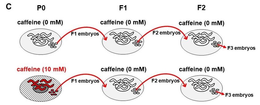
Nutrients 2020, 12, 1334                                                                              2 of 16

(30 mM), stress responses were induced, larval development was inhibited, and even food-avoidance
behavior was elicited when fed at the early larval stage of C. elegans [6–8]. However, at doses
Nutrients 2020, 12, 1334                                                                           3 of 16

Cruz Biotechnology, Dallas, TX, USA), and HRP-conjugated donkey anti-mouse IgG (1:1000; Jackson
ImmunoResearch, PA, USA).

2.4. Analysis of Oocyte and Eggshell Integrity
      To investigate oocyte and eggshell integrity after caffeine intake, membrane permeability was
assessed using FM4-64 dye (Sigma-Aldrich, St. Louis, MO, USA), as previously described [13]. In brief,
caffeine-ingested mothers were dissected in 150 mM KCl with 30 µM of FM4-64 dye to observe oocytes
and embryos. The proportion of either embryos or oocytes infiltrated by FM4-64 was measured using a
Zeiss microscope at 40× magnification. For each case, three independent experiments were performed.

2.5. RNA Interference (RNAi) Assays
     RNAi experiments were performed using the soaking method, as previously described [14].
dsRNAs of vit-1, vit-2, vit-3, vit-4, vit-5, vit-6, and unc-62 genes were synthesized in vitro using
the respective cDNA template. The cDNA templates flanked by T7 promoter sequences were
generated by PCR using T7 primer, 50 -GTAATACGACTCACTATAGGGC-30 and CMo422 primer,
50 -GCGTAATACGACTCACTATAGGGAACAAAAGCTGGAGCT-30 . Soaking buffer without dsRNA
was used as the negative mock RNAi control. L4-stage animals were soaked in dsRNA solution for
24 h, then transferred onto caffeine-containing NGM agar plates to grow for 24 h until the animals
reached the adult stage. The adult-stage animals were evaluated by membrane integrity assay.

2.6. DNA Staining in Oocytes
     To observe whether oocytes of caffeine-ingested mothers have six pairs of homologous
chromosomes (bivalents), DNA staining was performed, as previously described [14]. Animals
were dissected to extrude gonads in 10 µL of M9 buffer containing 100 µg/mL tetramisole on a
poly-L-lysine-coated slide, covered with a coverslip, freeze-cracked with liquid nitrogen, and fixed with
cold methanol and cold acetone. The specimens were then stained with 1 µM TO-PRO-3 (Molecular
Probes, Eugene, OR, USA) for 1 h at 20 ◦ C to stain DNA and then observed under a confocal microscope
(Olympus, FV1000 Spectral, Tokyo, Japan).

2.7. Real Time RT-PCR (qRT-PCR)
      Adult hermaphrodites of wild-type that were treated or not treated with caffeine (10 mM) were
collected in TRIzol (Invitrogen, Waltham, MA, USA), and total RNA was extracted using a phase
lock gel (MaXtract High Density, Qiagen, Germantown, MD, USA). cDNA was synthesized using
oligo-dT primer and M-MLV reverse transcriptase (Invitrogen, Waltham, MA, USA). qRT-PCR assays
were performed using SYBR Green PCR Master Mix (Applied Biosystems, Waltham, MA, USA). The
final PCR volume was 10 µL. act-1 mRNA was used as an endogenous control for data normalization.
The primers used for the measurement of expression of the unc-62 gene were as follows: forward,
50 -TAAGACATACCCAAGAGAATGCTG-30 and reverse, 50 -TTTGCCTTTCAGACAGACCA-30 .

2.8. Statistical Analysis
     All experiments were repeated more than three times for statistical evaluation of the data.
Two-tailed Student’s t-test was used to calculate p-values; p < 0.05 was considered significant. The
data are expressed as the mean ± standard deviation (SD).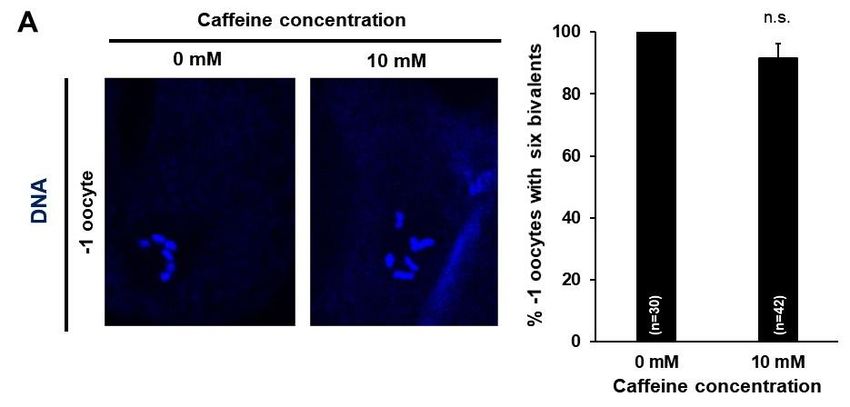
Nutrients 2020, 12, 1334 4 of 16
3. Results
3.1. Maternal Caffeine Intake Causes a Reduction in Fertility and Retardation in the Developmental Growth of
Progeny in C. elegans
To investigate whether caffeine intake by the mother has intergenerational effects on offspring, we
fed caffeine
Nutrients 2020,only
12, x to
FORP0PEER
mothers,
REVIEW as shown in Figure 1, and measured the fertility and the developmental 4 of 17
growth of offspring. It has been previously reported that the effects of caffeine treatment are dose
on reproduction
dependent by feeding
[6]. Therefore, we0,first
5, 10, and 30 mM
examined of caffeine
the effect to hermaphrodites
of doses of caffeine intakeofonwild-type L4-stage
reproduction by
animals for 24 h. The number of progenies was significantly decreased and embryonic
feeding 0, 5, 10, and 30 mM of caffeine to hermaphrodites of wild-type L4-stage animals for 24 h. The lethality was
increased
number of when
progenies mothers were fed 10 decreased
was significantly or 30 mM and
of caffeine (Figure
embryonic 2A,B).
lethality wasThese results
increased indicate
when that
mothers
>10 mM of caffeine intake seriously reduced fertility. In this study, the effects of
were fed 10 or 30 mM of caffeine (Figure 2A,B). These results indicate that >10 mM of caffeine intake 10 mM of caffeine
intake were
seriously examined
reduced in the
fertility. subsequent
In this experiments
study, the effects of 10because some mothers
mM of caffeine intake fed
were30examined
mM of caffeine
in the
became sick.
subsequent experiments because some mothers fed 30 mM of caffeine became sick.
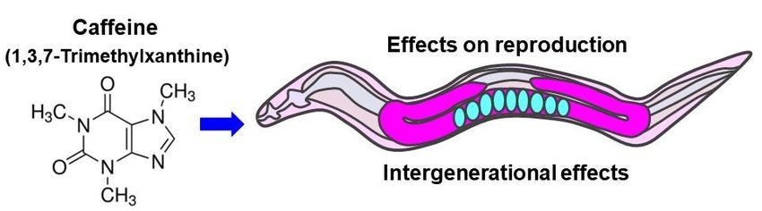
Figure Caenorhabditis
Figure1. 1. eleganselegans
Caenorhabditis P0 mothers
P0 were fed single
mothers werecompound caffeine
fed single (1,3,7-trimethylxanthine).
compound caffeine (1,3,7-
trimethylxanthine).
To determine whether caffeine intake by the mother has intergenerational effects, fertility including
the number of progenies
To determine and embryonic
whether lethality
caffeine intake by was assessedhas
the mother from P0 to F2 generation
intergenerational in C. fertility
effects, elegans
(Figure 2C). The number of progenies decreased in P0 mothers fed 10 mM caffeine,
including the number of progenies and embryonic lethality was assessed from P0 to F2 generation but not in F1 andin
F2
C. elegans (Figure 2C). The number of progenies decreased in P0 mothers fed 10 mM caffeine, but the
generation mothers. In addition, embryonic lethality increased in F1 embryos produced by not
caffeine-ingested P0 mother,
in F1 and F2 generation but not
mothers. in F2 andembryonic
In addition, F3 embryos (Figureincreased
lethality 2D,E). These
in F1results
embryossuggest that
produced
caffeine intake reduced fertility in P0 mothers, but not in F1 or F2 mothers.
by the caffeine-ingested P0 mother, but not in F2 and F3 embryos (Figure 2D,E). These results suggest
that Next, weintake
caffeine testedreduced
the possibility that
fertility in P0 caffeine
mothers,intake
but notcould
in F1affect growth of hatched offspring
or F2 mothers.
produced by caffeine-ingested mothers. We evaluated the developmental growth rate in F1 and F2
generations of caffeine-ingested P0 mothers. Interestingly, caffeine-ingested P0 mothers showed a
significantly retarded developmental growth rate in the F1 generation (Figure 3A). However, in the F2
generation, no growth retardation was observed (Figure 3B). These results suggest that caffeine intake
by the mother delays growth of the subsequent F1 generation (ca. 70% adult without caffeine intake,
but 0% adult with caffeine at 72 h growth), but not of the F2 generation (ca. 70% adult in both 0 mM
and 10 mM caffeine intake at 72 h growth).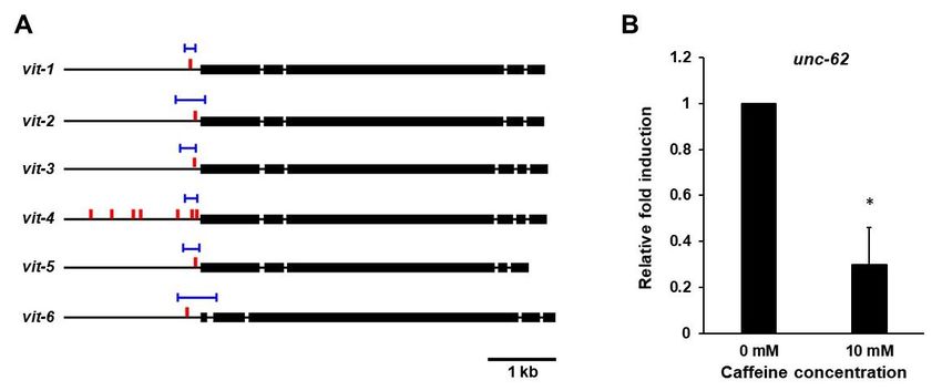
Nutrients 2020, 12, 1334                                                                                                             5 of 16
Nutrients 2020, 12, x FOR PEER REVIEW                                                                                               5 of 17

       Figure 2.
       Figure   2. Caffeine
                   Caffeine intake
                              intake caused
                                      caused aa reduction
                                                 reduction in in fertility  of P0
                                                                 fertility of  P0 mothers
                                                                                   mothers and  and an
                                                                                                     an increase
                                                                                                         increase inin F1
                                                                                                                       F1 embryonic
                                                                                                                           embryonic
       lethality  in Caenorhabditis    elegans. (A)   Total  number      of progenies     by   caffeine-ingested
       lethality in Caenorhabditis elegans. (A) Total number of progenies by caffeine-ingested mothers (5, 10,       mothers    (5,and
                                                                                                                                    10,
       and  30  mM)    compared     to  mothers    with  a caffeine-free     diet  (0  mM).    * p <  0.05.  (B)
       30 mM) compared to mothers with a caffeine-free diet (0 mM). * p < 0.05. (B) The percentage of embryonic  The   percentage    of
       embryonic     lethality  among    the total   number    of   progenies    produced      by  caffeine-ingested
       lethality among the total number of progenies produced by caffeine-ingested P0 mothers that were fed 0, 5,         P0  mothers
       thatand
      10,   were30 fed
                    mM0,of5,caffeine
                              10, andat30the
                                           mML4of   caffeine
                                                 stage  for 24ath.the
                                                                    * pL4  stage(C)
                                                                       < 0.05.    forA24scheme
                                                                                           h. * p
Nutrients 2020, 12, x FOR PEER REVIEW 6 of 17
intake, but
Nutrients 2020,0% adult with caffeine
12, 1334 at 72 h growth), but not of the F2 generation (ca. 70% adult in6both
of 16
0 mM and 10 mM caffeine intake at 72 h growth).
Figure
Figure 3. Caffeine intake
3. Caffeine intake by P0 mothers affected development in the the subsequent
subsequent generation
generation of
of
Caenorhabditis elegans. (A,B) Synchronized L4-stage animals (n =
Caenorhabditis elegans. = 30) of C. elegans wild-type were fed
fed
either
either00oror1010
mM mMof caffeine for 24for
of caffeine h, and
24 h,embryos were transferred
and embryos to respective
were transferred OP50-seeded
to respective nematode
OP50-seeded
growth medium plates and further ◦ C. The developmental stage of each individual in the
nematode growth medium platescultured at 20 cultured
and further at 20 °C. The developmental stage of each
F1 and F2 generations
individual in the F1was anddetermined based on
F2 generations its size
was and stage-specific
determined based onmorphological
its size and characteristics
stage-specific
(see Section 2 forcharacteristics
morphological details) during(seedevelopment
Section 2 by
foreither caffeine-ingested
details) (10 mM) or
during development bycaffeine-free diet
either caffeine-
P0 mother(10
ingested (0 mM)
mM).or caffeine-free diet P0 mother (0 mM).
3.2. Maternal Caffeine Intake Reduces Yolk Production in C. elegans
3.2. Maternal Caffeine Intake Reduces Yolk Production in C. Elegans
Vitellogenin (yolk proteins) plays an important role in the normal development of the animal’s
Vitellogenin (yolk proteins) plays an important role in the normal development of the animal’s
offspring by supplying nutrients [15–17]. To understand the molecular mechanism underlying the
offspring by supplying nutrients [15–17]. To understand the molecular mechanism underlying the
retarded growth rate of offspring produced by caffeine-ingested mothers, we investigated the possibility
retarded growth rate of offspring produced by caffeine-ingested mothers, we investigated the
of association with the expression of vitellogenin. We measured the expression level of vitellogenin gene
possibility of association with the expression of vitellogenin. We measured the expression level of
6 (vit-6) and vitellogenin gene 2 (vit-2) after caffeine intake using transgenic animals with a transgene
vitellogenin gene 6 (vit-6) and vitellogenin gene 2 (vit-2) after caffeine intake using transgenic animals
vit-6::gfp or vit-2::gfp. Adult-stage transgenic animals expressing either VIT-6::GFP or VIT-2::GFP were
with a transgene vit-6::gfp or vit-2::gfp. Adult-stage transgenic animals expressing either VIT-6::GFP
examined following exposure of caffeine and expression levels of both VIT-6::GFP and VIT-2::GFP were
or VIT-2::GFP were examined following exposure of caffeine and expression levels of both VIT-
found to be reduced (Figure 4A–D). We confirmed a significantly reduced level of VIT-6::GFP compared
6::GFP and VIT-2::GFP were found to be reduced (Figure 4A–D). We confirmed a significantly
to the control by Western blot analysis (Figure 4B). VIT-2 and VIT-6 are exclusively expressed in the
reduced level of VIT-6::GFP compared to the control by Western blot analysis (Figure 4B). VIT-2 and
intestine at the adult stage and VIT-2 is transported into oocytes and eventually to the embryonic cells,
VIT-6 are exclusively expressed in the intestine at the adult stage and VIT-2 is transported into
whereas VIT-6 remains in the intestine [18]. We thus observed oocytes and embryos for VIT-2::GFP
oocytes and eventually to the embryonic cells, whereas VIT-6 remains in the intestine [18]. We thus
expression and found a significantly decreased level of VIT-2::GFP both in oocytes and embryos after
observed oocytes and embryos for VIT-2::GFP expression and found a significantly decreased level
caffeine intake (Figure 4C,D). These results demonstrate that the expression of vitellogenin genes is
of VIT-2::GFP both in oocytes and embryos after caffeine intake (Figure 4C,D). These results
suppressed by caffeine intake.
demonstrate that the expression of vitellogenin genes is suppressed by caffeine intake.Nutrients 2020, 12, 1334 7 of 16
Nutrients 2020, 12, x FOR PEER REVIEW 7 of 17
Figure 4. Maternal caffeine intake reduces vitellogenin (yolk proteins) in Caenorhabditis elegans. (A)
Figure 4. Maternal
VIT-6::GFPcaffeine
transgenicintake reduces vitellogenin
animals synchronized (yolk
at the L4-stage were proteins)
exposed to caffeine in Caenorhabditis
for 24 h at 20 elegans.
(A) VIT-6::GFP°C.transgenic animals
VIT-6::GFP expresses synchronized
in the at thelevel
intestine. The reduced L4-stage wereinexposed
of VIT-6::GFP the intestinetowas
caffeine for 24 h
observed in caffeine-ingested mothers. (B) Western blot analysis of VIT-6::GFP protein levels in
at 20 ◦ C. VIT-6::GFP expresses in the intestine. The reduced level of VIT-6::GFP in the intestine
caffeine-free diet mother (0 mM) and caffeine-ingested mother (10 mM). Respective GFP band
was observed in caffeine-ingested
intensities were normalizedmothers.
against those(B) Western
of α-tubulin blot
in the sameanalysis
lane. Thenof
theVIT-6::GFP
normalized GFP protein levels in
band intensity was converted to a relative value compared to the normalized GFP band intensity of 0
caffeine-free diet mother (0 mM) and caffeine-ingested mother (10 mM). Respective GFP band intensities
mM, as shown in the right graph with mean ± SD values. These GFP band intensity values were
were normalized against
obtained fromthose of α-tubulin
three independent Westerninblot
theanalyses.
same Statistical
lane. Then the normalized
significance GFP band intensity
was calculated using
was converted Student’s t-test. * value
to a relative p < 0.05.compared
(C,D) VIT-2::GFP transgenic
to the animals synchronized
normalized GFP band at intensity
the L4-stage of
were0 mM, as shown
exposed to caffeine for 24 h at 20 °C. The caffeine-ingested mother showed a reduced level of VIT-
in the right graph with mean ± SD values. These GFP band intensity values were obtained from three
independent Western blot analyses. Statistical significance was calculated using Student’s t-test. * p <
0.05. (C,D) VIT-2::GFP transgenic animals synchronized at the L4-stage were exposed to caffeine for
24 h at 20 ◦ C. The caffeine-ingested mother showed a reduced level of VIT-2::GFP intensity in oocytes
and embryos, as shown in the right graph with mean ± SD values. (E) Synchronized L4-stage animals
(n = 30) of wild-type were treated with RNA interference (RNAi) of vit-3 gene for 24 h and recovered to
OP50-seeded nematode growth medium (NGM) plates for 24 h. The embryos produced by P0 mothers
were transferred to respective NGM plates and further cultured at 20 ◦ C. The developmental stage of
each individual in the progenies was determined based on its size and stage-specific morphological
characteristics (see Section 2 for details) during development by either the vit-3 RNAi-treated P0 mother
(10 mM) or non-treated (mock) P0 mother (0 mM). (F) VIT-6::GFP transgenic animals synchronized
at the L4-stage were exposed to caffeine for 24 h at 20 ◦ C. Then their F1 generation grew under the
caffeine-free diet (0 mM) condition, and VIT-6::GFP transgenic F1 animals were observed at the adult
stage. Statistical significance was calculated using Student’s t-test. n.s., p > 0.05.Nutrients 2020, 12, 1334 8 of 16
We further assessed a possible association between the reduction in vitellogenin and developmental
growth of offspring by examining the developmental stages of F1 worms produced by P0 mothers treated
with RNA interference (RNAi) of the vitellogenin gene vit-3. We observed a retarded developmental
growth rate in vit-3 RNAi depleted worms (Figure 4E), indicating that the presence of vitellogenin is
required for the normal growth of offspring. Taken together, these results suggest that caffeine intake
by the mother reduces the production of vitellogenin, which causes retarded growth of offspring. We
further examined whether the reduced level of vitellogenin observed in caffeine-ingested P0 mothers
sustains in the adult-stage F1 offspring. Interestingly, F1 generation adults expressed VIT-6::GFP
similarly to that in the caffeine-free diet control group (Figure 4F), suggesting that the effect of reduced
levels of vitellogenin on the development of offspring is limited to F1 generation. These findings
suggest that the decreased level of vitellogenin in the caffeine-ingested mother caused retarded larval
growth of the F1 offspring, but the expression of vitellogenin in the adult stage of the F1 generation
was not altered and no further effects were observed in F2 generation (Figures 3B and 4F).
3.3. Maternal Caffeine Intake Disrupts Eggshell Integrity in C. elegans
Caffeine-ingested mothers showed an increased level of embryonic lethality (Figure 2B).
Chromosomal alterations in oocytes have been reported as the primary cause of embryonic lethality in
C. elegans [19]. Therefore, we examined DNA morphology in −1 position oocytes of caffeine-ingested
mothers under a fluorescent microscope after DNA staining with TO-PRO-3 fluorescent dye. The
majority of mature oocytes in caffeine-ingested mothers contained six pairs of aligned and condensed
chromosomes in their nuclei, which are characteristic of the diakinesis stage in meiotic prophase I in
normal −1 position oocytes (Figure 5A). As little as 8.4% of oocytes showed a chromosomal abnormality,
which was not statistically significant (p = 0.068). This result indicates that caffeine intake by mothers
did not affect chromosomal integrity in oocytes.
The eggshell, which provides a protective structure with the extracellular matrix, is an important
factor in many early developmental events [13,20] and recent studies have suggested the pivotal
role of the eggshell during embryonic development and survival [21,22]. Therefore, we next
examined eggshell integrity in F1 embryos produced by caffeine-ingested mothers. We isolated
embryos from dissected caffeine-ingested and caffeine-free diet mothers and examined their integrity.
Approximately 17% of embryos isolated from caffeine-ingested mothers were ruptured (Figure 5B,C);
the remaining embryos were examined with lipophilic dye FM4-64 staining for visualizing embryonic
morphology [13,23]. Surprisingly, approximately 38% of the embryos out of the 83% non-ruptured
embryos from caffeine-ingested mothers were permeable to FM4-64 and their embryonic membrane
was stained, whereas embryos produced by caffeine-free diet mothers were not permeable to FM4-64
and their embryonic membrane was not stained at all (Figure 5D). Lipophilic FM4-64 dye binds to
the embryonic membrane and stains it if the eggshell is disrupted. Taken together, maternal caffeine
intake disrupted eggshell integrity, causing the eggshell to rupture and become permeable.
3.4. Reduced Vitellogenin Production in Caffeine-Ingested Mothers Causes Defective Oocyte and Eggshell
Integrity in C. elegans
We found that caffeine intake reduced vitellogenin production (Figure 4A–D) and disrupted
eggshell integrity (Figure 5B–D). On the basis of these findings, we investigated the relationship
between vitellogenin and eggshell integrity to determine whether disrupted eggshell integrity could
be due to the reduction in vitellogenin production. The vitellogenin family is comprised of six genes:
vit-1 to vit-6 [24]. We performed the respective RNAi of the six vit genes to knock down vitellogenin
production and observed eggshell permeability of embryos produced by RNAi-treated mothers
(Figure 6A). Approximately 20% of embryos were permeable to FM4-64 by depletion of vit genes,
whereas a majority of mock RNAi-treated embryos were not permeable to FM4-64 (Figure 6A). The
eggshell permeability defects in the vit RNAi experiments were likely due to simultaneous knockdown
of more than one vit gene, due to the RNAi target sequence similarity to more than one vit gene.Nutrients 2020, 12, 1334 9 of 16
Off target effects likely occur because vit-1 is 82% identical to vit-2, vit-3 and vit-4 are 99% identical,
vit-5 is 96% identical to vit-3, and vit-6 is 50% identical to vit-2 [17]. Indeed, a single vit mutant only
showed subtle phenotype, suggesting the functional redundancy among vit genes. We observed that
approximately 10% of embryos in vit-1(ok2616) and vit-2(ok3211) mutants, and approximately 1.7%
of embryos in vit-5(ok3239) mutant were permeable to FM4-64 dye (Figure 6B). Here we presented
results from vit RNAi and mutant analyses (Figure 6), which suggest that the reduction in yolk proteins
disrupts eggshell integrity.
Nutrients 2020, 12, x FOR PEER REVIEW 9 of 17
FigureFigure 5. Maternal
5. Maternal caffeine
caffeine intake
intake disruptedeggshell
disrupted eggshell integrity
integrity ininCaenorhabditis
Caenorhabditiselegans. (A) (A)
elegans. Oocytes
Oocytes
at theat−1
the –1 position
position in aingonad
a gonadarm arm
ofofwild-type
wild-typeadult
adult hermaphrodites
hermaphrodites growngrownwithout or with
without 10 mM
or with 10 mM
caffeine. DNA was stained with TO-PRO-3 and then six bivalents were examined. The percent ofofintact
caffeine. DNA was stained with TO-PRO-3 and then six bivalents were examined. The percent
intact six bivalents is shown in a bar graph on the right. (B–D) DIC images (B) and lipophilic dye FM4-
six bivalents is shown in a bar graph on the right. (B–D) DIC images (B) and lipophilic dye FM4-64
64 staining images (C,D) of embryos from the dissected caffeine-free diet (0 mM) and caffeine-
staining images (C,D) of embryos from the dissected caffeine-free diet (0 mM) and caffeine-ingested
ingested mothers (10 mM). The caffeine-free diet (0 mM) mother produced intact and ovoid embryos
mothers (10 mM). The caffeine-free diet (0 mM) mother produced intact and ovoid embryos and only
and only polar bodies (white arrowheads) were stained, but the caffeine-ingested mother produced
polarruptured
bodies (white
embryosarrowheads)
(C) and theirwere stained,
eggshells werebut the caffeine-ingested
permeable to FM4-64, and themother producedofruptured
cell membranes the
embryos (C) and
embryos weretheir eggshells
stained were permeable
(D). Statistical significanceto
was FM4-64, andusing
calculated the cell membranes
Student’s t-test. * pof< the
0.05.embryos
were stained (D). Statistical significance was calculated using Student’s t-test. * p < 0.05.
The eggshell, which provides a protective structure with the extracellular matrix, is an important
factor in many early developmental events [13,20] and recent studies have suggested the pivotal role
of the eggshell during embryonic development and survival [21,22]. Therefore, we next examined
eggshell integrity in F1 embryos produced by caffeine-ingested mothers. We isolated embryos from
dissected caffeine-ingested and caffeine-free diet mothers and examined their integrity.
Approximately 17% of embryos isolated from caffeine-ingested mothers were ruptured (Figure
5B,C); the remaining embryos were examined with lipophilic dye FM4-64 staining for visualizing
embryonic morphology [13,23]. Surprisingly, approximately 38% of the embryos out of the 83% non-3.4. Reduced Vitellogenin Production in Caffeine-Ingested Mothers Causes Defective Oocyte and Eggshell
Integrity in C. Elegans
We found that caffeine intake reduced vitellogenin production (Figure 4A–D) and disrupted
eggshell integrity (Figure 5B–D). On the basis of these findings, we investigated the relationship
Nutrients between vitellogenin and eggshell integrity to determine whether disrupted eggshell integrity could 10 of 16
2020, 12, 1334
be due to the reduction in vitellogenin production. The vitellogenin family is comprised of six genes:
vit-1 to vit-6 [24]. We performed the respective RNAi of the six vit genes to knock down vitellogenin
Next, we examined
production and observedwhether
eggshelldisrupted
permeabilityeggshell
of embryos integrity
produced is byassociated
RNAi-treated with oocytes in
mothers
(Figure 6A).mothers.
caffeine-ingested Approximately
We 20% of embryos were
hypothesized permeable to in
that disruption FM4-64 by depletion
eggshell integrity of vit genes,
is possibly due
whereas a majority of mock RNAi-treated embryos were not permeable to
to defective oocytes in caffeine-ingested mothers and thus we examined permeability of oocytes FM4-64 (Figure 6A). The
eggshell permeability defects in the vit RNAi experiments were likely due to simultaneous
using lipophilic dye FM4-64. Interestingly, caffeine-ingested mothers exhibited approximately 20% of
knockdown of more than one vit gene, due to the RNAi target sequence similarity to more than one
gonads vit with permeable
gene. oocytes,
Off target effects unlike
likely occur oocytes produced
because vit-1 by caffeine-free
is 82% identical diet
to vit-2, vit-3 mothers
and vit-4 are(Figure
99% 6C).
Furthermore, wevit-5
identical, alsoisperformed
96% identicalRNAi of respective
to vit-3, vitellogenin
and vit-6 is 50% genes
identical to vit-2 to test
[17]. whether
Indeed, the vit
a single reduction
mutant only
in vitellogenin canshowed subtleoocytes
also allow phenotype,to suggesting the functional
be permeable. redundancy
The respective among vit genes.
vitellogenin geneWe (vit-1, -2,
observed that approximately 10% of embryos in vit-1(ok2616) and vit-2(ok3211)
-3, -4, -5, and -6) RNAi-treated mothers produced approximately 18–22% of permeable oocytes in mutants, and
approximately 1.7% of embryos in vit-5(ok3239) mutant were permeable to FM4-64 dye (Figure 6B).
gonads (Figure 6D). These findings indicate that maternal caffeine intake caused defective oocytes and
Here we presented results from vit RNAi and mutant analyses (Figure 6), which suggest that the
disrupted eggshell integrity through the reduction in vitellogenin production.
reduction in yolk proteins disrupts eggshell integrity.
Figure 6.Figure 6. Vitellogenin
Vitellogenin is required
is required for eggshell
for eggshell and oocyte
and oocyte integrity
integrity in Caenorhabditis
in Caenorhabditis elegans.(A)
elegans. (A)Eggshell
Eggshell permeability was examined by lipophilic dye FM4-64 staining in
permeability was examined by lipophilic dye FM4-64 staining in the embryos produced by mockthe embryos produced by RNAi-
mock RNAi- or vit-1 to vit-6 RNAi-treated mothers. Statistical significance was calculated using
or vit-1 to vit-6 RNAi-treated mothers. Statistical significance was calculated using Student’s t-test.
Student’s t-test. * p < 0.05 against mock RNAi-treated animals. (B) Eggshell permeability was
* p < 0.05 againstby
examined mock
FM4-64RNAi-treated
staining in theanimals.
embryos(B) Eggshell
produced bypermeability was examined
vit-1(ok2616), vit-2(ok3211), and byvit-FM4-64
staining5(ok3239)
in the embryos
mutants. produced by vit-1(ok2616),
Statistical significance vit-2(ok3211),
was calculated and vit-5(ok3239)
using Student’s mutants.
t-test. * p < 0.05 against N2Statistical
significance was calculated using Student’s t-test. * p < 0.05 against N2 control. (C) Oocyte permeability
examined with lipophilic dye FM4-64 staining in the caffeine-ingested mother (10 mM) and caffeine-free
diet mother (0 mM). Gonads were extruded by dissecting adult animals in 150 mM of KCl. Statistical
significance was calculated using Student’s t-test. * p < 0.05. (D) Oocyte permeability was examined
by FM4-64 staining after treatment with RNAi of the vitellogenin genes from vit-1 to vit-6. Statistical
significance was calculated using Student’s t-test. * p < 0.05 against mock RNAi-treated animals.
3.5. Maternal Caffeine Intake Reduces unc-62 Expression and the Reduced Level of unc-62 Causes Defective
Oocyte and Eggshell Integrity in C. elegans
It has been reported that VPE1 (TGTCAAT) and VPE2 (CTGATAA), the cis-elements in the vit
promoter, are important for vitellogenin (vit) gene expression (Figure 7A), [25,26]. VPE1 is bound by
the UNC-62 (human Meis homeobox) transcription factor, which is highly expressed in the intestines
of adult C. elegans, where vit genes are specifically expressed; and expression of vit genes is suppressed
in unc-62 RNAi-treated animals [27]. Therefore, we investigated whether unc-62 expression is affected
by caffeine intake by measuring the mRNA level of unc-62 gene in caffeine-ingested and caffeine-free
diet mothers using qRT-PCR (Figure 7B). We found that the mRNA level of unc-62 gene significantly
decreased in caffeine-ingested mothers (Figure 7B) and the reduced unc-62 level by RNAi led to
a decreased level of VIT-2::GFP (Figure 7C). These results suggest that the reduced vitellogenin
production by caffeine intake is caused by the decreased level of transactivator unc-62.Nutrients 2020, 12, 1334 11 of 16
Nutrients 2020, 12, x FOR PEER REVIEW 12 of 17
Figure 7. Caffeine intake reduces unc-62 expression and the reduced level of unc-62 exhibits defects
Figure 7. Caffeine intake reduces unc-62 expression and the reduced level of unc-62 exhibits defects
in embryo and oocyte integrity in Caenorhabditis elegans. (A) UNC-62 binding sites in the genomic
in embryostructures
and oocyte integrity in Caenorhabditis elegans. (A) UNC-62 binding sites in the genomic
of vitellogenin genes. The thin black lines indicate the promoter and introns of each of six
structuresvitellogenin
of vitellogeninloci, andgenes. The
the thick thin
black black
lines lines
indicate indicate
exons the promoter
of six vitellogenin genes.and introns
The blue lines of each of
six vitellogenin loci, and the thick black lines indicate exons of six vitellogenin genes. The blue lines
indicate UNC-62 binding sites, and the red bars indicate VPE-1 (ATTGACA) vitellogenin regulatory
motif previously
indicate UNC-62 binding described [25,27].
sites, and the(B)
redFold induction
bars indicate of mRNA
VPE-1level of unc-62 in caffeine-ingested
(ATTGACA) vitellogenin regulatory
mothers (10 mM) than caffeine-free diet mothers (0 mM). The mRNA level of unc-62 was determined
motif previously described [25,27]. (B) Fold induction of mRNA level of unc-62 in caffeine-ingested
by three independent qRT-PCR using the mRNA level of act-1 in each sample as an internal control
mothers (10 for mM) than caffeine-free
normalization. dietSD.
T-bars represent mothers (0 mM).
Approximately 150The mRNA
adult animal level of unc-62
individuals was to
were used determined
by three independent
prepare total RNA qRT-PCR using
for respective the mRNA
conditions. Statistical of act-1 in
levelsignificance waseach sample
calculated asStudent’s
using an internal t- control
test. * p < 0.05. (C) VIT-2::GFP transgenic animals synchronized at the L4-stage
for normalization. T-bars represent SD. Approximately 150 adult animal individuals were used to were treated with unc-
62 RNAi. The unc-62 RNAi-treated mothers showed the reduced level of VIT-2::GFP intensity both in
prepare total RNA for respective conditions. Statistical significance was calculated using Student’s
t-test. * p < 0.05. (C) VIT-2::GFP transgenic animals synchronized at the L4-stage were treated with
unc-62 RNAi. The unc-62 RNAi-treated mothers showed the reduced level of VIT-2::GFP intensity both
in oocytes and embryos, as shown in the right graph with mean ± SD values. (D) Eggshell permeability
examined by lipophilic dye FM4-64 staining of the embryos produced by unc-62 RNAi-treated mothers.
Statistical significance was calculated using Student’s t-test. * p < 0.05 against mock RNAi-treated
animals. (E) Oocyte permeability examined by lipophilic dye FM4-64 staining in the dissected gonad
from the unc-62 RNAi-treated mothers. Statistical significance was calculated using Student’s t-test.
* p < 0.05 against mock RNAi-treated mothers.oocytes and embryos, as shown in the right graph with mean ± SD values. (D) Eggshell permeability
examined by lipophilic dye FM4-64 staining of the embryos produced by unc-62 RNAi-treated
mothers. Statistical significance was calculated using Student’s t-test. * p < 0.05 against mock RNAi-
treated animals. (E) Oocyte permeability examined by lipophilic dye FM4-64 staining in the dissected
gonad from the unc-62 RNAi-treated mothers. Statistical significance was calculated using Student’s
Nutrients 2020, 12, 1334 12 of 16
t-test. * p < 0.05 against mock RNAi-treated mothers.
Next, we
Next, we addressed
addressed whether
whether the
the reduced
reducedlevel
level of
of unc-62
unc-62 can
can cause
cause defects
defects in
in eggshell
eggshell and
and oocyte
oocyte
integrity. We
integrity. Weperformed
performedunc-62
unc-62RNAi
RNAi in in
mothers andand
mothers observed eggshell
observed and and
eggshell oocyte integrity
oocyte by FM4-
integrity by
64 dye staining. The unc-62 RNAi-treated embryos and oocytes also became permeable
FM4-64 dye staining. The unc-62 RNAi-treated embryos and oocytes also became permeable FM4-64 to FM4-64
dye (Figure
dye (Figure 7C,D),
7C,D), indicating
indicating that the reduced
reduced level
level of
of unc-62
unc-62 induced
induced by by caffeine
caffeine intake
intake results
results in
in
defective eggshell
defective eggshell and oocyte
oocyte integrity possibly through the reduction in vitellogenin production.
Takentogether,
Taken together,wewepropose
proposethat
thatmaternal
maternal caffeine
caffeineintake
intake affects
affectsthe
the survival
survival and
and growth
growth ofof offspring
offspring
through the
through the reduction
reduction inin yolk
yolk protein
protein production,
production, which
which is is mediated
mediated byby the
the repression
repression of
of unc-62
unc-62 gene
gene
expression (Figure
expression (Figure 8).8).
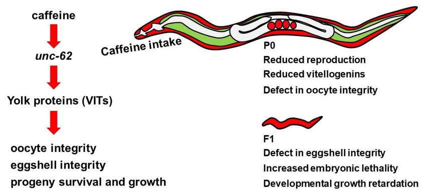
Figure
Figure 8.8. Model
Model ofof the
the intergenerational
intergenerational effects
effects of
of maternal
maternal caffeine
caffeine intake in Caenorhabditis
intake in Caenorhabditis elegans.
elegans.
Caffeine intake decreased the production of yolk proteins by reducing unc-62 expression.
Caffeine intake decreased the production of yolk proteins by reducing unc-62 expression. The decreased
The
levels of yolk
decreased proteins
levels disrupted
of yolk proteinsoocyte and eggshell
disrupted integrity
oocyte and andintegrity
eggshell induced and
embryonic lethality
induced and
embryonic
growth
lethalityretardation
and growth ofretardation
the next generation.
of the next generation.
4. Discussion
4. Discussion
The physiological effects of caffeine intake in C. elegans are highly dose dependent [6]. Previous
The
reports physiological
about the effectseffects of caffeine
of caffeine using intake in C. model
a C. elegans elegansindicate
are highly dose
that the dependent [6]. Previous
intake of a high dose of
reports about the effects of caffeine using a C. elegans model indicate that the intake of
caffeine (>10 mM) showed adverse effects, such as developmental arrest, activation of stress-responsea high dose of
caffeine (>10
pathways, mM)
and showed adverse
stimulation effects, such behavior
of food-avoidance as developmental arrest, activation
[6–8], whereas of stress-response
animals treated with a low
pathways, and stimulation of food-avoidance behavior [6–8], whereas animals treated with a low
dose of caffeine (Nutrients 2020, 12, 1334 13 of 16
the development of C. elegans. Considering that caffeine intake modulates the metabolic pathways and
changes nutritional state in an animal, the developmental regulators are the possible targets responding
to the alterations in metabolism. To investigate this possibility, the changes in the metabolic pathways
in caffeine-ingested mother and the relation between these pathways and unc-62 repression needs to
be determined. Vitellogenin (YP170) is transported from the intestine, where it is produced, to the
oocytes by receptor-mediated endocytosis; it becomes enriched in the yolk and provides nutrients to
the next generation in C. elegans [35]. Therefore, vitellogenin is considered as an intergenerational
molecule that is transferred from the mother to the offspring. Suppression of vit gene expression after
caffeine intake suggests that food signals are possibly involved in vitellogenesis. C. elegans vit genes
are homologous to those in vertebrates [36]. In vertebrates, vitellogenin is produced in the female
liver responding to estrogen signals and transported to the oocytes in the ovary through the blood.
Furthermore, environmental estrogenic chemicals can induce vitellogenin production. Therefore,
vitellogenin production is sensitive to the environmental status; thus, it can be used as a biomarker
of environmental stress during reproduction [37]. The novel finding that vitellogenin is required for
eggshell integrity and lack of vitellogenin causes embryonic lethality in approximately 20% of the
embryos observed in this study suggests that the presence of vitellogenin is important for embryonic
development of the next generation. Vitellogenin is mainly associated with lipid droplets in which
phosphatidylcholine (PC, 23%) and phosphoethanolamine (PE, 28.2%) are over half of the total lipid
content [17]. PC and PE are a major class of phospholipids that are the main constituents of the cell
membrane [38]. Therefore, the reduction of vitellogenin along with PC and PE in oocytes may cause
defects in the vitelline layer of oocytes and permeability. However, we propose that vitellogenin can
partially contribute to the survival of embryos and other factors can compensate for the requirement
of vitellogenin during embryogenesis. This is supported by the findings that 23% of live embryos
were retained even with complete loss of vitellogenin in oocytes in rme-2 mutants [35]; and that
vitellogenin production was remarkably decreased under dietary restriction in C. elegans, causing
increased embryonic lethality that was suppressed by methionine supplement [39]. In addition, larval
growth retardation in the F1 generation of caffeine-ingested mothers in the present study reveals that
vitellogenin is indeed an intergenerational protein. A large amount of vitellogenin from the mother
was observed in the early F1 larval stage of C. elegans, suggesting that maternal vitellogenin remains
in the larval stage and provides nutrients for larval development in the F1 progeny [40]. However,
in the present study, we found that the decreased level of vitellogenin in the mother affected the F1
generation, but not further generations, suggesting that it did not affect vitellogenin production in the
adult-stage intestine and germ cells in the F1 generation.
The eggshell plays an important role in protecting the embryo by maintaining proper osmotic
conditions and preventing the entry of potentially harmful molecules from the environment. In
addition, the permeability barrier of the embryo also allows it to maintain the substances required
for embryogenesis [41–43]. Previous studies have shown that fatty acid synthesis and modifications
of enzymes are required for the formation of the permeability barrier in the embryo [23], and lipid
metabolism is strongly associated with eggshell integrity [43]. It has been reported that in zebrafish,
caffeine intake has a role in the suppression of fatty liver by downregulation of genes associated with
lipogenesis, and enhancement of lipid oxidation and autophagy activity [44]. A relationship between
caffeine intake and fat metabolism has also been observed in a rat model system, which has shown
that caffeine intake reduces body fat by lipolysis and has an anti-obesity effect [45]. These findings
suggest that caffeine intake affects lipid metabolism, which in turn possibly controls eggshell formation.
Therefore, in addition to vitellogenin production, lipid metabolism appears to be involved in eggshell
and oocyte integrity after caffeine intake. Further analysis of the direct relationship between caffeine
intake and lipid metabolism in eggshell integrity remains to be determined. Dietary habit is one of
the external stimulations to induce internal physiological effects. Caffeine intake can be an effective
external stimulator. It has previously been described that caffeine intake causes global deacetylation of
proteins and mimics caloric restriction through autophagy induction [46]. It will be worthwhile toNutrients 2020, 12, 1334 14 of 16
examine whether the mode of action in oocyte and eggshell integrity after caffeine intake is related
to autophagy.
In summary, our results suggest that caffeine intake by the mother affects reproduction in a
dose-dependent manner. We demonstrated that maternal caffeine intake reduces yolk production by
regulating unc-62 expression. The reduced vitellogenin production by caffeine intake, in turn, decreases
embryonic survival by disrupting eggshell integrity, and inhibits larval development. As reported,
vitellogenin expression is highly specific at the adult stage of C. elegans females. Therefore, further
investigation of the consequences of caffeine intake in males to understand paternal effects is required.
5. Conclusion
This study provides several evidences showing the intergenerational effects of maternal caffeine
intake. These effects were attributed to the suppression of yolk protein production mediated by a
transcriptional activator, unc-62 (human Meis homeobox). These findings support that the mother’s
diet during pregnancy is critical for the survival and growth of progeny.
Author Contributions: H.M. and Y.-H.S. conceived and designed the study. H.M. and E.Y. conducted experiments.
H.M., E.Y., and Y.-H.S. analyzed and interpreted the data. H.M. and Y.-H.S. wrote the manuscript. Y.-H.S.
administrated the project and provided materials and reagents. All authors have read and agreed to the published
version of the manuscript.
Funding: This study was supported by a grant from the Konkuk University in 2017 to Y.-H.S.
Acknowledgments: C. elegans strains were provided by the Caenorhabditis Genetics Center, which is funded by
NIH Office of Research Infrastructure Programs (P40 OD010440). yk cDNA clones were provided by Yuji Kohara
(National Institute of Genetics, Japan).
Conflicts of Interest: The authors declare no conflicts of interest.
References
1. Li, J.; Zhao, H.; Song, J.M.; Zhang, J.; Tang, Y.L.; Xin, C.M. A meta-analysis of risk of pregnancy loss and
caffeine and coffee consumption during pregnancy. Int. J. Gynecol. Obestet. 2015, 130, 116–122. [CrossRef]
[PubMed]
2. Papadopoulou, E.; Botton, J.; Brantsæter, A.L.; Haugen, M.; Alexander, J.; Meltzer, H.M.; Bacelis, J.; Elfvin, A.;
Jacobsson, B.; Sengpiel, V. Maternal caffeine intake during pregnancy and childhood growth and overweight:
Results from a large Norwegian prospective observational cohort study. BMJ Open 2018, 8, e018895.
[CrossRef] [PubMed]
3. C. elegans Sequencing Consortium. Genome sequence of the nematode C. elegans: A platform for
investigating biology. Science 1998, 282, 2012–2018. [CrossRef] [PubMed]
4. Calvo, D.R.; Martorell, P.; Genovés, S.; Gosálbez, L. Development of novel functional ingredients: Need
for testing systems and solutions with Caenorhabditis elegans. Trends Food Sci. Technol. 2016, 54, 197–203.
[CrossRef]
5. Kimble, J.; Crittenden, S.L. Controls of germline stem cells, entry into meiosis, and the sperm/oocyte decision
in Caenorhabditis elegans. Annu. Rev. Cell Dev. Biol. 2007, 23, 405–433. [CrossRef] [PubMed]
6. Min, H.; Kawasaki, I.; Gong, J.; Shim, Y.H. Caffeine induces high expression of cyp-35A family genes and
inhibits the early larval development in Caenorhabditis elegans. Mol. Cells 2015, 38, 236–242. [CrossRef]
[PubMed]
7. Al-Amin, M.; Kawasaki, I.; Gong, J.; Shim, Y.H. Caffeine induces the stress response and up-regulates heat
shock proteins in Caenorhabditis elegans. Mol. Cells 2016, 39, 163–168. [CrossRef]
8. Min, H.; Youn, E.; Kawasaki, I.; Shim, Y.H. Caffeine-induced food-avoidance behavior is mediated by
neuroendocrine signals in Caenorhabditis elegans. BMB Rep. 2017, 50, 31–36. [CrossRef]
9. Bridi, J.C.; Barros, A.G.; Sampaio, L.R.; Ferreira, J.C.; Antunes Soares, F.A.; Romano-Silva, M.A. Lifespan
extension induced by caffeine in Caenorhabditis elegans is partially dependent on adenosine signaling. Front.
Aging Neurosci. 2015, 57, 220. [CrossRef]
10. Li, H.; Roxo, M.; Cheng, X.; Zhang, S.; Cheng, H.; Wink, M. Pro-oxidant and lifespan extension effects of
caffeine and related methylxanthines in Caenorhabditis elegans. Food Chem. X 2019, 1, 100005. [CrossRef]Nutrients 2020, 12, 1334 15 of 16
11. Kolahdouzan, M.; Hamadeh, M.J. The neuroprotective effects of caffeine in neurodegenerative diseases. CNS
Neurosci. Ther. 2017, 23, 272–290. [CrossRef] [PubMed]
12. Brenner, S. The genetics of Caenorhabditis elegans. Genetics 1974, 77, 71–94. [PubMed]
13. Johnston, W.L.; Krizus, A.; Dennis, J.W. The eggshell is required for meiotic fidelity, polar-body extrusion
and polarization of the C. elegans embryo. Genetics 2006, 4, 35. [CrossRef]
14. Min, H.; Kim, J.S.; Ahn, J.; Shim, Y.H. Gliadin intake causes disruption of the intestinal barrier and an increase
in germ cell apoptosis in a Caenorhabditis elegans model. Nutrients 2019, 11, E2587. [CrossRef]
15. Wahli, W.; Dawid, I.B.; Ryffel, G.U.; Weber, R. Vitellogenesis and vitellogenin gene family. Science 1981, 212,
298–304. [CrossRef]
16. Carducci, F.; Biscotti, M.A.; Canapa, A. Vitellogenin gene family in vertebrates: Evolution and functions. Eur.
Zool. J. 2019, 86, 233–240. [CrossRef]
17. Perez, M.F.; Lehner, B. Vitellogenins—Yolk gene function and regulation in Caenorhabditis elegans. Front.
Physiol. 2019, 10, 1067. [CrossRef]
18. Hall, D.H.; Winfrey, V.P.; Blaeuer, G.; Hoffman, L.H.; Furuta, T.; Rose, K.L.; Hobert, O.; Greenstein, D.
Ultrastructural features of the adult hermaphrodite gonad of Caenorhabditis elegans: Relations between the
germ line and soma. Dev. Biol. 1999, 212, 101–123. [CrossRef]
19. Min, H.; Sung, M.; Son, M.; Kawasaki, I.; Shim, Y.H. Transgenerational effects of proton beam irradiation on
Caenorhabditis elegans germline apoptosis. Biochem. Biophys. Res. Commun. 2017, 490, 608–615. [CrossRef]
20. Stein, K.K.; Golden, A. The C. elegans eggshell. WormBook 2018. Available online: http://www.wormbook.org
(accessed on 2 August 2018).
21. Johnston, W.L.; Dennis, J.W. The eggshell in the C. elegans oocyte-to-embryo transition. Genesis 2012, 50,
333–349. [CrossRef]
22. Fickentscher, R.; Struntz, P.; Weiss, M. Mechanical cues in the early embryogenesis of Caenorhabditis elegans.
Biophys. J. 2013, 103, 1805–1811. [CrossRef] [PubMed]
23. Olson, S.K.; Greenan, G.; Desai, A.; Müller-Reichert, T.; Oegema, K. Hierarchical assembly of the eggshell
and permeability barrier in C. elegans. J. Cell Biol. 2012, 198, 731–748. [CrossRef] [PubMed]
24. Spieth, J.; MacMorris, M.; Broverman, S.; Greenspoon, S.; Blumenthal, T. Regulated expression of a vitellogenin
fusion gene in transgenic nematodes. Dev. Biol. 1988, 130, 285–293. [CrossRef]
25. MacMorris, M.; Broverman, S.; Greenspoon, S.; Lea, K.; Madej, C.; Blumenthal, T.; Spieth, J. Regulation of
vitellogenin gene expression in transgenic Caenorhabditis elegans: Short sequences required for activation
of the vit-2 promoter. Mol. Cell Biol. 1992, 12, 1652–1662. [CrossRef] [PubMed]
26. Goszczynski, B.; Captan, V.V.; Danielson, A.M.; Lancaster, B.R.; McGhee, J.D. A 44bpintestine-specific
hermaphrodite-specific enhancer from the C. elegans vit-2 vitellogenin gene is directly regulated by
ELT-2,MAB-3, FKH-9 and DAF-16 and indirectly regulated by the germline, by daf-2/ insulin signaling and
by the TGF-β/Sma/Mab pathway. Dev. Biol. 2016, 413, 112–127. [CrossRef] [PubMed]
27. Van Nostrand, E.L.; Sánchez-Blance, A.; Wu, B.; Nguyen, A.; Kim, S.K. Roles of the developmental regulator
unc-62/ Homothorax in limiting longevity in Caenorhabditis elegans. PLoS Genet. 2013, 9, e1003325.
[CrossRef] [PubMed]
28. Sutphin, G.L.; Bishop, E.; Yanos, M.E.; Moller, R.M.; Kaeberlein, M. Caffeine extends life span, improves
healthspan, and delays age-associated pathology in Caenorhabditis elegans. Longev. Healthsapn. 2012, 1, 9.
[CrossRef]
29. Manalo, R.V.M.; Medina, P.M.B. Caffeine protects dopaminergic neurons from dopamine-induced
neurodegeneration via synergistic adenosine-dopamine D2-like receptor interactions in transgenic
Caenorhabditis elegans. Front. Neurosci. 2018, 12, 137. [CrossRef]
30. Moskow, J.J.; Bullrich, F.; Huebner, K.; Daar, I.O.; Buchberg, A.M. Meis1, a PBX1-related homeobox gene
involved in myeloid leukemia in BXH-2 mice. Mol. Cell Biol. 1995, 15, 5434–5443. [CrossRef]
31. Acheson, K.J.; Zahorska-Markiewicz, B.; Pittet, P.; Anantharaman, K.; Jéquier, E. Caffeine and coffee: Their
influence on metabolic rate and substrate utilization in normal weight and obese individuals. Am. J. Clin.
Nutr. 1980, 33, 989–997. [CrossRef]
32. Bracco, D.; Ferrarra, J.M.; Arnaud, M.J.; Jéquier, E.; Schutz, Y. Effects of caffeine on energy metabolism,
heart rate, and methylxanthine metabolism in lean and obese women. Am. J. Physiol. 1995, 269, E671–E678.
[CrossRef] [PubMed]Nutrients 2020, 12, 1334 16 of 16
33. Acheson, K.J.; Gremaud, G.; Meirim, I.; Montigon, F.; Krebs, Y.; Fay, L.B.; Gay, L.J.; Schneiter, P.; Schindler, C.;
Tappy, L. Metabolic effects of caffeine in humans: Lipid oxidation or futile cycling? Am. J. Clin. Nutr. 2004,
79, 40–46. [CrossRef] [PubMed]
34. Hanson, R.W. Nutrient control of gene transcription minireview series. J. Biol. Chem. 2000, 275, 30747.
[CrossRef]
35. Grant, B.; Hirsh, D. Receptor-mediated endocytosis in the Caenorhabditis elegans oocyte. Mol. Biol. Cell
1999, 10, 4311–4326. [CrossRef]
36. Spieth, J.; Blumenthal, T. The Caenorhabditis elegans vitellogenin gene family includes a gene encoding a
distantly related protein. Mol. Cell Biol. 1985, 5, 2495–2501. [CrossRef]
37. Denslow, N.D.; Chow, M.C.; Kroll, K.J.; Green, L. Vitellogenin as a biomarker of exposure for estrogen
mimics. Ecotoxicology 1999, 8, 385–398. [CrossRef]
38. van der Veen, J.N.; Kennelly, J.P.; Wan, S.; Vance, J.E.; Vance, D.E.; Jacobs, R.L. The critical role of
phosphatidylcholine and phosphatidylethanolamine metabolism in health and disease. Biochim. Biophys.
Acta Biomembr. 2017, 1859, 1558–1572. [CrossRef]
39. Zhou, G.; Huang, C.; Xing, L.; Li, L.; Jiang, Y.; Wei, Y. Methionine increases yolk production to offset the
negative effect of caloric restriction on reproduction without affecting longevity in C. elegans. Aging 2020, 12,
2680–2697. [CrossRef]
40. Kimble, J.; Sharrock, W.J. Tissue-specific synthesis of yolk proteins in Caenorhabditis elegans. Dev. Biol. 1983,
96, 189–196. [CrossRef]
41. Schierenberg, E.; Junkersdorf, B. The role of eggshell and underlying vitelline membrane for normal pattern
formation in the early C. elegans embryo. Roux. Arch. Dev. Biol. 1992, 202, 10–16. [CrossRef]
42. Lee, J.Y.; Goldstein, B. Mechanisms of cell positioning during C. elegans gastrulation. Development 2003, 130,
307–320. [CrossRef] [PubMed]
43. Carvalho, A.; Olson, S.K.; Gutierrez, E.; Zhang, K.; Noble, L.B.; Zanin, E.; Desai, A.; Groisman, A.; Oegema, K.
Acute drug treatment in the early C. elegans embryo. PLoS ONE 2011, 6, e24656. [CrossRef] [PubMed]
44. Zheng, X.; Dai, W.; Chen, X.; Wang, K.; Zhang, W.; Liu, L.; Hou, J. Caffeine reduces hepatic lipid accumulation
through regulation of lipogenesis and ER stress in zebrafish larvae. J. Biomed. Sci. 2015, 22, 105. [CrossRef]
[PubMed]
45. Kobayashi-Hattori, K.; Mogi, A.; Matsumoto, Y.; Takita, T. Effect of caffeine on the body fat and lipid
metabolism of rats fed on a high-fat diet. Biosci. Biotechnol. Biochem. 2005, 69, 2219–2223. [CrossRef]
46. Pietrocola, F.; Malik, S.A.; Mariño, G.; Vacchelli, E.; Senovilla, L.; Chaba, K.; Niso-Santano, M.; Maiuri, M.C.;
Madeo, F.; Kroemer, G. Coffee induces autophagy in vivo. Cell Cycle 2014, 13, 1987–1994. [CrossRef]
© 2020 by the authors. Licensee MDPI, Basel, Switzerland. This article is an open access
article distributed under the terms and conditions of the Creative Commons Attribution
(CC BY) license (http://creativecommons.org/licenses/by/4.0/).You can also read