Factor XII-Deficient Chicken Plasma as a Useful Target for Screening of Pro- and Anticoagulant Animal Venom Toxins - MDPI
←
→
Page content transcription
If your browser does not render page correctly, please read the page content below
toxins
Article
Factor XII-Deficient Chicken Plasma as a Useful
Target for Screening of Pro- and Anticoagulant
Animal Venom Toxins
Benedito C. Prezoto 1, *         and Nancy Oguiura 2
 1    Laboratory of Pharmacology, The Butantan Institute, Av. Dr. Vital Brazil 1500,
      São Paulo CEP 05503-900, Brazil
 2    Ecology and Evolution Laboratory, The Butantan Institute, Av. Dr. Vital Brazil 1500,
      São Paulo CEP 05503-900, Brazil; nancy.oguiura@butantan.gov.br
 *    Correspondence: benedito.prezoto@butantan.gov.br; Tel.: +55-11-2627-9753

 Received: 18 December 2019; Accepted: 20 January 2020; Published: 23 January 2020

 Abstract: The sensitivity of vertebrate citrated plasma to pro- and anticoagulant venom or toxins
 occurs on a microscale level (micrograms). Although it improves responses to agonists, recalcification
 triggers a relatively fast thrombin formation process in mammalian plasma. As it has a natural factor
 XII deficiency, the recalcification time (RT) of chicken plasma (CP) is comparatively long [≥1800 s].
 Our objective was to compare the ability of bee venom phospholipase A2 (bvPLA2 ) to neutralize clot
 formation induced by an activator of coagulation (the aPTT clot) in recalcified human and chicken
 plasmas, through rotational thromboelastometry. The strategy used in this study was to find doses
 of bvPLA2 that were sufficient enough to prolong the clotting time (CT) of these activated plasmas
 to values within their normal RT range. The CT of CP was prolonged in a dose-dependent manner
 by bvPLA2 , with 17 ± 2.8 ng (n = 6) being sufficient to displace the CT values of the activated
 samples to ≥ 1800 s. Only amounts up to 380 ± 41 ng (n = 6) of bvPLA2 induced the same effect in
 activated human plasma samples. In conclusion, the high sensitivity of CP to agonists and rotational
 thromboelastometry could be useful. For example, during screening procedures for assaying the
 effects of toxins in several stages of the coagulation pathway, such as clot initiation, formation, stability,
 strength, or dissolution.

 Keywords: thromboelastimetry; phospholipase; snake venom; insect venom

 Key Contribution: The speed of the mammalian clotting process after recalcification leads to a
 sensitivity to the pro- and anticoagulant effects of insect or snake venoms or toxins; this takes place
 on a microscale level (micrograms). In contrast, the low dynamics of the factor XII-deficient chicken
 coagulation process allows for recalcification, enhancing its sensitivity to these agonists.

1. Introduction
      Studies of animal venoms and toxins have focused on one or more of the following objectives:
(i) To determine the mode and mechanism of action of the toxins; (ii) to find ways and means to
neutralize the toxicity and adverse effects of accidents; (iii) to develop specific research tools that are
useful in understanding the normal physiological processes at both cellular and molecular levels;
(iv) to develop prototypes of pharmaceutical agents based on the structure of toxins [1].
      Tests to assess the in vivo effects of animal venoms, such as hemorrhage, myonecrosis, defibrination,
edema, and venom lethality/antivenom potencies, still largely rely on animal models (usually involving
rodents). However, these assays lead to animal suffering. Stringent regulations governing the use
of animals, limited research funds, and public pressure all drive the need for the development of

Toxins 2020, 12, 79; doi:10.3390/toxins12020079                                      www.mdpi.com/journal/toxins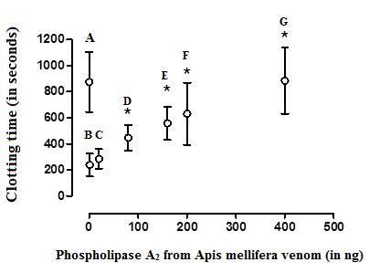
Toxins 2020, 12, 79                                                                                  2 of 11

alternative in vitro assays involving non-animal, or nonsentient research methods, as a way to achieve
the ‘Three Rs’ goals of animal experimentation, i.e., Reduce, Refine, and Replace animal tests. It is
widely known that animal venoms contain enzymes and non-enzymatic proteins that interfere with
hemostasis leading to hemorrhage or even thrombosis. Regarding hemotoxins, current scientific
attention is focused on the validation of alternative methods to the screening and characterization of
individual venom proteins, which should, in turn, enable novel in vitro assays to be designed, thus
reducing the number of animals required [2].
      Technologies that show a good correlation coefficient between the venom pro- and anticoagulant
effects on mammalian plasmas in vitro and lethality in vivo have been reported [3–15]. However, these
techniques present some limitations: (i) Many studies simply characterize the toxin conversion of
isolated substrates, giving a single parameter for one complex enzymatic process. As the full effects
of toxins on the coagulation pathway are rarely examined, even in vitro, our understanding of the
pathophysiology of envenoming is limited [16] and (ii) non-recalcified plasma samples, such as those
used in the minimum coagulant dose assay require relatively large amounts of venom (on the µg
or mg scale) [17]. If the potentiation of action of agonists could be achieved by the addition of the
cofactors Ca2+ and phospholipids [18], this strategy could accelerate some enzymatic reactions in
the coagulation cascade [19]. As a consequence, the time interval in which agonists can be assayed
becomes limited [to around 600 seconds (s)]. This limitation associated with mammalian plasma
becomes evident during screening strategies, when small amounts of the proteins being tested are
available, for example, during the purification procedures of individual toxins from crude venoms.
      Four constituents form the mammalian intrinsic pathway of coagulation: The trypsin-type serine
proteases factors XIIa, XIa, plasma kallikrein and the cofactor high molecular weight kininogen [20].
Extreme slowness in spontaneous in vitro thrombin/fibrin generation even after the addition of Ca2+
ions is a hallmark of the plasma of patients with a factor XII deficiency [21]. The main difference
between the human and chicken coagulation process is that the factor XII gene is completely missing
in the chicken genome [22]. Rotational thromboelastometry improves the evaluation of the clotting
process, since it monitors several parameters, such as the stages of clot initiation, formation, stability,
strength, and dissolution [23]. This technology has been used in various studies reporting on the
pro- and anticoagulant activities of several snake venoms on citrated human whole blood, as well as
plasma samples of rats and dogs [24–29]. By testing recalcified chicken plasma (CP) samples through
rotational thromboelastometry, we recently published two studies in which we describe the abilities of
the bothropic and crotalic antiserums in neutralizing the pro- and anticoagulant activities of Bothrops
jararaca (B. jararaca) venom and crotoxin, respectively. We found a positive correlation between the
data obtained with these in vitro assays and data from the in vivo lethality technique related to the
B. jararaca and Crotalus durissus terrificus (C. d. terrificus) venoms [30,31]. Predictably, addition of
the cofactors Ca2+ and phospholipids factor XII-deficient CP elicited a time lapse sufficient for the
elaboration of one typical dose–response curve, establishing its sensitivity to these agonists and to
antagonists in the nanoscale range.
      Phospolipase A2 (PLA2 s) (EC 3.1.1.4) are a large family of proteins found in various mammalian
tissues and in the venoms of snakes and arthropods. The toxins that are predominately responsible for
the neurotoxic effects of snake venoms are members of the diverse PLA2 and three-finger toxin (3FTX)
families [32–34]. One important pharmacological effect of certain secreted PLA2 toxins is the in vitro
anticoagulant activity on human plasma samples [35–37]. The bee (Apis mellifera) (A. mellifera) venom
resulted in no in vitro coagulant activity toward human plasma or fibrinogen, but had an anticoagulant
effect. It did not destroy fibrinogen, inactivate thrombin, nor interfere with the interaction between
thrombin and fibrinogen. Hence, its anticoagulant effect does not appear to occur in the final step
of blood coagulation (fibrinogen to fibrin transformation or proteolytic action of thrombin). The
anticoagulant action of this venom appears to be due to the inhibition of prothrombin activation, while
tissue thromboplastin, cephalin, and ruptured platelets were able to counteract the anticoagulant effect
of the venom [38]. Bee venom PLA2 (bvPLA2 ) and melittin are the most important and abundant
Toxins 2020, 12, x FOR PEER REVIEW 3 of 11
most important and abundant muscle-damaging components and key factors implicated in the
Toxins 2020, 12, 79 3 of 11
pathophysiology of envenomations induced by bee A. mellifera venom [39,40]. Recently, Nielsen
reported consistent anticoagulant activity of bvPLA2 when tested on recalcified and nonactivated
muscle-damaging
normal human plasma components
samples,andat akey factors implicated
concentration in the [41].
of 200 ng/mL pathophysiology of envenomations
induced
The by bee A. mellifera
unusually high venom [39,40].
sensitivity Recently, Nielsen
of recalcified CP to reported
the pro-consistent anticoagulant
and anticoagulant activity
effects of B.of
bvPLA2venom
jararaca when tested on recalcified
and crotoxin, and nonactivated
respectively, encouragednormal
us to human plasma
compare the samples, at a of
sensitivities concentration
recalcified
of 200 ng/mL
chicken [41]. plasma previously activated with a standardized doses of an ellagic acid- and
and human
The unusually reagent
phospholipid-based high sensitivity
(aPTT clot)ofinrecalcified
relation toCPthe to the pro-
in vitro and anticoagulant
anticoagulant activity ofeffects
bvPLAof 2,
B. jararaca
under venom
similar and crotoxin,
conditions, throughrespectively, encouraged us to compare the sensitivities of recalcified
rotational thromboelastometry.
chicken and human plasma previously activated with a standardized doses of an ellagic acid- and
2. Results
phospholipid-based reagent (aPTT clot) in relation to the in vitro anticoagulant activity of bvPLA2 ,
under similar conditions, through rotational thromboelastometry.
2.1. Standardization of the Activator aPTT Clot Mean Coagulant Dose on Chicken and Human Plasma
2. Results
Samples
The clotting time
2.1. Standardization (CT)
of the valuesaPTT
Activator of theClot
control-treated
Mean Coagulant samples
Dose on ofChicken
recalcified
and CP
Human[(corresponding
Plasma Samples to
the recalcification time (RT)] were significantly greater (mean = 2247 ± 319 s, n = 6) than those
The clotting
presented by human timeplasma
(CT) values
(874 ±of230 thes,control-treated samplesofof0.6,
n = 6). The addition recalcified
6, and 60 CPµ L[(corresponding
of the aPTT clot to
the recalcification
reagent induced CT timevalues
(RT)] were
of 1775significantly
± 323, 1262 greater
± 282, and =
(mean 2792247 ± 319
± 125 n = 6) than those
s, s,respectively (n = 6presented
in each
by human plasma (874 ± 230 s, n = 6). The addition of 0.6, 6, and 60 µL
experimental group), in the CP samples (Figure 1). For the elaboration of the dose–response of the aPTT clot reagent induced
curve
CT values of 1775 ± 323, 1262 ± 282, and 279 ± 125 s, respectively (n
using CP, reference values of 90, 900, and 1800 s were considered as the maximum, mean, and = 6 in each experimental group),
in the CP samples
minimum coagulant (Figure 1). Forrespectively,
responses, the elaboration of the
of the dose–response
plasma to the tested curve using
doses of CP,
thereference
activatorvalues
aPTT
of 90, 900, and 1800 s were considered as the maximum, mean, and minimum
clot reagent. Doses between 6 and 18 µ L (inducing to a CT value of 900 s) were then defined as the coagulant responses,
respectively,
mean of the
coagulant doseplasma
(MCD) to of
thethe
tested dosesaPTT
activator of theclot
activator
reagentaPTT clot reagent.
for each particularDoses
assaybetween
with CP. 6 and
For
18 µL (inducing to a CT value of 900 s) were then defined as the mean
human plasma, the addition of 0.6, 6, and 60 µ L of the aPTT clot reagent induced CT values of 454coagulant dose (MCD) of the
±
102, 347 ± 95, and 149 ± 60 s, respectively (n = 6 each experimental group) (Figure 2). Doses between 56,
activator aPTT clot reagent for each particular assay with CP. For human plasma, the addition of 0.6,
and 960µµL
and of the aPTT
L [placing the clot
CT reagent
parameter induced
in anCT valuesof
interval of 100 ± 102,
454 to 240 347 ± 95,
s, the and 149
normal ± 60values
range s, respectively
for the
(n = 6 each experimental group) (Figure 2). Doses between
intrinsic pathway thromboelastometry (INTEM) assay using human plasma] were then 5 and 9 µL [placing the CT parameter
defined as in
an interval
the MCD of of the100 to 240 aPTT
activator s, theclot
normal
reagentrange
for values for the intrinsic
each particular assay of pathway thromboelastometry
the experimental groups with
(INTEM) assay
human plasma. using human plasma] were then defined as the MCD of the activator aPTT clot reagent
for each particular assay of the experimental groups with human plasma.
Figure 1. The effect of crescent doses of the activated partial thromboplastin time reagent (aPTT clot)
Figure 1. The effect of crescent doses of the activated partial thromboplastin time reagent (aPTT clot)
(an ellagic acid- and phospholipid-based reagent) on the clotting time parameter of recalcified (with
(an ellagic acid- and phospholipid-based reagent) on the clotting time parameter of recalcified (with
20 µL of 0.2 M CaCl ) chicken plasma samples. Values are presented as means ± SD. * Statistically
20 µ L of 0.2 M CaCl22) chicken plasma samples. Values are presented as means ± SD. * Statistically
significant differences (p < 0.05) were observed between groups 0.6 and 60 µL, respectively (n = 6, each),
significant differences (p < 0.05) were observed between groups 0.6 and 60 µ L, respectively (n = 6,
treated with minor and major doses of this activator.
each), treated with minor and major doses of this activator.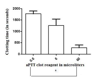
Toxins 2020, 12, x FOR PEER REVIEW                                                                                                     4 of 11

Toxins 2020,
Toxins 2020, 12,
             12, 79
                 x FOR PEER REVIEW                                                                                                     4 of 11

     Figure 2. The effect of crescent doses of the aPTT clot reagent (an ellagic acid- and
     phospholipid-based     ofreagent)  on  the clotting timeclot
                                                               parameter of    recalcified   (with 20 µ L of 0.2 M CaCl2)
     Figure 2.2.The
     Figure          effect
                   The         crescent
                          effect         doses
                                   of crescent  ofdoses
                                                   the aPTTof thereagent
                                                                     aPTT (an    ellagic
                                                                              clot       acid-
                                                                                     reagent    and
                                                                                                (an phospholipid-based
                                                                                                     ellagic acid- and
     human
     reagent) plasma   samples.
               on the clotting      Values
                                   timeon   are presented
                                         parameter          as  means
                                                      of recalcified     ± SD.
                                                                      (withof20No   statistically
                                                                                 µL of 0.2(with   significant
                                                                                              M CaCl             differences
     phospholipid-based      reagent)      the clotting  time parameter        recalcified         20 µ2 )L human
                                                                                                            of 0.2 Mplasma
                                                                                                                      CaCl2)
     (p < 0.05)
     samples.    were   observed
                Valuessamples.       between
                         are presented         groups   (n =
                                                     ± SD. as6, each)   treated  with   minor   and  major     doses
                                                                                                                 0.05)ofwere
                                                                                                                         this
                                                                                                            (p
Toxins2020,
Toxins 2020,12,
12,79
x FOR PEER REVIEW 55 of
of 11
11
Toxins 2020, 12, x FOR PEER REVIEW 5 of 11
Figure 4. Upper, typical intrinsic pathway thromboelastometry (INTEM) profile of human plasma
(260
Figureµ L)
Figure 4.4.recalcified
Upper, (withintrinsic
Upper, typical
typical 20 µ L of pathway
intrinsic 0.2 M CaCl
pathway 2) and treated with (INTEM)
thromboelastometry
thromboelastometry 60 µ L of phosphate-buffered
(INTEM) profile
profile ofof human
human plasma saline
plasma
(PBS)
(260 µsolution
(260 µL) containing:
L)recalcified
recalcified (with
(with20 A—PBS
20 µ Lof
µL solution;
of0.2
0.2 MMCaCl
CaCl B—a mean
22)) and
and coagulant
treated
treated with
with 60dose
60 (MCD)
µ L of
µL of the activatorsaline
of phosphate-buffered
phosphate-buffered aPTT
saline
(PBS)
clot
(PBS) solution
reagent;
solution containing:
and C—B in the
containing: A—PBS
A—PBS solution;
presence of 20B—a
solution; ng ofmean
B—a bee A.
mean coagulant
mellifera dose
coagulant venom
dose (MCD) of
of the
the activator
phospholipase
(MCD) aPTT
A2. Below,
activator aPTT
clot
clotreagent;
the typical and
andC—B
reagent;INTEM C—B ininthe
profile ofpresence
the chickenofplasma
presence 20 20
of ngng A. mellifera
ofrecalcified
bee
of bee venom
and treated
A. mellifera venomphospholipase
with 60 µ L ofAPBS
phospholipase 2 . Below,
A solution
2 . the
Below,
typical INTEM
containing: D— profile
PBS of chicken
solution; E—aplasma
MCD recalcified
of the and treated
activator aPTT with
clot 60 µL
reagent;
the typical INTEM profile of chicken plasma recalcified and treated with 60 µ L of PBS solutionof PBS
F—Esolution
in the containing:
presence of
D—PBS
20 solution;
ng of bee D—
containing: E—a
A. mellifera MCD of
venom E—a
PBS solution; the activator
phospholipase
MCD of the aPTT clot
A2,activatorreagent;
respectively. F—E in the presence of 20
aPTT clot reagent; F—E in the presence of ng of bee
A.
20mellifera
ng of bee venom phospholipase
A. mellifera A2 , respectively.
venom phospholipase A2, respectively.
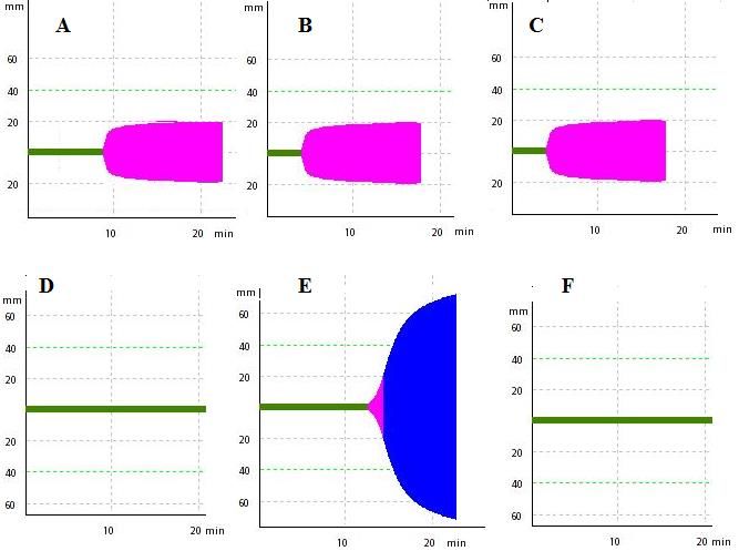
Figure 5. The effect of crescent doses of bee A. mellifera venom phospholipase A2 on the clotting time
Figure 5. The effect of crescent doses of bee A. mellifera venom phospholipase A2 on the clotting time
of recalcified (with 20 µL of 0.2 M CaCl2 ) and activated (with a mean coagulant dose (MCD) of the
of recalcified
Figure 5. The(with
effect20of µcrescent
L of 0.2doses
M CaCl ) and
of 2bee A. activated (with aphospholipase
mellifera venom mean coagulant dose
A2 on the(MCD)
clottingoftime
the
activator aPTT clot reagent) human plasma samples. A—clotting time related to recalcified plasma
activator aPTT(with
of recalcified clot reagent)
20 µ L of human plasma
0.2 M CaCl 2) andsamples. A—clotting
activated time coagulant
(with a mean related to dose
recalcified
(MCD)plasma
of the
samples (m= 874 ± 230 sec, control group); B—recalcified plasma samples activated with a MCD of
samples
activator(m=
aPTT874clot
± 230 sec, control
reagent) humangroup);
plasmaB—recalcified plasma samples
samples. A—clotting activated
time related with a MCD
to recalcified plasmaof
aPTT clot reagent (238 ± 87 sec); C (284 ± 75 sec), D (446 ± 98 sec), E (557 ± 127 sec), F (630 ± 238 sec), and
aPTT clot reagent (238 ± 87 sec); C (284 ± 75 sec), D (446 ± 98 sec), E (557 ± 127 sec),
samples (m= 874 ± 230 sec, control group); B—recalcified plasma samples activated with a MCD of F (630 ± 238 sec),
G (884 ± 255 sec)—B treated with 20, 80, 160, 200, and 400 ng of bee A. mellifera venom phospholipase
and
aPTTGclot(884 ± 255(238
reagent sec)—B treated
± 87 sec); with
C (284 ± 75 20,sec),
80, D160,
(446200,
± 98and
sec),400 ng ±of127
E (557 bee A. Fmellifera
sec), venom
(630 ± 238 sec),
A2 , respectively. Values are presented as means ± SD. * Statistically significant differences (p < 0.05) in
phospholipase
and G (884 ± A 2, respectively.
255 sec)—B treated Values
withare 20, presented as means
80, 160, 200, and 400 ± ng
SD. of* bee
Statistically significant
A. mellifera venom
relation to B (n = 6 each experimental group).
differences
phospholipase(p < 0.05) in relation to Values
A2, respectively. B (n = 6 are
eachpresented
experimental group).± SD. * Statistically significant
as means
differences (p < 0.05) in relation to B (n = 6 each experimental group).Toxins 2020, 12, 79 6 of 11
3. Discussion
The relatively large RT presented by CP allowed for the elaboration of a dose–response curve,
and the determination of the relative potencies of both the agonist (aPTT clot reagent) and antagonist
(bvPLA2 ), which caused its sensitivity to be almost 20-fold higher than that of human plasma. Nielsen
recently showed through thromboelastography that effective anticoagulant activity on normal human
plasma was achieved by using bvPLA2 at 200 ng/mL [41]. In our conditions, 380 ± 41 ng was sufficient
to displace the CT values of human plasma to values within the time intervals of their related RT in
100% of the assays. This discordance in results is likely due to this author using nonactivated human
plasma samples.
In summary, three clear differences can be observed in our study, when the INTEM profiles of
activated CP samples are compared with that of human plasma: (i) The CT parameter values of
the control recalcified CP is comparatively greater (mean = 2247 ± 319 s versus 874 ± 230 s); (ii) a
statistically significant difference between the minimum and maximal coagulant responses to the
activator aPTT clot reagent occur within two orders of magnitude (from 0.6 to 60 µL); and (iii) the
bvPLA2 anticoagulant effect occurs in a dose-dependent manner. This unusual sensitivity of activated
and recalcified CP to the bvPLA2 anticoagulant effect described herein reinforces our previous studies
with this pharmacological target [30,31].
Below, we present arguments justifying why, in our opinion, platelet-poor CP samples should
be considered in some studies using rotational thromboelastometry, for screening purposes of some
animal venom toxins that interfere with several steps of the clotting process, such as the stages of clot
initiation, formation, stability, strength, and dissolution.
The careful collection and centrifugation of blood samples from chickens is required. Foam
or bubble formation in the syringes during blood sample collection leads to cell lysis and possible
activation of the coagulation process, shortening the RT of the control-treated CP samples. Some
parameters of the ROTEM profile, such as the clotting time, clot formation time, α angle and maximal
amplitude, are particularly dependent on fibrin polymerization and platelet count [42–44]. Recalcified
CP samples obtained after centrifugation at greater rates render no typical ROTEM profile, probably as
a consequence of low concentrations of cofactors, such as, for example, platelet microparticles and
phospholipids [45,46]. To obtain one typical ROTEM profile, such as that shown in Figure 4, white
Leghorn whole blood samples must be centrifuged at lower rates (up to 4000× g). Vehicle-treated
recalcified CP samples are stable (no spontaneous clot formation) for at least 30 min. This prolonged RT
is essential for our main purpose: Elaboration of one sensitive dose-response curve to agonists. Pilot
assays with chicken whole blood samples or platelet-rich plasma present RT values of 705 ± 338 s and
1045 ± 428 s, respectively (n = 8, each) (data not shown). These restricted intervals become impracticable
for the construction of one dose-response curve to these agonists. Consequently, platelet-rich plasma or
whole blood samples presented sensitivities that were significantly smaller to the bvPLA2 anticoagulant
activity, when compared with that of platelet-poor plasma samples.
The time taken for spontaneous clot formation following recalcification is highly variable between
the plasma of mammal and nonmammal species. This may be due to a variation in the level of factor
XII (Hageman factor) present [47]. Although it has a natural factor XII deficiency [22], CP possesses a
fully functional extrinsic pathway, and its key coagulant proteins (factors V and X) could be considered
as suitable targets or substrates for both procoagulant [30,48,49] and anticoagulant toxins [31] from
several animal venoms.
Our findings indicate that RT can be considered as the main difference between chicken and human
plasma, in the conditions herein described. The relatively limited interval (almost 600 seconds) of RT
and the relatively large reference intervals presented by the values of the CT parameter of the INTEM
profile (from 100 to 240 s) in activated human plasma abrogates the possibility of the elaboration of a
true dose–response relationship for the agonists here described. The differences between the CT values
related to 0.6, 6 and 60 µL of the aPTT clot reagent were not statistically significant (Figure 2). On theToxins 2020, 12, 79 7 of 11
other hand, the relatively large RT presented by CP elicited the elaboration of one dose–response curve,
and the determination of the relative potencies of both the activator aPTT clot reagent and bvPLA2 .
4. Conclusions
The main limitations of most in vitro techniques designed for assaying pro- and anticoagulant
activity of animal venoms and toxins on mammalian plasma is that these assays give a single
parameter (fibrin formation) for one complex enzymatic process [16]. This limitation becomes evident,
for example, during screening procedures of animal venoms, when the effects of toxins, such as
fibrinolysis, fibrinogenolysis or fibrinolysis induction on the coagulation pathway should be examined.
In conclusion, we propose this functional assay as an alternative for (i) screening assays during the
purification of fibrinolytics, pro- or anticoagulant toxins from whole animal venoms, (ii) assessing
a specific antivenom’s relative potencies against venom toxic components with both coagulant and
lethal activities (such as B. jararaca venom [30] or with PLA2 -like substances, such as those present
in the venoms of the C. d. terrificus snake [31] or the A. mellifera bee, and (iii) studies for testing the
relative potencies of prototypes of pharmaceutical agents or natural inhibitors of PLA2 -like toxins from
animal venoms in more diluted solutions, thus with less potential to affect the normal coagulation
kinetic behavior.
5. Material and Methods
5.1. Reagents
An activated partial thromboplastin time reagent (aPTT) clot, containing ellagic acid and synthetic
phospholipids, was obtained from BIOS Diagnostica (SP, Brazil); sodium citrate was obtained from
Ecibra (Curitiba, Brazil); calcium chloride was obtained from E. Merck (German); pooled 4% citrated
normal human plasma (maintained at −80 ◦ C) and PLA2 , from A. mellifera venom, were obtained from
Sigma-Aldrich, St. Louis, MO, USA). PLA2 was dissolved in phosphate-buffered saline (PBS) at a
concentration of 1 mg/ml, aliquoted for single-use, and frozen at −80 ◦ C. The tested enzymes were
only frozen and thawed once for all subsequently described work. All chemicals were of an analytical
reagent grade.
5.2. Animals
Adult female or male white Leghorn chickens (1.0 to 1.7 kg) were used. All birds were a donation
from commercial breeding (Granja Ino, São Paulo, Brazil). The animals had free access to water and
food, and were kept in a 12 h light/dark cycle.
Ethical approval: All the procedures involving animals were carried out following the Guiding
Principles for the Use of Animals in Toxicology (International Society of Toxicology, http://www.
toxicology.org) and the Brazilian College of Animal Experimentation (COBEA). The experimental
protocol was approved by the Ethic Committee on Animal Use of the Butantan Institute (CEUAIB)
(protocol number CEUA 6259250918) in 11/08/2018.
5.3. Collection of Citrated Chicken Plasma Samples
The birds were restrained on their backs with their wings spread. After the use of xylocaine spray
as a local anesthetic agent, the feathers were removed, and small incisions were made for cleaning
around the brachial wing vein. Eight milliliters of whole blood samples were collected using syringes
containing 1:10 (v/v) 3.2% trisodium citrate and then closed with cotton-yarn. Chicken plasma was
obtained after centrifugation at 3000× g for 20 min at 4 ◦ C. Plasma samples were used immediately or
maintained at −80 ◦ C. The total blood volume of one particular organism is very difficult to determine
and depends on its species, sex, age, and health, as well as its nutritional condition. The total circulating
blood volume is in the range 55–70 mL/kg of the body weight, and the adult chickens provided at leastToxins 2020, 12, 79 8 of 11
8 mL of whole blood samples from each wing vein, without significant animal distress or the need
for euthanasia.
5.4. Thromboelastometric Assays with Chicken and Human Plasma Samples
5.4.1. Standardization of the Activator aPTT Clot Mean Coagulant Dose (MCD)
The amount of subsequently described plasmatic and other additives summed to a final volume
of 340 µL. Samples were composed of 260 µL of human or chicken plasmas, plus 20 µL of 200 mM
CaCl2 and 60 µL of PBS. Crescent doses of the activator aPTT clot reagent were included as a fraction
of the 60 µL of PBS. The final mixture was pipetted into a disposable cup in a computerized ROTEM
four-channel system (Pentapharm, Munich, Germany) at 37 ◦ C, and then rapidly mixed into the cup
against and then away from the plastic pipette three times. The parameter evaluated was clotting
time (CT, time from the start of the reaction to initial clot formation) [23]. Data were collected for one
hour. Two experimental groups were considered: (Group 1) - 60 µL of PBS solution (control-group,
standardized as RT), and (Group 2) - 60 µL of crescent doses of the activator aPTT clot reagent, for
determination of its MCD. The MCD of the aPTT clot for CP was considered as the amount (in µL) that
shortens the CT of the control group (2247 ± 319 sec) to 900 s (situated between the minimum and
maximum coagulant responses). In addition, the MCD of the aPTT clot reagent for human plasma was
considered as the amount that shortens the CT of the control group (874 ± 230 s) to an interval between
100 and 240 s (normal range values for the INTEM assay, according to the manufacturer’s instructions).
5.4.2. Standardization of the Effective and Mean Anticoagulant Doses (EAD and MAD, Respectively)
of Bee Venom Phospholipase A2 on Human and Chicken Activated Plasma Samples
Briefly, 60 µL of the PBS fraction of the assay containing the MCD of the activator aPTT clot in
absence or presence of crescent concentrations of bvPLA2 were incubated during 1 min with 260 µL of
human or chicken plasmas, before the addition of 20 µL of 200 mM CaCl2 . Two experimental groups
were then considered: (Group 3) - 60 µL of PBS solution containing the MCD of the activator aPTT
clot, standardized as activated samples and (Group 4) - 60 µL of PBS solution containing the MCD
of the activator aPTT clot plus crescent doses of bvPLA2 , for determining its EAD. If no change in
coagulation occurred compared to normal human or chicken plasma samples, the concentrations
of bvPLA2 were increased. However, if coagulation was not detectable, then the concentrations of
bvPLA2 were progressively diminished until at the very least coagulation was detectable. The EAD
of bvPLA2 was defined as the sufficient minimum dose (in ng) required to displace the CT values
induced by the MCD of the activator aPTT clot in chicken or human plasmas to values within the
time intervals of their related RT in 100% of assays. The MAD of bvPLA2 , defined as the sufficient
doses required to displace CT values induced by one MCD of the activator aPTT clot on CP to that
corresponding to values between the mean and minimum coagulant responses (in our study, 1350 s),
can be determined through linear regression.
6. Statistical Analysis
Values of the CT parameter were monitored in seconds and expressed as mean ± SD in six
independent experiments. One-way analysis of variance (ANOVA) comparisons were used, followed
by Newman–Keuls post hoc analysis. Values defining the MCD of the activator aPTT clot reagent,
EAD, and MAD of bvPLA2 were determined by means of linear regression analysis. The linear
regression plots were performed using GraphPad Prism 5.0 software (San Diego, CA, USA). p < 0.05
was considered statistically significant.
Author Contributions: Conceptualization, B.C.P. and N.O.; methodology, B.C.P. and N.O.; software, B.C.P. and
N.O.; validation, B.C.P. and N.O.; formal analysis, B.C.P. and N.O.; investigation, B.C.P. and N.O.; resources,
B.C.P. and N.O.; data curation, B.C.P. and N.O.; writing—original draft preparation, B.C.P.; writing—review and
editing, B.C.P. and N.O.; visualization, B.C.P and N.O.; supervision, B.C.P.; project administration, B.C.P. and N.O.;
funding acquisition, B.C.P. and N.O. All authors have read and agreed to the published version of the manuscript.Toxins 2020, 12, 79 9 of 11
Funding: This research was funded by Fundação de Amparo à Pesquisa do Estado de São Paulo (FAPESP), grant
number 2009/54620-4.
Acknowledgments: The authors would like to acknowledge (i) the staff within the Laboratory of Pharmacology,
from the Butantan Institute, (ii) Granja Ino, São Paulo, SP, Brazil for providing the chickens, and (iii) the grant
provided by the Butantan Foundation, of the Butantan Institute.
Conflicts of Interest: The authors declare no conflict of interest. The funding body for this study had no role in
the design of the study; in the collection, analyses, or interpretation of data, in the writing of the manuscript, or in
the decision to publish the results.
References
1. Kini, R.M. Molecular moulds with multiple missions: Functional sites in three-finger toxins. Clin. Exp.
Pharmacol. Physiol. 2002, 29, 815–822. [CrossRef]
2. Purchase, I.F.; Botham, P.A.; Bruner, L.H.; Frazier, J.M.; Stokes, W.S. Workshop overview: Scientific and
regulatory challenges for the reduction, refinement, and replacement of animals in toxicity testing. Toxicol.
Sci. 1998, 43, 86–101.
3. Gene, J.A.; Roy, A.; Rojas, G.; Gutiérrez, J.M.; Cerdas, L. Comparative study on coagulant, defibrinating,
fibrinolytic and fibrinogenolytic activities of Costa Rican crotaline snake venoms and their neutralization by
a polyvalent antivenom. Toxicon 1989, 27, 841–848. [CrossRef]
4. Laing, G.D.; Theakston, R.D.; Leite, R.P.; da Silva, W.D.; Warrell, D.A. Comparison of the potency of
three Brazilian Bothrops antivenoms using in vivo rodent and in vitro assays. BIASG (Butantan Institute
Antivenom Study Group). Toxicon 1992, 30, 1219–1225. [CrossRef]
5. Clemens, R.; Lorenz, R.; Pukrittayakamee, S.; Punpoowang, B.; Vanijanonta, S.; Charoenlarp, P. Effects of
antithrombin III and antivenom on procoagulant activity of Russell’s viper venom in a whole blood model.
Southeast Asian J. Trop. Med. Public Health 1995, 26, 143–148. [PubMed]
6. Sprivulis, P.; Jelinek, G.A.; Marshall, L. Efficacy and potency of antivenoms in neutralizing the procoagulant
effects of Australian snake venoms in dog and human plasma. Anaesth. Intensive Care 1996, 24, 379–381.
[CrossRef]
7. Sells, P.G. Animal experimentation in snake venom research and in vitro alternatives. Toxicon 2003, 42,
115–133. [CrossRef]
8. Isbister, G.K.; O’Leary, M.A.; Schneider, J.J.; Brown, S.G.; Currie, B.J. ASP Investigators. Efficacy of antivenom
against the procoagulant effect of Australian brown snake (Pseudonaja sp.) venom: In vivo and in vitro
studies. Toxicon 2007, 49, 57–67. [CrossRef]
9. Isbister, G.K.; Woods, D.; Alley, S.; O’Leary, M.A.; Seldon, M.; Lincz, L.F. Endogenous thrombin potential as a
novel method for the characterization of procoagulant snake venoms and the efficacy of antivenom. Toxicon
2010, 56, 75–85. [CrossRef]
10. Segura, A.; Villalta, M.; Herrera, M.; Léon, G.; Harrison, R.; Durfa, N.; Nasidi, A.; Calvete, J.J.; Theakston, R.D.;
Warrell, D.A.; et al. Preclinical assessment of the efficacy of a new antivenom (EchiTAb-Plus-ICP) for the
treatment of viper envenoming in sub-Saharan Africa. Toxicon 2010, 55, 369–374. [CrossRef]
11. Pla, D.; Paiva, O.K.; Sanz, L.; Beutler, M.; Wright, C.E.; Calvete, J.J.; Williams, D.J.; Gutiérrez, J.M. Preclinical
efficacy of Australian antivenoms against the venom of the small-eyed snake, Micropechis ikaheka, from
Papua New Guinea: An antivenomics and neutralization study. J. Proteom. 2014, 110, 198–208. [CrossRef]
[PubMed]
12. Pla, D.; Bande, B.W.; Welton, R.E.; Paiva, O.K.; Sanz, L.; Segura, A.; Wright, C.E.; Calvete, J.J.; Gutiérrez, J.M.;
Williams, D.J. Proteomics and antivenomics of Papuan black snake (Pseudechis papuanus) venom with
analysis of its toxicological profile and the preclinical efficacy of Australian antivenoms. J. Proteom. 2017,
150, 201–215. [CrossRef] [PubMed]
13. Pornmuttakun, D.; Ratanabanangkoon, K. Development of an in vitro potency assay for antivenom against
Malayan pit viper (Calloselasma rhodostoma). Toxicon 2014, 77, 1–5. [CrossRef] [PubMed]
14. Sanchez, L.V.; Pla, D.; Herrera, M.; Chippaux, J.P.; Calvete, J.J.; Gutiérrez, J.M. Evaluation of the preclinical
efficacy of four antivenoms, distributed in sub-Saharan Africa, to neutralize the venom of the carpet viper,
Echis ocellatus, from Mali, Cameroon, and Nigeria. Toxicon 2015, 106, 97–107. [CrossRef]Toxins 2020, 12, 79 10 of 11
15. Calvete, J.J.; Arias, A.S.; Rodríguez, Y.; Quesada-Bernat, S.; Sánchez, L.V.; Chippaux, J.P.; Pla, D.; Gutiérrez, J.M.
Preclinical evaluation of three polyspecific antivenoms against the venom of Echis ocellatus: Neutralization
of toxic activities and antivenomics. Toxicon 2016, 119, 280–288. [CrossRef]
16. Isbister, G.K. Procoagulant snake toxins: Laboratory studies, diagnosis, and understanding snakebite
coagulopathy. Semin. Thromb. Hemost. 2009, 35, 93–103. [CrossRef]
17. Theakston, R.D.; Reid, H.A. Development of simple assay standard procedures for the characterization of
snake venoms. Bull. World Health Organ. 1983, 61, 949–956.
18. O’Leary, M.A.; Isbister, G.K. A turbidimetric assay for the measurement of clotting times of procoagulant
venoms in plasma. J. Pharmacol. Toxicol. Methods 2010, 61, 27–31. [CrossRef]
19. Mann, K.G.; Jenny, R.J.; Krishnaswamy, S. Cofactor proteins in the assembly and expression of blood clotting
enzyme complexes. Annu. Rev. Biochem. 1988, 57, 915–956. [CrossRef]
20. Weidmann, H.; Heikaus, L.; Long, A.T.; Naudin, C.; Schlüter, H.; Renné, T. The plasma contact system, a
protease cascade at the nexus of inflammation, coagulation and immunity. Biochim. Biophys. Acta Mol. Cell
Res. 2017, 864, 2118–2212. [CrossRef]
21. Ratnoff, O.D.; Colopy, J.E. A familial hemorrhagic trait associated with a deficiency of a clot-promoting
fraction of plasma. J. Clin. Investig. 1955, 34, 602–613. [CrossRef] [PubMed]
22. Ponczek, M.B.; Gailani, D.; Doolittle, R.F. Evolution of the contact phase of vertebrate blood coagulation.
J. Thromb. Haemost. 2008, 6, 1876–1883. [CrossRef] [PubMed]
23. Whiting, D.; DiNardo, J.A. TEG and ROTEM: Technology and clinical applications. Am. J. Hematol. 2014, 89,
228–232. [CrossRef] [PubMed]
24. Dambisya, Y.M.; Lee, T.L.; Gopalakrishnakone, P. Action of Calloselasma rhodostoma (Malayan pit viper)
venom on human blood coagulation and fibrinolysis using computerized thromboelastography (CTEG).
Toxicon 1994, 32, 1619–1626. [CrossRef]
25. Dambisya, Y.M.; Lee, T.L.; Gopalakrishnakone, P. Anticoagulant effects of Pseudechis australis (Australian
king brown snake) venom on human blood: A computerized thromboelastography study. Toxicon 1995, 33,
1378–1382. [CrossRef]
26. Nagel, S.S.; Schoeman, J.P.; Thompson, P.N.; Wiinberg, B.; Goddard, A. Hemostatic analysis of dogs naturally
envenomed by the African puffadder (Bitis arietans) and snouted cobra (Naja annulifera). J. Vet. Emerg. Crit.
Care (San Antonio) 2014, 24, 662–671. [CrossRef]
27. Hiremath, V.; Nanjaraj Urs, A.N.; Joshi, V.; Suvilesh, K.N.; Savitha, M.N.; Urs Amog, P.; Rudresha, G.V.;
Yariswamy, M.; Vishwanath, B.S. Differential action of medically important Indian BIG FOUR snake venoms
on rodent blood coagulation. Toxicon 2016, 110, 19–26. [CrossRef]
28. Nielsen, V.G.; Boyer, L.V.; Redford, D.T.; Ford, P. Thrombelastographic characterization of the thrombin-like
activity of Crotalus simus and Bothrops asper venoms. Blood Coagul. Fibrinolysis 2017, 28, 211–217. [CrossRef]
29. Nielsen, V.G.; Sanchez, E.E.; Redford, D.T. Characterization of the rabbit as an in vitro and in vivo model to
assess the effects of fibrinogenolytic activity of snake venom on coagulation. Basic Clin. Pharmacol. Toxicol.
2018, 122, 157–164. [CrossRef]
30. Oguiura, N.; Kapronezai, J.; Ribeiro, T.; Rocha, M.M.; Medeiros, C.R.; Marcelino, J.R.; Prezoto, B.C. An
alternative micromethod to access the procoagulant activity of Bothrops jararaca venom and the efficacy of
antivenom. Toxicon 2014, 90, 148–154. [CrossRef]
31. Prezoto, B.C.; Tanaka-Azevedo, A.M.; Marcelino, J.R.; Tashima, A.K.; Nishiduka, E.S.; Kapronezai, J.;
Mota, J.O.; Rocha, M.M.T.; Serino-Silva, C.; Oguiura, N. A functional and thromboelastometric-based
micromethod for assessing crotoxin anticoagulant activity and antiserum relative potency against Crotalus
durissus terrificus venom. Toxicon 2018, 148, 26–32. [CrossRef] [PubMed]
32. Fry, B.G.; Wüster, W.; Kini, R.M.; Brusic, V.; Khan, A.; Venkataraman, D.; Rooney, A.P. Molecular evolution
and phylogeny of elapid snake venom three-finger toxins. J. Mol. Evol. 2003, 57, 110–129. [CrossRef]
[PubMed]
33. Lynch, V.J. Inventing an arsenal: Adaptive evolution and neofunctionalization of snake venom phospholipase
A2 genes. BMC Evol. Biol. 2007, 7, 2. [CrossRef] [PubMed]
34. Casewell, N.R.; Wüster, W.; Vonk, F.J.; Harrison, R.A.; Fry, B.G. Complex cocktails: The evolutionary novelty
of venoms. Trends Ecol. Evol. 2013, 28, 219–229. [CrossRef]
35. Kini, R.M.; Evans, H.J. The role of enzymatic activity in inhibition of the extrinsic tenase complex by
phospholipase A2 isoenzymes from Naja nigricollis venom. Toxicon 1995, 33, 1585–1590. [CrossRef]Toxins 2020, 12, 79 11 of 11
36. Kerns, R.T.; Kini, R.M.; Stefansson, S.; Evans, H.J. Targeting of Venom Phospholipases: The strongly
anticoagulant phospholipase A2 from Naja nigricollis venom binds to coagulation Factor xa to inhibit the
prothrombinase complex. Arch. Biochem. Biophys. 1999, 369, 107–113. [CrossRef]
37. Kini, R.M. Structure—Function relationships and mechanism of anticoagulant phospholipase A2 enzymes
from snake venoms. Toxicon 2005, 45, 1147–1161. [CrossRef]
38. Ouyang, C.; Lin, S.C.; Teng, C.M. Anticoagulant properties of Apis mellifera (honey bee) venom. Toxicon
1979, 17, 197–201. [CrossRef]
39. Ownby, C.L.; Powell, J.R.; Jiang, M.S.; Fletcher, J.E. Melittin and phospholipase A2 from bee (Apis mellifera)
venom cause necrosis of murine skeletal muscle in vivo. Toxicon 1997, 35, 67–80. [CrossRef]
40. Prado, M.; Solano-Trejos, G.; Lomonte, B. Acute physiological effects of honeybee (Apis mellifera) envenoming
by subcutaneousroute in a mouse model. Toxicon 2010, 56, 1007–1017. [CrossRef]
41. Nielsen, V.G. The anticoagulant effect of Apis mellifera phospholipase A2 is inhibited by CORM-2 via a
carbon monoxide-independent mechanism. J. Thromb. Thrombolysis 2020, 49, 100–107. [CrossRef]
42. Orlikowski, C.E.; Rocke, D.A.; Murray, W.B.; Gouws, E.; Moodley, J.; Kenoyer, D.G.; Byrne, S.
Thromboelastography changes in preeclampsia and eclampsia. Br. J. Anaesth. 1996, 77, 157–161. [CrossRef]
[PubMed]
43. Apelseth, T.O.; Bruserud, O.; Wentzel-Larsen, T.; Hervig, T. Therapeutic efficacy of platelet transfusion in
patients with acute leukemia: An evaluation of methods. Transfusion 2010, 50, 766–775. [CrossRef]
44. Rumph, B.; Bolliger, D.; Narang, N.; Molinaro, R.J.; Levy, J.H.; Szlam, F.; Tanaka, K.A. In vitro comparative
study of hemostatic components in warfarin-treated and fibrinogen-deficient plasma. J. Cardiothorac. Vasc.
Anesth. 2010, 24, 408–412. [CrossRef] [PubMed]
45. Owens, A.P.; Mackman, N. Microparticles in hemostasis and thrombosis. Circ. Res. 2011, 108, 1284–1297.
[CrossRef] [PubMed]
46. Wu, Z.H.; Ji, C.L.; Li, H.; Qiu, G.X.; Gao, C.J.; Weng, X.S. Membrane microparticles and diseases. Eur. Rev.
Med. Pharmacol. Sci. 2013, 17, 2420–2427. [PubMed]
47. Ratnoff, O.D. The Biology and Pathology of the Initial Stages of Blood Coagulation; Brown, E.B., Moore, C.V., Eds.;
Heinemann: London, UK, 1966; Volume 5, pp. 204–245.
48. Johnson, G.S.; Turrentine, M.A.; Swayne, D.E. Coagulation of plasma from the chicken (Gallus domesticus):
Phospholipids influence clotting rates induced by components from Russell’s viper venom. Comp. Biochem.
Physiol. B 1985, 82, 647–653. [CrossRef]
49. Bernardoni, J.L.; Sousa, L.F.; Wermelinger, L.S.; Lopes, A.S.; Prezoto, B.C.; Serrano, S.M.; Zingali, R.B.;
Moura-da-Silva, A.M. Functional variability of snake venom metalloproteinases: Adaptive advantages in
targeting different prey and implications for human envenomation. PLoS ONE 2014, 9, e109651. [CrossRef]
© 2020 by the authors. Licensee MDPI, Basel, Switzerland. This article is an open access
article distributed under the terms and conditions of the Creative Commons Attribution
(CC BY) license (http://creativecommons.org/licenses/by/4.0/).You can also read