DRUG RELEASE FROM POLYMER-COATED TIO 2 NANOTUBES ON ADDITIVELY MANUFACTURED TI-6AL-4V BONE IMPLANTS: A FEASIBILITY STUDY
←
→
Page content transcription
If your browser does not render page correctly, please read the page content below

Nano Express

PAPER • OPEN ACCESS

Drug release from polymer-coated TiO2 nanotubes on additively
manufactured Ti-6Al-4V bone implants: a feasibility study
To cite this article: Chiara Micheletti et al 2021 Nano Ex. 2 010018

View the article online for updates and enhancements.

                                This content was downloaded from IP address 46.4.80.155 on 09/09/2021 at 09:36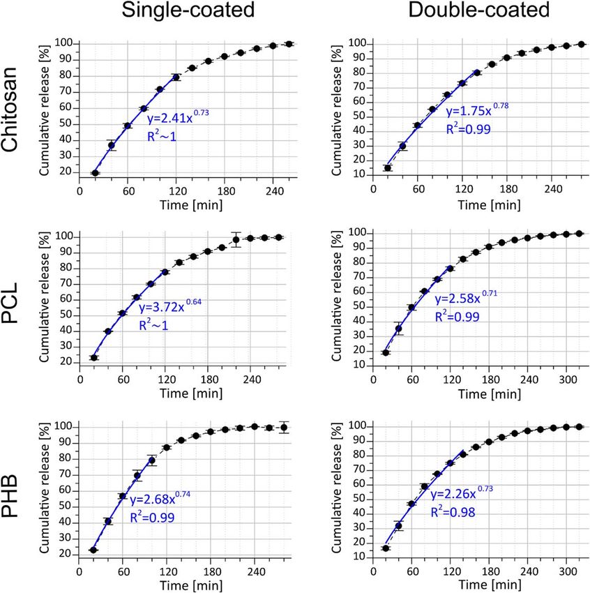
Nano Express 2 (2021) 010018                                                                https://doi.org/10.1088/2632-959X/abe278

                              PAPER

                              Drug release from polymer-coated TiO2 nanotubes on additively
OPEN ACCESS
                              manufactured Ti-6Al-4V bone implants: a feasibility study
RECEIVED
10 October 2020
                              Chiara Micheletti1,2 , Raffaella Suriano1 , Kathryn Grandfield2,3                       and Stefano Turri1
REVISED                       1
28 January 2021                   Department of Chemistry, Materials and Chemical Engineering, Politecnico di Milano, Milan, Italy
                              2
                                  Department of Materials Science and Engineering, McMaster University, Hamilton, ON, Canada
ACCEPTED FOR PUBLICATION      3
                                  School of Biomedical Engineering, McMaster University, Hamilton, ON, Canada
2 February 2021
PUBLISHED
                              E-mail: michelec@mcmaster.ca
10 February 2021
                              Keywords: bone implant, SLM, titania nanotubes, surface topography, local drug delivery, polymer coating

Original content from this
work may be used under
the terms of the Creative     Abstract
Commons Attribution 4.0
licence.                      Insufficient osseointegration, inflammatory response and bacterial infection are responsible for the
Any further distribution of   majority of bone implant failures. Drug-releasing implants subjected to adequate surface modification
this work must maintain
attribution to the            can concurrently address these challenges to improve the success of implant surgeries. This work
author(s) and the title of
the work, journal citation
                              investigates the use of Ti-6Al-4V (Ti64) with a dual-scale surface topography as a platform for local
and DOI.                      drug delivery. Dual-scale topography was obtained combining the inherent microscale roughness of
                              the Ti64 samples manufactured by selective laser melting (SLM) with the nanoscale roughness of TiO2
                              nanotubes (TNTs) obtained by subsequent electrochemical anodization at 60 V for 30 min. TNTs
                              were loaded with a solution of penicillin-streptomycin, a common antibiotic, and drug release was
                              tested in vitro. Three biocompatible and biodegradable polymers, i.e. chitosan, poly(ε-caprolactone)
                              and poly(3-hydroxybutyrate), were deposited by spin coating, while preserving the microscale
                              topography of the substrate underneath. The presence of polymer coatings overall modified the drug
                              release pattern, as revealed by fitting of the experimental data with a power-law model. A slight
                              extension in the overall duration of drug release (about 17% for a single layer and 33% for two layers of
                              PCL and PHB) and reduced burst release was observed for all polymer-coated samples compared to
                              uncoated, especially when two layers of coatings were applied.

                              1. Introduction

                              Titanium and its alloy Ti-6Al-4V (Ti64) are widely employed materials for orthopaedic and dental implants due
                              to their high excellent biocompatibility and corrosion resistance provided by the thin oxide layer (TiO2, also
                              termed titania) that spontaneously forms on their surface [1]. This surface oxide layer is also considered
                              responsible for the bioactivity of titanium-based implants and hence their natural ability to osseointegrate [2, 3].
                                  Successful osseointegration, i.e. the formation of a structural and functional connection between implant
                              and the host bone tissue [4], relies on several aspects, among which surface topography has been shown to play
                              an important role [5]. In particular, several studies have concluded that microscale topography can improve
                              osseointegration [6, 7]. Recently, the processability of titanium alloys by selective laser melting (SLM), an
                              additive manufacturing (AM) technique, has emerged as a method to obtain parts with an inherent microrough
                              surface without subsequent post-processing steps. Such microroughness is conferred by the presence of
                              randomly distributed microspherical particles on the surface as a result of unmelted and unsintered process
                              powders [8] and balling effects [9]. Moreover, AM components can be highly customized, hence patient-specific
                              bone implants can be fabricated.
                                  Not only microscale, but also nanoscale roughness has been shown to be beneficial for osseointegration [10].
                              Among the several strategies to produce nanoscale features, a well-investigated possibility is the generation of
                              well-ordered arrays of TiO2 nanotubes (TNTs) by electrochemical anodization [11]. TNTs have demonstrated
                              excellent biocompatibility and cell responses in vitro [12]. Furthermore, promising results in terms of improved
                              osseointegration have been obtained in vivo [13].

                              © 2021 The Author(s). Published by IOP Publishing Ltd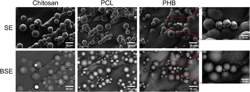
Nano Express 2 (2021) 010018 C Micheletti et al
Not only are TNTs believed to be beneficial for osseointegration, but they could also serve as platforms for
local drug delivery [14, 15]. In addition to insufficient osseointegration, other factors still pose challenges to the
success of bone implants, i.e. inflammatory responses and bacterial infections. Treatments for reducing the risk
of inflammation and infection post-surgery currently involve systemic administration of inflammatory drugs
and antibiotics, respectively. However, conventional systemic drug therapy in bone presents some limitations,
including low efficacy, lack of selectivity, poor bioavailabilty and biodistribution, and toxicity [14]. While drug
delivery to specific skeletal sites remains challenging, drug-releasing bone implants have emerged as a possibility
to overcome the limitation of conventional drug administration [14, 16, 17]. In fact, thanks to local drug
delivery, site-specific and optimal drug concentration can be achieved, without dilution across the entire body,
hence leaving other sites unaffected and avoiding toxicity and side effects. Given their hollow nature, (nano)
tubes closed at the bottom and open at the top, therapeutic agents can be accommodated inside the TNTs. Drug
release kinetics and overall duration are influenced by the nanotube size [18], which in turn can be adjusted by
the electrochemical anodization process parameters (e.g. anodization time and voltage) [19]. Different strategies
to control and extend the drug release have been proposed, including the use of polymer coatings to cap the
nanotube top opening [20–22].
Electrochemical anodization of SLM substrates was first proposed by Gulati et al to combine the inherent
microscale topography of SLM parts and the nanoscale of the TNTs [23]. The resulting dual-scale surface
topography can benefit from both microscale and nanoscale in improving osseointegration [10]. Moreover, the
viability of this type of substrate for local drug delivery has been tested in vitro [24].
In this work, the possibility to employ drug-releasing bone implants with a dual-scale surface topography
was further investigated. Ti64 samples were manufactured by SLM and electrochemically anodized to generate
TNTs on their surface. The release of the model antibiotic drug penicillin-streptomycin (pen-strep) loaded in
the TNTs was assessed in vitro. This paper presents several new approaches not previously investigated. First, our
work is the first example of drug release from polymer-coated TiO2 nanotubes on SLM substrates, aiming to
combine the benefits of antibacterial capabilities and enhanced osseointegration thanks to drugs delivered from
a surface with nano- and microscale features. While previous studies have investigated the use of polymer
coatings to control drug release duration and kinetics from TNTs on flat samples [20–22, 25], this has never been
examined for samples with a dual-scale surface topography. Second, two biodegradable polyesters never
employed in local drug delivery studies with TNTs, i.e. poly(ε-caprolactone) (PCL) and poly(3-hydroxybutyrate)
(PHB), were used. These were compared to the more commonly employed chitosan. Third, while most studies
employ dip coating [20–22], in our work spin coating was used instead to deposit the polymer coatings. Fourth,
we examined the drug release pattern by fitting with the Korsmeyer-Peppas’s power law model to compare the
diffusion behaviour of uncoated and coated TNTs. Both single- and double-coated nanotubes were investigated
to evaluate the role of coating thickness on drug release. Surface topography and morphology, roughness and
wettability were characterized for uncoated and coated substrates.
2. Materials and methods
2.1. Fabrication of Ti64 samples with dual-scale surface topography
Ti64 samples with a dual-scale surface topography were obtained combining SLM and electrochemical
anodization as previously described [26]. Briefly, Ti64 samples were manufactured by SLM (EOSINT M280
machine, EOS GmbH, Munich, Germany) as 10 mm×10 mm×1 mm squares with a 3 mm×10 mm×
1 mm handle. The samples were then electrochemically anodized at 60 V for 30 min by immersing their square
portion in a solution of ethylene glycol (certified grade, Sigma Aldrich) with 0.3% (w/w) of ammonium fluoride
(certified grade, Fisher Chemical) and 2% (v/v) of deionized water, under mild magnetic stirring. Afterwards,
they were ultrasonicated for 30 s in ethanol. The handle was then detached from the square base and discarded.
The specimens were tested as-printed in this study, without heat treatment prior or post anodization.
2.2. Drug loading
After rinsing in acetone and drying, anodized Ti64 samples were plasma treated for 60 s with atmospheric air
using a plasma system (Kenosistec Srl, Perugia, Italy) operated at a power of 150 W, to ensure high surface
hydrophilicity and facilitate drug loading. Afterwards, a solution of pen-strep (10000 IU ml−1 of penicillin and
10 mg ml−1 of streptomycin, Sigma Aldrich) was loaded in the samples by progressively drop casting 5 μl of pen-
strep solution until 1 ml of antibiotic solution was loaded. The sample surface was allowed to dry in air in
between subsequent loading steps. Samples were then gently rinsed with phosphate-buffered saline (PBS)
(Sigma Aldrich; pH=7.4) to remove any surface-bound drug and allowed to dry in air at room temperature.
2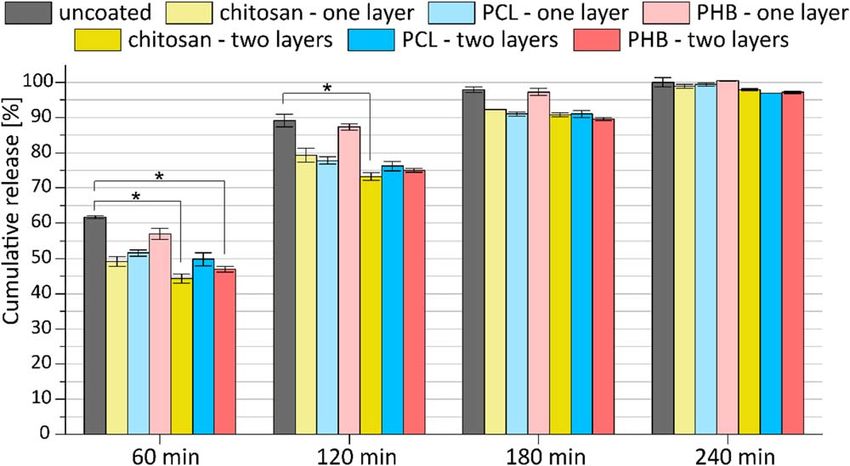
Nano Express 2 (2021) 010018 C Micheletti et al
2.3. Preparation of polymer solutions and spin coating
Chitosan powder (molecular weight 50,000–190,000 Da, degree of deacetylation 75%–85%, Sigma Aldrich) was
dissolved at 2% (w/v) in 0.8% (v/v) of acetic acid (glacial, Sigma Aldrich) and distilled water. PCL pellets
(number average molecular weight 80000, Sigma Aldrich) was dissolved at 5% (w/w) in chloroform (analytical
reagent grade, Fisher Chemical). PHB pellets (P209, Biomer) was dissolved at 4% (w/v) in chloroform
(analytical reagent grade, Fisher Chemical) at 50 °C under magnetic stirring. The PHB-based solution was then
centrifugated at 3500 rpm for 45 min (Rotofix 32A centrifuge, Hettich, Beverly, USA).
150 μl of polymer solution of either chitosan, PCL or PHB, was deposited on the surface of drug-loaded
samples, and they were subsequently spin coated for 15 s (WS-400BZ-6NPP/LITE spin coater, Laurell
Technologies Corp., North Wales, USA). Spin coating angular velocity was set to 500 rpm for chitosan, 500 rpm
for PHB and 5000 rpm for PCL. Once dry, some samples were spin coated a second time with 150 μl of the same
polymer solution and process parameters used for the first coating layer.
2.4. Characterization of uncoated and coated substrates
Surface morphology and topography of the SLM Ti64 samples before and after anodization was imaged with
scanning electron microscopy (SEM) (JSM-7000F SEM, JEOL, Peabody, USA). Average size of the
microspherical particles of the SLM samples and average TNT diameter of the anodized sample was measured
from the SEM micrographs using ImageJ (NIH, Bethesda, USA). More details on how these measurements were
carried out are provided in our previous work [26]. SEM was also used to collect both secondary electron (SE)
and backscattered electron (BSE) images of spin coated samples (EVO 50 SEM, Zeiss, Jena, Germany). Coating
thickness was estimated from mass variation before and after spin coating, assuming that the coating material
was homogeneously distributed on the substrate. To better account for the surface roughness, the ‘true’ surface
area was estimated from the geometrical area of the sample by means of the developed interfacial area ratio (Sdr).
Sdr was measured using a focus variation instrument (Alicona Infinite Focus G5, Alicona Imaging GmbH, Graz,
Austria), averaging the values of Sdr obtained for three different samples. Surface roughness of anodized and
single-coated samples was evaluated by laser profilometry (UBM Microfocus, UBM Messtechnik GmbH,
Ettlingen, Germany). Linear roughness parameters (e.g. Ra, Rq, Rz) were measured in three different spots per
sample and the values obtained were statistically averaged. Both evaluation of coating thickness and surface
roughness were repeated in duplicates, and results were statistically averaged. Hydrophilicity of polymer
coatings was evaluated by measuring the water contact angle (OCA 20, DataPhysics Instruments GmbH,
Filderstadt, Germany) in triplicates using two single-coated samples per coating material. From the measured
water contact angles, values of equilibrium (Young) contact angle were obtained by applying the Wenzel’s model
for homogeneous wetting [27], in order to take into account the surface roughness. The roughness factor (r) in
the Wenzel’s model was computed from Sdr (r=1+Sdr [28]). Roughness and wettability were assessed for
single-coated samples only, and it is assumed these properties were not significantly affected by the presence of a
second coating layer.
2.5. Characterization of drug release
Drug-loaded uncoated and spin coated (one or two coating layers of chitosan, PCL and PHB) were immersed in
1 ml of PBS and incubated at 37 °C. Every 20 min, 200 μl of release medium was withdrawn and replaced with
fresh PBS. Subsequently, 20 μl of each withdrawn sample was mixed with 200 μl of BCA working reagent (Pierce
BCA protein assay kit, ThermoFisher) in a 96-well plate and incubated at 37 °C for 30 min. Absorbance was
measured at 570 nm with a microplate reader (GENios Plus, Tecan, Männedorf, Switzerland) and the
corresponding pen-strep concentration was quantified using a calibration curve previously constructed for pen-
strep. Absorbance was measured for two withdrawn samples per release time point for each sample, and the
absorbance values were statistically averaged, after subtraction of blank (100% PBS) absorbance. The cumulative
drug release was obtained by dividing the amount of drug released at a given time by the total amount of drug
released. Two samples per type (uncoated; chitosan-, PCL- and PHB-coated; single and double coatings) were
used to test the drug release, and the values of cumulative drug release obtained for the two samples were
statistically averaged. Finally, the cumulative drug release profile was fitted using the Korsmeyer-Peppas’s power
law model, i.e. Mt/M∞ =kt n (Mt/M∞ =fraction of drug released at time t, k=release rate constant,
n=release exponent) [18, 29, 30], using OriginPro 8 (OriginLab Corporation, Northampton, USA). Only the
experimental data up to a cumulative release around 80% were considered for the fitting, in order to exclude the
plateau-like region from this analysis. Statistical significance in cumulative drug release at fixed time intervals for
different groups was determined using a one-way ANOVA with a post-hoc Tukey’s HSD test (α=0.05).
3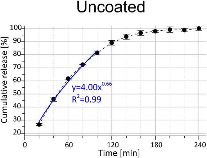
Nano Express 2 (2021) 010018 C Micheletti et al
Figure 1. SEM images of the surface of Ti64 samples before (A) and after (B), (C) electrochemical anodization. (A) reveals the spherical
microparticles characteristic of manufacturing by SLM. (B) shows the typical cracks appearing on the surface of the samples after
anodization: these cracks are attributed to the formation of separate arrays of TNTs growing on curved surfaces (more details about
anodization and cracks formation can be found in [31]). (C) provides a zoomed-in view of one of the well-ordered arrays of TNTs
present in (B).
Table 1. Values of coating thickness for single and double coatings of
chitosan, PHB and PCL. Spin coating angular velocities employed are
also reported.
Coating thick- Spin coating angular
Coating ness [μm] velocity [rpm]
Chitosan—one 2.0±0.2 500
layer
Chitosan—two 3.6±0.2 500
layers
PHB—one layer 2.5±0.2 500
PHB—two layers 5.1±0.2 500
PCL—one layer 1.4±0.4 5000
PCL—two layers 2.9±0.2 5000
3. Results
3.1. Dual-scale surface topography
Ti64 samples manufactured by SLM showed the presence of microspherical particles on the surface
characteristic of this AM process (figure 1(A)). These microparticles had an average diameter of 26 μm. Imaging
by SEM of the SLM Ti64 samples after anodization confirmed the presence of TNTs on both the microparticles
and the flatter areas of the samples, and thus the creation of a dual-scale surface topography (figures 1(B)–(C)).
In this work, TNTs with a diameter of around 70 nm were obtained. Comprehensive characterization of Ti64
samples with a dual-scale surface topography, including TNT diameter calculation and high-resolution SEM
images, is available in our previous work [26].
3.2. Polymer coatings
Samples were successfully spin coated with one or two layers of chitosan, PCL and PHB. Table 1 reports the
values of coating thickness estimated by mass variation for the three polymers for both single and double spin
coatings. A roughly two-fold increase in coating thickness was obtained with the second spin coating step, as
expected.
From SE imaging in SEM, chitosan and PCL coatings appeared to be uniform and homogeneous, while PHB
coating displayed higher porosity and heterogeneity (figure 2). The presence of brighter regions in the BSE
images of PCL coatings compared to chitosan coatings (figure 2) may indicate detection of more signal from the
Ti substrate underneath. This suggests a lower thickness of PCL coatings compared to chitosan coatings,
confirming what was assessed by the measurement of coating thickness (table 1). PHB coatings appeared less
uniform from BSE imaging, supporting the film heterogeneity observed in the SE images (figure 2). Overall,
SEM images indicated that the polymer coatings tended to reproduce the morphology and topography of the
underlying substrate. This was further confirmed by measurement of surface roughness, as coated samples
displayed roughness values analogous to the uncoated ones (table 2).
Chitosan coatings resulted in a slightly hydrophilic surface, as an average water contact angle of
(89.7±10.3)° was obtained. Both PHB and PCL coatings appeared to be more hydrophobic, with water contact
angles equal to (107.5±8.4)° and (96.5±8.7)°, respectively. These values of equilibrium contact angle
4Nano Express 2 (2021) 010018 C Micheletti et al
Figure 2. SE and BSE images of chitosan, PCL and PHB coatings deposited on SLM Ti64 samples by spin coating. While chitosan and
PCL coatings appeared homogeneous, higher porosity was observed for PHB coatings in SE images (top inset), and a more
heterogeneous BSE signals was also collected (bottom inset).
Table 2. Values of Ra, Rq and Rz roughness parameters for
uncoated and single-coated Ti64 samples as measured by laser
profilometry.
Ra [μm] Rq [μm] Rz [μm]
Uncoated 7.0±0.5 8.6±0.7 37.8±3.9
Chitosan-coated 6.1±0.5 7.6±0.5 33.6±2.7
PHB-coated 7.2±0.7 8.7±0.9 37.7±2.9
PCL-coated 7.0±0.3 8.6±0.5 36.8±3.1
(Young) were computed by applying the Wenzel’s model for homogeneous wetting [27] to the water contact
angles measured experimentally. The roughness ratio in Wenzel’s equation was computed considering an
average Sdr of 186%, which was determined by focus variation. All the coatings led to a significant decrease in
wettability compared to uncoated samples, which displayed a super-hydrophilic behaviour (contact angle close
to 0°) after air plasma treatment.
3.3. Drug release
A drug release profile from uncoated samples is shown in figure 3. Drug release lasted a total of four hours and
was characterized by an initial burst release, as 90% of pen-strep was released in the first 120 min.
A slightly more extended drug release was obtained for single-coated samples (figure 4). In particular, a total
drug release time of 280 min was reached with chitosan coatings. Drug release time was extended to 300 min for
both PCL- and PHB-coated samples. Therefore, an increase in the release time equal to about 8% and 17% was
registered using chitosan and both PCL and PHB, respectively. Use of a second coating layer allowed for further
increase in the total duration of drug release (figure 4). In fact, for double-coated samples, drug release lasted
300 min in presence of chitosan coatings, and 320 min with both PCL and PHB coatings. Therefore, compared
to uncoated substrates, the increase in drug release time was approximately equal to 17% for chitosan and 33%
for PCL and PHB double coatings.
Both single- and double- coated samples displayed a release pattern analogous to uncoated samples, with an
initial burst release followed by a slower release stage, until reaching a plateau. However, the initial burst release
was reduced by the presence of coatings, as can be seen from the bar graph in figure 5. In particular, the reduction
in burst release was more significant in the first 60 min, while over time the cumulative drug release from coated
samples tended to reach the same value as for the uncoated ones. Among single-coated samples, those with
chitosan exhibited the highest decrease in initial drug release, as 20% less pen-strep was released in the first
60 min compared to uncoated substrates. This reduction in initial drug release became equal to 28% when using
two layers of chitosan, which was statistically significant (Tukey’s HSD, pNano Express 2 (2021) 010018 C Micheletti et al
Figure 3. Cumulative drug release from uncoated samples. The blue line represents the power law fitting of the experimental data. The
equation resulting from this fitting is reported on the graph in the form y = kxn, where y corresponds to the cumulative drug release
(Mt/M∞ in the Korsmeyer-Peppas model) and x represents time.
Power law fitting using the Korsmeyer-Peppas model [29, 30] revealed a non-Fickian release behaviour for
all the groups, as values of the release exponent (n) were greater than 0.5 (Fickian diffusion) [32]. This deviation
was more pronounced for both single- and double- coated samples, compared to uncoated nanotubes, as greater
values of n were obtained when polymer coatings were present (fitting equations are reported on the graphs in
figures 3 and 4 for uncoated and coated samples, respectively).
4. Discussion
4.1. Dual-scale surface topography
Samples with a dual-scale topography were obtained by combining the inherent microscale of parts produced by
SLM with the nanoscale features of TNTs generated by electrochemical anodization (figure 1), using the
approach first proposed by Gulati et al [23], but employing different anodization conditions (electrolyte, time
and voltage) already tested in our previous work [26]. This method is considered a promising strategy to improve
osseointegration of Ti-based bone implants, as both microscale and nanoscale topography have been proved to
be beneficial to this regard [10]. Moreover, this method combines the merits of both AM and electrochemical
anodization. In fact, the use of AM processes, such as SLM, offers high customization since samples of any size
and shape can be manufactured considering each patient’s specific needs [33, 34]. On the other hand,
electrochemical anodization is a facile approach to obtain TNTs and to easily tune their diameter and length by
adjusting the process parameters (e.g. anodization time and voltage) [11, 19].
4.2. Polymer coatings
Three biocompatible polymers, i.e. chitosan, PCL and PHB, were successfully deposited on the SLM and
anodized Ti64 samples by spin coating (figure 2). Dip coating and plasma polymerization have been previously
investigated as methods to polymer-coat nanoporous structures [21, 35]. Although spin coating is more
commonly employed with planar substrates [36], it was identified as an easy and effective approach to deposit
polymer coatings on TNTs, despite the roughness of the substrate underneath. In the spin coating process,
coating thickness depends on solution concentration (viscosity) and spin coating angular velocity [37]. To
compensate for the different viscosities displayed by the solutions of chitosan, PCL and PHB, different spin
coating angular velocities were employed, which in turn produced coatings with comparable thicknesses in the
range of few μm (table 1).
The microscale surface topography of the SLM substrates was preserved by the polymer coatings, which
tended to reproduce the morphology of the substrate underneath, as indicated by the comparable roughness
values measured by laser profilometry (table 2). This is an important aspect to consider for the potential use of
these substrates as bone implants, as microscale surface topography has been shown to improve
osseointegration [6, 7]. On the other hand, the presence of the polymer coatings may mask the nanoscale surface
topography created by the anodized TNTs. However, this shall be regarded as a temporary limitation, as polymer
6Nano Express 2 (2021) 010018 C Micheletti et al
Figure 4. Cumulative drug release from samples coated with one (left) or two (right) layers of chitosan, PCL and PHB. The blue lines
represent the power law fitting of the experimental data. The equation resulting from this fitting is reported on the graph in the form
y=kxn, where y corresponds to the cumulative drug release (Mt/M∞ in the Korsmeyer-Peppas model) and x represents time.
coatings would eventually degrade over time, unveiling the dual-scale surface topography, which, therefore,
could contribute to long-term osseointegration.
While chitosan coatings displayed hydrophilic properties, PHB and PCL resulted in slightly hydrophobic
surfaces. This may limit their application as coatings for bone implants, as hydrophilic surfaces have been shown to
better promote the initial biological cascade that ultimately leads to osseointegration [28]. However, hydrophilicity
of PHB and PCL could be improved by, for example, functionalization or copolymerization [38, 39].
4.3. Drug release
As dual-scale surface topography has shown promising results for improved osseointegration, the use of this
type of substrates as local drug delivery platforms could further enhance their potential as bone implants.
Although the potential of TiO2 nanotubes for local drug delivery has been intensively investigated [14, 15, 18],
studies have been limited to flat Ti substrates and little attention has been paid to using samples with a dual-scale
surface topography. In fact, to our knowledge, only one attempt has been reported [24]. Our study further
investigates this possibility and, for the first time for this type of substrate, explores the use of polymer coatings to
modify the drug release pattern.
TNTs nanotubes were successfully loaded with pen-strep, a commonly used antibiotic. Drug release lasted a
total of 4 h (figure 3), which is significantly shorter than what was obtained in a study by Maher et al where
vancomycin was released for 5 days from nanotubes on samples with analogous dual-scale surface topography
[24]. Other studies using TNTs on flat Ti substrates have achieved drug release of different time length, spanning
7Nano Express 2 (2021) 010018 C Micheletti et al Figure 5. Bar chart representing the cumulative drug release from uncoated, single-coated and double-coated samples at fixed time intervals, i.e. 60 min, 120 min, 180 min and 240 min. It can be noticed that coatings reduced the amount of drug released compared to uncoated samples in the earlier time points. * indicates statistical significance (p
Nano Express 2 (2021) 010018 C Micheletti et al
mechanism was overall altered by the presence of polymer coatings, as the deviation from Fick diffusion (n=0.5)
was more significant. As can be seen from the equations reported in figures 3 and 4, the release exponent (n) had
values ranging from 0.71 to 0.78 for polymer-coated samples, while it was equal to 0.66 for uncoated nanotubes.
Mathematical models to describe drug release kinetics are important tools to understand and control the drug
release rate and thus achieve the optimal dosage within the time frame required by a specific therapy [32]. A zero-
order release kinetics is often desirable to release drug at a uniform and constant rate [15]. The release exponent
closer to 1 observed in this work for the polymer-coated samples seems indicating that use of polymer coatings can
be an effective way to achieve zero-order kinetics, as confirmed by others as well [21]. Finally, future work with
in vitro studies should be carried out to assess cell responses for both uncoated and coated substrates and investigate
the efficacy of TNTs-released pen-strep in reducing bacteria proliferation.
5. Conclusion
The combination of surface modification and local drug delivery have the potential to create drug-releasing
bone implants able to simultaneously address major post-surgery challenges, i.e. poor osseointegration,
inflammatory responses and bacterial infections. Ti64 samples with a dual-scale surface topography were
obtained combining SLM and electrochemical anodization. TNTs with a diameter of around 70 nm were loaded
with a solution of penicillin-streptomycin, which was released in vitro over four hours, displaying a significant
initial burst release of 90% in 120 min. For the first time on dual-scale topography samples, we investigated the
effect of chitosan, PCL and PHB single and double-layer coatings on drug release. Total drug release time was
slightly extended by the presence of coatings, especially for samples double-coated with PCL and PHB (320 min
from 240 min). In addition, polymer coatings reduced the initial burst release by 8% (PHB—one layer) to 28%
(chitosan—two layers) and altered the overall drug release pattern to be closer to a zero-order, as indicate by
fitting with the Korsmeyer-Peppas power law equation. Therefore, the feasibility of using spin coated polymer
coatings to control local drug delivery from dual-scale AM implants was demonstrated. Different drug-polymer
combinations could be explored in future studies, aiming to extend the drug release and optimize its kinetics.
Acknowledgments
3D printing was completed at the Additive Manufacturing Innovation Centre at Mohawk Collage (Hamilton,
ON). SEM imaging using the JEOL microscope was performed at the Canadian Centre for Electron Microscopy
(Hamilton, ON). Laser profilometry and SEM imaging with the Zeiss microscope were carried out at the facility
‘Servizio di Analisi Microstrutturali dei Materiali’ (SAMM) at Politecnico di Milano (Milan, Italy). The authors
would like to thank Prof. Gabriele Candiani for the support and help for the drug release tests.
Data availability statement
The data that support the findings of this study are available upon reasonable request from the authors.
Conflict of interest
The authors declare no potential conflict of interest.
ORCID iDs
Chiara Micheletti https://orcid.org/0000-0001-5823-6399
Raffaella Suriano https://orcid.org/0000-0002-7448-359X
Kathryn Grandfield https://orcid.org/0000-0002-0188-9580
Stefano Turri https://orcid.org/0000-0001-8996-0603
References
[1] Liu X, Chu P K and Ding C 2004 Surface modification of titanium, titanium alloys, and related materials for biomedical applications
Mater. Sci. Eng. R Reports 47 49–121
[2] Hanawa T 1991 Titanium and its oxide film: a substrate for formation of apatite The Bone-Biomaterial Interface ed J E Davies (Toronto,
Canada: University of Toronto Press) pp 49–619781442671508
[3] Van N R 1987 Titanium: the implant material of today J. Mater. Sci. 22 3801–11
9Nano Express 2 (2021) 010018 C Micheletti et al
[4] Albrektsson T, Brånemark P-I, Hansson H-A and Lindström J 1981 Osseointegrated titanium implants: requirements for ensuring a
long-lasting, direct bone-to-implant anchorage in man Acta Orthop. Scand. 52 155–70
[5] Puleo D A and Nanci A 1999 Understanding and controlling the bone implant-interface Biomaterials 20 2311–21
[6] Davies J E, Mendes V C, Ko J C H and Ajami E 2014 Topographic scale-range synergy at the functional bone/implant interface
Biomaterials 35 25–35
[7] Bauer S, Schmuki P, von der Mark K and Park J 2013 Engineering biocompatible implant surfaces. Part I: materials and surfaces Prog.
Mater. Sci. 58 261–326
[8] Pyka G et al 2012 Surface modification of Ti6Al4V open porous structures produced by additive manufacturing Adv. Eng. Mater. 14
363–70
[9] Gu D and Shen Y 2009 Balling phenomena in direct laser sintering of stainless steel powder: metallurgical mechanisms and control
methods Mater. Des. 30 2903–10
[10] Brånemark R, Emanuelsson L, Palmquist A and Thomsen P 2011 Bone response to laser-induced micro- and nano-size titanium
surface features Nanomedicine Nanotechnology, Biol. Med. 7 220–7
[11] Roy P, Berger S and Schmuki P 2011 TiO2 nanotubes: synthesis and applications Angew. Chemie - Int. Ed. 50 2904–39
[12] Oh S, Daraio C, Chen L-H, Pisanic T R, Finõnes R R and Jin S 2006 Significantly accelerated osteoblast cell growth on aligned TiO2
nanotubes J. Biomed. Mater. Res. 78A 97–103
[13] Bjursten L M, Rasmusson L, Oh S, Smith G C, Brammer K S and Jin S 2010 Titanium dioxide nanotubes enhance bone bonding in vivo
J. Biomed. Mater. Res. 92A 1218–24
[14] Gulati K, Aw M S, Findlay D and Losic D 2012 Local drug delivery to the bone by drug-releasing implants: perspectives of nano-
engineered titania nanotube arrays Ther. Deliv. 3 857–73
[15] Losic D, Aw M S, Santos A, Gulati K and Bariana M 2015 Titania nanotube arrays for local drug delivery: recent advances and
perspectives Expert Opin. Drug Deliv. 12 103–27
[16] Buchholz H W, Elson R A, Engelbrecht E, Lodenkämper H, Röttger J and Siegel A 1981 Management of deep infection of total hip
replacement J. Bone Jt. Surg. 63 342–53
[17] Jain A K and Panchagnula R 2000 Skeletal drug delivery systems Int. J. Pharm. 206 1–12
[18] Aw M S, Kurian M and Losic D 2014 Non-eroding drug-releasing implants with ordered nanoporous and nanotubular structures:
concepts for controlling drug release Biomater. Sci. 2 10–34
[19] Khudhair D et al 2016 Anodization parameters influencing the morphology and electrical properties of TiO2 nanotubes for living cell
interfacing and investigations Mater. Sci. Eng. C 59 1125–42
[20] Aw M S, Gulati K and Losic D 2011 Controlling drug release from titania nanotube arrays using polymer nanocarriers and biopolymer
coating J. Biomater. Nanobiotechnol. 2 477–84
[21] Gulati K, Ramakrishnan S, Aw M S, Atkins G J, Findlay D M and Losic D 2012 Biocompatible polymer coating of titania nanotube
arrays for improved drug elution and osteoblast adhesion Acta Biomater. 8 449–56
[22] Kumeria T et al 2015 Advanced biopolymer-coated drug-releasing titania nanotubes (TNTs) implants with simultaneously enhanced
osteoblast adhesion and antibacterial properties Colloids Surfaces B Biointerfaces 130 255–63
[23] Gulati K et al 2017 Anodized 3D-printed titanium implants with dual micro- and nano-scale topography promote interaction with
human osteoblasts and osteocyte-like cells J. Tissue Eng. Regen. Med. 11 3313–25
[24] Maher S et al 2016 3D printed titanium implants with nano-engineered surface titania nanotubes for localized drug delivery Chemeca 2016:
Chemical Engineering-Regeneration, Recovery and Reinvention p 65
[25] Jia H and Kerr L L 2013 Sustained ibuprofen release using composite poly(lactic-co-glycolic acid)/titanium dioxide nanotubes from Ti
implant surface J. Pharm. Sci. 102 2341–8
[26] Micheletti C et al 2020 Ti-5Al-5Mo-5V-3Cr bone implants with dual-scale topography: a promising alternative to Ti-6Al-4V
Nanotechnology 31 235101
[27] Wenzel R N 1936 Resistance of solid surfaces to wetting by water Ind. Eng. Chem. 28 988–94
[28] Rupp F et al 2014 A review on the wettability of dental implant surfaces I: theoretical and experimental aspects Acta Biomater. 10
2894–906
[29] Korsmeyer R W, Gurny R, Doelker E, Buri P and Peppas N A 1983 Mechanisms of solute release from porous hydrophilic polymers Int.
J. Pharm. 15 25–35
[30] Peppas N A 1985 Analysis of Fickian and non-Fickian drug release from polymers Pharm. Acta Helv. 60 110–1 (PMID: 4011621)
[31] Gulati K, Santos A, Findlay D and Losic D 2015 Optimizing anodization conditions for the growth of titania nanotubes on curved
surfaces J. Phys. Chem. C 119 16033–45
[32] Costa P and Sousa Lobo J M 2001 Modeling and comparison of dissolution profiles Eur. J. Pharm. Sci. 13 123–33
[33] Ventola C L 2014 Medical applications for 3D printing: current and projected uses P&T 39 704–11 (PMID: 25336867)
[34] Bandyopadhyay A, Bose S and Das S 2015 3D printing of biomaterials MRS Bull. 40 108–14
[35] Simovic S, Losic D and Vasilev K 2010 Controlled drug release from porous materials by plasma polymer deposition Chem. Commun.
46 1317–9
[36] Norrman K and Larsen N B 2005 Studies of spin-coated polymer films Annu. Reports Prog. Chem. Sect. C 101 174–201
[37] Lawrence C J 1988 The mechanics of spin coating of polymer films Phys. Fluids 31 2786–95
[38] Mondal D, Griffith M and Venkatraman S S 2016 Polycaprolactone-based biomaterials for tissue engineering and drug delivery:
current scenario and challenges Int. J. Polym. Mater. Polym. Biomater. 65 255–65
[39] Hazer D B, Kiliçay E and Hazer B 2012 Poly(3-hydroxyalkanoate)s: diversification and biomedical applications. A state of the art review
Mater. Sci. Eng. C 32 637–47
[40] Hamlekhan A et al 2015 Fabrication of drug eluting implants: study of drug release mechanism from titanium dioxide nanotubes
J. Phys. D: Appl. Phys. 48 275401
[41] Armentano I, Dottori M, Fortunati E, Mattioli S and Kenny J M 2010 Biodegradable polymer matrix nanocomposites for tissue
engineering: a review Polym. Degrad. Stab. 95 2126–46
[42] Scholz C 2000 Poly(β-hydroxyalkanoates) as potential biomedical materials: an overview,’ in Polymers from Renewable Resources ACS
Sympos. ed C Scholz and R A Gross vol 764 (Washington, DC: American Chemical Society) pp 328–34
[43] Najdahmadi A, Lakey J R and Botvinick E 2018 Structural characteristics and diffusion coefficient of alginate hydrogels used for cell
based drug delivery MRS Adv. 3 2399–408
10You can also read