Open approaches for cruciate ligament reconstruction in knee dislocations: A technical note and case series
←
→
Page content transcription
If your browser does not render page correctly, please read the page content below
SICOT-J 2021, 7, 17
Ó The Authors, published by EDP Sciences, 2021
https://doi.org/10.1051/sicotj/2021016
                                                                                                                                   Available online at:
                                                                                                                                     www.sicot-j.org

    SURGICAL TECHNIQUE                                                                                                               OPEN        ACCESS

Open approaches for cruciate ligament reconstruction in knee
dislocations: A technical note and case series

Michael Held1,*, Martiz Laubscher1, Richard von Bormann2, Dustin L. Richter3, Daniel C. Wascher3,
and Robert C. Schenck3
1
    Department of Orthopaedic Surgery, Groote Schuur Hospital, Orthopaedic Research Unit, University of Cape Town, 7925 Cape Town,
    South Africa
2
    Knee Unit, Groote Schuur Hospital and Christiaan Barnard Memorial Hospital, University of Cape Town, 7700 Cape Town, South
    Africa
3
    Department of Orthopaedics & Rehabilitation, The University of New Mexico Health Sciences Center, Albuquerque, 87131-0001 NM,
    USA

              Received 15 September 2020, Accepted 28 February 2021, Published online 22 March 2021

              Abstract – Introduction: Arthroscopic surgery is the gold standard for cruciate ligament reconstruction in multi-
              ligament knee injuries. However, hospitals in limited-resource settings often lack arthroscopic-trained surgeons or
              equipment. Open approaches for treating knee dislocations can overcome many of these limitations. Methodology: This
              study aims to describe techniques for open approaches in a supine patient to address the cruciate ligaments in multi-
              ligament knee injuries and to review associated complications and clinical outcomes in a retrospective case series.
              Results: Ten patients with multi-ligament knee injuries who had undergone open cruciate ligament reconstruction
              between July 2016 and November 2018 were retrospectively identified. Open approaches were performed owing to
              the extravasation of arthroscopy fluid into the posterior compartment (3) or a large traumatic arthrotomy (7). Compli-
              cations and patient-reported outcomes were analysed. Eight of the 10 patients were followed up at 10 months postop-
              eratively (range, 5–23 months). None had iatrogenic neurovascular damage. Median outcomes scores were: visual
              analogue scale, 45 (range, 0–100); Knee Injury and Osteoarthritis Outcome Score-Physical Function Short Form,
              81.4 (range, 75–100); Lysholm, 85 (range, 67–92). Discussion: Open approaches were safe and useful in treating cru-
              ciate ligaments and should be considered in arthroscopy fluid extraversion and large traumatic arthrotomies.

              Key words: Knee dislocation, Multi-ligament knee injury, Multiple ligamentous injuries, Open approach, Limited
              resource settings (LRS).

Introduction                                                                      tially unnecessary. In some circumstances, such as an irreducible
                                                                                  knee dislocation, an open approach is the only safe initial
    For multi-ligament knee injuries (MLKIs), most authors                        approach [5–7]. Thus, even surgeons well versed in arthroscopic
promote an arthroscopic reconstruction of cruciate ligaments                      techniques need to be familiar with alternatives such as open
and open surgical treatment of lateral and medial structures to                   approaches for cruciate ligament reconstruction. Open
achieve good outcomes [1–4]. Arthroscopy can also help to                         approaches for treating knee dislocations can overcome many
assess and treat associated meniscal and cartilaginous injuries,                  of these limitations and allow surgeons to stabilize the knee with-
decrease the risk of arthrofibrosis, and result in less injury to                  out the need for specialized arthroscopic equipment or skills.
the articular cartilage.                                                               There is a paucity of studies on open cruciate reconstruction
    But the risk of arthroscopy, especially in acute MLKIs, is                    from state hospitals in a limited resource setting (LRS). Yet,
fluid extravasation and the concomitant risk of compartment                        these centres often lack arthroscopic-trained surgeons and
syndrome or vascular compromise. Also, in knee dislocations                       equipment for which open techniques play a vital role to pro-
with a large Morel-type lesion in which traumatic dissection                      vide trauma care for a large patient population [8]. Also, a com-
can be used to access ligaments (Figure 1), arthroscopy is poten-                 prehensive description of open access to cruciate ligaments
                                                                                  through various approaches in a single publication is not
    *Corresponding author: michael.held@uct.ac.za                                 available.
     This is an Open Access article distributed under the terms of the Creative Commons Attribution License (https://creativecommons.org/licenses/by/4.0),
                  which permits unrestricted use, distribution, and reproduction in any medium, provided the original work is properly cited.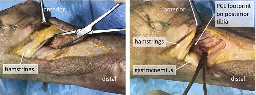
2                                                M. Held et al.: SICOT-J 2021, 7, 17

     The purpose of this study was to present open approaches
used to address both cruciate ligaments in patients with MLKIs.
Our aims were as follows: (1) describe surgical approaches that
can be useful to treat patients with acute injuries and in hospi-
tals in LRS, and (2) report short-term clinical outcomes of ten
patients with knee dislocations treated with open cruciate liga-
ment reconstruction by a single surgeon (MH) in an LRS.

Methods
    The primary aim of this work is to provide a technical note
for open approaches in a supine patient to address the cruciate
ligaments in multi-ligament knee injuries. Furthermore, a case
series of patients treated with these techniques were reviewed
retrospectively to describe associated complications and clinical
outcomes.
                                                                       Figure 1. Large medial capsular tear with exposed medial condyle
                                                                       after skin incision in an irreducible knee dislocation. This allows
Surgical technique                                                     access to the tibial insertion of the posterior cruciate ligament
                                                                       through the traumatic arthrotomy without extensive surgical
    Patients are positioned supine to allow full knee flexion and       dissection.
extension. A radiolucent table enables fluoroscopy if necessary.
A foot bolsters placed under the heel keeps the knee flexed 70°,
allowing easier access to the tibial footprint of the anterior cru-    injuries facilitated the approach to the PCL through a lateral
ciate ligament (ACL). With the toes on the bolster, knee flexion        incision [9].
can be increased to allow easier access to the femoral insertion
of the cruciate ligaments. A high side bolster prevents the leg
from rotating at the hip. Tourniquets, when used, are placed           Notch access
as proximal as possible to allow insertion of guidewires without
compromise.                                                                 Access to the notch is achieved through a medial parapatel-
                                                                       lar dissection (Figure 2, Video 2). This can be extended subvas-
                                                                       tus, midvastus, or proximally into the quadriceps tendon to
Incision
                                                                       enable patella sublaxation, greatly increasing exposure [10].
    The primary incision is curvilinear anteromedial beginning         Although the fat pad may be retained [11, 12], it is frequently
halfway between the medial border of the patella and the medial        debulked to increase visualization. The cruciate ligament
epicondyle, extending distally along the anteromedial surface of       stumps are excised to allow better visualization of the insertion
the tibia to below the pes anserine attachment. This incision          sites for guide pin placement. The knee is placed in slight exten-
allows access to the intercondylar notch, the proximal and distal      sion to enable visualization of the tibial insertion of the ACL, as
attachments of the medial collateral ligament, and the tibial          well as the anterior horns and roots of the menisci. It also facil-
insertion of the PCL (i.e., Lobenhoffer approach, Video 1). It         itated the placement of the ACL tibial and PCL femoral tunnels
also allows harvesting of the patella tendon and hamstring ten-        by removing tension off the extensor mechanism. If cruciate
dons and could be extended proximally for a quadriceps tendon          ligament guides are available, the use of Z-retractors and fat
harvest. For lateral side injuries, the lateral incision begins over   pad excision can provide acceptable visualization of the notch
the lateral epicondyle and extended distally posterior and infe-       even in more limited incisions. A headlamp can provide clearer
rior to the fibular head. This incision gives access to the per-        visualization.
oneal nerve, the posterolateral structures, and the PCL tibial              Guide pins for the femoral attachments for the ACL and
attachment. It also facilitates outside-in drilling and fixation        PCL are placed in an “inside-out” fashion. When performing
of the ACL femoral attachment.                                         a single bundle PCL reconstruction, the guide pin is started
    The decision of whether to approach the posterior part of          in the centre of the anterolateral bundle. The ACL pin is started
the knee laterally or medially is dictated by the collateral liga-     at the centre of the ACL femoral footprint. The ACL tibial
ment involvement. This is usually confirmed via magnetic res-           guide pin is placed in an “outside-in” fashion.
onance imaging or stress radiographs. The approach to the PCL
through the medial Lobenhoffer interval is facilitated by trau-        Medial approach
matic dissection of posteromedial structures and capsules.
Access through the Lobenhoffer interval is performed by the                The posterior aspect of the knee can be approached through
takedown of the semimembranosus muscle as needed, and then             the Lobenhoffer interval [9, 13]. The superficial dissection
using the plane between the medial collateral ligament (MCL)           should expose the hamstrings and MCL. To increase access,
and medial head of the gastrocnemius with an elevation of              the knee was flexed to 90° with the ipsilateral hip externally
the popliteus muscle. Similarly, posterolateral corner (PLC)           rotated. Visualization is improved by working from the
M. Held et al.: SICOT-J 2021, 7, 17                                                        3

Figure 2. Intraoperative access to the notch of left through a medial parapatellar dissection with the patella retracted. This gives appropriate
access to important structures in the notch. Left: guidewire drilled through the femoral footprint of the anterolateral bundle of the posterior
cruciate ligament. Right: guidewire drilled through the femoral footprint of the anterior cruciate ligament.

Figure 3. Medial Lobenhoffer approach in a left cadaveric knee. Left: the haemostat shows access to the posterior part of the knee, inferior to
the attachments of the hamstrings. Right: the gastrocnemius muscle is reflected posteriorly to give access to the posterior capsule and posterior
cruciate ligament. Note the haemostat is spreading fibres of the popliteus muscle. Leaving the pes intact and working above and below the pes
in knee flexion allows for safe access to the back of the tibia.

opposite side of the table. A sponge can be used to clean resid-              Use of a Cobb elevator, electrocautery, identifications of
ual fat off these structures to identify the proximal and distal          anatomic landmarks of the medial joint line and the inner edge
border of the pes anserine.                                               of the posteromedial femoral condyle, and exposure through the
     For acute injuries to the posteromedial corner, the traumatic        Lobenhoffer interval allow improved visualization and safe
dissection often enables palpation of the champagne glass drop-           access to the posterior knee in these multiple trauma patients.
off and PCL stump around the posteromedial part of the knee.              This helps avoid prone positioning and subsequent physiologic
To gain further access, a Hohman retractor is placed anterior to          risk to already compromised patients.
the medial head of the gastrocnemius muscle.
     External rotation of the tibia and blunt dissection of the           Lateral approach
fibres of the popliteus muscle gives access to the posterior cap-
sule and PCL tibial insertion (Figure 3, Video 3). Working with               Using a direct lateral incision, access to the posterior aspect
a blunt instrument, such as a Cobb (Skylar Surgical Instru-               of the tibia and insertion of the PCL is achieved (Figure 4,
ments, West Chester, Pennsylvania), the residual capsule was              Video 4). This has been described for the reconstruction of
stripped until full access and visualization of the PCL insertion         the PLC [14–17]. The incision passes just anterior to the lateral
is possible. It is critical to keep the retractors anterior to the gas-   condyle and distally just posterior to the fibular head. With a
trocnemius and popliteal muscles, hugging the posterior surface           separate anteromedial incision to access the notch, a sufficient
of the tibia to protect the neurovascular structures during tibial        skin bridge of at least 8 cm is maintained to avoid skin necrosis.
tunnel drilling or through creation. Knee flexion to 60–90° dur-           Three fascial “windows” are created to gain access to the pos-
ing dissection of the back of the tibia relaxes the neurovascular         terolateral structures [17].
structures. Adhesions and scarring in chronic injuries can make               An incision is first made posterior to the biceps tendon
the anatomic differentiation of structures challenging.                   (window 1) to identify the peroneal nerve about 1 cm distal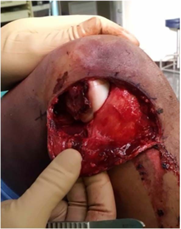
4 M. Held et al.: SICOT-J 2021, 7, 17
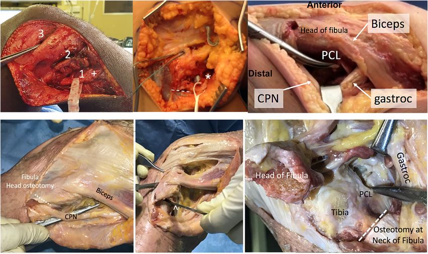
Figure 4. Lateral approach to careful dissection and retraction of common peroneal nerve (CPN). Top left: release of the nerve through
window “2”, in which the nerve has (+) a drop-down appearance with appropriate release below window “1” and the forceps are placed into
window “3”. Top middle: release of peroneal nerve (*) in a chronic condition with extensive scarring and release to peroneus longus fascia
(white line). Top right: lateral Lobenhoffer approach without fibula head osteotomy in a cadaver, in which protecting the CPN can be achieved
through access distal to the biceps tendon, retracting the gastrocnemius (“gastroc”) tendon posterior. The tibial insertion of the posterior
cruciate ligament (“PCL”) can be reached posterior to the head of the fibula. Bottom left: lateral Lobenhoffer approach to the posterior part of
the tibia in a cadaver. Initially, the CPN is identified and protected. Bottom middle and right: An osteotomy at the neck of the fibula (^)
increases access to the PCL when the gastrocnemius and popliteus muscles are retracted posteriorly.
and posterior to the tip of the fibular head or just distal around The tibial insertion of the PCL can be exposed by internal
the fibular neck. The nerve is released distally into the anterior rotation of the tibia and placing a Hohmann retractor past the
compartment with the careful division of the overlying muscle midline of the tibia, just proximal to the champagne glass
fascia to achieve a tension-free “drop-down sign” of the nerve. drop-off. Careful posterior retraction and knee flexion to 90°
Careful dissection and protection of the nerve throughout the will protect neurovascular structures and enable appropriate
procedure are crucial. access and visualization.
Window 2 is created next by developing the interval
between the iliotibial band and the biceps femoris tendon. This Closure
window gave access to the posterolateral capsule, the lateral
meniscus, the fibular collateral ligament (FCL) attachment, Medially and laterally, the interval of the posterior approach
and the attachment of the popliteofibular ligament. is mainly intermuscular, and no deep sutures are necessary for
Finally, window 3 is created by splitting the iliotibial band closure. On the lateral side, window 1 should be allowed to heal
fibres overlying the lateral epicondyle. This allowed visualiza- without repair to avoid any compression or injury to the per-
tion of the femoral attachments of the FCL and popliteus oneal nerve. The interval between the biceps femoris and ili-
tendon. otibial band can be reapproximated with an absorbable suture.
The posterior tibia is accessed through the lateral approach The window 3 splits of the iliotibial band should be closed with
[9]. Window 1 or 2 is used to gain access posterior to the fibular strong absorbable braided sutures.
head. The lateral head of the gastrocnemius is identified and Medially, no suture of deep structures is necessary [9]. The
retracted posteriorly with a Hohman retractor by sliding under popliteus muscle is split in line with the fibres during the medial
the popliteus tendon beneath the lateral head of the gastrocne- approach and does not require approximation. Postoperatively,
mius muscle. Blunt dissection is used to avoid venous bleeders. it is critical to monitor limb perfusion and peripheral nerve
A fibular neck osteotomy can be used at the metaphyseal– function. Lastly, tourniquet time must be monitored, and con-
diaphyseal junction to increase access [9]. However, with trau- tinuous ischemia longer than 120 min should be avoided. It
matic tears of the lateral collateral ligament and popliteus is often difficult to perform bicruciate and lateral collateral liga-
tendon, the exposed tibia can often be palpated around the fibu- ment reconstructions in less than 2 h. Surgeons should either
lar head and osteotomy is usually not necessary [18]. allow reperfusion for at least 15 min followed by tourniquetM. Held et al.: SICOT-J 2021, 7, 17 5
Table 1. Categorization of knee dislocations according to Schenck classification, and description of associated injuries.
Patient no. Sex Age (years) KDC Associated injuries Associated IA pathology
1 Male 38 KDIIIM – Medial meniscus bucket handle tear
2 Female 37 KDIV – Medial and lateral meniscus posterior
root avulsion, lateral meniscus tear
bucket handle
3 Male 59 KDI – –
4 Male 31 KDIV – –
5 Male 64 KDIIIL Femur fracture, closed head injury, CPN Arcuate fracture
6 Male 58 KDV Open book pelvic injury, ipsilateral Patella tendon rupture
foot fracture, contralateral MLKI, CPN
7 Male 17 KDII – –
8 Male 32 KDIIIM – –
9 Female 33 KDIIIL CPN –
10 Male 25 KDIIIM Contralateral MLKI with CPN, Medial meniscus anterior
traumatic aortic dissection root avulsion
Abbreviations: KDC, knee dislocation classification; IA, intraarticular; CPN, common peroneal nerve; MLKI, multi-ligament knee injury;
CPNP, common peroneal nerve palsy; –, not applicable.
re-inflation or perform the remainder of the procedure (or the All of these patients underwent a posteromedial approach to
entire operation) without tourniquet control. the posterior cruciate ligament (PCL) tibial insertion. In seven
patients, a large traumatic arthrotomy enabled sufficient access
to the posterior tibia through the medially based injuries (five
Case series patients) and laterally based injuries (two patients). Two
For this case series, consecutive patients who had under- patients had bilateral MLKIs, of which only one side was trea-
gone surgery for MLKI between July 2016 and November ted through an open approach.
2018 were retrospectively identified. All patients who had an Eight of the 10 patients were able to be contacted for a med-
open cruciate reconstruction were included. Patients below ian follow-up of 24 months (range, 17–33 months; IQR, 20.5).
the age of 18 years were excluded. Demographic data, injury No iatrogenic damage to neurovascular structures occurred.
mechanism, and classification, associated injuries, time delay Three patients developed arthrofibrosis with decreased flex-
to surgery, as well as the indication for open surgery were ion of less than 80°. Two of these patients showed heterotopic
collected. ossification on radiographs (Figure 5). One of these patients
The primary outcome measure was the presence of any improved after manipulation under anaesthesia to 100°,
major complications such as neurovascular injury, stiffness, whereas the other two patients opted not to have a further inter-
delayed wound healing, or infection. Secondary patient- vention. In one patient with a KDV and ipsilateral patella ten-
reported outcome measures (PROMs) included the following: don rupture, impaired wound healing necessitated reoperation
the visual analogue scale (VAS), Knee Injury and Osteoarthritis after 6 weeks, which resolved after removal of the protective
Outcome Score-Physical Function Short Form (KOOS-PS), and cerclage wire.
Lysholm scale. PROMs were assessed at the time of final follow-up. The
All procedures performed in studies involving human par- KOOS-PS median score was 81.4 (range, 75–100; IQR, 12.2)
ticipants were in accordance with the ethical standards of the and the median Lysholm score was 85 (range, 67–92; IQR,
institutional and/or national research committee (HREC REF 13.3). The median VAS pain rating was 45 (range, 0–50;
050/2018) and with the 1964 Helsinki declaration and its later IQR 15).
amendments or comparable ethical standards.
Discussion
Results
Arthroscopic single-stage surgery is the gold-standard for
Ten patients (two female) with a mean age of 35 years cruciate reconstruction in multi-ligament knee injuries. But, open
(range: 18–64, IQR: 21.5) were identified. All injuries, except cruciate surgery avoids fluid extravasation and can overcome
one (ultra-low energy fall), were high-energy injuries caused challenges faced by the hospital with limited arthroscopy-trained
by road traffic collisions. Injuries were further categorized by surgeons or equipment in an LRS (Table 2). The findings of this
the Schenck classification system. Eight patients had a KDIII study show that open cruciate surgery in 10 patients with severe
or higher injury and most had associated fractures or soft tissue MLKIs resulted in acceptable PROMs, with an acceptable
and neurovascular injuries (Table 1). All procedures but one incidence of complications. Notably, arthrofibrosis and hetero-
were performed within 3 months of injury. topic ossification (HO) occurred in three patients.
Three patients were converted to open surgical treatment This study has several limitations. Although the short-term
owing to fluid extravasation during diagnostic arthroscopy. follow-up allows assessment of complications, long-term6 M. Held et al.: SICOT-J 2021, 7, 17
Alternatively, an anterior midline incision can be used to
address all cruciate and collateral ligaments, thus avoiding mul-
tiple scars and potential wound healing problems; however, it
requires extensive undermining of the skin to reach the collat-
eral ligaments and tibial attachment of the PCL. Furthermore,
accessing and protecting the peroneal nerve during PLC recon-
struction can be challenging.
Other authors have described an open approach to the PCL
tibial insertion with similar outcomes but this was usually done
in a prone patient [7, 21–23]. The described approach allows
maximum visualization of the PCL tibial footprint while pro-
tecting the neurovascular structures. Our technique can be per-
formed in supine patients, enabling improved access to other
injuries in patients with multiple traumas. Although most cruci-
ate ligament reconstructions are performed arthroscopically
[24], acceptable long-term follow-up can be achieved after open
reconstruction [25]. Furthermore, a detailed summary of results
of studies using open PCL [26] and ACL reconstructions [27]
has been made in a meta-analysis and no difference in PROMs
or complications was found when compared to arthroscopic
surgery. Also, reported stiffness and HO after arthroscopic or
Figure 5. Radiographic valgus stress view of a patient with a
closed treatment of knee dislocations are similar [19, 20].
KDIIIM, showing heterotopic ossification around the medial
epicondyle at 9 months postoperatively.
Another important point to consider is that preoperative
imaging is important to confirm lateral or medial laxity. This
dictates the approach to the posterior part of the knee. MRI
follow-up is necessary to understand the risk of increased post- has high diagnostic accuracy in acute knee dislocations [28],
traumatic osteoarthritis [19]. Because of the low-resource set- but when MRI is not available, similar accuracy can be
ting and difficulty contacting patients, we were unable to achieved with comparative clinician-assisted varus and valgus
perform any instrumented ligamentous testing. But the fol- stress radiographs in 20° of flexion [29–31]. Here the tibiofe-
low-up was sufficient to assess perioperative complications moral distance at the medial or lateral joint line is measured
and describe a safe technique to perform this procedure. Other in millimetres and compared to the contralateral side. A similar
limitations include the retrospective design, small number of accuracy has also been found with PCL stress views. These can
patients, and lack of a standardized treatment protocol, all of be done with posteriorly directed force by the clinician, or in
which are common challenges in knee dislocation research. form of kneeling radiographs [32]. Here the line of the posterior
Our preferred approach is an extended anteromedial inci- tibial cortex is referenced to the most posterior point of the Blu-
sion for bicruciate injuries involving the medial side (KDIII- mensaat line and this distance is compared to the contralateral
M). A lateral incision is included if the PLC structures are side. A further advantage of stress radiographs is their dynamic
injured (KDIII-L) or when all ligaments are affected (KDIV). component of assessment and the possibility to grade the laxity
Table 2. Key points for open cruciate procedures.
No. Description
1 Use traumatic dissection of posterolateral and posteromedial corners to gain access to the PCL insertion of the tibia
2 Minimize tourniquet use to prevent prolonged ischemia
3 For the retraction of posterior structures, stay subperiosteal and dissect past the midline prior to placing the Hohmann retractor
4 Even a small shift or subluxation of the patella out of the notch increases access and visualization of the cruciate ligaments greatly
5 Decreasing flexion of the knee allows better access to the notch
6 Sit during the dissection of the posterior approach to the tibia or stand on the opposite side of the table
7 Rotation of the tibia is often increased in collateral ligament injuries and facilitates access to the posterior tibia
Table 3. Evaluation of posterior, varus, and valgus knee instability using stress radiographs [3].
Kneeling stress Injury Grade Varus stress Injury Valgus stress Injury
7 mm Normal or partial tear I 2.6 mm Normal or partial tear 3.1 mm Normal or partial tear
8–11 mm Complete PCL tear II 2.7–3.9 mm Isolated LCL tear 3.2–9.7 mm Complete sMCL tear
12 mm Combined ligament injury III 4 mm Complete PLC injury 9.8 mm Complete tear of all medial structures
LCL, lateral collateral ligament; PCL, posterior collateral ligament; PLC, posterolateral corner; PTT, posterior tibial translation; sMCL,
superficial medial collateral ligament.M. Held et al.: SICOT-J 2021, 7, 17 7
based on the distance measured (Table 3). Although preopera- 5. Berg EE (1995) Posterior cruciate ligament tibial inlay recon-
tive assessment prior to surgery is essential for adequate plan- struction. Arthroscopy 11(1), 69–76.
ning, stress radiographs done under anaesthesia and before 6. Jakob R, Rüegsegger M (1993) Therapy of posterior and
the incision can therefore provide crucial information even if posterolateral knee instability. Orthopade 22(6), 405–413.
MRI is available. 7. Burks RT, Schaffer JJ (1990) A simplified approach to the tibial
Overall, our findings support the concept that open attachment of the posterior cruciate ligament. Clin Orthop Relat
approaches to knee dislocations can be a useful tool for cruciate Res 254, 216–219.
ligament reconstruction in special circumstances. The most 8. Richter DL, Held M, Campos T, et al. (2020) The management
important future potential for it is the management of MLKIs of knee dislocations in the limited-resource setting. J Bone Joint
Surg 10, 2106.
in LRS.
9. Lobenhoffer P, Gerich T, Bertram T, et al. (1997) Particular
posteromedial and posterolateral approaches for the treatment of
tibial head fractures. Unfallchirurg 100(12), 957–967.
Supplementary materials
10. Hoppenfeld S (2012) The Knee, in Surgical exposures in
Supplementary material is available at https://www.sicot-j. orthopaedics: The anatomic approach. Hoppenfeld S, DeBoer P,
org/10.1051/sicotj/2021016/olm Buckley R, Editors. Lippincott Williams & Wilkins, pp. 520–
Video 1. Posteromedial approach cadaver JSICOT. 526.
Video 2. Notch access JSICOT. 11. Gallagher J, Tierney P, Murray P, et al. (2005) The infrapatellar
fat pad: Anatomy and clinical correlations. Knee Surg Sports
Video 3. Posterolateral approach cadaver JSICOT.
Traumatol Arthrosc 13(4), 268–272.
Video 4. Posteromedial approach surgery JSICOT.
12. Maculé F, Sastre S, Lasurt S, et al. (2005) Hoffa’s fat pad
Acknowledgements. The authors thank Sahar Freedman for editorial resection in total knee arthroplasty. Acta Orthop Belg 71(6),
assistance. 714.
13. Richter D, Wascher DC, Schenck RC (2004) A novel postero-
medial approach for tibial inlay PCL reconstruction in KDIIIM
Conflict of interest injuries: Avoiding prone patient positioning. Clin Orthop Relat
Res 472(9), 2680–2690.
DW receives funding from Smith & Nephew and Axogen 14. Arciero RA (2005) Anatomic posterolateral corner knee recon-
in the form of educational grants unrelated to the submission. struction. Arthroscopy 21(9), 1141–1147.
MH receives funding from Smith & Nephew in the form of 15. LaPrade RF, Johansen S, Wentorf FA, et al. (2004) An analysis
educational grants unrelated to the submission. ML, RvB, of an anatomical posterolateral knee reconstruction: An in vitro
DR, and RS certified that they have no financial conflict of biomechanical study and development of a surgical technique.
interest (e.g., consultancies, stock ownership, equity interest, Am J Sports Med 32(6), 1405–1414.
patent/licensing arrangements, etc) in connection with this 16. Larsen MW, Moinfar AR, Moorman CT (2004) Posterolateral
article. corner reconstruction–fibular-based technique. J Knee Surg 18
(02), 163–166.
17. Stannard JP, Brown SL, Robinson JT, et al. (2005) Recon-
struction of the posterolateral corner of the knee. Arthroscopy
Authorship statement 21(9), 1051–1059.
All authors have contributed substantially to the conception 18. Frosch K-H, Balcarek P, Walde T, et al. (2010) A new
posterolateral approach without fibula osteotomy for the treat-
of the work, the drafting of the manuscript and have approved
ment of tibial plateau fractures. J Orthop Trauma 24(8), 515–
the final version to be published and agree to be accountable for 520.
the work.
19. Bodendorfer BM, Keeling LE, Michaelson EM, et al. (2019)
Predictors of knee arthrofibrosis and outcomes after arthroscopic
lysis of adhesions following ligamentous reconstruction: A
References retrospective case–control study with over two years’ average
follow-up. J Knee Surg 32(06), 536–543.
1. Engebretsen L, Risberg MA, Robertson B, et al. (2009) 20. Fanelli GC, Edson CJ (2012) Surgical treatment of combined
Outcome after knee dislocations: A 2–9 years follow-up of 85 PCL-ACL medial and lateral side injuries (global laxity):
consecutive patients. Knee Surg Sports Traumatol Arthrosc 17 Surgical technique and 2- to 18-year results. J Knee Surg 25(4),
(9), 1013–1026. 307–316.
2. Giannoulias CS, Freedman KB (2004) Knee dislocations: 21. Jazayeri SM, Esmaili Jah AA, Karami M (2009) A safe postero-
management of the multiligament-injured knee. Am J Orthop medial approach to posterior cruciate ligament avulsion fracture.
33(11), 553–559. Knee Surg Sports Traumatol Arthrosc 17(3), 244–247.
3. Moatshe G, Chahla J, LaPrade RF, et al. (2017) Diagnosis and 22. Nicandri GT, Klineberg EO, Wahl CJ, et al. (2008) (2008)
treatment of multiligament knee injury: State of the art. J Treatment of posterior cruciate ligament tibial avulsion fractures
ISAKOS 2(3), 152–161. through a modified open posterior approach: Operative tech-
4. Stuart MJ (2001) Evaluation and treatment principles of knee nique and 12- to 48-month outcomes. J Orthop Trauma 22(5),
dislocations. Oper Tech Sports Med 9(2), 91–95. 317–324.8 M. Held et al.: SICOT-J 2021, 7, 17
23. Trickey EL (1980) Injuries to the posterior cruciate ligament: 28. Halinen J, Koivikko M, Lindahl J, et al. (2009) The efficacy of
Diagnosis and treatment of early injuries and reconstruction of magnetic resonance imaging in acute multi-ligament injuries. Int
late instability. Clin Orthop Relat Res 147, 76–81. Orthop 33(6), 1733–1738.
24. Boutefnouchet T, Bentayeb M, Qadri Q, et al. (2013) Long-term 29. James EW, Williams BT, LaPrade RF (2014) Stress radiography
outcomes following single-bundle transtibial arthroscopic for the diagnosis of knee ligament injuries: A systematic review.
posterior cruciate ligament reconstruction. Int Orthop 37(2), Clin Orthop Relat Res 472(9), 2644–2657.
337–343. 30. LaPrade RF, Bernhardson AS, Griffith CJ, et al. (2010)
25. Adler T, Friederich NF, Amsler F, et al. (2015) Clinical and Correlation of valgus stress radiographs with medial knee
radiological long-term outcome after posterior cruciate ligament ligament injuries: An in vitro biomechanical study. Am J Sports
reconstruction and nonanatomical popliteus bypass. Int Orthop Med 38(2), 330–338.
39(1), 131–136. 31. LaPrade RF, Heikes C, Bakker AJ, et al. (2008) The
26. Hooper PO III, Silko C, Malcolm TL, et al. (2018) (2018) reproducibility and repeatability of varus stress radiographs in
Management of posterior cruciate ligament tibial avulsion the assessment of isolated fibular collateral ligament and grade-
injuries: A systematic review. Am J Sports Med 46(3), 734– III posterolateral knee injuries: An in vitro biomechanical study.
742. J Bone Joint Surg 90(10), 2069–2076.
27. Levy DM, Erickson BJ, Bach BR (2018) Open versus 32. Jackman T, LaPrade RF, Pontinen T, et al. (2008) Intraobserver
arthroscopic anterior cruciate ligament reconstruction: A sys- and interobserver reliability of the kneeling technique of stress
tematic review of randomized controlled trials. Curr Orthop radiography for the evaluation of posterior knee laxity. Am J
Pract 28(5), 449–452. Sports Med 36(8), 1571–1576.
Cite this article as: Held M, Laubscher M, von Bormann R, Richter DL, Wascher DC & Schenck RC (2021) Open approaches for cruciate
ligament reconstruction in knee dislocations: A technical note and case series. SICOT-J 7, 17You can also read