Highly Efficient Bicolor (Green-Blue) Fluorescence Imaging in Polymeric Films
←
→
Page content transcription
If your browser does not render page correctly, please read the page content below
790                                                     Chem. Mater. 2002, 14, 790-796

                   Highly Efficient Bicolor (Green-Blue) Fluorescence
                               Imaging in Polymeric Films
                           G. Pistolis,*,† S. Boyatzis,‡ M. Chatzichristidi,† and P. Argitis*,†
                    Institute of Physical Chemistry and Institute of Microelectronics, NCSR Demokritos,
                     Athens 153 10, Greece, and Department of Materials Science, University of Patras,
                                                    26500 Patras, Greece
                             Received July 19, 2001. Revised Manuscript Received October 5, 2001

              A novel approach for photochemical definition of distinct, basic color, emission areas in
            thin polymeric films is introduced. Two-color, blue and green, fluorescence imaging is
            demonstrated using an amino diphenylhexatriene derivative that upon protonation shows
            sizable shifts in both absorbance and fluorescence spectra. Protons are generated photo-
            chemically in selected areas of the thin polymeric films containing the probe by a typical
            onium salt photoacid generator. Significant “flow” of transferred electronic energy from a
            typical aromatic polymer, epoxy novolac, to the mentioned dye was manifested permitting
            amplification and color tunability of fluorescent signals.

                           Introduction                                         On the other hand, the investigation of fluorescence
                                                                             patterning possibilities in thin polymeric films emerges
   Patterning using films of photosensitive polymeric                        as an important topic of research in the area of plastic
materials, photoresists, is one of the most critical areas                   materials suitable for image recording and display
in modern semiconductor industry where the capability                        applications.5-7 The exploration of novel strategies for
for IC dimension shrinkage still defines the level of the                    the formation of full color displayssby defining blue,
whole technology. Advanced photoresist materials ca-                         green, and red emissive pixelssis in particular a very
pable of manufacturing sub-0.2 micron images with                            hot subject nowadays.8
optical lithography are already in the market, while the                        Fluorescent organic dyes dispersed in the polymer
research efforts in this area target these days to the sub-                  film or moieties pending to the polymer itself have been
tenth micron regime. The most successful strategy                            used so far in fluorescence imaging, either in the area
adopted in the development of new photoresists during                        of lithographic processing or in one of the organic
the last 15 years is based on “chemical amplification”,                      displays, including fluoresceins,2 acridins,3 cumarins,4
where a photoacid generator (PAG) usually provides                           anthracene derivatives,7 quinizarins,6 and recently py-
protons upon optical e-beam or X-ray exposure inside                         ridylbenzoxazoles.5 The acid-induced changes of emis-
the resist film, which then catalyze or initiate the                         sion properties in all these cases result in production
necessary for solubility change chemical reactions.1 In                      of fluorescent and nonfluorescent areas (acid-induced
addition to the development of new photoresist materi-                       dye quenching).
als capable for exposure at deep, and lately vacuum, UV                         In this paper, we introduce a new approach for the
spectral regions, increased interest has also arisen in                      fluorescence patterning of thin films on the basis of
the development of suitable metrology tools and meth-                        the photochemical definition of distinguishable green
odolologies to monitor and control physicochemical                           and blue fluorescent areas. This new approach results
changes occurring inside the resist films. One of the                        to photochemically activated color-changing media
areas where intense activity is reported lately is related                   (PACCM) by using an appropriate luminescent probe
with the introduction of suitable probes, including                          and offers new opportunities both for fluorescence
fluorescence probes, for the study of photogenerated acid                    probing of acid generation and diffusion inside photo-
formation and diffusion aiming at the optimization of                        resists and for full-color display applications in the area
lithographic materials and processes.2-4                                     of organic light-emitting diodes (OLEDs).

   * To whom correspondence should be addressed. Fax: +301 6511                 (3) Okoroanyanwu, U.; Byers, J. D.; Cao, T.; Webber, S. E.; Wilson,
766. E-mail: pistolis@chem.demokritos.gr. or Fax: +301 6511723.              C. G. Proc. SPIE 1998, 3333, 747.
E-mail: argitis@imel.demokritos.gr.                                             (4) Coenjarts, C.; Cameron, J.; Deschamps, N.; Hamply, D.; Pohlers,
   † NCSR Demokritos.                                                        G.; Scaiano, J. C.; Sinta, R.; Virdee, S.; Zampini, A.. Proc. SPIE 1999,
   ‡ University of Patras.                                                   3678, 1062.
   (1) (a) Reichmanis, E. F.; Houlihan, M.; Nalamasu, O.; Neenan, T.            (5) Kim, J. M.; Chang, T. E.; Kang, J. H.; Park, K. H.; Han, D. K.;
X. Chem. Mater. 1991, 3, 394. (b) MacDonald, S. A.; Wilson, C. G.;           Ahn, K. D. Angew. Chem., Int. Ed. 2000, 39, 1780.
Frechet, J. M. Acc. Chem. Res. 1994, 24, 151.                                   (6) (a) Kim, J. M.; Chang, T. E.; Kang, J. H.; Hang, D. K.; Ahn, K.
   (2) (a) Lu, B.; Taylor, J. W.; Cerrina, F.; Soo, C. P.; Bourdillion, A.   D. Adv. Mater. 1999, 11, 1499. (b) Kim, J. M.; Kang, J. H.; Han, D. K.;
J. J. Vac. Sci. Technol., B 1999, 17 (6), 33450. (b) Zhang, P. L.; Webber,   Lee, C. W.; Ahn, K. D. Chem. Mater. 1998, 10, 2332.
S.; Mendelhall, J.; Byers, J.; Chao, K. Proc. SPIE 1998, 3333, 794. (c)         (7) Vekselman, A. M.; Zhang, C. Chem. Mater. 1997, 9, 1942.
Jessop, J. L. P.; Goldie, S. N.; Scranton, A. B.; Blanchard, G. J.;             (8) (a) Forrest, S.; Burrows, P.; Thompson, M. IEEE Spectrum
Rangarajan, B.; Capodieci, L.; Subramanian, R.; Templeton, M. K. Proc.       August 2000, 29-34. (b) Parthasarathy G.; Gu, G.; Forrest, S. R. Adv.
SPIE 1999, 3678, 914.                                                        Mater. 1999, 11, 907.

                                  10.1021/cm0111783 CCC: $22.00 © 2002 American Chemical Society
                                                    Published on Web 01/26/2002
Bicolor (Green-Blue) Fluorescence Imaging Chem. Mater., Vol. 14, No. 2, 2002 791
Experimental Section Scheme 1
Materials and Instrumentation. 1-(4′-dimethyl-ami-
nophenyl)-6-phenyl-1,3,5-hexatriene (DMA-DPH) was pur-
chased from Lambda Probes and Diagnostics. The parent
molecule, all-trans-1,6-diphenyl-1,3,5-hexatriene (DPH) was
purchased from Fluka. The PMMA polymer used is Elvacite
2041 purchased from DuPont, whereas the epoxy novolac was
obtained after fractionation of Shell Epikote 164.9For absorp-
tion spectra, the Perkin-Elmer Lambda-16 spectrophotometer
was employed. Fluorescence and excitation spectra were
recorded on a Perkin-Elmer LS-50B fluorometer. Steady-state
and time-resolved fluorescence anisotropy measurements were
determined using the time-correlated single-photon counter
FL900 of Edinburgh Instruments equipped with Glan-
Thompson polarizing prisms. The steady-state, ⟨r⟩, and time-
dependent fluorescence anisotropy, r(t), were calculated by
using the expressions 1 and 2, respectively:
GIVV - IVH
⟨r⟩ ) (1)
GIVV + 2IVH
environment. The observed sizable spectral shifts of
and DMA-DPH arise from charge delocalization in the
extended conjugated system of the molecule induced by
GRVV(t) - RVH(t)
r(t) ) (2) the electron-donating effect of the p-dimethylamino
GRVV(t) + 2RVH(t) group. By keeping this in mind, we made the hypothesis
that photoinduced protonation of the dimethylamino
where I and R stand for the integrated spectrum intensity and group in DMA-DPH may disrupt the conjugation effect
time-resolved decays, the symbols VV and VH correspond to and as a consequence could hypsochromically shift the
parallel and perpendicular plane of the analyzer with respect DMA-DPH absorption and emission maxima toward
to that of excited light, while the plane of the polarizer is fixed
to that of incidence. Finally, G, also called G-factor, is a those of the parent DPH molecule (Scheme 1). As a
correction factor dependent on the emission wavelength and result, information concerning the photogeneration of
is given by G ) IHH/IHV and G ) RHH(t)/RHV(t) for static and acid in thin polymeric films can be suitably transmitted
time-dependent anisotropy expressions, respectively. to us via intense light signals. Moreover, if photoinduced
Polymeric Film Preparation and Processing. Solutions protonation of DMA-DPH into the polymer films is
containing either the polymer poly(methyl methacrylate) efficient, then by exposing selected areas through a
(PMMA) (7% w/w) or the epoxy novolac (EPNOV) (22% w/w),
a photoacid generator triphenylsulfonium hexafluoroanti-
photomask, patterned bicolor fluorescent images could
monate (TPSHFA), in various contents (2%, 3%, and 10% w/w) be obtained.
and the fluorescence probe DMA-DPH (1% w/w) were prepared In an effort to verify this hypothesis, we examined
using propylene glycol methyl ether acetate PGMEA as the the spectral changes of DMA-DPH (1) incorporated into
solvent. Films were spin coated at 4000 rpm and then baked selected polymer matrixes. The formation of the proto-
on a hot plate at 110 °C for 1 min. Film thicknesses were nated form (compound 2) and the photoinduced color
measured with a Dektak profilometer (typical film thickness
changes were tested by both UV-vis and fluorescence
0.53 µm). Photoacid generation was induced by exposing films
with a 500 W Oriel Hg-Xe exposure tool through a 248 nm spectroscopy. Two representative polymer matrixes
narrow-band filter (6.5 nm half-bandwidth) for assessed times were employed in this study: poly(methyl methacrylate)
(see text). The incident power was 0.21 ( 0.02 mJ/s. (PMMA), an almost transparent matrix in the tested
Fluorescence Imaging. Solutions similar to the ones wavelength range (248 ( 7 nm) which is also chemically
described above were used for fluorescence imaging. Solutions inert toward photogenerated acid, and an epoxy novolac
after spin coating and baking as above were exposed to 248 polymer (EPNOV), which absorbs significantly in the
nm UV light for 15 min through a patterned photomask. The
fluorescence image pattern was photographed using a Nikon mentioned wavelength range and emits fluorescence in
Microphot-FXA fluorescence microscope. the ultraviolet-near-visible region. It is also well docu-
mented that photoacid generated by a sulfonium salt
Results and Discussion in this epoxy matrix causes cross-linking reactions.9
I. UV-Vis Absorption Spectroscopy. The UV-vis
Recently, we have reported an extensive spectroscopic absorption spectrum of a thin (0.53 µm) film prepared
study10 of a derivative of the fluorescence probe 1,6- by spin coating a propylene glycol methyl ether acetate
diphenyl-1,3,5-hexatriene (DPH) which carries the elec- (PGMEA) solution of PMMA containing DMA-DPH (1%
tron donating para-dimethyl-amino group on one of the w/w) and a photoacid generator, triphenyl sulfonium
phenyl rings, namely 1-[4-(dimethylamino)phenyl]-6- hexafluoroantimonate (3% w/w), on quartz substrate is
phenylhexa-1,3,5,-triene (DMA-DPH) (compound 1). shown in Figure 1a (curve 1). The para substitution
Briefly, it was found that in contrast to the parent effect in the DMA-DPH molecule induces broadening
molecule DPH, the emission wavelength of DMA-DPH of the absorption spectrum and shifts the wavelength
is very sensitive to the micropolarity of its immediate maximum to ca. 400 nm. This maximum is shifted 24
nm hypsochromically with respect to that of DPH (see
(9) Argitis, P.; Raptis, I.; Aidinis, C. J.; Glezos, N.; Baciocchi, M.; Figure 1a dotted line). Upon subsequent irradiation
Everett J.; Hatzakis, M. J. Vac. Sci. Technol., B 1995, 13 (6), 3030.
(10) (a) Pistolis, G.; Malliaris, A. Chem. Phys. 1998, 226, 83-99. with 248 nm light (curves 2, 3, and 4), photoacid
(b) Pistolis, G.; Malliaris, A. Langmuir 1997, 13, 1457-1462. generation is triggered and the absorption at the792 Chem. Mater., Vol. 14, No. 2, 2002 Pistolis et al.
Figure 1. (a) UV-vis spectra of compound 1 in poly(methyl
methacrylate) plus TPSHFA (3 wt %) after 248 nm UV
irradiation; irradiation time: (1) 0, ( 2) 30 s, (3) 240 s, and (4)
600 s. Dotted line: Absorption spectrum of DPH in PMMA.
(b) UV-vis spectra of a PMMA film containing equimolar
quantities of compound 1 and TPSHFA; irradiation time: (1)
0 s, (2) 10 s, (3) 60 s, (4) 240 s, (5) 540 s, and (6) 1140 s.
Figure 2. UV-vis spectra of compound 1 in PMMA after UV
irradiation with (a) 248-nm narrow-band filter (6.5 nm half-
wavelength maximum (400 nm) of the neutral DMA- bandwidth) and (b) 254-nm wide-band filter (50 nm half-
DPH form (compound 1) progressively decreases, whereas bandwidth). Dashed line corresponds to absorption spectrum
of PMMA alone. Assessed times are as hereupon: (1) 0 s, (2)
a new structured band appears with a maximum at 361 180 s, (3) 360 s, (4) 660 s, and (5) 1080 s.
nm. The new absorption features resemble very closely
those of DPH in accordance to our hypothesis. On the One could argue that the absorption spectral change
other hand, the evident isosbestic point occurring at observed after UV exposure is not related to photoacid
374 nm strongly supports a mechanism that proceeds generation but is rather due either to photooxidation
between two distinguishable species involving no in- products, since aromatic amines are easily oxidized by
termediates. Furthermore, fluorescence excitation spec- air, or most likely to the well-known photoisomerization
tra (Figure 3, inset) of compound 1 and compound 2 that polyenes undergo after exposure to light even when
agree satisfactorily with the corresponding absorp- they are enclosed in well-defined molecular cavities.12
tion spectra of the same compounds. It is also docu- In an effort to clarify this point and safely eliminate the
mented that 1-[4-(trimethylamino)phenyl]-6-phenylhexa- possibility of such artifacts, we performed control ex-
1,3,5,-triene (TMA-DPH), an analogous cation to com- periments having exposed the polymeric film to UV
pound 2, which is also a derivative of DPH with a irradiation without using the photoacid generator. Two
cationic moiety (trimethylamino) affixed to the para filters were employed in this part of study; the first one
position of one of the phenyl rings, has photophysical was the narrow-band filter which we had used in the
properties that are generally very similar to those of experiments where the spectrum shift was noticed (248
DPH.11 nm, 6.5 nm half-bandwidth), and the second was a
Even though the polymer film contains TPSHFA and broad-band filter (254 nm, 50 nm half-bandwidth) that
DMA-DPH in near 1:1 molar ratio, the yield of photo- permits direct irradiation into the short- and near long-
protonation approaches 100% even in such highly stiff wave absorption band of the DMA-DPH molecule. As
matrixes as those under investigation (see section III). can be seen in Figure 2a, irradiation using the narrow
In fact, in a PMMA film containing equimolar concen- filter did not induce any significant change in the UV-
trations of each component, namely, compound 1 and vis spectrum of the DMA-DPH molecule even after a
TPSHFA, ([compound 1] ≈ [TPSHFA] = 2 × 10⁻² mol/ prolonged illumination time period of about 20 min.
l), the conversion of compound 1 to the protonated form
On the contrary, under irradiation with the broad-
(compound 2) proceeded quantitatively after irradiation
band filter, the fundamental all-trans absorption band
for 15 min as manifested by absorption and fluorescence
of compound 1, centered at 395 nm, gradually decreased
spectra (Figure 1b).
to 65% of the original value, after a lapse of 18 min and,
(11) Prendergast, F. G.; Haugland, R. P.; Callahan, P. J. Biochem-
istry 1981, 20, 7333. (12) Kohler, B. E. Chem. Rev. 1993, 93, 41.Bicolor (Green-Blue) Fluorescence Imaging Chem. Mater., Vol. 14, No. 2, 2002 793
Figure 3. Fluorescence spectra of neutral (compound 1) and
fully protonated form of DMA-DPH (compound 2). Excitation
spectra of both forms are given in inset
simultaneously, an increase in absorbance at around
200 nm was observed as shown in Figure 2b. At the
same time several isosbestic points were detected at 295,
240, and 228 nm, the initial maximum of all-trans
conformer of compound 1, located at 395 nm, progres-
sively shifted toward the short-wavelengths spectral
region. Clearly, these spectroscopic observations dem-
onstrate the existence of structural rearrangement(s)
occurring after illumination of the dye just with the
broad-band filter. Although the verification of the
identity of those new species is out of the scope of the
present work, however, taking into account the similar-
ity of our spectroscopic data with those concerning
photoisomerization of the parent molecule DPH (Diphe-
nylhexatriene) or related diphenylpolyenes in fluid
media 13, it can be speculated that the primary photo-
products cis-trans-trans andr trans-cis-trans are respon-
sible for the observed spectral changes. Photochemical
destruction, however, should not be excluded as an
additional factor to the aforementioned changes, under
conditions of direct probe illumination.
Therefore, when triphenylsulfonium hexafluoroanti-
monate (TPSHFA) sensitizer is present in the film, as
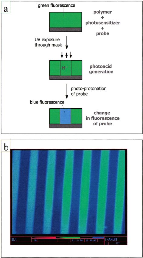
in the case of film illumination through the narrow 248 Figure 4. (a) Illustration of color patterning process. (b)
nm filter, the shifts in DMA-DPH absorbance spectra Fluorescence patterns obtained with a PMMA thin film
containing compound 1 (1 wt %) and the photoacid generator
clearly indicate that the photogenerated protons attack TPSHFA (3 wt %). The green areas are the regions exposed
the para-dimethylamino group of compound 1. through the photomask. The actual colors may differ somewhat
II. Fluorescence Spectra and Imaging. On going because of computer image process and printing quality. (Film
to fluorescence spectral changes, the results are even thickness: 0.53 µm. Line width: 50 µm)
more interesting. As can be seen in Figure 3 (curve 1),
the initial neutral form of DMA-DPH (compound 1) containing the polymer (PMMA), the photoacid genera-
fluoresces strongly in the green spectral region. After tor (3% w/w), and the fluorescent compound 1 (1% w/w)
irradiation for 10 min, green fluorescence totally disap- was irradiated for 15 min in contact with a photomask
peared and, by exciting the film into the new absorption at 248 nm (using the narrow-band filter mentioned
band of the protonated form (compound 2), blue fluo- above). The photograph shown in Figure 4b reveals two
rescence is observed as the single component (curve 2). clearly resolved fluorescent regions under illumination
The quantum yields of both forms are identical whereas with a 365-nm broad UV lamp adapted to a fluorescence
further illumination does not induce changes on elec- microscope. As expected, the blue fluorescent areas are
tronic spectra. the regions exposed through the photomask.
Having established effective signaling of photoacid The color patterning process is also illustrated sche-
generation in the above system, our next effort focused matically in Figure 4a. The exposure through a litho-
on the demonstration of the capability for the definition graphic mask results in the generation of acid in
of bicolor fluorescence patterns. A thin film (0.53 µm) selected film areas according to a well established
microlithography process. Using the suitable fluores-
(13) (a) Savondal, A.; Zechmeister, L. J. Am. Chem. Soc. 1947, 69, cence probe dispersed in the polymeric film, this pho-
553. (b) Pinckard, J. H.; Wille, B.; Zechmeister, L. J. Am. Chem. Soc. toacid generation leads to the definition of distinct color
1948, 70, 1938. (c) Lunde, K.; Zechmeister, L. J. Am. Chem. Soc. 1954,
76, 2308. (d) Saltiel J.; Ko, D. H.; Fleming, S. A. J. Am. Chem. Soc. areas, in our example blue and green, in the polymeric
1994, 116, 4099. film. Further extension of the technique for the defini-794 Chem. Mater., Vol. 14, No. 2, 2002 Pistolis et al.
tion of red areas as well is conceivable. Nevertheless,
suitable probes and ideas for the definition of three
different color areas, possibly at the same level, are
needed. In any case, the resolution capabilities of the
technique are much higher and depend mostly on the
lithographic equipment used for the patterning.
On the other hand, a reference to related techniques
should be made at this point to facilitate further
discussion on the potential of the presented methodol-
ogy. In the area of OLEDs, the effect of protonation-
deprotonation processes on tuning optical properties of
a new type, PPV-like, conjugated polymer containing
bipyridylene-vinylene subunits has been recently dem-
onstrated by Y. Eichen et al.14 They observed optical
tunability both in concentrated solution of the above
polymers in formic acid and in films saturated mainly
by using different acid/base vapors in the atmosphere
surrounding the film. According to the scheme presented
here, however, color patterning using in situ photoge-
neration of acid in selected areas of the polymer matrix
could be also effective upon suitable material modifica-
tions and the use of suitable probes. Analogous pro-
cesses based on well-established processes adopted from
semiconductor microlithography have been also pro-
posed for the in situ modification of conductivity in
polyaniline films and for the fabrication of polymer-
based transistors.15 Figure 5. (a) Variation of steady-state fluorescence anisotropy
III. Fluorescence Depolarization in Polymeric (SSFA) on DPH concentration in a PMMA film. Inset: time-
Films. The definition of bicolor patterns according to resolved fluorescence anisotropy (TRFA) trace. (b) A plot of
the proposed scheme is based on the presence of 1/P vs concentration of DPH. Solid line indicates computer fit
protonated and unprotonated probe molecules in distinct (R2 ) 0.9999) in the concentration region in which energy
film areas. Pattern stability issues, related to the probe migration is present. (c) Dependence of fluorescence intensity
on DPH concentration. Linearity was observed below critical
diffusion, could prove important for the possible use of concentration (2.7 × 10⁻³M).
the technique in image recording or display applications.
In a first attempt to study the motion of the unbound ization, respectively, R is the critical distance between
probe molecules in the typical polymer matrixes used
the two molecules such that the energy transfer prob-
in this investigation, we used the technique of fluores-
ability equals the emission probability, a is the molec-
cence depolarization.
ular radius, and C is the concentration in moles/l. By
Fluorescence depolarization gives an estimation of the
introducing a molecular radius of 8 Å for DPH in the
free volume inside the matrix if energy transfer from
above equation, a value of 26 Å for R is estimated from
the originally excited dye molecule to a neighboring one
the line slope, which is in the typical range of 15-40 Å
is negligible. Figure 5a shows the effect of dilution upon
the fluorescence anisotropy of the diphenylhexatriene for small molecules in viscous solvents. A linear depen-
(DPH) incorporated in PMMA matrix. The concentration dence of the fluorescence intensity versus concentration
region at which energy migration takes place and the is also observed exactly below 2.7 × 10⁻³ M (Figure 5c),
region limit at which this is practically absent (below in perfect agreement with depolarization experiments.
2.7 × 10⁻³ M) are clearly seen. Figure 5b shows the Under these conditions, steady-state anisotropy val-
linear dependence of 1/P (P is the fluorescence polariza- ues ⟨r⟩ as high as 0.36 and 0.33 for DPH in PMMA and
tion) upon dye concentration as has been well verified EPNOV, respectively, were found, whereas the DPH
for small molecules in viscous solvents.16 The working time-dependent anisotropy profiles decay before leveling
eq 3 for the concentration range where intermolecular off to residual anisotropy values of r∞ ) 0.30 and r∞ )
energy transfer between dye molecules is present is 0.26, respectively (see inset Figure 5a). The static ⟨r⟩
given by and limiting time-dependent anisotropy values (r∞) for
both matrixes are much larger than zero and close to
1 1
- =
P 3
1 1
-
P0 3 ( )(
1 + 1.68
R6
2a
C ( ) ) (3)
the value measured in frozen solutions (ro ) 0.38 10)
demonstrating that diffusive and rotational motions of
the dye are strongly hindered in the above matrixes.
where P and P0 are the observed and maximum polar- Even though such stiff matrixes contain the dye and
the photosensitizer at stoichiometry level, the photo-
(14) Eichen, Y.; Nakhmanovich, G.; Gorelic, V.; Epshtein, O.; protonation yield approaches 100%.
Poplawski, J. M.; Ehrenfreund, E. J. Am. Chem. Soc. 1998, 120, 10463.
(15) (a) Drury, C. J.; Mutsaers, C. M. J.; Hart, C. M.; Matters, M.; IV. Energy Transfer in an Aromatic Polymer
Leeuw, D. M. Appl. Phys. Lett. 1998, 73, 108. (b) Angelopoulos, M.; Matrix. The possibility for energy transfer to the probe
Shaw, J. M.; Lee, K. L.; Huang, W. S.; Lecorre, M. A. J. Vac. Sci.
Technol., B 1991, 9, 3428. in the typical aromatic polymer matrix (EPNOV) em-
(16) Weber, G. Biochem. J. 1960, 75, 335. ployed in the present contribution was investigated.Bicolor (Green-Blue) Fluorescence Imaging Chem. Mater., Vol. 14, No. 2, 2002 795
excitation spectrum relative to the absorption spectrum
should reflect the energy lost during the transfer step.
In our case, efficient intermolecular energy transfer
through dipole-dipole interaction18 of about 63% was
manifested as seen in Figure 6c. The energy transfer
efficiency was also estimated by the degree of the donor
(polymer) emission quenching in the presence of the
acceptor (dye). Figure 6b shows a comparison between
the fluorescence spectrum of the epoxy novolac units,
without and in the presence of compound 1. It is roughly
62% at maximal loading, which is within experimental
error in perfect agreement with the former method. In
the matrixes consisting of the epoxy novolac polymer
(EPNOV) plus the photosensitizer TPHSFA (3% w/w in
our experiments), referred also in the literature as
epoxy-resist (EPR),9 the DMA-DPH probe shows similar
absorption and fluorescence spectra shifts with those
described in PMMA. Although the addition of the
photosensitizer TPSHFA in the EPNOV polymer matrix
induced a partial quenching of energy transfer, the
transfer efficiency remained effectual and equally dis-
tributed in the two dye forms, the neutral (compound
1) and the protonated (compound 2)
The energy transfer observed in an aromatic polymer
matrix, namely, EPNOV, as described above, permits
excitation at the wavelength of the polymer absorption,
that is, around 275 nm, resulting not only in fluorescent
signal amplification but also in prevention of the chro-
mophore photobleaching.19 The results from the energy
transfer experiments provide also further support for
the potential of color patterning via in situ photoacid
Figure 6. (a) UV-vis absorption spectra of various thin films. generation in the area of organic light-emitting devices
Dash-dot line: absorption spectrum of EPNOV. Dash line: (OLEDs), provided that similar results could be obtained
absorption spectrum of compound 1 in PMMA matrix. Solid
line: Absorption spectrum of compound 1 in EPNOV. (b) in blue electroluminescent polymer matrixes using
Fluorescence emission spectra of thin films: (curve 1) EPNOV suitable color-shifting probes. Photochemical alteration
alone, exc. at 275 nm; (curve 2) EPNOV + compound 1, exc. of emission properties in selected film areas could
at 275 nm; (curve 3) exc. at the absorption maximum (402 nm) greatly simplify the processes toward full color display
of I. (c) Absorption (curve 1) and fluorescence excitation microfabrication by reducing the necessary layers or
spectrum (curve 2) of compound 1 in EPNOV, recorded at the even by totally eliminating the need for multilayer
λem of the dispersed dye. Spectra have been normalized at the
absorption maximum (402 nm) of the dye.
structures.
Summary
Transfer of energy in this system can be easily
recognized because around 275 nm, where the polymer Photochemically induced color patterning in polymeric
absorbs strongly, the absorption of the probe is negli- films has been demonstrated. In particular, the ef-
gible, as shown from the absorption spectra of thin films fectuality and applicability of the fluorescent dye DMA-
containing compound 1 dispersed in the EPNOV matrix DPH (compound 1), in photoacid generation monitoring
on one hand and in an optically transparent matrix such as well as in color and fluorescent imaging in polymer
as PMMA on the other (Figure 6a). An inspection of films was investigated. Well-resolved two-color fluores-
Figure 6b clearly shows that when the epoxy polymer cent image patterns of high sensitivity were readily
units are excited at λexc ) 275 nm, its own fluorescence obtained.
(λem ) 310 nm) is quenched and green fluorescence Several advantages characterize the dye studied.
centered at 500 nm of compound 1 is observed, indicat- First, it has a high extinction coefficient (=70,000 M-1
ing energy transfer from the polymer backbone to the cm-1) and a high fluorescence quantum yield,10 allowing
dispersed dye molecule. Upon excitation at λexc ) 275 effective signaling at low concentration levels in the
nm, no fluorescence was detected at 500 nm from the polymer matrix so that the perturbation of the photo-
polymer backbone alone, whereas the fluorescence resist film properties is minimized. Second, the changes
signal at 500 nm is amplified up to a nearly 400% higher in electronic spectra of compound 1 that accompanied
value than the one obtained upon direct excitation of the photoacid generation procedure are very well dis-
chromophore 1 at its maximum (402 nm).
The energy transfer efficiency was first determined (17) (a) Bourson, J.; Mugnier, J.; Valeur, B. Chem. Phys. Lett. 1982,
by comparing the absorption and the fluorescence 92, 430. (b) Mugnier, J.; Pouget, J.; Bourson, J. J. Lumin. 1985, 33,
excitation spectrum recorded at the λem of the dispersed 273.
(18) Forster, T. Discuss. Faraday Soc. 1959, 27, 7-17.
dye.17 In fact, when these two spectra are normalized (19) Chattopadhyay, S. K.; Das, P. K.; Hug, G. L. J. Am. Chem.
at the λmax of the dye, any “loss” in intensity in the Soc. 1982, 104, 4507.796 Chem. Mater., Vol. 14, No. 2, 2002 Pistolis et al.
tinguishable permitting two-color imaging in fluores- This work is considered as the starting point in the
cence and “on to of” color in absorption. Third, the investigation of photosensitive polymeric matrixes with
selectivity of the dimethylamino group of the probe DPH-DMA probe and other homologues. Fluorescence
toward photogenerated protons is as high as 100% even imaging by exposure at shorter wavelengths (e.g., 193,
in such highly stiff matrixes as those under investiga- 157 nm and X-rays) and e-beam in suitable photoresist
tion, allowing also its use as an acid diffusion control matrixes is conceivable. On the other hand, a similar
additive at optimized concentrations in photoresist
strategy in electroluminescent polymer matrixes can
formulations. Fourth, in polymeric matrixes containing
aromatic moieties that absorb and emit fluorescence in lead to photochemically activated color changing media
UV spectral region, for example, epoxy novolac polymer, suitable for organic display applications.
significant “flow” of transferred energy from polymer to
the probe was manifested, permitting amplification of
Acknowledgment. The authors wish to thank NCSR
fluorescent imaging signals as well as protection from
dye photobleaching. Fifth, its absorption around 248 nm Demokritos for financial support and Ms. G. Terzoudi
is negligible, and this allows the photosensitizer to for help in fluorescence imaging.
absorb practically all incident light at exposure in this
wavelength range. CM0111783You can also read