Towards Development of an Anti-Vampire Bat Vaccine for Rabies Management: Inoculation of Vampire Bat Saliva Induces Immune-Mediated Resistance
←
→
Page content transcription
If your browser does not render page correctly, please read the page content below
viruses
Article
Towards Development of an Anti-Vampire Bat Vaccine for
Rabies Management: Inoculation of Vampire Bat Saliva Induces
Immune-Mediated Resistance
Horacio A. Delpietro 1, *, Roberto G. Russo 1 , Charles E. Rupprecht 2 and Gabriela L. Delpietro 1

                                          1   Servicio Nacional de Sanidad y Calidad Agroalimentaria (SENASA), Padre Serrano 1116,
                                              3300 Posadas, Argentina; reviroseco@hotmail.com (R.G.R.); gabi.delpietro@gmail.com (G.L.D.)
                                          2   LYSSA LLC, 309 Pirkle Ferry Rd., Cumming, GA 30040, USA; charleserupprechtii@gmail.com
                                          *   Correspondence: hadelpietro@arnet.com.ar

                                          Abstract: The common vampire bat (Desmodus rotundus) is a hematophagous species responsible for
                                          paralytic rabies and bite damage that affects livestock, humans and wildlife from Mexico to Argentina.
                                          Current measures to control vampires, based upon coumarin-derived poisons, are not used exten-
                                          sively due in part to the high cost of application, risks for bats that share roosts with vampires and
                                          residual environmental contamination. Observations that vampire bat bites may induce resistance in
                                          livestock against vampire bat salivary anticoagulants encourage research into novel vaccine-based
                                          alternatives particularly focused upon increasing livestock resistance to vampire salivary components.
                                We evaluated the action of vampire bat saliva-Freund’s incomplete adjuvant administered to sheep

                                          with anticoagulant responses induced by repeated vampire bites in a control group and examined
Citation: Delpietro, H.A.; Russo,         characteristics of vampire bat salivary secretion. We observed that injections induced a response
R.G.; Rupprecht, C.E.; Delpietro, G.L.
                                          against vampire bat salivary anticoagulants stronger than by repeated vampire bat bites. Based upon
Towards Development of an
                                          these preliminary findings, we hypothesize the utility of developing a control technique based on
Anti-Vampire Bat Vaccine for Rabies
                                          induction of an immunologically mediated resistance against vampire bat anticoagulants and rabies
Management: Inoculation of Vampire
                                          virus via dual delivery of appropriate host and pathogen antigens. Fundamental characteristics of
Bat Saliva Induces Immune-Mediated
Resistance. Viruses 2021, 13, 515.
                                          host biology favor alternative strategies than simple culling by poisons for practical, economical, and
https://doi.org/10.3390/v13030515         ecologically relevant management of vampire populations within a One Health context.

Academic Editors: Ryan M. Wallace,        Keywords: anticoagulant; blood; control; lyssavirus; rabies; saliva; vampire bat; zoonosis
Charles E. Rupprecht and Amy
T. Gilbert

Received: 9 February 2021                 1. Introduction
Accepted: 12 March 2021
                                               The common vampire bat (Desmodus rotundus) is a strict hematophagous species
Published: 20 March 2021
                                          that ranges from northern Mexico to Uruguay and to central Chile and Argentina [1,2].
                                          Today, vampires abound, feeding mainly on the blood of livestock, poultry, wildlife and
Publisher’s Note: MDPI stays neutral
                                          humans [3–6]. Vampire bat hematophagy is unique among mammals. This adaptation
with regard to jurisdictional claims in
                                          is inherent to anatomical and physiological characteristics of the bat’s digestive system.
published maps and institutional affil-
                                          Briefly, vampire bats have specialized canine and incisor teeth that allow removal of a piece
iations.
                                          of skin from prey in a single bite (Figure 1), exposing the subcutaneous tissue (sometimes
                                          deep within the muscle) from which they lick the flowing blood of the host [1,6–12].
                                               Vampire bat salivary glands secrete saliva containing anticoagulants and fibrinolytic
                                          enzymes that prevent blood clotting, both during ingestion as well as inside the gastroin-
Copyright: © 2021 by the authors.
                                          testinal tract during the processes of digestion and elimination of excess water [13–21].
Licensee MDPI, Basel, Switzerland.
                                          The tubular cecum and extensibility of the gastrointestinal tract allow for rapid ingestion
This article is an open access article
                                          and digestion of a large blood volume (Figure 2). During each feeding bout that lasts
distributed under the terms and
                                          20–30 min, vampire bats ingest a blood quantity close to their own weight (roughly 30 mL),
conditions of the Creative Commons
                                          and in less than 120 min, defecation begins [6–9,22,23].
Attribution (CC BY) license (https://
creativecommons.org/licenses/by/
4.0/).

Viruses 2021, 13, 515. https://doi.org/10.3390/v13030515                                                    https://www.mdpi.com/journal/viruses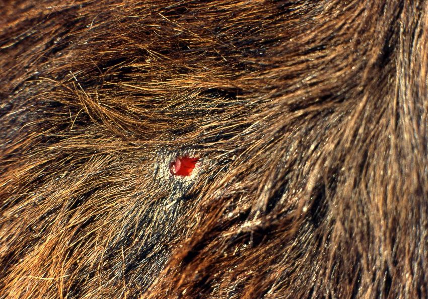
Viruses 2021, 13, 515 2 of 11
Viruses 2021, 13, x FOR PEER REVIEW 2 of 12
Figure 1. A typical lesion after a feeding bite of the common vampire bat on cattle. In a single bite, the vampire bat removes
Figure
a piece of skin1. ~6Amm,
typical lesion
exposing after a feeding
the subcutaneous tissue bite
of theof theand
prey, common vampire
licks the blood. bat on cattle.
The anticoagulants In bat’s
in the a single
bite,
saliva the vampire
facilitate bat removes
the maintenance a piece
of bleeding duringof skin ~6 mm, exposing the subcutaneous tissue of the prey,
feeding.
and
Viruses 2021, 13, xlicks theREVIEW
FOR PEER blood. The anticoagulants in the bat’s saliva facilitate the maintenance of bleeding
3 of 12
Vampire bat salivary glands secrete saliva containing anticoagulants and fibrinolytic
during feeding. enzymes that prevent blood clotting, both during ingestion as well as inside the gastroin-
testinal tract during the processes of digestion and elimination of excess water [13–21].
The tubular cecum and extensibility of the gastrointestinal tract allow for rapid ingestion
and digestion of a large blood volume (Figure 2). During each feeding bout that lasts 20–
30 min, vampire bats ingest a blood quantity close to their own weight (roughly 30 mL),
and in less than 120 min, defecation begins [6–9,22,23].
Figure 2. A male vampire bat captured while feeding in the wild, with a distended abdomen, not yet totally filled with
Figure
blood. A male vampire
2. anticoagulants
Salivary bat
facilitate thecaptured while
maintenance of thefeeding in during
blood flow the wild, withand
ingestion a distended abdomen,
in a liquid state within not
yet totally
the digestive filled
tract of with
the bat. blood.
Blood of prey,Salivary
with stronganticoagulants facilitate
resistance to vampire the maintenance
bat anticoagulants induced by of the blood flow
an appropriate
delivery, could coagulate within the digestive tract of the bat, making both digestion and elimination of excess water
during ingestion and in a liquid state within the digestive tract of the bat. Blood of prey, with strong
difficult.
resistance to vampire bat anticoagulants induced by an appropriate delivery, could coagulate within
Elimination
the digestive tract of the of excess
bat, making bothwater through
digestion andurination beginsof2 excess
elimination to 4 minwater
after the blood intake
difficult.
begins and continues for more than two hours [10–12]. Vampire bats feed nightly [10–12].
Elimination Searching
of excessforwater
suitable prey expends
through a high energetic
urination begins 2cost
to [24,25].
4 min After
afterfeeding, the wound
the blood intake
on the for
begins and continues preymore
continues
thantotwo
bleedhours
(Figure[10–12].
3) due to Vampire
the persistent effect
bats feedof salivary
nightlyanticoag-
[10–12].
ulants [6–9,26].
Searching for suitable prey expends a high energetic cost [24,25]. After feeding, the wound
on the prey continues to bleed (Figure 3) due to the persistent effect of salivary anticoagu-
lants [6–9,26].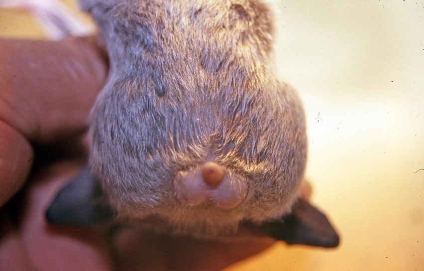
Viruses 2021, 13, 515 3 of 11
Viruses 2021, 13, x FOR PEER REVIEW 4 of 12
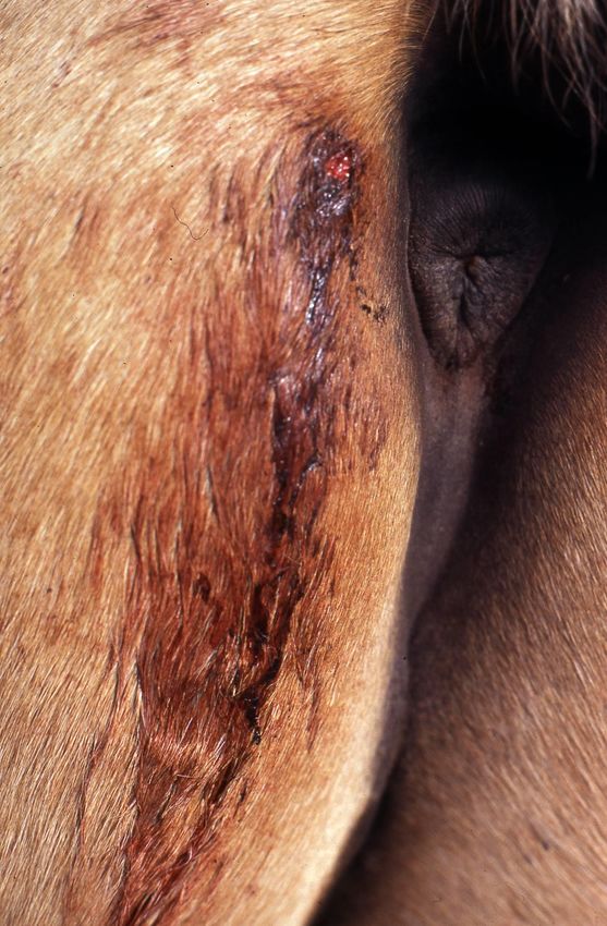
Figure 3. Residual hemorrhage in a horse. Blood continues to flow from the bite after the vampire
Figure 3. Residualbathemorrhage
finishes feedingindue
a horse. Bloodeffect
to the residual continues to flow
of its salivary from the bite after the vampire
anticoagulants.
bat finishes feeding due to the residual effect of its salivary anticoagulants.
Vampire bats are the primary reservoir of the rabies virus in tropical and subtropical
Vampire areas of the
bats are the Americas.
primary Annually,
reservoir“paralytic rabies”virus
of the rabies kills tens of thousands
in tropical andofsubtropical
livestock,
dozens of humans and an indeterminate quantity of wildlife [4–6,26–32]. Additionally,
areas of the Americas. Annually, “paralytic rabies” kills tens of thousands of livestock,
dozens of humans and an indeterminate quantity of wildlife [4–6,26–32]. Additionally,
wounds and blood loss due to vampire bat bites cause serious damage (Figure 3), in-
cluding anemia, infections, physical pain and psychological suffering in humans; anemia,
loss of vision, loss of weight and productivity and predisposition to infections and myiasis
in livestock; and mortality from excessive bleeding in poultry, wild birds and other small
prey [33–37]. While the risk of rabies acquisition may be minimized by vaccination of hu-
mans, domestic animals or wildlife, such prevention does not impact the burden associated
with the outcome from overt vampire bat feeding upon prey.
Current measures to control vampire bats based on the use of coumarin-derived
poisons [38–40] are generally used only at a local level because their high application cost
makes it difficult or impossible to maintain treatment over time in large areas such asViruses 2021, 13, 515 4 of 11
provinces or countries [41,42]. Furthermore, due to the toxic risk for non-target species
of bats and other mammals that share roosts with vampires [4–6,43–48], such use is
recommended to be reduced to the minimum possible and to seek other control mea-
sures [4,5,27,28,41,49].
Observations that repeated vampire bat bites induce in cattle immunologically me-
diated resistance against vampire salivary anticoagulants [50], and of the preference of
vampire bats for biting those animals apparently less resistant to their salivary anticoag-
ulants, such as young individuals and newcomers from vampire bat-free sites [50–53],
encourage applied research for control alternatives to present methods, particularly those
focused on increasing resistance of livestock against vampire salivary anticoagulants.
In this study, we analyzed aspects of the antigenic behavior of vampire bat saliva inocu-
lated parenterally in sheep. Our main objectives were to: determine whether subcutaneous
inoculation of vampire bat saliva-Freund’s incomplete adjuvant could induce an immuno-
logically mediated resistance against vampire bat anticoagulants; evaluate the response of
inoculated sheep compared with that induced by repeated vampire bat bites in a control
group; compare the interference capacity against vampire bat salivary anticoagulants of
sheep sera before and after inoculation with bat saliva-Freund’s incomplete adjuvant, and
observe characteristics of vampire bat salivary secretion during saliva collection.
2. Materials and Methods
2.1. Abbreviations
CT: clotting time (min); VSA: vampire salivary anticoagulants; mix 1: a sample of
sheep blood mixed with vampire saliva in a 29:1 ratio, used to observe its CT when
evaluating the resistance of sheep against VSA (see below Section 2.7); mix 2: a sheep
serum sample mixed with vampire saliva, and blood from the reference sheep in a 6:1:20
ratio, used to observe its CT when evaluating the neutralizing capacity of sera before and
after sheep were injected with vampire saliva-Freund’s incomplete adjuvant (see below
Section 2.8).
2.2. Sheep
We used 24 Hampshire Down ewes raised in our Laboratory of 12/24 months of age
and a mean weight of 46 kg. Sheep had an inbreeding rate >80%, daughters from the same
father, and descendant by maternal lines from the same grandfather and great-grandfather,
and they were immunologically naive with respect to vampire bat saliva since they had not
been bitten previously by vampire bats nor injected with vampire bat saliva. Sheep were
divided into groups A, B and C, composed of 12, 11 and 1 sheep each, respectively. Taking
advantage that sheep left their corral one-by-one crossing a narrow working chute, on
8 July 2019, we formed the experimental groups, proceeding as follows: group C was
the first sheep that left the corral; group A consisted of those sheep from even-numbered
departures: 2◦ ,4◦ . . . 24◦ ; and group B with those from odd-numbered departures: 3◦ , 5◦
. . . 23◦ , which were identified by numbered ear tags. Sheep of group A were used in the
assay of inoculation of vampire bat saliva, those of group B in the assay of exposure to
vampire bat bites and the single individual of group C was used as a blood source in the
tests to evaluate the neutralizing capacity of sheep serum against VSA. All procedures
used in the experiments with sheep were approved by the Ethics Committee of the Consejo
Profesional de Médicos Veterinarios de la Provincia de Misiones, Argentina.
2.3. Vampire Bats
We used 62 adult common vampire bats captured, quarantined, and held in captivity
during the study, as described [50]. Briefly, they were caught during 3–8 March 2019,
in areas where there were no reported livestock rabies outbreaks during the previous
2 years. After a 70-day quarantine in which they remained healthy, and rabies virus
was not isolated from 2 saliva samplings made 26 and 60 days after capture, the bats
were considered appropriate for use in the study. The bats were divided into 2 groups:Viruses 2021, 13, 515 5 of 11
48 vampire bats were used for the extraction of saliva, and 14 of them were used for
feeding on sheep. Both the collection in the field, as well as the management in captivity of
vampire bats, were made under Provincial and Federal permits and followed the guidelines
approved by the American Society of Mammalogists for the use of wild mammals in
research [54].
2.4. Extraction, Preservation, and Manipulation of Vampire Bat Saliva
We collected vampire bat saliva during May–July (Table 1), proceeding as follows:
a bat was restrained, and to stimulate salivation, their mouth was rinsed 3–4 times with
0.5 mL of 0.4% pilocarpine solution, administered with a plastic syringe without a needle.
Afterward, the bat was hung head down for 10 min, and the saliva secretion was collected
in a Petri dish placed on ice under the bat. The quantity of saliva secreted by 8–11 bats
(about 7–9 mL) was pipetted from the Petri dish and combined in a tube for the elimination
of cells and other impurities by centrifugation. The supernatant was kept in another
labeled tube and maintained at −30 ◦ C. When the quantity of saliva was accumulated,
the tubes were thawed, and saliva was placed together in a dish on wet ice, where it was
mixed carefully for 15 min and redistributed in tubes that were kept at −30 ◦ C until use.
In this way, we were able to use saliva of the same origin and with the same handling
(i.e., 2 freezing periods and 2 thaws).
Table 1. Tasks performed during the study in experimental sheep. Groups: A (injected with vampire
bat saliva) and B (exposed to vampire bat bites).
Date Group Task
10 July A Blood sampling for resistance testing against VSA
11 July B Blood sampling for resistance testing against VSA
12 July A Blood sampling for extraction of sera
13 July A Tests of the neutralizing capacity of sera against VSA
15 July B Start of exposure sessions to vampire bat bites
First injection of vampire bat saliva–incomplete
17 July A
Freund’s adjuvant
Second injection of vampire bat saliva–incomplete
20 August A
Freund’s adjuvant
Third injection of vampire bat saliva–incomplete
21 October A
Freund’s adjuvant
12 November B Completion of exposure sessions to vampire bat bites
2 December A Blood sampling for resistance testing against VSA
3 December B Blood sampling for resistance testing against VSA
5 December A Blood sampling for extraction of sera
6 December A Tests of the neutralizing capacity of sera against VSA
2.5. Inoculation of Sheep with Vampire Bat Saliva
Vampire bat saliva was injected mixed in equal parts with incomplete Freund’s adju-
vant (Sigma-Aldrich, #F5506). The mixture was prepared 15/20 min before its application
by mixing the components with a syringe until obtaining an emulsion. Inoculations were
performed subcutaneously in group A sheep, as follows: 2 mL during July and 1 mL during
August and during October 2019 (Table 1).
2.6. Exposure of Sheep to Vampire Bat Bites
Between 15 July and 12 November 2019, sheep of group B were exposed to vampire
bat bites (Table 1). In each exposure session, 5–6 sheep were placed into a room where
12 vampires were kept from dusk to 23 h. Exposure sessions were held every 2–3 days.Viruses 2021, 13, 515 6 of 11
After exposure, bites suffered by sheep were counted, and a topical insecticide was applied
to avoid myiasis.
2.7. Evaluation of the Resistance of Sheep against Vampire Bat Salivary Anticoagulants
We observed the CT values of sheep blood mixed with vampire bat saliva in a 29:1
ratio (mix 1), proceeding as follows: we placed 0.1 mL of vampire bat saliva in a tube
and added 2.9 mL of the freshly drawn blood sample, which was mixed by inverting and
rotating the tube, which was then placed vertically at 38 ◦ C, tilting every 3–4 min to observe
the CT; as the relationship between these parameters were negative, high values of mix
1 CT indicated scarce resistance of sheep against VSA and vice versa. We observed the
CT of mix 1 to evaluate and compare the resistance against VSA in the sheep of group A,
before and after inoculation with saliva and in those of group B before and after exposing
them to vampire bat bites (Table 2).
Table 2. Observation of the clotting time (min) of * mix 1 to evaluate resistance against ** VSA in sheep of groups A and B
after they were administered vampire bat saliva–incomplete Freund’s adjuvant or exposed to vampire bat bites, respectively.
Group A of 12 Sheep Injected with Vampire Group B of 11 Sheep Exposed to Vampire
Clotting Time (min) of Saliva-Incomplete Freund’s Adjuvant Bat Bites
mix 1, Statistical Data
before Shots after Shots before Bites after Bites
Minimum 32 13 34 22
Maximum 47 30 47 36
Median 36 22 37 29
Mean 37.7 21 38.6 28.3
Skewness/kurtosis 1/−0.1 0.5/−0.2 1/0–0.4 0.2/0.4
Conf. int. of mean 34.6–40.7 17.9–24 35.6–41.6 25.6–30.9
Standard deviation 4.8 4.8 4.4 4
Sum 452 252 425 311
Comparison by paired
t = 8.1, p = 0.000006 t = 5.3, p = 0.00004
t-test
* mix 1: a mixture of a sample of sheep blood freshly drawn and vampire saliva in a ratio 29:1, respectively (see Section 2.7 in the text)
** VSA: vampire salivary anticoagulants.
2.8. Evaluation of the Interference Capacity of sheep against Vampire Bat Salivary Anticoagulants
To evaluate this aspect, we observed the CT values of serum mixed with vampire saliva
and blood from the reference sheep (mix 2), proceeding as follows: we placed in a tube
0.6 mL of sheep serum sample and 0.1 mL of vampire bat saliva, mixing and incubating at
38 ◦ C for 30 min; then, to enable this mixture for coagulation and CT observation, we added
2 mL of freshly drawn blood from the reference sheep, mixing and observing the CT, as
previously described. Although we did not know if the blood of reference sheep used in
these tests to achieve the coagulation could have influenced the CT results, this does not
invalidate the observations, since when using blood from the same sheep, the influence
would have been the same in all tests as an in-house control. We used these tests to evaluate
and compare the interfering capacity of serum in group A sheep before and after they were
injected with vampire bat saliva and incomplete Freund’s adjuvant (Tables 1 and 3).Viruses 2021, 13, 515 7 of 11
Table 3. Observation of the clotting time (min) of * mix 2 to evaluate the interference capacity against
** VSA of the sera of group A sheep before and after administration of vampire bat saliva–incomplete
Freund’s adjuvant.
Clotting Time (min),
Before Inoculation After Inoculation
Statistical Data
Minimum 19 9
Maximum 29 15
Median 22.5 11
Mean 22.8 11.3
Skewness/kurtosis 0.9/0.4 0.6/−0.7
Conf. int. of mean 21/24.7 10/12.5
Standard deviation 2.9 2.0
Sum 274 135
Comparison by paired t-test: t = 19.9, pViruses 2021, 13, 515 8 of 11
3.4. Interference Capacity of Ser against VSA.
The observation of a decrease from 22.8 to 11.3 min (t = 19.9, pViruses 2021, 13, 515 9 of 11
applications, which may justify such efforts, given the expanding burden throughout the
region. The utilization of recombinant rabies viruses as expression vectors may provide one
strategy as a candidate for dual vaccination against both the pathogen and the predator [62].
Such a control technique would protect livestock directly and negatively impact vampire
bats, taking into account the close relationship between the size of vampire populations
with the intensity, duration and frequency of rabies virus outbreaks [3,6,41]. As rabies
remains a core vaccine, ideally, all domestic animals at risk of exposure, including livestock,
should be vaccinated, especially in areas where vampire bat rabies is enzootic. Future
research may result in the development of a new safe and effective concomitant rabies and
anti-vampire bat vaccine for relevant prevention and control in a One Health context.
Author Contributions: Conceptualization, H.A.D.; methodology, H.A.D., R.G.R., G.L.D.; formal
analysis, H.A.D., R.G.R., G.L.D.; investigation, H.A.D., R.G.R., G.L.D.; writing—original draft
preparation, H.A.D., R.G.R., G.L.D.; writing—review and editing, H.A.D., R.G.R., G.L.D., C.E.R.
All authors have read and agreed to the published version of the manuscript.
Funding: This research was supported by the SENASA of Argentina.
Institutional Review Board Statement: Our research was carried out with direct permission from
Servicio Nacional de Sanidad y Calidad Agroalimentaria (SENASA).
Informed Consent Statement: Not applicable.
Data Availability Statement: Not applicable.
Acknowledgments: We are grateful for the collaboration of Marta Malvina Lanari de Elizalde, Ruben
Emilio Díaz, Ricardo Florentin and Gert Simon.
Conflicts of Interest: The authors declare no conflict of interest. The funders had no role in the design
of the study, in the collection, analyses, or interpretation of data, in the writing of the manuscript, or
in the decision to publish the results.
References
1. Greenhall, A.M.; Joermann, G.; Schmidt, U. Desmodus rotundus. Mamm. Species 1983, 202, 1–6. [CrossRef]
2. Koopman, K.F. Systematics and distribution. In Natural history of Vampire Bats; Greenhall, A.M., Schmidt, U., Eds.; CRC Press:
Boca Raton, FL, USA, 1988; pp. 7–17.
3. Delpietro, H.; Marchevsky, N.; Simonetti, E. Relative population densities and predation of the common vampire bat (Desmodus
rotundus) in natural and cattle-raising areas in north-east Argentina. Prev. Veter. Med. 1992, 14, 13–20. [CrossRef]
4. Johnson, N.; Aréchiga-Ceballos, N.; Aguilar-Setien, A. Vampire Bat Rabies: Ecology, Epidemiology and Control. Viruses 2014,
6, 1911–1928. [CrossRef] [PubMed]
5. Streicker, D.G.; Allgeier, J.E. Foraging choices of vampire bats in diverse landscapes: Potential implications for land-use change
and disease transmission. J. Appl. Ecol. 2016, 53, 1280–1288. [CrossRef]
6. Delpietro, H.A.; Russo, R.G.; Carter, G.G.; Lord, R.D.; Delpietro, G.L. Reproductive seasonality, sex ratio and philopatry in
Argentina’s common vampire bats. R. Soc. Open Sci. 2017, 4, 160959. [CrossRef]
7. Paradiso, J.L.; Goodwin, G.G.; Greenhall, A.M. A Review of the Bats of Trinidad and Tobago: Descriptions, Rabies Infection, and
Ecology. J. Mammal. 1961, 42, 559. [CrossRef]
8. Greenhall, A.M. The biting and feeding habits of the Vampire bat, Desmodus rotundus. J. Zoöl. 1972, 168, 451–461. [CrossRef]
9. Greenhall, A.M.; Schmidt, U.; Lopez-Forment, W. Field observations on the mode of attack of the vampire bat Desmodus ro-tundus
in Mexico. An. Inst. Biol. Univ. Aut. México 1969, 40, 245–252.
10. Wimsatt, W.A. Transient Behavior, Nocturnal Activity Patterns, and Feeding Efficiency of Vampire Bats (Desmodus rotundus)
under Natural Conditions. J. Mammal. 1969, 50, 233–244. [CrossRef]
11. Wimsatt, W.A.; Guerriere, A. Observations on the Feeding Capacities and Excretory Functions of Captive Vampire Bats. J. Mammal.
1962, 43, 17–27. [CrossRef]
12. McFarland, W.N.; Wimsatt, W.A. Renal function and its relation to the ecology of the vampire bat, Desmodus rotundus. Comp.
Biochem. Physiol. 1969, 28, 985–1006. [CrossRef]
13. Disanto, P.E. Anatomy and histochemistry of the salivary glands of the vampire bat, desmodus rotundus murinus. J. Morphol.
1960, 106, 301–335. [CrossRef]
14. Hawkey, C. Inhibitor of Platelet Aggregation Present in Saliva of the Vampire Bat Desmodus rotundus. Br. J. Haematol. 1967,
13, 1014–1020. [CrossRef]Viruses 2021, 13, 515 10 of 11
15. Gardell, S.J.; Duong, L.T.; Diehl, R.E.; York, J.D.; Hare, T.R.; Register, R.B.; Jacobs, J.W.; Dixon, R.A.; Friedman, P.A. Isolation,
characterization, and cDNA cloning of a vampire bat salivary plasminogen activator. J. Biol. Chem. 1989, 264, 17947–17952.
[CrossRef]
16. Krätzschmar, J.; Haendler, B.; Langer, G.; Boidol, W.; Bringmann, P.; Alagon, A.; Donner, P.; Schleuning, W.D. The plasminogen
activator family from the salivary gland of the vampire bat Desmodus rotundus: Cloning and expression. Gene 1991, 105, 229–237.
[CrossRef]
17. Krätzschmar, J.; Haendler, B.; Bringmann, P.; Dinter, H.; Hess, H.; Donner, P.; Schleuning, W.D. High-level secretion of the four
salivary plasminogen activators from the vampire bat Desmodus rotundus by stably transfected baby hamster kidney cells. Gene
1992, 116, 281. [CrossRef]
18. Fernandez, A.Z.; Tablante, A.; Bartoli, F.; Beguin, S.; Hemker, H.; Apitz-Castro, R. Expression of biological activity of draculin,
the anticoagulant factor from vampire bat saliva, is strictly dependent on the appropriate glycosylation of the native molecule.
Biochim. Biophys. Acta (BBA) Gen. Subj. 1998, 1425, 291–299. [CrossRef]
19. Fernandez, A.Z.; Tablante, A.; Beguín, S.; Hemker, H.; Apitz-Castro, R. Draculin, the anticoagulant factor in vampire bat saliva, is
a tight-binding, noncompetitive inhibitor of activated factor X. Biochim. Biophys. Acta (BBA) Protein Struct. Mol. Enzym. 1999,
1434, 135–142. [CrossRef]
20. Low, D.H.; Sunagar, K.; Undheim, E.A.; Ali, S.A.; Alagon, A.C.; Ruder, T. Dracula’s children: Molecular evolution of vampire bat
venom. J. Proteomics. 2013, 89, 95–111. [CrossRef] [PubMed]
21. Ware, F.L.; Luck, M.R. Evolution of salivary secretions in haematophagous animals. Biosci. Horizons Int. J. Stud. Res. 2017, 10.
[CrossRef]
22. Mitchell, G.C.; Tigner, J.R. The route of the ingested blood in the common vampire bat (Desmodus rotundus). J. Mammal. 1970,
51, 814–817. [CrossRef]
23. Rouk, C.S.; Glass, B.P. Comparative Gastric Histology of Five North and Central American Bats. J. Mammal. 1970, 51, 455–472.
[CrossRef] [PubMed]
24. Young, A.M. Foraging of vampire bats Desmodus rotundus in Atlantic wet lowland Costa Rica. Rev. Biol. Trop. 1971, 18, 73–88.
25. Breidenstein, C.P. Digestion and Assimilation of Bovine Blood by a Vampire Bat (Desmodus rotundus). J. Mammal. 1982, 63, 482–484.
[CrossRef]
26. Baer, G.M. The biology and control of vampire bats. In The Natural History of Rabies; Baer, G.M., Ed.; Academic Press: New York,
NY, USA, 1975; Volume 2, pp. 1–387.
27. World Health Organization. WHO Expert Consultation on Rabies: First Report; WHO: Geneva, Switzerland, 2004.
28. World Health Organization. WHO Expert Consultation on Rabies: Second Report; WHO: Geneva, Switzerland, 2013.
29. Rupprecht, C.E.; Turmelle, A.; Kuzmin, I.V. A perspective on lyssavirus emergence nd perpetuation. Curr Opin Virol. 2015, 1,
662–670. [CrossRef] [PubMed]
30. Rupprecht, C.E.; Kuzmin, I.V. Why we can prevent, control and possibly treat, but will not eradicate rabies. Future Virol. 2015,
10, 517–535. [CrossRef]
31. Da Rosa, E.S.; Kotait, I.; Barbosa, T.F.; Carrieri, M.L.; Brandão, P.E.; Pinheiro, A.S.; Begot, A.L.; Wada, M.Y.; De Oliveira, R.C.;
Grisard, E.C.; et al. Bat-transmitted human rabies outbreaks, Brazilian Amazon. Emerg. Infect. Diseases 2006, 12, 1197–1202.
[CrossRef]
32. Delpietro, H.A.; Lord, R.; Russo, R.G.; Gury-Dhomen, F. Observations of sylvatic rabies in Northern Argentina during out-breaks
of paralytic cattle rabies transmitted by vampire bats (Desmodus rotundus). J. Wildl. Dis. 2009, 45, 1169–1173. [CrossRef]
33. Kverno, N.B.; Mitchell, G.C. Los murciélgos vampiros y la producción pecuaria en. Am. Lat. Rev. Mund. Zoot. 1976, 17, 1–7.
34. Thompson, R.D.; Elias, D.J.; Mitchell, G.C. Effects of Vampire Bat Control on Bovine Milk Production. J. Wildl. Manag. 1977,
41, 736. [CrossRef]
35. Greenhall, A.M. Feeding behavior. In Natural History of Vampire Bats; Greenhall, A.M., Schmidt, U., Eds.; CRC Press: Boca Raton,
FL, USA, 1988; pp. 111–132.
36. Crespo, J.; Vanella, J.; Blood, B.; De Carlo, J.M. Observaciones ecológicas del vampiro Desmodus rotundus rotundus (Geoffroy)
en el norte de Córdoba. Rev. Mus. Arg. Cien. Nat. B Rivadavia 1961, 6, 131–160.
37. Delpietro, H.A.; Russo, R.G.; Schwieters, H.H.G. Observaciones sobre el ataque del vampiro común (Desmodus rotundus) al
ganado en el norte de Argentina. Rev. Med. Vet. 1999, 80, 460–464.
38. Linhart, S.B.; Flores Crespo, R.; Mitchell, G.C. Control de murciélagos vampiros por medio de un anticoagulante. Bol. Oficina
Sanit. Panam. 1972, 73, 100–109.
39. Schmidt, U.; Schmidt, C.; Lopez-Forment, W.; Flores Crespo, R. Rückfunde beringter Vampirfledermaüse (Desmodus rotundus) in
Mexico. Z. Säugetierkunde 1978, 43, 70–75.
40. Flores Crespo, R.; Ibarra, V.F.; De Anda, D.L. Vampirinip II: Un producto utilizable en tres métodos para el combate del
mur-ciélago hematófago. Téc. Pecu. Méx. 1976, 30, 67–75.
41. Delpietro, H.A.; Nader, A.J. Rabies of herbivores transmitted by vampire bats in north-eastern Argentina. Rev. Sci. Tech. Off. Int.
Epiz. 1989, 8, 189–198. [CrossRef] [PubMed]
42. Seetahal, J.F.R.; Vokaty, A.; Carrington, C.V.; Adesiyun, A.A.; Mahabir, R.; Hinds, A.Q.J.; Rupprecht, C.E. The History of Rabies in
Trinidad: Epidemiology and Control Measures. Trop. Med. Infect. Dis. 2017, 2, 27. [CrossRef] [PubMed]Viruses 2021, 13, 515 11 of 11
43. Greenhall, A.M. Ecology and bionomics of vampire bats in Latin America. In Bats and Rabies; Greenhall, A.M., Artois, M., Fekadu,
M., Eds.; Fondation Marcel Mérieux: Lyon, France, 1993; pp. 3–57.
44. Walker, S.; Medellín, M.R.A.; Aguirre, L.A.; Mann, A.; Ochoa, J.R. Conservation progress in Latin America. Bat. Mag. 2001, 19, 1–7.
Available online: https://www.batcon.org/article/conservation-progress-in-latin-america/ (accessed on 19 March 2021).
45. Mayen, F. Haematophagous Bats in Brazil, Their Role in Rabies Transmission, Impact on Public Health, Livestock Industry and
Alternatives to an Indiscriminate Reduction of Bat Population. J. Veter Med. Ser. B 2003, 50, 469–472. [CrossRef] [PubMed]
46. Asprilla-Aguilar, A.A.; Mantilla-Meluk, H.; Jiménez Ortega, A.M. Analysis of the non-hematophagous bat species captured
within the plan of eradication of Desmodus rotundus (E. Geoffroy, 1810) in the Colombian Biogeographic Chocó. Rev. Inst. Univ.
Tecnol. Chocó Invest. Biod. Des. 2007, 26, 42–48.
47. Oprea, M.; Aguliar, L.M.S.; Wilson, D.E. Anoura caudifer (Chiroptera: Phyllostomidae). Mamm. Species 2009, 844, 1–8. [CrossRef]
48. Aguiar, L.M.S.; Brito, D.; Machado, R.B. Do Current Vampire Bat (Desmodus rotundus) Population Control Practices Pose a Threat
to Dekeyser’s Nectar Bat’s (Lonchophylla dekeyseri) Long-Term Persistence in the Cerrado? Acta Chiropterologica 2010, 12, 275–282.
[CrossRef]
49. Rocha, F.; Dias, R.A. The common vampire bat Desmodus rotundus (Chiroptera: Phyllostomidae) and the transmission of the
rabies virus to livestock: A contact network approach and recommendations for surveillance and control. Prev. Veter. Med. 2020,
174, 104809. [CrossRef]
50. Delpietro, H.A.; Russo, R.G. Acquired resistance to saliva anticoagulants by prey previously fed upon by vampire bats (Des-modus
rotundus): Evidence for immune response. J. Mammal. 2009, 90, 1132–1138. [CrossRef]
51. Arellano, C.S.; Sureau, P.; Greenhall, A.M. Preferencia de la predación del vampiro en relación a la edad y la raza del ganado y la
época del año. Téc. Pec. Méx. 1971, 17, 23–29.
52. Dalquest, W.W. Natural History of the Vampire Bats of Eastern Mexico. Am. Midl. Nat. 1955, 53, 79. [CrossRef]
53. Acosta y Lara, E.F. Quirópteros del Uruguay. Com. Zool. Mus. Hist. Nat. Montev. 1950, 3, 1–71.
54. Gannon, W.L.; Sikes, R.S. Guidelines of the American Society of Mammalogists for the Use of Wild Mammals in Research.
J. Mammal. 2007, 88, 809–823. [CrossRef]
55. Delpietro, H.; Konolsaisen, F.; Marchevsky, N.; Russo, G. Domestic cat predation on vampire bats (Desmodus rotundus) while
foraging on goats, pigs, cows and human beings. Appl. Anim. Behav. Sci. 1994, 39, 141–150. [CrossRef]
56. Willadsen, P.; Bird, P.; Cobon, G.S.; Hungerford, J. Commercialization of a recombinant vaccine against Boophilus microplus.
Parasitology 1995, 110, 43–50. [CrossRef] [PubMed]
57. Willadsen, P. Anti-tick vaccines. Parasitology 2004, 129, 367–387. [CrossRef]
58. Trimnell, A.R.; Hails, R.S.; Nuttall, P.A. Dual action ectoparasite vaccine targeting ‘exposed’ and ‘concealed’ antigens. Vaccine
2002, 20, 3560–3568. [CrossRef]
59. Nuttall, P.A.; Trimnell, A.R.; Kazimirova, M.; Labuda, M. Exposed and concealed antigens as vaccine targets for controlling ticks
and tick-borne diseases. Parasite Immunol. 2006, 28, 155–163. [CrossRef] [PubMed]
60. De la Fuente, J.; Contreras, M. Tick vaccines: Current status and future directions. Expert Rev. Vaccines 2015, 14, 1367–1376.
[CrossRef]
61. Rego, R.O.M.; Trentelman, J.J.A.; Anguita, J.; Nijhof, A.M.; Sprong, H.; Klempa, B.; Ehajdusek, O.; Tomás-Cortázar, J.; Azagi, T.;
Strnad, M.; et al. Counterattacking the tick bite: Towards a rational design of anti-tick vaccines targeting pathogen transmission.
Parasites Vectors 2019, 12, 229. [CrossRef]
62. Scher, G.; Schnell, M.J. Rhabdoviruses as vectors for vaccines and therapeutics. Curr. Opin. Virol. 2020, 44, 169–182. [CrossRef]
[PubMed]You can also read