Original Article EGCG suppresses NF-κB activation induced by gastroesophageal reflux contents in human esophageal epithelial cells
←
→
Page content transcription
If your browser does not render page correctly, please read the page content below
Int J Clin Exp Pathol 2016;9(5):5421-5428
www.ijcep.com /ISSN:1936-2625/IJCEP0024048

Original Article
EGCG suppresses NF-κB activation induced by
gastroesophageal reflux contents in human
esophageal epithelial cells
Chang Liu1*, Jie Dai2*, Heng Lu1, Hui Shi1, Youke Lu1, Fangyu Wang1
1
 Department of Gastroenterology and Hepatology, Jinling Hospital, Nanjing Clinical School of Southern Medical
University, Nanjing, China; 2Department of Gastroenterology and Hepatology, The PLA Navy Anqing Hospital,
Anqing, China. *Equal contributors.
Received January 14, 2016; Accepted March 26, 2016; Epub May 1, 2016; Published May 15, 2016

Abstract: Background and aims: Epigallocatechin-3-gallate (EGCG) is a natural component of green tea that has
been shown to have inhibitory effects against the inflammation-induced onset and the development of carcinogen-
induced tumors in animal models at different organ sites, including the esophagus. This study investigates the
effect of mixed refluxate (acid, bile acids and trypsin) on the expression of the nuclear factor kappa-light-chain-
enhancer of activated B cells (NF-κB) signaling pathway in normal human esophageal epithelial cells (HEECs) and
the effect of EGCG pretreatment of cells on activation of NF-κB induced by the mixed refluxate. Methods: HEECs
were cultured in vitro and treated with varying concentrations of EGCG in the absence or presence of GER contents.
NF-κB DNA-binding activity was examined using an electrophoretic mobility shift assay (EMSA) and intracellular lev-
els of NF-κB were evaluated using an ELISA. NF-κB reporter gene activity was measured using a luciferase reporter
gene assay. The expression levels of NF-κB/p65, p-NFκB/p65, IκBα, p-IκBα, p-IKKα and proinflammatory cytokines,
such as IL-6, IL-8, iNOS and COX-2 proteins, were examined using Western blot analysis. Results: Exposure of GER
to HEECs results in a significant increase in NF-κB DNA-binding activity, intracellular levels of NF-κB and luciferase
reporter activity compared to the control group. GER also induced the activation and nuclear translocation of NF-
κB/p65, phosphorylation of IκBα and IKKα, and upregulated the expression of NF-κB-regulated proteins IL-6, IL-8,
COX-2 and iNOS all of which were significantly downregulated by EGCG pretreatment. Conclusions: Our data suggest
that EGCG can suppress GER-induced NF-κB activation and can downregulate the expression of NF-κB-regulated
proteins in HEECs.

Keywords: EGCG, gastroesophageal refluxate(GER), HEECs, NF-κB

Introduction                                                   ry and the effects of the refluxate on the patho-
                                                               genesis of GERD. There is a strong relationship
Gastroesophageal reflux disease (GERD) is                      between inflammation and cancer, and now, it
common in Western countries, and the inci-                     is recognized that the tumor microenvironment
dence in United States alone is approximately                  is largely orchestrated by inflammatory cells
7-10% [1]. A mild form of this disease may cau-                and contributes to the neoplastic process by
se no apparent mucosal damage, but chronic                     promoting cell proliferation, survival and migra-
GERD can lead to severe lesions, erosion,                      tion [5]. Exposure to the carcinogens present in
ulceration, stricture formation and Barrett’s                  the GER contents can cause structural and
esophagus (BE), a precancerous lesion that                     functional changes in the esophageal squa-
could potentially develop into esophageal ade-                 mous epithelium, including abnormal prolifera-
nocarcinoma [2]. Bile and acid are major con-                  tion and differentiation of these cells, BE and
stituents of the gastroesophagealrefluxate                     Barrett’s-related adenocarcinoma (BAA).
(GER) [3], and this mixture is closely associated
with tumor progression [4]. Therefore, it is                   NF-κB is recognized as a key transcription fac-
important to understand the molecular and cel-                 tor involved in initiating and regulating inflam-
lular mechanisms of esophageal mucosal inju-                   matory responses. NF-κB activity is regulated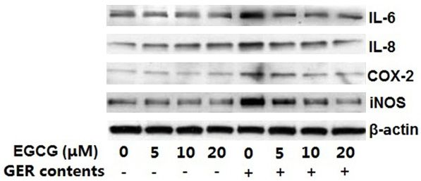
EGCG suppresses NF-κB activation in EEC

by the cytoplasmic degradation of a related         were purchased from Cell Signaling Technology
inhibitory protein known as IκBα. Once IκBα is      (Beverly, MA, USA). IL-6, IL-8, COX-2 and iNOS
inactivated and the Rel proteins are phosphory-     antibodies were purchased from KeyGENBio-
lated, the NF-κB dimers relocate to the nucleus     TECH Co. Ltd (Nanjing, China).
[6]. The activated NF-κB binds to specific target
sites in the nucleus and regulates the expres-      Cell culture
sion of genes related to inflammation, immune
                                                    HEECs were purchased from American Type
responses and cell survival [7]. Recently, both
                                                    Culture Collection (Manassas, VA, USA). HEECs
esophageal cell lines and biopsy cultures
                                                    were cultured in RPMI-1640 medium obtained
showed that GER contents can induce the
                                                    from Gibco (Rockville, MD, USA) and supple-
expression of NF-κB dependent genes [8].
                                                    mented with 10% fetal bovine serum, 100
Epigallocatechin-3-gallate (EGCG), the major        μg/mL streptomycin and 100 U/mL penicillin
polyphenol found in green tea, is reported to       G, which were purchased from Invitrogen
have antioxidant, anti-inflammatory, anti-muta-      (Carlsbad, CA, USA). Cells were harvested with
genic, anti-angiogenic and chemopreventive          0.05% trypsin and maintained at 37°C in an
effects [9-13]. The mechanisms used to des-         environment containing 5% CO2.
cribe EGCG’s anti-tumor activity include the
                                                    Cell treatments
induction of detoxifying enzymes, inhibition of
the activation of carcinogens and inhibition of     A solution of EGCG dissolved in 1X PBS was
signaling pathways that control cell prolifera-     used for this study. Cells (80-90% confluent)
tion and tumor growth [14]. Although there is       were treated with various concentrations of
extensive literature describing the inhibitory      EGCG (5, 10, 20 µM) at 37°C for 4 h in RPMI-
effects of EGCG, most of these studies were         1640 medium following which, the media was
conducted in models in which the esophageal         removed, the cells were washed with PBS and
carcinogenesis was already well established. It     treated with GER contents (including acid,
is well known that esophageal tumors result in      CDCA, and trypsin) for 12 h and harvested. The
irreversible structural and functional damage       acidic culture conditions (pH 6.5), the CDCA
to several tissues and organs. Therefore, it is     concentration (200 µmol/L), and the trypsin
necessary to investigate the effects of EGCG        concentration (10 U/mL) were recommended
on healthy esophageal cells, which are exposed      by Kawabe et al. [15].
to GER contents, and assess the resulting
effects that are involved in esophageal carci-      Preparation of total cell lysate
nogenesis.
                                                    After the treatment of HEECs with EGCG, GER
In this study, we investigated the relationships    or both, the culture medium was aspirated, and
between the activated NF-κB induced by GER          cells were washed thrice with PBS. The cells
contents and the anti-inflammatory effect of         were incubated in 0.4 ml ice-cold lysis buffer
EGCG in human esophageal epithelial cells           (150 mM NaCl, 100 mM Na3VO4, 50 mM Tris-
(HEECs). Additionally, the non-malignant HEECs      HCl, 20 mM NaF, 1 mM PMSF (pH 7.4), 1 mM
used in this study provide clear evidence           EDTA, 1 mM EGTA, 1% Triton X-100 and 0.5%
of specific molecular mechanisms involved in         NP-40) containing freshly added protease
GER -induced esophageal carcinogenesis.             inhibitor cocktail (Cell Signaling Technology,
                                                    Beverly, MA, USA). The cells were centrifuged
Materials and methods                               for 20 min (12,000 g) at 4°C, and the superna-
                                                    tant (total cell lysate) was collected and stored
Antibodies and reagents                             at -80°C. The protein concentration was deter-
                                                    mined using the BCA protein assay kit (Pierce,
Chenodeoxycholic acid (CDCA) and EGCG               Rockford, IL, USA).
(>98% pure) were obtained from Wako Pure
Chemical Industries (Osaka, Japan). Trypsin         Preparation of cytosolic and nuclear lysates
(1:250) was obtained from Gibco (Rockville,
MD, USA). IκBα, p-IκBα (Ser32/36), IKKα,            Following the treatment of HEECs with EGCG,
NF-κB/p65, p-NFκB/p65 (Ser536) antibodies           GER contents or both, the culture medium was

5422                                                    Int J Clin Exp Pathol 2016;9(5):5421-5428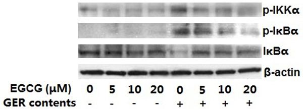
EGCG suppresses NF-κB activation in EEC
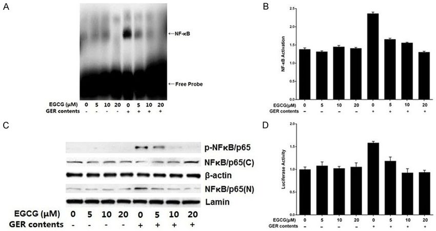
Figure 1. EGCG inhibited GER content-induced activation of NF-κB in HEECs. A. EGCG treatment of HEECs inhibits
GER content-induced NF-κB DNA-binding activity. Cells were pretreated with various concentrations of EGCG (5-20
μM) for 4 h and then exposed to GER contents; nuclear extracts were prepared and assayed for NF-κB transcrip-
tional activity using EMSA. The gel is representative of three experiments with similar results. B. EGCG inhibited GER
content-induced NF-κB/p65 activity. The effect of EGCG on NF-κB/p65 was evaluated using an ELISA. The values
are presented as the mean ± SEM. C. EGCG inhibited GER content-induced NF-κB/p65 phosphorylation, as shown
by a decrease in nuclear NF-κB/p65 levels with a concomitant increase in the total cytosolic NF-κB/p65 levels
in EGCG treated samples. Cytosolic and nuclear extracts were prepared for Western blot analysis. Equal protein
loading was achieved using β-actin and laminin as loading controls. D. EGCG inhibited GER content-induced NF-
κB-dependent reporter gene expression. HEECs were seeded at a concentration of 1.0×10⁵ cells per well in 6-well
plates and co-transfected with an NF-κB-driven luciferase reporter construct and pSV40-β-gal plasmid. After 48 h,
cells were treated with GER contents with or without EGCG and Luciferase activity was measured and normalized
with respect to β-galactosidase activity. Values are presented as the mean ± SEM. GER contents: acidified media
(pH 6.5) treated with CDCA (200 µmol/L) for a total of 12 h along with trypsin (10 U/mL) during the final hour.
aspirated, and the cells were washed thrice the BCA protein assay kit (Pierce, Rockford, IL,
with PBS. The cells were incubated in 0.4 ml USA).
ice-cold lysis buffer (10 mM KCl, 10 mM HEPES
(pH 7.9), 1 mM PMSF, 1 mM DTT, 0.1 mM EDTA Electrophoretic mobility shift assay
and 0.1 mM EGTA) containing freshly added
The nuclear protein extracts from HEECs were
protease inhibitor cocktail for 20 min, following
prepared as described earlier. For the DNA
which 12.5 µl of 10% NP-40 was added and the
binding assay, an end-labeled biotinylated dou-
contents were mixed. The cells were centri-
ble-stranded NF-κB oligonucleotide (5’-AGT
fuged at 13,000 g for 2 min at 4°C. The super- TGA GGG GAC TTT CCC AGG C-3’) was used.
natant (cytosolic lysate) was collected and Binding reactions were conducted using the
stored at -80°C. The pellet (nuclear lysate) was nonradioactive LightshiftTM chemiluminescent
resuspended in 50 µl of ice-cold nuclear extrac- EMSA kit (Pierce, Rockford, IL, USA) according
tion buffer (20 mM HEPES (pH 7.9), 1 mM to the manufacturer’s directions. Reaction
PMSF, 1 mM DTT, 1 mM EDTA, 1 mM EGTA and products were separated through a 6% DNA
0.4 M NaCl) containing freshly added protease retardation gel (Invitrogen, Carlsbad, CA, USA)
inhibitor cocktail for 40 min with intermittent and transferred to a Biodyne B membrane
mixing. The tubes were centrifuged at 13,000 g (Pierce, Rockford, IL, USA). The membrane was
for 6 min at 4°C. The supernatant (nuclear exposed to X-ray film and developed using the
extract) was collected and stored at -80°C. The Chemiluminescent Nucleic Acid Detection
protein concentration was determined using Module (Pierce, Rockford, IL, USA).
5423 Int J Clin Exp Pathol 2016;9(5):5421-5428EGCG suppresses NF-κB activation in EEC
Enzyme-linked immunosorbent assay viability of HEECs. The results indicate that the
NF-κB DNA-binding activity was significantly
Quantitative analysis of NF-κB/p65 activity in enhanced after HEECs were exposed to GER
the culture media of treated cells was conduct- contents (Figure 1A: lane 5). EGCG pretreat-
ed using the NF-κBTransAM ELISA kit from Active ment significantly inhibited GER content-
Motif (Carlsbad, CA, USA). All procedures were induced NF-κB DNA-binding activity of HEECs in
performed according to the manufacturer’s a dose-dependent manner (Figure 1A: lane 6,
protocol. 7, 8). In addition, the levels of NF-κB DNA-
binding activity of cells treated with EGCG alone
Western blot analysis
(Figure 1A: lane 2, 3, 4) were not significantly
different from those of the untreated group
70 µg of protein was resolved using PAGE
(Figure 1A: lane 1). This indicates that the ph-
(5-10% gels), transferred to PVDF membranes
ysiological concentrations of EGCG may not
and blocked with 5% non-fat milk in TBS (150
substantially affect the NF-κB DNA-binding
mM NaCl and 20 mM Tris, pH 7.6) containing
activity.
0.05% Tween-20. The membranes were incu-
bated overnight at 4°C with the appropriate EGCG suppresses GER content-induced phos-
monoclonal/polyclonal primary antibodies, and phorylation and nuclear translocation of NF-
then incubated with corresponding HRP-con- κB/p65
jugated secondary antibody for 2 h. The protein
bands were visualized using the ECL kit (Pierce, Western blot analysis shows that GER conten-
Rockford, IL, USA) and imaged using X-ray films. ts induce NF-κB/p65 (Ser536) phosphorylation
leading to the nuclear translocation of NF-κB/
Luciferase activity
p65. EGCG pretreatment of HEECs inhibited
HEECs (1.0×10⁵) were plated onto 6-well plates this GER content-induced NF-κB/p65 phos-
and co-transfected with 6 µg NF-κB-driven phorylation in a dose-dependent manner (Fig-
luciferase reporter construct and 8 µg pSV40- ure 1C). In addition, there is an increase in total
β-gal plasmid using Lipofectamine 2000 (In- cytosolic NF-κB/p65 levels with a concomitant
vitrogen, Carlsbad, CA, USA). After 48 h of expo- decrease in nuclear NF-κB/p65 levels in EGCG
sure to the transfection mixture, cells were pretreated samples, suggesting that EGCG sup-
incubated in medium containing EGCG for 4 h presses GER content-mediated NF-κB/p65
and subsequently exposed to GER contents for migration from the cytosol to the nucleus
12 h. The wells were washed with PBS, incu- (Figure 1C). ELISA analysis further confirmed
bated with Passive Lysis Buffer (Pierce, Ro- this effect. The activity of nuclear NF-κB/p65
ckford, IL, USA) for 20 min on a shaker, and the that was induced by the GER contents was
lysates were collected. Luciferase activity was effectively suppressed by EGCG (Figure 1B).
determined using the Reporter Luciferase
EGCG suppresses GER content-induced NF-κB
Assay System (Promega, Madison, WI, USA)
reporter activity
according to the methods provided by the man-
ufacturer and the values obtained were normal-
GER contents can activate NF-κB, which can
ized to the β-galactosidase activity (Clontech,
lead to the transcriptional activation of NF-κB-
Mountain View, CA, USA).
inducible genes. To determine whether EGCG
Results could inhibit GER content-induced NF-κB pro-
moter activity, HEECs were transiently trans-
EGCG suppresses GER content-induced NF-κB fected with the NF-κB promoter-luciferase
DNA-binding activity reporter plasmid and were exposed to GER con-
tents in the absence and presence of EGCG. An
To exclude the possibility of DNA damage increase in luciferase reporter expression was
caused due to high concentrations of EGCG, observed in GER content-exposed cells com-
the effects of EGCG on cell viability were pared to the control group (Figure 1D), suggest-
assessed using the MTT assay. We found that ing that NF-κB was activated. We found that
EGCG pretreatment at concentrations of 5-20 EGCG pretreatment of HEECs not only sup-
μM for 4 h had an insignificant effect on the pressed NF-κB DNA-binding activity but also
5424 Int J Clin Exp Pathol 2016;9(5):5421-5428EGCG suppresses NF-κB activation in EEC
treatment of HEECs with EGCG led to the inhibi-
tion of GER content-induced IKKα activation,
phosphorylation and degradation of IκBα, and
subsequent NF-κB activation.
EGCG suppresses GER content-induced activa-
tion of inflammatory markers
Figure 2. EGCG inhibited GER content-induced phos-
phorylation of IκBα. HEECs were pretreated with vari- To determine whether EGCG could decrease
ous concentrations of EGCG (5-20 μM) for 4 h and the production of proinflammatory cytokines,
then exposed to GER contents. To examine the effect we used Western blotting to analyze the whole
of EGCG on the expression levels of IKKα and IκBα cell extracts. The results showed that GER con-
proteins, cytosolic lysates were prepared for Western
blot analysis. The gel is representative of three ex- tents exposure to HEECs led to an increased
periments with similar results. GER contents: acidi- expression of inflammatory markers such as
fied media (pH 6.5) treated with CDCA (200 µmol/L) IL-6, IL-8, COX-2 and iNOS proteins. Pretreat-
for a total of 12 h along with trypsin (10 U/mL) during ment of HEECs with EGCG led to a significant
the final hour. inhibition of GER content-induced increase in
the expression of these proteins (Figure 3) indi-
cating that EGCG potentially has anti-inflamma-
tory properties.
Discussion
Many studies have been conducted on esopha-
geal cancer cell lines to evaluate whether EGCG
could reduce their viability and invasiveness. In
our study, normal esophageal cells pretreat-
Figure 3. EGCG inhibited GER content-induced ac- ed with EGCG showed downregulation of GER
tivation of inflammatory markers. HEECs were pre-
treated with various concentrations of EGCG (5-20
content-activated NF-κB. The development of
μM) for 4 h and then exposed to GER contents, after esophageal inflammation and cancer is associ-
which the cells were harvested and total cell lysates ated with oxidative stress induced by reflux
were prepared for Western blot analysis. The gel is contents such as gastric acid, bile acids and
representative of three experiments with similar re- trypsin [16]. Dvorak et al. showed that exposing
sults. GER contents: acidified media (pH 6.5) treated
with CDCA (200 µmol/L) for a total of 12 h along with
immortalized human esophageal epithelial
trypsin (10 U/mL) during the final hour. cells HET-1A to pH 2 or 4 for 1 minute led to the
production of reactive oxygen species (ROS)
and low pH in combination with bile acids
led to a significant decrease in GER content- induced mitochondrial oxidative stress and
induced NF-κB promoter activity. DNA damage [17, 18]. These changes may
cause cancer. Indeed, previous studies have
EGCG suppresses GER content-induced phos- shown that mitochondrial DNA mutations fre-
phorylation of IKKα and IκBα quently occur in dysplastic BE and esophageal
adenocarcinoma, probably due to oxidative
A key step in NF-κB activation is the phosphory-
damage [19].
lation of IκBα by the IκB kinase (IKK) complex
(IKKα, IKKβ and IKKγ/NEMO). To determine Oxidative stress is closely related to the activa-
whether EGCG’s inhibition of NF-κB activation tion of several signaling pathways, including
was because of its impact on IκBα degrada- the NF-κB, Janus kinase/Signal Transducer and
tion, the cytosolic levels of IKKα and IκBα were Activator of Transcription signal transducer and
measured using immunoblotting. The results activator of transcription (JAK/STAT) and MAPK
showed that GER content exposure led to an pathways, that can upregulate the expression
increased phosphorylation of IKKα and IκBα, of pro-survival, anti-apoptotic and angiogenic
and EGCG pretreatment of HEECs suppressed proteins [20, 21]. Activation of these pathways
these phosphorylations in a dose-dependent and expression of anti-apoptotic proteins was
manner (Figure 2). Our data indicate that pre- shown to be increased in BE [22-24].
5425 Int J Clin Exp Pathol 2016;9(5):5421-5428EGCG suppresses NF-κB activation in EEC
We speculate that the oxidative stress induced Cytokine-induced neutrophil accumulation ine-
by the mixed refluxate (acid and bile acids) may sophageal inflammation induced by the reflux
lead to the activation of anti-apoptotic and pro- of gastroduodenal contents has been previous-
survival pathways in BE and BAA. However, ly demonstrated [28]. Cytokines, such as IL-1,
DNA damage in anti-apoptosis cells is danger- IL-6, IL-8 and MCP-1, may be secreted during
ous and can result in cell mutations due to rep- this inflammatory reaction. Rafiee et al. [1]
lication errors, clonal expansion of mutated showed that an acidic environment results in
cells and tumor progression. Acquired somatic increased IL-6 and IL-8 release by normal
mutations may be an important determining esophageal epithelial cells. Transcriptional fac-
factor in esophageal carcinogenesis and may tors, especially those regulated by NF-κB acti-
alter the signal transduction in the esophagus. vation, play an important role in the induction
The squamous epithelium of distal esophagus of cytokines, COX-2, iNOS, acute-phase pro-
is frequently exposed to acid and bile acids dur- teins, cell adhesion molecules and growth fac-
ing a reflux episode. An analysis of the esopha- tors [29]. These inflammatory mediators attract
geal aspirates of GERD patients suggests that inflammatory cells to the tissues damaged by
bile acids are found in the esophageal aspi- GER contents and can activate other enzymes.
rates of 86% of the patients [25]. Therefore, it The inducible isoform of nitric oxide synthase
is necessary to develop chemopreventive strat- (iNOS) is considered more important during
egies for high-risk patients with GERD or for tumor progression than the other isoforms
those suffering from chronic reflux. (eNOS and nNOS) because it is involved in
maintaining the nitric oxide levels during inflam-
Pathways activated by EGCG in healthy cells, matory responses. Nitric oxide can directly
compared to tumor cells, create a different oxi- damage DNA, inhibit the repair of damaged
dizing environment, which is conducive to the DNA, regulate transcriptional factors, enhance
survival of normal cells and the destruction of oncogene expression, block cell apoptosis and
tumor cells. Studies have shown that epithelial promote angiogenesis and is therefore associ-
cells in skin, oral mucosa and gastrointestinal ated with the processes of tumor initiation and
regions, which are in frequent contact with development [30]. Moreover, studies have fo-
plant-derived polyphenols, are able to develop und that a selective nitric oxide synthase inhibi-
mechanisms to reduce the toxicity from these tor prevents the progression of rat esophageal
compounds [26]. However, high doses of green tumors induced by nitrosomethylbenzylamine
tea polyphenols in other healthy human cells [31]. Soma et al. [32] showed that GER con-
(which lack these antitoxic mechanisms) or tents induced the expression of COX-2 in both
tumor cells (which may have lost these protec- normal and cancerous esophageal cells. Ab-
tive mechanisms) may have cytotoxic effects. del-Latif et al. [33] speculated that the COX-2
Therefore, it is important to find an optimal expression in the esophagus may be directly
dose of EGCG that can selectively promote the related to the signal transduction pathways
death of tumor cells while protecting the nor- involved in tumor progression. Our results sh-
mal cells. ow that EGCG suppresses the activation of
inflammatory markers (IL-6, IL-8, COX-2 and
The activation of NF-κB can be induced by iNOS) induced by GER contents, suggesting
microenvironmental signals, such as acid, bile, that EGCG can prevent the formation of an
hypoxia, cytokines, and genetic factors [27]. inflammatory environment and thus, inhibit
NF-κB activation is closely associated with an pathogenesis of GER contents-induced esoph-
increase in the anti-apoptotic responses and ageal diseases.
the growth-promoting potential of cells leading
to a broad spectrum of stresses, which may Conclusions
lead to a malignant transformation of the cells.
In this study, GER content-induced NF-κB acti- In this study, we have shown that EGCG inhibits
vation resulted in the NF-κB/p65 nuclear trans- the GER content-mediated activation of NF-κB
location, which was suppressed by EGCG pre- and reduces the production of inflammatory
treatment, suggesting that EGCG has the markers such as IL-6, IL-8, COX-2 and iNOS in
potential to suppress carcinogenesis induced HEECs. According to our results, long-term
by GER contents. exposure to low levels of green tea derived
5426 Int J Clin Exp Pathol 2016;9(5):5421-5428EGCG suppresses NF-κB activation in EEC
EGCG could play a chemopreventive role in Ikappa B and in phosphorylating the p65 sub-
patients with chronic GERD and in those suffer- unit of NF-kappa B. J Biol Chem 2002; 277:
ing from chronic reflux. Further studies to test 3863-3869.
the efficacy of EGCG in animal models with [7] Lin A and Karin M. NF-kappaB in cancer: a
marked target. Semin Cancer Biol 2003; 13:
inflammation-associated esophagus injury and
107-114.
to elucidate the mechanisms by which this nat- [8] Cronin J, Alhamdani A, Griffiths AP, Baxter JN,
ural product modulates proinflammatory signal Brown T and Jenkins GJ. In vitro and ex vivo
transduction pathways will be conducted. models of extended reflux exposure demon-
strate that weakly acidic mixed reflux height-
Acknowledgements ens NF-kB-mediated gene expression. Dis
Esophagus 2011; 24: 360-370.
This work was supported by the National Na- [9] Higdon JV and Frei B. Tea catechins and poly-
tural Science Foundation of China (No. 812- phenols: health effects, metabolism, and anti-
70453) to Fang-Yu Wang. oxidant functions. Crit Rev Food Sci Nutr 2003;
43: 89-143.
Disclosure of conflict of interest [10] Kada T, Kaneko K, Matsuzaki S, Matsuzaki T
and Hara Y. Detection and chemical identifica-
None. tion of natural bio-antimutagens. A case of the
green tea factor. Mutat Res 1985; 150: 127-
Address correspondence to: Fangyu Wang, Depart- 132.
ment of Gastroenterology and Hepatology, Jinling [11] Nechuta S, Shu XO, Li HL, Yang G, Ji BT, Xiang
Hospital, Nanjing Clinical School of Southern Me- YB, Cai H, Chow WH, Gao YT and Zheng W.
dical University, 305 East Zhongshan Road, Nan- Prospective cohort study of tea consumption
and risk of digestive system cancers: results
jing 210002, Jiangsu Province, China. Tel: (0086)
from the Shanghai Women’s Health Study. Am
25-80861051; Fax: (0086) 25-80861051; E-mail:
J ClinNutr 2012; 96: 1056-1063.
wangfangyu1965@163.com [12] Dryden GW, Lam A, Beatty K, Qazzaz HH and
McClain CJ. A pilot study to evaluate the safety
References and efficacy of an oral dose of (-)-epigallocate-
chin-3-gallate-rich polyphenon E in patients
[1] Rafiee P, Nelson VM, Manley S, Wellner M, with mild to moderate ulcerative colitis. Infla-
Floer M, Binion DG and Shaker R. Effect of cur- mm Bowel Dis 2013; 19: 1904-1912.
cumin on acidic pH-induced expression of IL-6 [13] Jung YD and Ellis LM. Inhibition of tumour inva-
and IL-8 in human esophageal epithelial cells sion and angiogenesis by epigallocatechin gal-
(HET-1A): role of PKC, MAPKs, and NF-kappaB. late (EGCG), a major component of green tea.
Am J PhysiolGastrointest Liver Physiol 2009; Int J Exp Pathol 2001; 82: 309-316.
296: G388-398. [14] Yang CS, Liao J, Yang GY and Lu G. Inhibition of
[2] Thrift AP, Kramer JR, Qureshi Z, Richardson PA lung tumorigenesis by tea. Exp Lung Res 2005;
and El-Serag HB. Age at onset of GERD symp- 31: 135-144.
toms predicts risk of Barrett’s esophagus. Am [15] Kawabe A, Shimada Y, Soma T, Maeda M, Itami
J Gastroenterol 2013; 108: 915-922. A, Kaganoi J, Kiyono T and Imamura M. Pro-
[3] Champion G, Richter JE, Vaezi MF, Singh S and duction of prostaglandin E2 via bile acid is en-
Alexander R. Duodenogastroesophageal re- hanced by trypsin and acid in normal human
flux: relationship to pH and importance in esophageal epithelial cells. Life Sci 2004; 75:
Barrett’s esophagus. Gastroenterology 1994; 21-34.
107: 747-754. [16] Yoshida N. Inflammation and oxidative stress
[4] Erichsen R, Robertson D, Farkas DK, Pedersen in gastroesophageal reflux disease. J Clin
L, Pohl H, Baron JA and Sorensen HT. Erosive Biochem Nutr 2007; 40: 13-23.
reflux disease increases risk for esophageal [17] Dvorak K, Fass R, Dekel R, Payne CM, Chavarria
adenocarcinoma, compared with nonerosive M, Dvorakova B, Bernstein H, Bernstein C and
reflux. Clin Gastroenterol Hepatol 2012; 10: Garewal H. Esophageal acid exposure at pH <
475-480, e471. or = 2 is more common in Barrett’s esopha-
[5] Coussens LM and Werb Z. Inflammation and gus patients and is associated with oxidative
cancer. Nature 2002; 420: 860-867. stress. Dis Esophagus 2006; 19: 366-372.
[6] Sizemore N, Lerner N, Dombrowski N, Sakurai [18] Dvorak K, Payne CM, Chavarria M, Ramsey
H and Stark GR. Distinct roles of the Ikappa B L, Dvorakova B, Bernstein H, Holubec H,
kinase alpha and beta subunits in liberating Sampliner RE, Guy N, Condon A, Bernstein C,
nuclear factor kappa B (NF-kappa B) from Green SB, Prasad A and Garewal HS. Bile acids
5427 Int J Clin Exp Pathol 2016;9(5):5421-5428EGCG suppresses NF-κB activation in EEC
in combination with low pH induce oxidative [26] Yamamoto T, Hsu S, Lewis J, Wataha J,
stress and oxidative DNA damage: relevance Dickinson D, Singh B, Bollag WB, Lockwood P,
to the pathogenesis of Barrett’s esophagus. Ueta E, Osaki T and Schuster G. Green tea
Gut 2007; 56: 763-771. polyphenol causes differential oxidative envi-
[19] Miyazono F, Schneider PM, Metzger R, ronments in tumor versus normal epithelial
Warnecke-Eberz U, Baldus SE, Dienes HP, cells. J Pharmacol Exp Ther 2003; 307: 230-
Aikou T and Hoelscher AH. Mutations in the mi- 236.
tochondrial DNA D-Loop region occur frequent- [27] Karin M, Cao Y, Greten FR and Li ZW. NF-
ly in adenocarcinoma in Barrett’s esophagus. kappaB in cancer: from innocent bystander to
Oncogene 2002; 21: 3780-3783. major culprit. Nat Rev Cancer 2002; 2: 301-
[20] Yu H and Jove R. The STATs of cancer--new mo- 310.
lecular targets come of age. Nat Rev Cancer [28] Yamaguchi T, Yoshida N, Tomatsuri N,
2004; 4: 97-105. Takayama R, Katada K, Takagi T, Ichikawa H,
[21] Angelo LS, Talpaz M and Kurzrock R. Autocrine Naito Y, Okanoue T and Yoshikawa T. Cytokine-
interleukin-6 production in renal cell carci- induced neutrophil accumulation in the patho-
noma: evidence for the involvement of p53. genesis of acute reflux esophagitis in rats. Int J
Cancer Res 2002; 62: 932-940. Mol Med 2005; 16: 71-77.
[22] Souza RF, Shewmake K, Terada LS and [29] Sahnoun Z, Jamoussi K and Zeghal KM. [Free
Spechler SJ. Acid exposure activates the mi- radicals and antioxidants: physiology, human
togen-activated protein kinase pathways in pathology and therapeutic aspects (part II)].
Barrett’s esophagus. Gastroenterology 2002; Therapie 1998; 53: 315-339.
122: 299-307. [30] Rao CV. Nitric oxide signaling in colon cancer
[23] Abdel-Latif MM, O’Riordan J, Windle HJ, Carton chemoprevention. Mutat Res 2004; 555: 107-
E, Ravi N, Kelleher D and Reynolds JV. NF- 119.
kappaB activation in esophageal adenocarci- [31] Chen T, Nines RG, Peschke SM, Kresty LA and
noma: relationship to Barrett’s metaplasia, Stoner GD. Chemopreventive effects of a se-
survival, and response to neoadjuvant chemo- lective nitric oxide synthase inhibitor on carcin-
radiotherapy. Ann Surg 2004; 239: 491-500. ogen-induced rat esophageal tumorigenesis.
[24] Dvorakova K, Payne CM, Ramsey L, Holubec H, Cancer Res 2004; 64: 3714-3717.
Sampliner R, Dominguez J, Dvorak B, Bernstein [32] Soma T, Shimada Y, Kawabe A, Kaganoi J,
H, Bernstein C, Prasad A, Fass R, Cui H and Kondo K, Imamura M and Uemoto S. Induction
Garewal H. Increased expression and secre- of prostaglandin E synthase by gastroesopha-
tion of interleukin-6 in patients with Barrett’s geal reflux contents in normal esophageal epi-
esophagus. Clin Cancer Res 2004; 10: 2020- thelial cells and esophageal cancer cells. Dis
2028. Esophagus 2007; 20: 123-129.
[25] Kauer WK, Peters JH, DeMeester TR, Feussner [33] Abdel-Latif MM, Duggan S, Reynolds JV and
H, Ireland AP, Stein HJ and Siewert RJ. Com- Kelleher D. Inflammation and esophageal car-
position and concentration of bile acid reflux cinogenesis. Curr Opin Pharmacol 2009; 9:
into the esophagus of patients with gastro- 396-404.
esophageal reflux disease. Surgery 1997;
122: 874-881.
5428 Int J Clin Exp Pathol 2016;9(5):5421-5428You can also read