Ex situ Normothermic Split Liver Machine Perfusion: Protocol for Robust Comparative Controls in Liver Function Assessment Suitable for Evaluation ...
←
→
Page content transcription
If your browser does not render page correctly, please read the page content below
ORIGINAL RESEARCH
                                                                                                                                          published: 17 February 2021
                                                                                                                                      doi: 10.3389/fsurg.2021.627332

                                             Ex situ Normothermic Split Liver
                                             Machine Perfusion: Protocol for
                                             Robust Comparative Controls in
                                             Liver Function Assessment Suitable
                                             for Evaluation of Novel Therapeutic
                                             Interventions in the Pre-clinical
                                             Setting
                                             Joseph A. Attard 1,2,3 , Daniel-Clement Osei-Bordom 1,2 , Yuri Boteon 1,2,3 , Lorraine Wallace 1,2 ,
                                             Vincenzo Ronca 1,2 , Gary Reynolds 1,2 , M. T. P. R. Perera 3 , Ye Htun Oo 1,2,3,4 ,
                                             Hynek Mergental 1,2,3 , Darius F. Mirza 3 and Simon C. Afford 1,2*
                           Edited by:
                                             1
                                              National Institute for Health Research (NIHR) Birmingham Biomedical Research Centre, University Hospitals Birmingham
                     Arash Nickkholgh,
                                             National Health Service (NHS) Foundation Trust, University of Birmingham, Birmingham, United Kingdom, 2 Centre for Liver
                  Heidelberg University
                                             and Gastrointestinal Research, Institute of Immunology and Immunotherapy, University of Birmingham, Birmingham,
                    Hospital, Germany
                                             United Kingdom, 3 Liver Unit, Queen Elizabeth Hospital, University Hospitals Birmingham National Health Service (NHS)
                       Reviewed by:          Foundation Trust, Birmingham, United Kingdom, 4 Centre for Rare Disease, European Reference Network Centre (ERN
          Annemarie Weissenbacher,           RARE-LIVER), Hamburg, Germany
 Innsbruck Medical University, Austria
                     Gian Luca Grazi,
Regina Elena National Cancer Institute       Background: Ex situ donor liver machine perfusion is a promising tool to assess
                         (IRCCS), Italy
                                             organ viability prior to transplantation and platform to investigate novel therapeutic
                    Cristiano Quintini,
       Cleveland Clinic, United States       interventions. However, the wide variability in donor and graft characteristics between
                   *Correspondence:          individual donor livers limits the comparability of results. We investigated the hypothesis
                        Simon C. Afford      that the development of a split liver ex situ machine perfusion protocol provides the
                s.c.afford@bham.ac.uk
                                             ideal comparative controls in the investigation of machine perfusion techniques and
                    Specialty section:       therapeutic interventions, thus leading to more comparable results.
          This article was submitted to
                       Visceral Surgery,
                                             Methods: Four discarded human donor livers were surgically split following identification
                a section of the journal     and separation of right and left inflow and outflow vessels. Each lobe, on separate
                    Frontiers in Surgery     perfusion machines, was subjected to normothermic perfusion using an artificial
       Received: 09 November 2020            hemoglobin-based oxygen carrier solution for 6 h. Metabolic parameters as well as
        Accepted: 06 January 2021
       Published: 17 February 2021           hepatic artery and portal vein perfusion parameters monitored.
                              Citation:      Results: Trends in hepatic artery and portal vein flows showed a general increase in both
           Attard JA, Osei-Bordom DC,
                                             lobes throughout each perfusion experiment, even when normalized for tissue weight.
        Boteon Y, Wallace L, Ronca V,
   Reynolds G, Perera MTPR, Oo YH,           Progressive decreases in perfusate lactate and glucose levels exhibited comparable
 Mergental H, Mirza DF and Afford SC         trends in between lobes.
    (2021) Ex situ Normothermic Split
 Liver Machine Perfusion: Protocol for       Conclusion: Our results demonstrate comparability between right and left lobes
 Robust Comparative Controls in Liver
                                             when simultaneously subjected to normothermic machine perfusion. In the pre-clinical
     Function Assessment Suitable for
       Evaluation of Novel Therapeutic       setting, this model provides the ideal comparative controls in the investigation of
        Interventions in the Pre-clinical    therapeutic interventions.
       Setting. Front. Surg. 8:627332.
      doi: 10.3389/fsurg.2021.627332         Keywords: normothermic, liver function, organ preservation, split liver technique, machine perfusion

Frontiers in Surgery | www.frontiersin.org                                         1                                         February 2021 | Volume 8 | Article 627332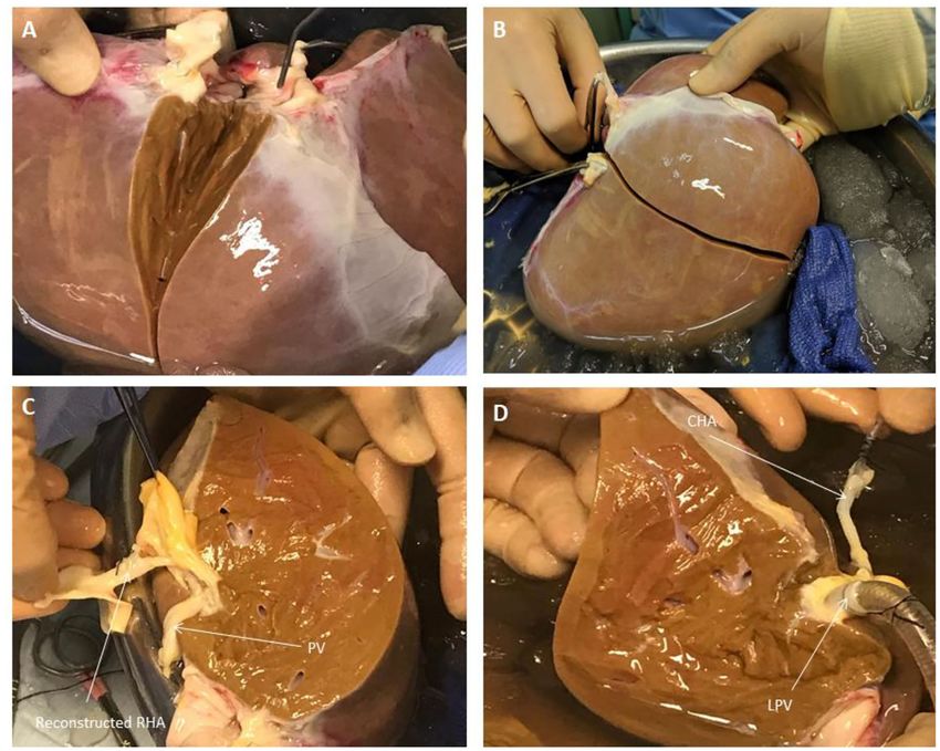
Attard et al.                                                                                                  Ex-situ Normothermic Split-Liver Machine Perfusion

INTRODUCTION                                                                          viability testing of donor livers prior to transplantation in a near-
                                                                                      physiological environment (5, 11). In the context of scientific
The main purpose of ex situ donor liver machine perfusion                             research, this makes NMLP a promising pre-clinical platform
has been the development of superior modality of organ                                for the investigation of therapeutics and mechanistic studies. We
preservation to conventional static cold storage as well as                           therefore sought to develop a normothermic machine perfusion
a method to assess organ viability prior to transplantation                           protocol for the application of the split liver model to pre-
(1). In the United Kingdom, between April 2018 and March                              clinical research. Our hypothesis was that individual lobes from
2019, 15% of donor livers retrieved were not transplanted,                            the same liver would recover and function similarly to one
representing a significant pool of potentially viable grafts (2)                      another when subjected to ex situ end-ischaemic NMLP. In
A recent randomized controlled clinical trial demonstrated that                       addition to providing more liver units for perfusion experiments,
normothermic machine liver perfusion (NMLP) reduced discard                           the protocol enables each liver to act as its own internal
rates of donor organs when compared to static cold storage,                           control, thus eliminating the inherent heterogeneity of whole
without jeopardizing transplant outcomes (3, 4). A number                             organ perfusions.
of perfusion devices are currently available. The principal
components include a blood reservoir, centrifugal pump, oxygen
concentrator, heat exchanger and a circuit which continuously
pumps perfusate through the liver via the organ’s inflow vessels                      MATERIALS AND METHODS
(hepatic artery and/or portal vein) and recirculates this following
drainage from the inferior vena cava (1). These systems allow                         Study Design
for extraction of perfusate for blood gas analysis, thus enabling                     This study was designed to investigate and compare the
real-time monitoring of oxygen and carbon dioxide levels,                             performance of right and left lobes from the same human
acid-base homeostasis as well as glucose levels (1) All these                         liver. Each lobe has their own inflow and outflow vessels as
parameters have been described as markers for organ viability                         well as bile drainage, during ex situ end-ischaemic NMLP on
during machine perfusion (5). Several pre-clinical and clinical                       separate perfusion devices, thereby demonstrating their suitable
non-randomized studies have investigated different perfusion                          utilization as comparative controls in the pre-clinical setting.
modalities with variations in perfusion temperature, perfusate                        The primary endpoints were assessment of liver function and
composition, perfusion duration, vessel cannulation as well as                        evaluation of perfusion parameters. The perfusion machines used
other technical considerations (1).                                                   for this study both were Liver Assist devices (Organ Assist,
    The technology provides a unique opportunity for assessing                        Groningen, The Netherlands).
liver metabolic and synthetic function. This has been recognized
by pre-clinical studies investigating the effect of novel therapeutic
interventions on machine perfused donor livers (6, 7). However,                       Donor Liver Source and Selection
donor livers available for research in machine perfusion have                         Four donor livers were included in this study, resulting in
been, so far, in short supply. Furthermore, the inherent variation                    a total of eight perfusions. All donor livers included in this
in donor characteristics and liver quality that exist between                         study were originally retrieved with the primary intention
different livers have limited their capacity as suitable comparative                  for transplantation as per the policy of the National Health
controls and, therefore, the interpretability of data from small                      Service Blood and Transplant (NHSBT). The organs were
series of whole organ perfusions (8).                                                 subsequently rejected for transplantation by all UK liver
    Splitting of the donor liver is a well-established strategy                       transplant centers and, following that, were offered nationally
used in transplantation to increase organ availability by                             for research by NHSBT. The authors had no influence in
allowing the same organ to be “shared” between two recipients                         the process of declining donor livers. This was done by the
(9). The surgical technique splits the donor organ into                               transplant surgeons at each center. Informed consent for research
two separate independently-functioning units (9). A pre-                              use of donor organs was obtained by specialist nurses in
clinical study by Huang et al. reported that, during ex situ                          organ donation from the donor’s next of kin during informed
subnormothermic machine perfusion, split livers demonstrated                          consent for organ donation. Authorisation for research was
comparable perfusion and functional characteristics, providing                        mediated by each centre’s specialist nurse in organ donation.
a controlled comparison between split lobes, thus allowing each                       All methods described were performed in accordance with
liver to act as its own internal control (10). However, this has yet                  NHSBT guidelines and regulations. Study ethical approval was
to be demonstrated in a normothermic machine perfusion model.                         obtained from the London-Surrey Borders National Research
NMLP has been shown to enable the functional assessment and                           Ethics Service (Reference Number 13/LO/1926) and the NHSBT
                                                                                      ethics committee (Reference Number 06/Q702/61). No organs
Abbreviations: BD, Bile duct; DBD, Donor after Brainstem Death; DCD, Donor            used in this study originated from executed prisoners.
after Cardiac Death; CHA, Common Hepatic Artery; LHV, Left Hepatic Vein; LPV,            Donor liver exclusion criteria for this study were: gross
Left Branch of Portal Vein; LL, Left Lobe; MHV, Middle Hepatic Vein; NHSBT,           macroscopic appearance indicative of moderate or severe
National Health Service Blood and Transplant; NMLP, Normothermic Machine
Liver Perfusion; PAS, Periodic Acid Schiff; PV, Portal Vein; RHA, Right Hepatic
                                                                                      steatosis; asymmetric or poor perfusion demonstrated during
Artery; RHV, Right Hepatic Vein; RL, Right Lobe; RPV, Right Branch of Portal          organ retrieval; presence of hepatic malignancy; organ subjected
Vein; T0, Start of Perfusion; T6, End of Perfusion after 6 h; VC, Vena Cava.          to machine perfusion prior to being discarded.

Frontiers in Surgery | www.frontiersin.org                                        2                                     February 2021 | Volume 8 | Article 627332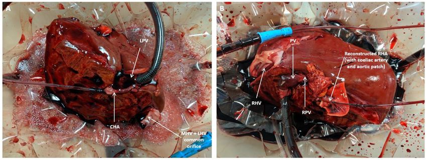
Attard et al.                                                                                       Ex-situ Normothermic Split-Liver Machine Perfusion

Liver Splitting Protocol                                                    transplantation) (Figure 2). Which technique was adopted was
During transportation to our center, the organs were preserved              dependent on the conduit length required as well as the caliber
in University of Wisconsin fluid at hypothermic temperatures in             of the vessels to be anastomosed together. The hepatic arteries
ice as per current standard clinical practice for static cold storage       were cannulated with 10−14 F cannulas. Each portal vein was
in the United Kingdom. Donor livers were split while in cold                cannulated with a 24 F cannula.
storage prior to commencement of perfusion in order to ensure
that both lobes were subjected to similar ischaemic times and
assessed simultaneously when placed in the perfusion devices.               Ex-situ Perfusion Protocol
Upon receipt by our center, the liver was initially cleared of excess       Both perfusion devices were primed with our previously
tissue in order to assess the quality of the organ. This also allowed       described (12) perfusion protocol using Hemopure [HBOC-201,
for assessment of the organ’s outflow, with respect to the hepatic          hemoglobin glutamer-250 (bovine); HBOC-201, Hemoglobin
veins and post-hepatic inferior vena cava, as well as the inflow, in        Oxygen Therapeutics LLC, Cambridge, MA] instead of packed
terms of the hepatic arterial tree and portal vein (Figure 1). Since        red blood cells as the oxygen carrier (Supplementary Material).
the perfusion device allows for open drainage of the perfusate              The former is a polymerised bovine hemoglobin-based acellular
from the organ’s outflow via the post-hepatic inferior vena cava,           oxygen carrier of low immunogenicity and an oxygen-carrying
the latter was incised in order to create two separate patches              capacity similar to that of human hemoglobin at normothermic
with direct visualization of left, middle and right hepatic veins           temperatures. Its efficacy as an alternative to blood-based
(Figure 1). This was the first step in determining the line of              machine perfusion fluid has been demonstrated in pre-clinical
demarcation for parenchymal division. For the purposes of this              and clinical studies (12, 13).
study, we sought to divide right and left halves of the liver along            The pre-perfusion weight of each lobe was recorded post-
Cantlie’s line—an extrapolated line, used when planning right or            splitting. The arterial and portal venous supplies were cannulated
left hepatectomies (liver resections), extending from the middle            with each lobe positioned inside the organ reservoir such that
of the post-hepatic inferior vena cava across the diaphragmatic             the open drainage from the hepatic veins could be visualized
(superior) surface of the liver to the point where the fundus of the        directly. Perfusion was commenced at 36–37◦ C with oxygenated
gallbladder on the inferior surface typically contacts the antero-          pulsatile flow and non-pulsatile flow in the hepatic artery
inferior margin of the liver (Figure 2). This approximates the              and PV, respectively. Perfusion of each lobe was commenced
plane of the middle hepatic vein and demarcates the right and               within five to 15 min of each other (Figure 3). Perfusion
left lobes of the liver.                                                    pressures and flow parameters were monitored continuously. An
    Once the vena cava was opened, the liver hilum was cleared              epoprostenol infusion pump was connected to each perfusion
further of excess tissue in order to identify the left and right            circuit and commenced at an initial rate of 4 ml/h. The
branches of the portal vein and hepatic artery, respectively                rate of prostaglandin infusion was adjusted according to the
(Figure 1). Particular attention was paid to identification of              flow readings to maintain physiological parameters. Hepatic
the arterial branch supplying segment 4 of the liver as this                artery pressures were maintained at 60–70 mm Hg while portal
would influence the level of division of the branches during                vein pressures were maintained at 10 mmHg. Oxygen supply
splitting. The left and right hepatic ducts were also identified            was adjusted in order to maintain a perfusate oxygen partial
to ensure that each half would have adequate biliary drainage               pressure >10 kPa in the arterial circuit. Serial perfusate samples
(Figure 1). A cholecystectomy was performed, and the cystic                 were analyzed in real-time using the Cobas b 221 point of
duct identified and ligated. Following satisfactory identification          care system (Roche Diagnostics, USA) Blood Gas Analyser in
of all relevant anatomy, the vessels and bile duct were divided             order to monitor metabolic parameters including oxygen partial
as follows. The hepatic arterial tree was divided such that the             pressures, lactate and glucose levels. These parameters have been
main trunk was retained by the left lobe of the liver with the right        previously described as appropriate methods of monitoring liver
lobe keeping the right hepatic branch, anatomy-permitting. The              function and viability during the perfusion process (5). Each lobe
right lobe retained the main trunks of the portal vein and bile             underwent NMLP for a total of 6 h. For histological analysis, core
duct with the left halve keeping its respective branches. At this           needle biopsies from each lobe were obtained at the beginning
point, parenchymal division was performed and completed along               and end of perfusion, fixed in formalin and embedded in paraffin.
Cantlie’s line (Figure 2).                                                  These biopsies were subsequently stained with haematoxylin
    If vessel length and diameter was deemed to be inadequate               and eosin for conventional examination and periodic acid schiff
for safe cannulation of either lobe, particularly with respect to
                                                                            (PAS) for glycogen content and distribution.
the isolated branches, then a decision was made to reconstruct
the vessels to enable proper positioning of the cannulas for
perfusion. This was done by fashioning a conduit using native
vessels for extra length in one of two ways: (1) resecting the              Statistical Analysis
distal portion of the main arterial or venous trunk retained by             Data analysis was carried out using Prism 7 (GraphPad Inc.,
either lobe, which was then anastomosed onto the branch of                  CA). Continuous data at each timepoint was compared using
the other; (2) utilizing the length of donor iliac vessels for the          Wilcoxon signed-rank test. Statistical significance was set at
reconstruction (received with the donor liver for the purposes              p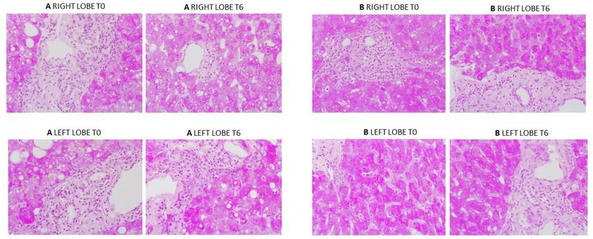
Attard et al. Ex-situ Normothermic Split-Liver Machine Perfusion
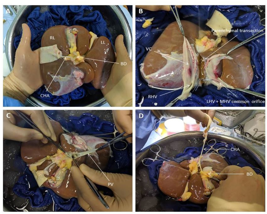
FIGURE 1 | Liver Splitting Procedure: (A) Inspection of liver hilum (B) Opening inferior vena cava to identify hepatic venous drainage (C) Identification of portal vein
branches (D) Identification of hepatic arterial branches and hepatic duct transection. BD, bile duct; CHA, common hepatic artery; LHV, left hepatic vein; LL, left lobe;
MHV, middle hepatic vein; PV, portal vein; RL, right lobe; RHV, right hepatic vein; VC, vena cava.
RESULTS perfusate lactate was observed in both lobes at the same timepoint
during the perfusion experiment. Perfusate glucose levels were
Donor Liver Characteristics initially high and trended downwards during the course of the
Four livers were included in the study. Information relating to experiment. Lactate and glucose levels were comparable at all
donor characteristics and liver characteristics can be found in timepoints (Figure 4).
(Table 1). Right lobe mass was consistently significantly higher
than their left lobe counterparts. Perfusion Hemodynamics
Hepatic artery and portal venous flows were relatively consistent
Assessment of Liver Function in both left and right lobes with a trend toward an increase
Lactate levels were comparable at the start of perfusion, T0, in flow rate as the perfusion experiment progressed (Figure 5).
and decreased significantly in all lobes up until the end of the Differences in flow rates for hepatic artery and portal vein were
perfusion experiment, T6 (Figure 4). Trends in perfusate lactate statistically insignificant across all time points.
clearance were similar in both lobes. When normalized for pre-
perfusion tissue weight, perfusate lactate levels tended to be Liver Histology
higher in the left lobe. However, the rate of reduction in lactate Overall histological analysis showed that both liver lobes
levels was observed to be similar in both lobes across all perfusion behaved in a similar manner throughout the perfusions. Lobar
experiments. Interestingly, in two of the four livers, an increase in architecture was well-preserved in both pre- and post-perfusion
Frontiers in Surgery | www.frontiersin.org 4 February 2021 | Volume 8 | Article 627332Attard et al. Ex-situ Normothermic Split-Liver Machine Perfusion
FIGURE 2 | Liver Splitting Procedure: (A) Parenchymal transection across gallbladder bed (B) Parenchymal transection complete (C) Reconstruction of hepatic artery
using donor celiac artery trunk (D) Cannulation of hepatic artery and portal vein branches. CHA, common hepatic artery; LPV, left portal vein; PV, portal vein; RHA,
right hepatic artery.
biopsies. Some variable centrilobular necrosis was observed at transplantation. However, results are compounded by small
both time points. However, this was present to the same degree sample size as a consequence of the limited availability of
before and during NMLP. All biopsies were PAS positive with discarded donor livers for research, further compounded by
staining showing a range of intensity from mild to strong and the inherent differences between donor livers. This limits
ranging from even distribution throughout the parenchyma to comparability between groups of individual livers.
patchy. Whatever the variability between liver, each of the paired Splitting of the donor liver is a well-known procedure in liver
lobes showed strong similarity, with no change throughout the transplantation to optimize the use of grafts (9). Human split liver
perfusion. In addition, patterns of histology did not correlate with machine perfusion data in the normothermic setting is limited
any parameters of functional assessment. Only one liver showed to case reports (14, 15). With this in mind, we sought to adapt
evidence of significant macrovesicular steatosis which did not the split liver technique to the normothermic machine perfusion
change between lobes or throughout the perfusion (Figure 6). model with the aim of providing a more robust perfusion
protocol with each liver providing its own internal control. Our
DISCUSSION results show that split livers recover functionality and perform
similarly to each other when subjected NMLP. The development
Several studies have demonstrated that NMLP enables the of a split liver model to ex situ end-ischaemic NMLP, therefore,
functional recovery of discarded donor livers and provides has the potential to provide a platform for suitable comparative
a window of opportunity for viability assessment prior to controls for the investigation and assessment of therapeutic
Frontiers in Surgery | www.frontiersin.org 5 February 2021 | Volume 8 | Article 627332Attard et al. Ex-situ Normothermic Split-Liver Machine Perfusion
FIGURE 3 | Split lobe perfusion: (A) Left lobe (B) Right lobe. CHA, common hepatic artery; LPV, left portal vein; MHV, middle hepatic vein; PV, portal vein; RHA, right
hepatic artery; RPV, right portal vein.
TABLE 1 | Donor liver demographics and characteristics. closed circuit was being used or if clinical use of these segmental
grafts was under consideration.
Liver 1 Liver 2 Liver 3 Liver 4
The incorporation of NMLP to the split liver model has
Donor age (years) 31 75 57 73 the benefit of enabling liver functional assessment under near-
Gender Male Male Female Female physiological conditions. The use of an artificial acellular
DBD/DCD DCD DBD DCD DBD hemoglobin-based perfusion fluid also has its advantages. Unlike
Cold ischaemia time (min) 902 799 1,108 1,026
blood products (derived from various donors), it has low
Weight (g)
immunogenicity and may therefore be of greater value in the
Right lobe 715 1,120 957 739
study of liver-specific immune cell populations and mechanistic
Left lobe 488 851 851 363
studies (12). Secondly, while the utility of blood products
as perfusion fluid is limited to physiological temperatures,
DBD, donor after brainstem death; DCD, donor after cardiac death. Hemopure can be deployed as a perfusion fluid under
hypothermic conditions (12). Subsequently, our proposed split
liver model can be applied to a range of perfusion temperatures.
interventions prior to being subjected to the rigor of a clinical It must be noted that there are limitations to the design
trial. Research published previously by our group allowed for
of this perfusion model. First, splitting of the liver requires
an initial four-hour window of NMLP before a full viability
considerable surgical expertise and increases the cold ischaemia
assessment of the perfused organ is carried out (11). This period
is important in the setting of split liver machine perfusion for time beyond what is usual for the preparation of a whole liver
two reasons. First and foremost, it will allow for the functional for machine perfusion. Secondly, due to the liver’s anatomy, the
recovery of both lobes following a variable period of cold right lobe is invariably larger than the left lobe and therefore they
ischaemia. Secondly, it will enable the investigator to assess and cannot be split into entities of equal mass without compromising
monitor differences in the parameters and performance of the blood supply. Furthermore, the shorter and smaller caliber of
two lobes prior to therapeutic intervention. hepatic artery and portal vein branches makes them much
The model described in the paper was developed solely more difficult to cannulate. We attempted to circumvent this
for experimental purposes without considerations for clinical issue by retaining the main trunk of the vessel with one lobe
adoption. The inflow portal venous anatomy was without while performing vascular reconstructions on the other, using
variations which allowed us to develop a standardized donor iliac vessels or larger caliber sections from the proximal
approached used in all livers. As the outflow from the liver celiac trunk or splenic artery (if included in the specimen).
was on free drainage to the reservoir, the site of caval division In all experiments this provided the additional length for safe
did not have any impact on function. In view of the open circuit cannulation. However, this technique further increases the cold
perfusion system, the liver outflow veins (including segmental ischaemia time of the liver and would also require the appropriate
veins draining into the middle hepatic vein) did not require any surgical expertise for a robust reconstruction that would not
reconstruction as the blood was freely drained and collected in compromise lobar perfusion. All these aforementioned factors
the device reservoir. As such, hepatic veins anatomy and inferior may lead to differences in the perfusion characteristics of each
vena cava accessories did not influence the surgical technique or lobe and must be considered. Finally, not all livers can be split.
the perfusion parameters. The situation would be different if a This is often a result of variations in arterial anatomy due to
Frontiers in Surgery | www.frontiersin.org 6 February 2021 | Volume 8 | Article 627332Attard et al. Ex-situ Normothermic Split-Liver Machine Perfusion FIGURE 4 | Perfusate lactate (A) and glucose (B) levels for each individual split lobe over 6 h of end-ischaemic normothermic machine perfusion. FIGURE 5 | Hepatic artery (A) and portal vein (B) flows for each individual split lobe over 6 h of end-ischaemic normothermic machine perfusion. Frontiers in Surgery | www.frontiersin.org 7 February 2021 | Volume 8 | Article 627332
Attard et al.                                                                                                       Ex-situ Normothermic Split-Liver Machine Perfusion

  FIGURE 6 | PAS staining from two representative perfusion experiments. (A) (from liver case number 2) shows mild to moderate macrovesicular steatosis, portal
  inflammatory cell infiltration and patchy PAS staining which was the same at commencement and end of perfusion. (B) (from liver case number 4) shows a liver with
  evenly distributed PAS staining throughout. Again, a degree of portal inflammation was seen. T0: pre-perfusion, T6: end of perfusion.

branching patterns which would compromise the inflow of a                             DATA AVAILABILITY STATEMENT
segment of the lobe if divided.
    We acknowledge that the data presented in this manuscript                         The raw data supporting the conclusions of this article will be
is from a small number of human donor livers. Nevertheless,                           made available by the authors, without undue reservation.
the results of this proof-of-concept study indicate that trends
in the functional recovery and metabolic parameters between                           ETHICS STATEMENT
lobes were very similar and may therefore prove useful in the
assessment of responses to therapeutic interventions in the pre-                      The studies involving human participants were reviewed and
clinical normothermic setting. Additionally, this model addresses                     approved by the London-Surrey Borders National Research
the variability that exists between individual donor livers by                        Ethics Service (Reference Number 13/LO/1926) and the NHSBT
enabling the same donor liver to be placed in the treatment                           Ethics Committee (Reference Number 06/Q702/61). Informed
and control groups. This also maximizes use of resources as one                       consent for research use of donor organs was obtained by
donor liver for pre-clinical studies.                                                 specialist nurses in organ donation from the donor’s next of kin
    Other benefits observed during these experiments came                             during informed consent for organ donation.
from the use of a perfusion machine with an open circuit.
These included: facilitated manipulation of the graft for direct
visualization, direct visualization of and access to graft outflow
                                                                                      AUTHOR CONTRIBUTIONS
and superior access to graft for direct therapeutic interventions                     JA, YB, LW, and SA designed the study and established
when compared to closed circuit alternatives.                                         the methodology. JA, LW, DO-B, and VR conducted the
                                                                                      experiments. JA and LW collected the data. DM and HM
                                                                                      supervised the liver splitting procedure. JA performed the data
                                                                                      analysis and drafted the manuscript. SA and GR reviewed the
CONCLUSION
                                                                                      histological specimens. YO, MP, HM, and DM contributed to
Liver splitting is a feasible model for providing comparative                         critical revision and editing of the final manuscript. All authors
controls for pre-clinical normothermic machine perfusion                              approved the final version of this manuscript.
research. Further work should involve the optimisation of this
protocol to minimize the need for surgical expertise when                             FUNDING
preparing the liver for machine perfusion as well as its application
to other perfusion modalities, namely, hypothermic and sub-                           This manuscript represents independent academic research
normothermic perfusion. This novel split liver model can be                           funded by the Wellcome Trust (200121/Z/15/Z), Vital Therapies,
tested for cellular therapies to investigated cellular phenotype                      Inc. and Medical Technologies Associates II, Inc. YO receives
and lineage changes and future pharmacological interventions of                       funding from Sir Jules Thorn Biomedical Research Award,
donor liver before implantation.                                                      Trans-Bioline grant from Innovative medicine initiative,

Frontiers in Surgery | www.frontiersin.org                                        8                                          February 2021 | Volume 8 | Article 627332
Attard et al. Ex-situ Normothermic Split-Liver Machine Perfusion
Medical Research Council and QEHB charity. This paper of Health and Social Care. We are also grateful for the
presents independent research supported by the NIHR support of the staff at the Liver Unit in the Queen Elizabeth
Birmingham Biomedical Research Center at the University Hospital in Birmingham as well as the research staff at the
Hospitals Birmingham NHS Foundation Trust, the University Center for Liver and Gastrointestinal Research. YO would
of Birmingham and the Wellcome Trust. The authors declare like to acknowledge the support from Sir Jules Thorn
that this study received funding from Vital Therapies, Inc. and Charitable Trust, Innovative Medicine Initiative TransBioLine
Medical Technologies Associates II, Inc. SA would like to further and the Medical Research Council (MRC). SA would like to
acknowledge the financial support from Ochre Bio, Oxford, UK. further acknowledge the financial support from Ochre Bio,
The funders were not involved in the study design, collection, Oxford, UK.
analysis, interpretation of data, the writing of this article or the
decision to submit it for publication.
SUPPLEMENTARY MATERIAL
ACKNOWLEDGMENTS
The Supplementary Material for this article can be found
The views expressed are those of the authors and not online at: https://www.frontiersin.org/articles/10.3389/fsurg.
necessarily those of the NHS, the NIHR or the Department 2021.627332/full#supplementary-material
REFERENCES 10. Huang V, Karimian N, Detelich D, Raigani S, Geerts S, Beijert I, et al.
Split-liver ex situ machine perfusion: a novel technique for studying organ
1. Laing RW, Mergental H, Mirza DF. Normothermic ex-situ liver preservation: preservation and therapeutic interventions. J Clin Med. (2020) 9:269.
the new gold standard. Curr Opin Organ Transplant. (2017) 22:274–80. doi: 10.3390/jcm9010269
doi: 10.1097/MOT.0000000000000414 11. Mergental H, Stephenson BTF, Laing RW, Kirkham AJ, Neil DAH, Wallace
2. Statistics and Clinical Studies NHS Blood and Transplant. Organ Donation LL, et al. Development of clinical criteria for functional assessment to
and Transplantation Activity Report 2018-2019. Available online at: https:// predict primary nonfunction of high-risk livers using normothermic machine
nhsbtdbe.blob.core.windows.net/umbraco-assets-corp/16537/organ- perfusion. Liver Transplant. (2018) 24:1453–69. doi: 10.1002/lt.25291
donation-and-transplantation-activity-report-2018-2019.pdf (accessed 12. Laing RW, Bhogal RH, Wallace L, Boteon Y, Neil DAH, Smith A,
November 2, 2020). et al. The use of an acellular oxygen carrier in a human liver model
3. Nasralla D, Coussios CC, Mergental H, Akhtar MZ, Butler AJ, Ceresa of normothermic machine perfusion. Transplantation. (2017) 101:2746–56.
CDL, et al. A randomized trial of normothermic preservation in liver doi: 10.1097/TP.0000000000001821
transplantation. Nature. (2018) 557:50–6. doi: 10.1038/s41586-018-0047-9 13. Matton APM, Burlage LC, van Rijn R, de Vries Y, Karangwa SA, Nijsten MW,
4. Laing RW, Mergental H, Yap C, Kirkham A, Whilku M, Barton D, et al. et al. Normothermic machine perfusion of donor livers without the need for
Viability testing and transplantation of marginal livers (VITTAL) using human blood products. Liver Transpl. (2018) 24:528–38. doi: 10.1002/lt.25005
normothermic machine perfusion: study protocol for an open-label, non- 14. Stephenson BTF, Bonney GK, Laing RW, Bhogal RH, Marcon F, Neil
randomised, prospective, single-arm trial. BMJ Open. (2017) 7:e017733. DAH, et al. Proof of concept: liver splitting during normothermic machine
doi: 10.1136/bmjopen-2017-017733 perfusion. J Surg Case Rep. (2018) 2018:rjx218. doi: 10.1093/jscr/rjx218
5. Watson CJE, Jochmans I. From “Gut Feeling” to objectivity: machine 15. Brockmann JG, Vogel T, Coussios C, Friend PJ. Liver splitting during
preservation of the liver as a tool to assess organ viability. Curr Transplant normothermic organ preservation. Liver Transpl. (2017) 23:701–6.
Rep. (2018) 5:72–81. doi: 10.1007/s40472-018-0178-9 doi: 10.1002/lt.24693
6. Boteon YL, Attard J, Boteon A, Wallace L, Reynolds G, Hubscher
S, et al. Manipulation of lipid metabolism during normothermic Conflict of Interest: JA is a clinical research fellow at the Queen Elizabeth Hospital
machine perfusion: effect of defatting therapies on donor liver in Birmingham and employed by University Hospitals Birmingham.
functional recovery. Liver Transpl. (2019) 25:1007–22. doi: 10.1002/lt.
25439 The remaining authors declare that the research was conducted in the absence of
7. Green CJ, Parry SA, Gunn PJ, Ceresa CGL, Rosqvist F, Piché ME, et al. any commercial or financial relationships that could be construed as a potential
Studying non-alcoholic fatty liver disease: the ins and outs of in vivo, ex vivo conflict of interest.
and in vitro human models. Hormone Mol Biol Clin Invest. (2018) 41:1–22.
doi: 10.1515/hmbci-2018-0038 Copyright © 2021 Attard, Osei-Bordom, Boteon, Wallace, Ronca, Reynolds, Perera,
8. Boteon YL, Afford SC, Mergental H. Pushing the limits: machine preservation Oo, Mergental, Mirza and Afford. This is an open-access article distributed under the
of the liver as a tool to recondition high-risk grafts. Curr Transplant Rep. terms of the Creative Commons Attribution License (CC BY). The use, distribution
(2018) 5:113–20. doi: 10.1007/s40472-018-0188-7 or reproduction in other forums is permitted, provided the original author(s) and
9. Lauterio A, Di Sandro S, Concone G,De Carlis R, Giacomoni A, De Carlis the copyright owner(s) are credited and that the original publication in this journal
L, et al. Current status and perspectives in split liver transplantation. World J is cited, in accordance with accepted academic practice. No use, distribution or
Gastroenterol. (2015) 21:11003–15. doi: 10.3748/wjg.v21.i39.11003 reproduction is permitted which does not comply with these terms.
Frontiers in Surgery | www.frontiersin.org 9 February 2021 | Volume 8 | Article 627332You can also read