Target protein-oriented isolation of Hes1 dimer inhibitors using protein based methods - Nature
←
→
Page content transcription
If your browser does not render page correctly, please read the page content below
www.nature.com/scientificreports

            OPEN           Target protein-oriented isolation of
                           Hes1 dimer inhibitors using protein
                           based methods
                           Midori A. Arai 1*, Kaori Morita1, Haruka Kawano1, Yuna Makita1, Manami Hashimoto1,
                           Akiko Suganami2, Yutaka Tamura2, Samir K. Sadhu3, Firoj Ahmed4 & Masami Ishibashi1*
                           Natural products isolation using protein based methods is an attractive for obtaining bioactive
                           compounds. To discover neural stem cell (NSC) differentiation activators, we isolated eight inhibitors
                           of Hes1 dimer formation from Psidium guajava using the Hes1-Hes1 interaction fluorescent plate assay
                           and one inhibitor from Terminalia chebula using the Hes1-immobilized beads method. Of the isolated
                           compounds, gallic acid (8) and 4-O-(4”-O-galloyl-α-L-rhamnopyranosyl)ellagic acid (11) showed potent
                           Hes1 dimer formation inhibitory activity, with IC50 values of 10.3 and 2.53 μM, respectively. Compound
                           11 accelerated the differentiation activity of C17.2 NSC cells dose dependently, increasing the number
                           of neurons with a 125% increase (5 μM) compared to the control.

                           Neurodegenerative diseases such as dementia and spinal cord injury severely compromise the quality of life. The
                           discovery of neural stem cells (NSCs) in human adult brain1,2 led to the expectation of novel treatments involving
                           NSC transplantation or accelerated differentiation of internal NSCs in the brain to overcome neurodegenerative
                           diseases. However, the development of these approaches and small molecule NSC activators has been slow, possi-
                           bly due to the complex mechanism underlying the differentiation of NSCs to neural cells.
                               Basic helix-loop-helix (bHLH) factors play crucial roles in the differentiation of neural cells such as neu-
                           rons, astrocytes and oligodendrocytes3. For example, proneural bHLH factors, such as Ascl1 (formerly Mash1),
                           Neurog1 and Neurog2 regulate the generation of neurons. Olig1 and Olig2 are bHLH factors important for the
                           maturation of oligodendrocytes. Hairy and enhancer of split (hes) bHLH factors promote the self-renewal of
                           NSCs and the generation of astrocytes, as well as inhibiting the differentiation of NSCs by suppressing the expres-
                           sion of proneural genes. Recently, Kageyama et al. reported that oscillation in the concentration of bHLH factors
                           directly controls the fate of NSCs4,5. Hes1 and Ascl1 proteins oscillate with 2- to 3-hour periods, whereas Olig2
                           oscillates with a 5- to 8-hour period. During cell fate choice, one bHLH factor is expressed in a sustained manner
                           while the others are repressed. In neurons, Ascl1 is expressed sustainably while Hes1 and Olig2 expression is
                           inhibited.
                               Hes1 dimerizes and binds to the promoter region of Ascl1 and other proneural genes (Fig. 1), making the
                           inhibition of Hes1 an attractive approach to enhance neuronal differentiation. If the interested target protein
                           was decided, it would be faster and effective to isolate the bioactive natural products using a “protein-based assay
                           system”6. However, the number of reports of isolation using protein-based assay system was still limited (affinity
                           chromatography7–9, protein-immobilized beads10–18, ultrafiltration19–21, and protein-protein interactions22,23). In
                           our attempts to find Hes1 inhibitors effectively, we constructed a new Hes1-Hes1 interaction fluorescent plate
                           assay which gave the first Hes1 dimer inhibitors24. Also, we have developed “target protein oriented natural prod-
                           ucts isolation (TPO-NAPI) methods” using protein-immobilized beads11,15,17,18. Lindbladione15, agalloside15,
                           isomicromonolactam17 and inohanamine17 were isolated by using Hes1 protein beads and accelerated the differ-
                           entiation of NSCs to neurons. Here, we report the isolation of eight Hes1 dimer inhibitors from Psidium guajava
                           using the Hes1-Hes1 interaction fluorescent plate assay, which is the first example of natural products isolation
                           using fluorescent plate assay of protein-protein interactions (PPIs). Moreover, the isolation using the TPO-NAPI
                           method with Hes1 beads gave a Hes1 dimer inhibitor from Terminalia chebula.

                           1
                            Graduate School of Pharmaceutical Sciences, Chiba University, 1-8-1 Inohana, Chuo-ku, Chiba, 260-8675, Japan.
                           2
                            Graduate School of Medicine, Chiba University, 1-8-1 Inohana, Chuo-ku, Chiba, 260-8670, Japan. 3Pharmacy
                           Discipline, Khulna University, Khulna, 9208, Bangladesh. 4Department of Pharmaceutical Chemistry, University of
                           Dhaka, Dhaka, 1000, Bangladesh. *email: midori_arai@chiba-u.jp; mish@chiba-u.jp

Scientific Reports |   (2020) 10:1381 | https://doi.org/10.1038/s41598-020-58451-3                                                           1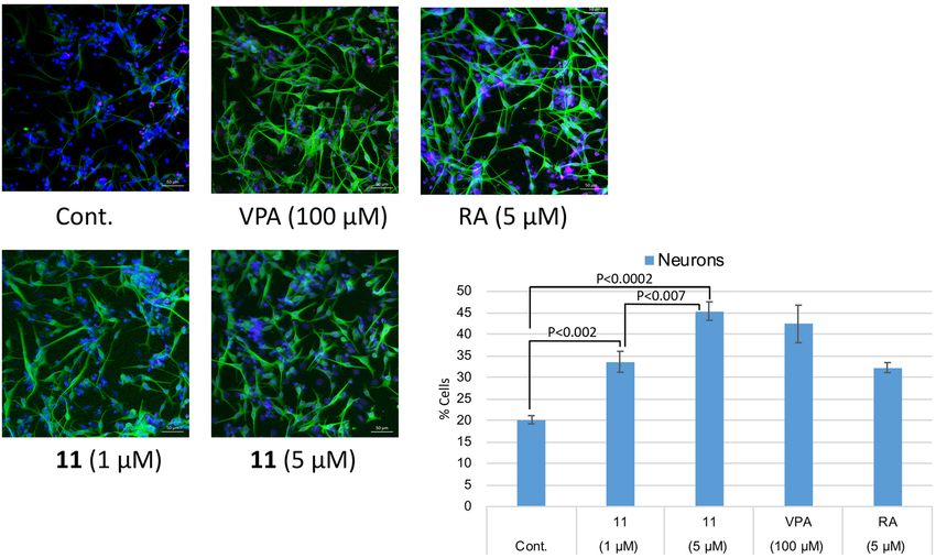
www.nature.com/scientificreports/                                                            www.nature.com/scientificreports

                           Figure 1. Neural stem cell differentiation and fate control by bHLH factors.

                           Figure 2. Target protein-oriented isolation methods. (A) Hes1-Hes1 interaction fluorescent plate assay,
                           (B) Hes1 immobilized beads method.

                           Results and Discussion
                           It is essential to inhibit undesired PPIs in drug development, and bioactivity-guided isolation using the inhib-
                           itory activity of PPIs is an attractive method to obtain effective inhibitors. However, currently there are only a
                           few examples of such approaches using PPI assay systems22,23. We previously developed a Hes1-Hes1 interaction
                           fluorescent plate assay and reported the screening results of our natural products compound library (Fig. 2A)24.
                           Glutathione-S-transferase (GST) fused rat Hes1(3–281) protein was expressed in Escherichia coli and purified
                           rat Hes1 protein was immobilized on the bottom of 96 well plates. We prevented GST-GST interactions, which
                           would result in false positives, by preparing GST-free Hes1 protein by GST cleavage with Turbo3C protease. After

Scientific Reports |   (2020) 10:1381 | https://doi.org/10.1038/s41598-020-58451-3                                                         2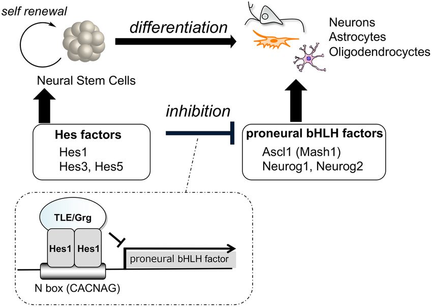
www.nature.com/scientificreports/                                                                  www.nature.com/scientificreports

                           Figure 3. Structures of the isolated compounds.

                           labeling Hes1 with Cy3, this florescent Hes1 protein was added to the wells of the above plate and incubated
                           for 24 h at 4 °C. Hes1-Hes1 interaction was successfully detected as Cy3 fluorescence intensity. Using this assay
                           system, we screened our 118 plant extract library and identified the MeOH extract of Psidium guajava leaves to
                           contain naturally occurring compounds that inhibit Hes1 dimer formation. The MeOH extract (29.9 g) was frac-
                           tionated using Diaion HP-20 with a MeOH-acetone solvent system to afford fractions 1A to 1C. Active fraction
                           1A (27.2 g) was suspended in 10% aq. MeOH and partitioned with hexane, EtOAc and BuOH to obtain hexane
                           (1.1 g), EtOAc (5.7 g), BuOH (4.7 g) and aqueous (18.9 g) soluble fractions. Part of the active BuOH soluble frac-
                           tion was subjected to ODS column chromatography and reverse-phase HPLC. Activity-guided separation yielded
                           ten compounds (1–10; Fig. 3). The isolated compounds were identified as morin (1)25, isoquercitrin (2)26, methyl
                           gallate (3)27, (+)-catechin (4)28,29, dihydrophaseic acid (5)30, quercetin (6)26,31, avicularin (7)32,33, gallic acid (8)34,
                           protocatechuic acid (9)35 and 4-hydroxybenzoic acid (10)36 based on comparisons of their spectral data with spec-
                           tra in the literature. The Hes1-Hes1 interaction inhibitory activities of the isolated compounds were examined
                           (Fig. 4) and 3, 7, 8 and 9 produced moderate inhibition (IC50 12.7, 26.5, 10.3 and 23.8 μM). The most potent inhib-
                           itor was gallic acid (8). Commercially available gallic acid also exhibited comparable inhibition (IC50 8.9 μM).
                           Inhibition by the gallic acid derivatives 3, 8, 9 and 10 showed that the number of phenolic hydroxyl groups affects
                           inhibitory activity, with activity decreasing as the number of phenolic hydroxyl groups decrease.
                               We recently developed another protein-based screening method, the target protein oriented natural products
                           isolation method (TPO-NAPI) using protein beads (Fig. 2B). Agalloside, inohanamine, α-mangostine, BE-14106,
                           isomicromonolactam, staurosporin and linarin were isolated as Hes1 binding compounds using the TPO-NAPI
                           method15,17. Rat Hes1 (1–95) containing basic and helix-loop-helix domains was immobilized because the
                           helix-loop-helix domain is known to be important for Hes1-Hes1 interaction; therefore, utilizing this domain in
                           the beads method would likely be effective for screening Hes1 dimer inhibitors. GST-Hes1 immobilized beads
                           were prepared by mixing freshly prepared GST-Hes1 protein with glutathione Sepharose 4B beads. GST-only
                           beads were prepared as a control. After incubating the beads with plant MeOH extracts at 4 °C for 2 h, bound
                           compounds were eluted by adding EtOH and heating at 100 °C for 3 min, then the eluted compounds were ana-
                           lyzed by HPLC. Of the 105 plant MeOH extracts screened using this method, the Bangladesh plant Terminalia
                           chebula was found to contain a Hes1 binding compound. The MeOH extract (64.6 g) of Terminalia chebula bark
                           was partitioned with hexane, EtOAc and BuOH to obtain hexane (1.5 g), EtOAc (3.6 g), BuOH (42.6 g), and aque-
                           ous (20.5 g) soluble fractions. The EtOAc fraction contained the target peak and was subjected to silica gel col-
                           umn chromatography to give eight fractions (1A-H). Fraction 1D contained the target peak and was separated

Scientific Reports |   (2020) 10:1381 | https://doi.org/10.1038/s41598-020-58451-3                                                                   3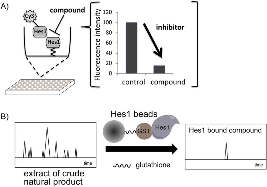
www.nature.com/scientificreports/                                                             www.nature.com/scientificreports

                           Figure 4. Hes1 dimer formation inhibitory activities of the isolated compounds.

                           by ODS column chromatography and reverse-phase HPLC to give compound 11 (0.4 mg). Compound 11 was
                           identified as 4-O-(4″-O-galloyl-α-L-rhamnopyranosyl)ellagic acid by comparison of its NMR data with reported
                           spectral data37. Next, we evaluated the Hes1 dimer inhibitory activity of compound 11 (Fig. 4) and found that
                           it exhibited the most potent inhibition of Hes1 dimer formation of the compounds tested, with an IC50 value of
                           2.53 μM. This is the strongest Hes1 dimer inhibitor reported to date. The ability of 11 to accelerate the differen-
                           tiation of C17.2 mouse neural stem cells was evaluated (Fig. 5). C17.2 cells (2 × 10⁵ cells/mL) were seeded in a
                           poly-L-lysine-coated 24 well plates and incubated for 24 h, then the cells were treated with DMSO (control), 100
                           μM valproic acid (VPA), 5 μM retinoic acid (RA) (positive controls) or 11 (1 or 5 μM) for 6 days. A confocal
                           microscope was used to obtain images of differentiated neural cells after immunostaining class III β-tubulin
                           (Tuj1) in neurons, and glial fibrillary acidic protein (GFAP) in astrocytes and nuclei (TO-PRO-3). The number
                           of neurons increased following the addition of VPA, RA or compound 11, suggesting that these compounds
                           enhanced the number of C17.2 neurons. In contrast, the control dish containing cells treated with DMSO con-
                           tained small number of neurons. The differentiated neurons with 11 were 33.6% (1 µM) and 45.4% (5 µM), which

Scientific Reports |   (2020) 10:1381 | https://doi.org/10.1038/s41598-020-58451-3                                                          4
www.nature.com/scientificreports/ www.nature.com/scientificreports
Figure 5. Effect of 11 on NSC differentiation into neurons. Green: neurons, blue: nuclei, red: astrocytes. The
number of neurons increased following the addition of compound 11 dose dependently. P values were analyzed
by Student’s t test. P < 0.05 is considered statistically significant.
are 67% and 125% increase compared to those of control. These results suggested that compound 11 accelerates
the differentiation of NSCs to neurons by inhibiting Hes1 dimer formation.
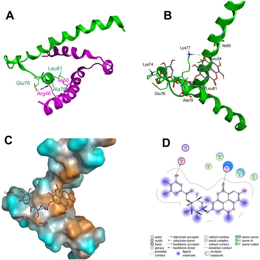
We also predicted the interaction between the HLH domain of Hes1 protein and compound 11 (4-O-(4″-
O-galloyl-α-L-rhamnopyranosyl)ellagic acid) by performing in silico docking analysis of compound 11 with the
HLH domain of Hes1. As shown in Fig. 6A,B, the galloyl site of compound 11 might interact with the loop region
of Hes1, aiding the formation of Hes1(Arg46 of helix region)-Hes1(Glu76 of loop region) and preventing mutual
recognition by Hes1 molecules. On the other hand, the ellagic acid site of compound 11 might bind with the helix
region of Hes1, which consists of Ile50, Leu54 and Leu81, preventing hydrophobic core formation in the Hes1
dimer. Orange shows the hydrophobic region in Hes1 (Fig. 6C). Moreover, hydrogen bond formation between the
ellagic acid site of compound 11 with Lys77 might obstruct Hes1(loop region)-Hes1(loop region) formation. Blue
shows the hydrophilic region. As shown in Fig. 6C, the interaction of galloyl moiety with the hydrophilic region
seems to be important. Therefore, the decrease of inhibition with decrease of number of phenolic hydroxyl groups
in galloyl group would be reasonable. In addition, the α-L-rhamnopyranosyl unit appears to be an efficient linker,
enabling tight interaction between compound 11 with the Hes1 monomer via its galloyl and ellagic acid sites.
Conclusion
In conclusion, several Hes1 binding natural products were isolated using two target protein-oriented isolation
methods: a Hes1-Hes1 interaction fluorescent plate assay and a Hes1-immobilized beads method. Ten com-
pounds were isolated from Psidium guajava, of which eight showed Hes1 dimer inhibitory activity. 4-O-(4″
-O-Galloyl-α-L-rhamnopyranosyl)ellagic acid (11) isolated using the Hes1 beads method showed potent Hes1
dimer formation inhibitory activity, with an IC50 of 2.53 μM. Compound 11 accelerated the differentiation of
C17.2 NSC cells into neurons dose dependently, increasing the number of neurons with a 125% increase (5 μM)
compared to the control. There are few reports of Hes1 dimer inhibitors and thus these results will be useful for
evaluating Hes1-related mechanisms in cells. We believe that protein-based isolation is an effective approach for
identifying bioactive compounds.
Methods
General experimental procedures. NMR spectra were recorded on JEOL ECZ600 spectrometers in a
deuterated solvent whose chemical shift was used as an internal standard. Column chromatography was per-
formed using Silica Gel 60N (Kanto Chemical Co., Tokyo, Japan), Chromatorex ODS (Fuji Silysia Chemical Ltd.,
Kasugai, Japan), Diaion HP-20 (Mitsubishi Chemical Co., Tokyo, Japan) and Sephadex LH-20 (GE Healthcare
Japan, Tokyo, Japan). Preparative HPLC was performed using YMC-Pack SIL-06 (YMC Co. Ltd., Kyoto, Japan),
COSMOSIL 5C18-AR-II, COSMOSIL Cholester, COSMOSIL πNAP (Nacalai Tesque, Inc. Kyoto, Japan) and
CAPCELL PAK C18 MGII (OSAKA SODA Ltd., Osaka, Japan).
Plant materials. Psidium guajava (Myrtaceae) and Terminalia chebula (Combretaceae) were collected in
Bangladesh in 2013 and were identified by one of the authors (S. K. Sadhu). Voucher specimens (KKB348 and
KKB364) were deposited at the Department of Natural Products Chemistry, Graduate School of Pharmaceutical
Sciences, Chiba University.
Scientific Reports | (2020) 10:1381 | https://doi.org/10.1038/s41598-020-58451-3 5www.nature.com/scientificreports/ www.nature.com/scientificreports
Figure 6. Docking study of compound 11 to the Hes1 HLH domain. (A) NMR structure of the HLH domains
of Hes1 dimers (PDB code: 2MH3). Green and pink show individual Hes1 HLH domains. (B–D) Docking study
results of 11 with the HLH domain.
Plate assay for Hes1 dimer inhibitors. Hes1-bound microplate wells24 (Nunc ImmobilizerTM Amino
Plate, Thermo) were incubated with 50 μL of Cy3-labeled-Hes1 in NET-N buffer (NET buffer (20 mm Tris-HCl,
®
pH 7.5, 200 mm NaCl, 1 mm EDTA) containing 0.05% Nonidet P-40, ca. 7 mg/L, dye/protein = 0.4) for 24 h at
4 °C. After removing the protein solution, the wells were washed twice with 200 μL of PBST, then each compound
solution (in NET-N buffer, 50 μL) was added and the plates were incubated for 1 h at RT in the dark. The wells
were washed twice with 200 μL of PBST, then dried under reduced pressure for 1 h in the dark. The Cy3 dye was
excited at 544 nm and emission was monitored at 590 nm using a microplate reader (Fluoroskan Ascent, Thermo
Labsystems, Vantaa, Finland). The assays were typically carried out in three individual wells, and the mean value
and SD were calculated. The equilibrium between dimer formation between Cy3-Hes1/Cy3-Hes1 (in solution)
and Cy3-Hes1/immobilized-Hes1 allowed the detection of Cy3-Hes1/immobilized-Hes1.
Typical screening procedure for the Hes1 beads method. To prepare GST-Hes1 beads,
GST-ratHes11-95 (ca. 3.6 nmol) in PBS was added to pre-washed glutathione Sepharose 4B beads (bed volume
100 μL, GE Healthcare) and mixed at 4 °C for 1 h. The GST-Hes1 beads were washed five times with NET buffer,
then suspended in 250 μL NET buffer. A MeOH or EtOAc plant extract (125 μg in EtOH, 25 μL) was added to
the above GST-Hes1 freshly prepared beads (bed volume 100 μL) and gently mixed for 2 h at 4 °C. The beads
were then washed by tapping the container with fingers three times with NET-N buffer. EtOH (70%, 150 μL) was
added to the washed beads and the suspension was heated at 100 °C for 3 min in a heating block. The beads were
collected by centrifugation (2000 rpm, 4 °C, 1 min) and the supernatant was centrifuged at 15000 rpm for 15 min.
One-third of the supernatant was analyzed by HPLC. Control GST-beads were prepared in the same manner
as GST-Hes1 beads by adding GST (ca. 3.8 nmol) in PBS to pre-washed glutathione Sepharose 4B beads (bed
volume 100 μL, GE Healthcare). Lindobladione was used as a positive control in each experiment. Obvious differ-
ences in peak intensity for the results obtained using GST-Hes1-beads and GST-beads (control) were considered
“hit” extracts containing a Hes1 binding natural product.
Scientific Reports | (2020) 10:1381 | https://doi.org/10.1038/s41598-020-58451-3 6www.nature.com/scientificreports/ www.nature.com/scientificreports
Extraction and isolation. The MeOH extract (29.9 g) of Psidium guajava (leaves; 139 g) was subjected to
Diaion HP-20 (ϕ 70 × 210 mm) chromatography with a MeOH-acetone solvent system (1:0–0:1) to afford frac-
tions 1A to 1 C. Active fraction 1A (27.2 g) was suspended in 10% aq. MeOH (150 mL) and partitioned with hex-
ane, AcOEt and BuOH (400 mL × 3) to obtain hexane (1.1 g), AcOEt (5.7 g), BuOH (4.7 g) and aqueous (18.9 g)
soluble fractions. An aliquot of the active BuOH soluble fraction was subjected to ODS column chromatography
(ϕ 40 × 210 mm; MeOH-H2O = 3:7–1:0) to afford fractions 3A to 3E. Fraction 3B (134.6 mg) was subjected to
ODS HPLC (COSMOSIL 5C18-AR-II; 40% MeOH + 0.1% HCOOH, flow rate 3.0 mL/min) to give compound
1 (4.6 mg). An aliquot of fraction 3D (124.5 mg) was subjected to ODS HPLC (COSMOSIL 5C18-AR-II; 40%
MeOH + 0.1% HCOOH, flow rate 5.0 mL/min) to give compound 2 (7.3 mg). An aliquot of the active AcOEt
soluble fraction was subjected to ODS column chromatography (ϕ 40 × 210 mm; MeOH-H2O = 3:7–1:0) to
afford fractions 2A to 2I. Fraction 2B (236.5 mg) was subjected to ODS column chromatography (ϕ 20 × 460 mm;
MeOH-H2O = 4:6–1:0) to give compound 3 (10.9 mg) and to afford fractions 4A to 4E. Fraction 4 C (32.9 mg)
was subjected to silica gel column chromatography (ϕ 20 × 280 mm; CHCl3-MeOH = 30:1-0:1) to give compound
4 (10.3 mg). Fraction 4A (10.0 mg) was subjected to HPLC (YMC-Pack SIL-06, CHCl3-MeOH = 6:1, flow rate
3.0 mL/min) to give compound 5 (0.3 mg). Fraction 2 H (83.3 mg) was subjected to silica gel column chroma-
tography (ϕ 15 × 310 mm; AcOEt-MeOH = 150:1–0:1) to give compound 6 (21.6 mg). An aliquot of fraction 2 F
(68.7 mg) was subjected to HPLC (YMC-Pack SIL-06, AcOEt-MeOH = 7:1, flow rate 1.0 mL/min) to afford frac-
tions 8 A to 8F. Fraction 8B (12.4 mg) was subjected to Sephadex LH-20 column chromatography (ϕ 15 × 400 mm;
MeOH) to give compound 7 (0.8 mg). An aliquot of fraction 2A (192.6 mg) was subjected to Sephadex LH-20
column chromatography (ϕ 30 × 420 mm; MeOH) to afford fractions 15A to 15E. Fraction 15B (15.7 mg) was
subjected to ODS HPLC (COSMOSIL 5C18-AR-II; 40% MeOH + 0.1% HCOOH, flow rate 3.0 mL/min) to give
compound 8 (7.0 mg), compound 9 (2.0 mg) and compound 10 (0.8 mg).
Terminalia chebula (bark; 395 g) MeOH extract was partitioned with hexane, EtOAc and BuOH to obtain hex-
ane (1.5 g), EtOAc (3.6 g), BuOH (42.6 g) and aqueous (20.5 g) soluble fractions. The EtOAc fraction containing
the target peak was subjected to silica gel column chromatography (ϕ 50 × 250 mm; CHCl3-MeOH = 1:0–0:1)
to give fractions 1A-H. Fraction 1D contained the target peak and was separated by ODS column chromatogra-
phy (ϕ 25 × 200 mm; MeOH-H2O = 7:3–0:1) to give fractions 2A–F. Fraction 2A was subjected to ODS column
chromatography (ϕ 20 × 200 mm; MeOH-H2O = 7:3-0:1) to give fractions 6A–G. Fraction 6E was separated by
reverse-phase HPLC (CAPCELL PAK C18 MGII; 50% MeOH) to give compound 11 (0.4 mg). Compound 11 was
identified as 4-O-(4″-O-galloyl-α-L-rhamnopyranosyl)ellagic acid by comparison of its NMR data with reported
spectral data.
Cell culture. C17.2 cells were cultured in proliferation medium [DMEM (DS Pharma Biomedical Co., Ltd.
Osaka, Japan) supplemented with 10% fetal bovine serum (Biowest, Nuaillé, France), 5% horse serum (Gibco, Life
Technologies, Tokyo, Japan)]. Neural differentiation of C17.2 cells was initiated by transferring the cells to dif-
ferentiation medium [Neurobasal Medium (Gibco) supplemented with 2% B-27 without vitamin A (Gibco), 1%
antibiotic-antimycotic (Gibco)]. All cultures were maintained in a humidified incubator at 37 °C in 5% CO2/95%
air.
Neural stem cell differentiation assay. C17.2 cells were dissociated using the proliferation medium
2 × 10⁵ cells/mL on cover glass (MATSUNAMI GLASS) coated with poly-L-lysine and fibronectin/laminin. After
incubation for 24 h, cells were washed with D-MEM and treated with individual compounds in differentiation
medium for 6 days.
Immunofluorescence staining. C17.2 cells were fixed with 4% paraformaldehyde in PBS for 20 min at
room temperature (r.t.) and washed with 1% BSA in PBS three times. After blocking with 10% BSA in 0.3% Triton
X-100 in PBS for 45 min at r.t., fixed cells were incubated with primary antibodies (anti-βIII-Tubulin; Neuronal
Class III, Mouse-Mono, 1:400, R&D SystemsTM; anti-GFAP, 1:400, VERITAS) for 12 h at 4 °C. After washing
three times with 1% BSA in PBS (500 μL/well), cells were incubated with secondary antibodies (Alexa Fluor ®
®
488 goat anti-mouse IgG (H + L), 1:400, Life Technologies; Alexa Fluor 555 goat ant-rabbit IgG (H + L), 1:200,
Life Technologies) for 1 hr at r.t. in the dark. After washing three times with 1% BSA in PBS (500 μL/well), cells
were treated with 200 μg/mL RNase (Nacalai Tesque) in PBS containing 1% BSA and 0.1% Triton X-100 for 1 hr at
®
37 °C in the dark. After washing three times with 500 μL/well PBS, cells were incubated in 30 μM TO-PRO -3, 1%
BSA, and 0.1% Triton X-100 in PBS for 10 min at r.t. in the dark. The cover glass on which the cells were attached
®
was transferred onto slide glass and mounted with ProLong Gold Antifade Reagent with DAPI (Invitrogen).
Slide glasses were viewed and photographed with an LSM 780 (Carl Zeiss). Four pictures were taken per well, and
the assays were carried out with three individual wells per condition.
In silico analysis. The initial 3D structures of 4-O-(4″-O-galloyl-α-L-rhamnopyranosyl)ellagic acid were
constructed using Build in MOE (version 2016; Chemical Computing Group, Montreal, Canada) with standard
geometric parameters. The ligand was then minimized using Energy Minimize in MOE with the Amber force
field until the root-mean-square (rms) energy gradient was less than 0.001 kcal mol−1Å−1. The protein model
was constructed based on the structure of HLH domain taken from the Protein Data Bank (PDB) (PDB code:
2MH3)38. Water molecules in the structure were removed. All hydrogen atoms were added and Amber all-atom
charges were assigned for the whole protein. The molecular docking simulations were performed using the
Induced Fit methods in MOE-Dock.
Received: 9 August 2019; Accepted: 15 January 2020;
Published: xx xx xxxx
Scientific Reports | (2020) 10:1381 | https://doi.org/10.1038/s41598-020-58451-3 7www.nature.com/scientificreports/ www.nature.com/scientificreports
References
1. Eriksson, P. S. et al. Neurogenesis in the adult human hippocampus. Nat. Med. 4, 1313–1317 (1998).
2. Goldman, S. A. Adult neurogenesis: from canaries to the clinic. J. Neurobiol. 36, 267–286 (1998).
3. Imayoshi, I. & Kageyama, R. bHLH factors in self-renewal, multipotency, and fate choice of neural progenitor cells. Neuron 82, 9–23
(2014).
4. Imayoshi, I. & Kageyama, R. Oscillatory control of bHLH factors in neural progenitors. Trends Neurosci. 37, 531–538 (2014).
5. Kageyama, R., Shimojo, H. & Ohtsuka, T. Dynamic control of neural stem cells by bHLH factors. Neurosci. Res. 138, 12–18 (2019).
6. Arai, M. A. & Ishibashi, M. Target protein oriented natural products isolation. Comprehensive Natural Products III: Chemistry and
Biology (ed. Saito, K.), Elsevier, in press.
7. Luo, H., Chen, L., Li, Z., Ding, Z. & Xu, X. Frontal immunoaffinity chromatography with mass spectrometric detection: A method
for Finding active compounds from traditional Chinese herbs. Anal. Chem. 75, 3994–3998 (2003).
8. Rivera, G. S. M., Beamish, C. R. & Wencewicz, T. A. Immobilized FhuD2 siderophore-binding protein enables purification of
salmycin sideromycins from Streptomyces violaceus. DSM 8286ACS Infect. Dis. 4, 845–859 (2018).
9. Wu, W. S., Cheng, W. C., Cheng, T. R. & Wong, C. H. Affinity-based screen for inhibitors of bacterial transglycosylase. J. Am. Chem.
Soc. 140, 2752–2755 (2018).
10. Ohtsu, Y. et al. Selective ligand purification using high-performance affinity beads. Anal. Biochem. 338, 245–252 (2005).
11. Arai, M. A. et al. A method for the rapid discovery of naturally occurring products by proteins immobilized on magnetic beads and
reverse affinity chromatography. Chem. Asian J. 4, 1802–1808 (2009).
12. Choi, Y. & van Breemen, R. B. Development of a screening assay for ligands to the estrogen receptor based on magnetic
microparticles and LC-MS. Comb. Chem. High Throughput Screen 11, 1–6 (2008).
13. Kang, M. J. et al. Functional chromatography reveals three natural products that target the same protein with distinct mechanisms
of action. ChemBioChem 15, 2125–2131 (2014).
14. Wubshet, S. G., Brighente, I. M. C., Moaddel, R. & Staerk, D. Magnetic ligand fishing as a targeting tool for HPLC-HRMS-SPENMR:
α-Glucosidase inhibitory ligands and alkylresorcinol glycosides from Eugenia catharinae. J. Nat. Prod. 78, 2657–2665 (2015).
15. Arai, M. A. et al. Hes1 inhibitor isolated by target protein oriented natural products isolation (TPO-NAPI) of differentiation
activators of neural stem cells. Chem. Sci. 7, 1514–1520 (2016).
16. Rush, M. D., Walker, E. M., Burton, T. & van Breemen, R. B. Magnetic microbead affinity selection screening (MagMASS) of
botanical extracts for inhibitors of 15-lipoxygenase. J. Nat. Prod. 79, 2898–2902 (2016).
17. Arai, M. A. et al. Hes1 binding compounds isolated by target protein oriented natural products isolation (TPO-NAPI). J. Nat. Prod.
80, 538–543 (2017).
18. Arai, M. A. et al. GLI1 inhibitors isolated by target protein oriented natural products isolation (TPO-NAPI) with hedgehog
inhibition. ACS Chem. Biol. 13, 2551–2559 (2018).
19. Chen, G. L., Tian, Y. Q., Wu, J. L., Li, N. & Guo, M. Q. Antiproliferative activities of Amaryllidaceae alkaloids from Lycoris radiata
targeting DNA topoisomerase I. Sci. Rep. 6, 38284 (2016).
20. Ma, C. et al. Rapid screening of potential α-amylase inhibitors from Rhodiola rosea by UPLC-DAD-TOF-MS/MS-based
metabolomic method. J. Funct. Foods 36, 144–149 (2017).
21. Wang, L., Liu, Y., Luo, Y., Huang, K. & Wu, Z. Quickly screening for potential α-glucosidase inhibitors from Guava leaves tea by
bioaffinity ultrafiltration coupled with HPLC-ESI-TOF/MS method. J. Agric. Food Chem. 66, 1576–1582 (2018).
22. Hassig, C. A. et al. Ultra-high-throughput screening of natural product extracts to identify proapoptotic inhibitors of Bcl-2 family
proteins. J. Biomol. Screen. 19, 1201–1211 (2014).
23. Arai, M. A. et al. Naturally occurring FANCF-Hes1 complex inhibitors from Wrightia religiosa. Med. Chem. Commun. 6, 455–460
(2015).
24. Arai, M. A., Masada, A., Ohtsuka, T., Kageyama, R. & Ishibashi, M. The first Hes1 dimer inhibitors from natural products. Bioorg.
Med. Chem. Lett. 19, 5778–5781 (2009).
25. Tachakittirungrod, S., Ikegami, F. & Okonogi, S. Antioxidant active principles isolated from Psidium guajava grown in Thailand. Sci.
Pharm. 75, 179–193 (2007).
26. Park, B. J. et al. Phenolic compounds from the leaves of Psidium guajava II. Quercetin and its glycosides. Chem. Nat. Comp. 48,
477–479 (2012).
27. Bailey, A. E., Asplund, R. O. & Ali, M. S. J. Nat. Prod. 49, 1149–1150 (1986).
28. Seto, R., Nakamura, H., Nanjo, F. & Hara, Y. Preparation of epimers of tea catechins by heat treatment. Biosci. Biotech. Biochem. 61,
1434–1439 (1997).
29. Takagaki, A. & Nanjo, F. Catabolism of (+)-catechin and (−)-epicatechin by rat intestinal microbiota. J. Agric. Food Chem. 61,
4927–4935 (2013).
30. Zhang, Z. et al. Gynostemosides A–E, megastigmane glycosides from Gynostemma pentaphyllum. Phytochemistry 71, 693–700
(2010).
31. Metwally, A. M., Omar, A. A., Harraz, F. M. & Sohafy, S. M. Phytochemical investigation and antimicrobial activity of Psidium
guajava L. leaves. Pharmacogn. Mag. 6, 212–218 (2010).
32. Schieber, A., Hilt, P., Conrad, J., Beifuss, U. & Carle, R. Elution order of quercetin glycosides from apple pomace extracts on a new
HPLC stationary phase with hydrophilic endcapping. J. Sep. Sci. 25, 361–364 (2002).
33. Ahmedu, A. A., Hassan, H. S. & Abudakar, M. U. Flavonoid glycosides from Byrsocarpus coccineus leaves. Schum and thonn
(Connaraceae). Afr. J. Trad. CAM 4, 257–260 (2007).
34. Ban, J. Y. et al. Neuroprotective properties of gallic acid from Sanguisorbae Radix on amyloid β protein (25–35)-Induced toxicity in
cultured rat cortical neurons. Biol. Pharm. Bull. 31, 149–153 (2008).
35. Chang, S. W. et al. Phytochemical Constituents of Bistorta manshuriensis. Nat. Prod. Sci. 15, 234–240 (2009).
36. Peungvicha, P. et al. 4-Hydroxybenzoic acid: a hypoglycemic constituent of aqueous extract of Pandanus odorus root. J.
Ethnopharmacol. 62, 79–84 (1998).
37. Pfundstein, B. et al. Polyphenolic compounds in the fruits of Egyptian medicinal plants (Terminalia bellerica, Terminalia chebula and
Terminalia horrida): Characterization, quantitation and determination of antioxidant capacities. Phytochemistry 7, 1132–1148
(2010).
38. Popovic, M. et al. The basic helix–loop–helix region of the transcriptional repressor hairy and enhancer of split 1 is preorganized to
bind DNA. Proteins 82, 537–545 (2014).
Acknowledgements
This study was supported by a Grant-in-Aid for Scientific Research from the Japan Society for the Promotion
of Science (JSPS), Strategic Priority Research Promotion Program, Chiba University, “Phytochemical Plant
Molecular Sciences”, Takeda Science Foundation, the Naito Foundation, Astellas Foundation for Research on
Metabolic Disorders, the Uehara Memorial Foundation, and a Workshop on Chirality at Chiba University
(WCCU). This work was inspired by the international and interdisciplinary environments of the JSPS Core-to-
Core Program “Asian Chemical Biology Initiative” and the JSPS A3 Foresight Program.
Scientific Reports | (2020) 10:1381 | https://doi.org/10.1038/s41598-020-58451-3 8www.nature.com/scientificreports/ www.nature.com/scientificreports
Author contributions
Midori A. Arai designed the research. Midori A. Arai and Masami Ishibashi prepared the manuscript, Kaori Morita,
Haruka Kawano, Yuna Makita, Manami Hashimoto, Akiko Suganami, and Yutaka Tamura did experiments. Samir K.
Sadhu and Firoj Ahmed collected the plants.
Competing interests
The authors declare no competing interests.
Additional information
Supplementary information is available for this paper at https://doi.org/10.1038/s41598-020-58451-3.
Correspondence and requests for materials should be addressed to M.A.A. or M.I.
Reprints and permissions information is available at www.nature.com/reprints.
Publisher’s note Springer Nature remains neutral with regard to jurisdictional claims in published maps and
institutional affiliations.
Open Access This article is licensed under a Creative Commons Attribution 4.0 International
License, which permits use, sharing, adaptation, distribution and reproduction in any medium or
format, as long as you give appropriate credit to the original author(s) and the source, provide a link to the Cre-
ative Commons license, and indicate if changes were made. The images or other third party material in this
article are included in the article’s Creative Commons license, unless indicated otherwise in a credit line to the
material. If material is not included in the article’s Creative Commons license and your intended use is not per-
mitted by statutory regulation or exceeds the permitted use, you will need to obtain permission directly from the
copyright holder. To view a copy of this license, visit http://creativecommons.org/licenses/by/4.0/.
© The Author(s) 2020
Scientific Reports | (2020) 10:1381 | https://doi.org/10.1038/s41598-020-58451-3 9You can also read