In-silico Computational Docking Study of Curry Leaves Biomolecules (Murraya koenigii) : A Potent
←
→
Page content transcription
If your browser does not render page correctly, please read the page content below
© 2021 JETIR May 2021, Volume 8, Issue 5                                                              www.jetir.org (ISSN-2349-5162)

   “In-silico Computational Docking Study of Curry
   Leaves Biomolecules (Murraya koenigii) : A Potent
                 Anticancerous Agent”
                           *1Alok   Kumar Singh, *2Hira Singh Gariya and *3Dr. Arun Bhatt
   *1   M. Tech, Biotechnology Department, G.B.P.I.E.T Ghurdauri, Pauri Garwal (246194) Uttarakhand,
                                                    India.
           *2 Junior Research Fellow, Department of Biotechnology India, 110001, New Delhi, India.
        *2Assistant Prof. Department of Biotechnology, G.B.P.I.E.T, Ghurdauri Pauri, Garwal (246194)

                                             Uttarakhand, India.
                                                Email address- alok55244@gmail.com

ABSTRACT

Due to advancement in the bioinformatics, there is a rapid increase in the computational method to predict the interaction between the
interface of two biological origin molecules. Bioinformatics reduces the tedious task to perform the repeated analysing of various
molecules interaction and gives the best interface interaction as an output. Prediction and experiment are the ways that undergo
simultaneously and provides best route. It gives the promising result with a good precision value. The virtual screening method has been
broadly acceptable as it omits the undesirable molecules from the compound repositories and gives a platform with a low cost and time-
consuming process. In our present study we have carry out the computational approach to predict and find out the anticancerous protein
from curry leaves. We have selected the target from PDB and the ligand from the PubChem data base. For the preparation of target, we
have removed the water molecules and added the polar hydrogen group. And for the preparation of ligand, we have detected the torsion
root where docking can be processed. All the files of target and ligand were saved in pdbqt format.

 We have taken three breast cancerous protein into consideration for the molecular docking against the three anticancerous drugs
 obtained from PubMed are Oestradiol (PDB ID- 3HB5), HER2 (PDB ID- 1N8Z) and NUDT-5(PDB ID-5NQR). Oestradiol is a well
 characterizedsex hormone that stimulates breast cancer in female. HER2 protein, when inappropriately activated leads to proliferation
 and differentiation of breast cancer cells. NUDT 5 has the importance in the gene regulation and the proliferation of breast cancer cells.
 After the molecular docking via Auto dock Vina Mahanine and Pyrayafoline D showed least interaction energy with the breast
 cancerous protein. The cancerous protein taken into consideration is also responsible for the other types of cancer but we are mainly
 focused on the breast cancer.

 Our present study concludes that the Murraya koenigii may serve as a potential source of bioactive compounds in the prevention of
 cancer. The potential for developing an anticancerous drugs from higher eukaryotic plants appears rewarding for the mankind as it
 leads to the development of new drugs that will be helpful for the cancer patient inpresent date.

 Keywords- Anticancerous, Bioactive, Docking, Drugs, Repositories, Signalling etc.

1. INTRODUCTION

Breast cancer is one of the leading cause of deaths on women globally (Parkin DM et. al. 2000). Breast cancers contributed 12.3% of the
total number of new cases diagnosed in 2020.There were nearly 2.26 million cases of breast cancer in 2020 in the world. In India for
every 2 women newly diagnosed with breast cancer, one of them dies. Some common breast cancerous proteins are estradiol, HER2 and
NUDT-5. Conventional and allopathic medication has a lot of side effects and are very expensive, so which makes herbal medicine a
preferable alternative. Also there has been tremendous demand of drugs from natural sources. The plant Murraya koenigii L. belongs to
the family Rutaceae, commonly called “curry leaf” in English and is native to Asia. Curry leaf tree was originally grown in India for its
aromatic leaves. It slowly made a way to many Asian kitchens because of its amazing and distinct flavour. It is reported to possess a
wide variety of medicinal importance such as anti-cancer, anti-inflammatory, anti-fungal, anti-bacterial and anti-oxidant (Makri and
Kintzios,2007; Negi et al.,2011).

Recent studies found that curry leaf contains some carbazole alkaloids like Grinimbine, Mahanine, Pyrafoline-D, Mahanimbine are said
to be possessed anti-cancerous and anti-oxidant activity (Iman V et al.,2016; Kumar VS. et. al. 1999). The curry leaf contains
carbohydrates, fiber, magnesium, iron, copper and minerals. It also contains various vitamins such as nicotinic acid, vitamin A, vitamin
C, vitamin B and vitamin E (Jain M. et. al. 2017). Curry leaf are a rich source of iron and folic acid. Folic acid is mainly responsible for
carrying and helping the body absorb iron. Recently Syam et al. reported that girinimbine, a carbazole alkaloid isolated from this plant
inhibited the growth and induced apoptosis in human hepatocellular carcinoma, hepg2 cells.

  JETIR2105436         Journal of Emerging Technologies and Innovative Research (JETIR) www.jetir.org                            d346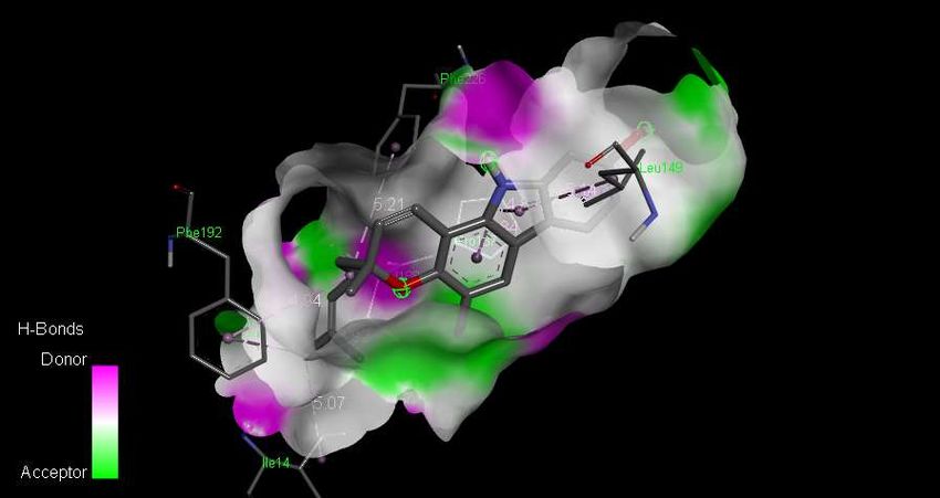
© 2021 JETIR May 2021, Volume 8, Issue 5                                                                 www.jetir.org (ISSN-2349-5162)

Different in vitro, in vivo and computational methods were employed to assess the anti-cancer potential of carbazole alkaloids. Among
these methods, docking has been used widely in drug designing of breast cancer. Role of these carbazole alkaloids found in curry leaf is
well studied by different scientists from time to time and their inhibition justifies their role in anti-cancer potential. The selected alakloids
were Girnimbine(PubChem CID:96943), Mahanine(PubChem CID:36689305) and Pyrafoline-D(PubChem CID:375148).

Research on breast cancer treatment using curry leaves is limited and thus this study is important in providing information about breast
cancer treatment by herbal medicine.

BOTANICAL DESCRIPTION OF MURRAYA KOENIGII

 Murraya koenigii, also known as curry-leaf tree, is mainly grown in the Indian subcontinent, mostly found in the southern parts of
 India. A perennial tree, being handy as a flavoring agent for various food preparations, has a wide variety of medicinal importance such
 as Anti-bacterial, Anti-fungal, Anti-cancer, Anti-inflammatory, etc. (Makri and Kintzios, 2007; Negi et al., 2011). It belongs to the
 family Rutaceae and is native to Asia.

Taxonomy of plant

Kingdom-                Plantae
Sub-kingdom-            Tracheobionta
Superdivision-          Spermatophyta
Division-               Magnoliophyta
Class-                  Magnoliospida
Subclass-                Rosidae
Order-                  Sapindales
Family-                 Rutaceae
Genus-                  Murraya J.Koenig ex. L.
Species-                Murraya Koenigii L. Spreng.

MORPHOLOGY OF CURRY PLANT

A small spreading shrub, about 2.5 metres high; the main stem, dark green to brownish, with numerous dots on it; its bark can be peeled
off longitudinally, exposing the white wood underneath; the girth of the main stem is 16 cm (Mhaskar et. al. 2000).

Leaves, exstipulate (with no stipules), bipinnately compound, 30 cm long, each bearing 24 leaflets, having reticulate venation; leaflets,
lanceolate, 4.9 cm long, 1.8 cm broad, having 0.5-cm-long petiole.

Flowers, bisexual, white, funnel-shaped, sweetly scented, stalked, complete, ebracteate, regular, actinomorphic, pentamerous,
hypogynous, the average diameter of a fully opened flower being 1.12 cm; inflorescence, a terminal cyme, each bearing 60 to 90 flowers;
calyx, 5-lobed, persistent, inferior, green; corolla, white, polypetalous, inferior, with 5 petals, lanceolate; length, 5 mm; androecium,
polyandrous, inferior, with 10 stamens, densified, arranged into circles of five each; smaller stamens, 4 mm. long whereas the longer
ones, 5 to 6 mm; gynoecium, 5 to 6 mm long; stigma, bright, sticky; style, short; ovary, superior.

Fruits, round to oblong, 1.4 to 1.6 cm long, 1 to 1.2 cm in diameter; weight, 880 mg; volume, 895 microlitres; fully ripe fruits, black
with a very shining surface; pulp, Wistaria blue 640/2; the number of fruits per cluster varying from 32 to 80.
  JETIR2105436          Journal of Emerging Technologies and Innovative Research (JETIR) www.jetir.org                                d347
© 2021 JETIR May 2021, Volume 8, Issue 5                                                              www.jetir.org (ISSN-2349-5162)

Seed, one in each fruit, 11 mm long, 8 mm in diameter, colour spinach green 0960/3; weight, 445 mg; volume, 460 microliters (Prajapati
et. al. 2003).

INTRODUCTION TO MOLECULAR DOCKING

Molecular docking is the study of how two or more molecular structures (e.g., drug and enzyme or protein) fit together to
form a stable complex with minimum overall energy. In a simple definition, docking is a molecular modelingtechnique that
is used to predict how a protein (enzyme) interacts with small molecules (ligands).

2. REVIEW OF LITERATURE

2.1 Properties of Murraya koenigii

Murraya koenigii L. belongs to the Rutaceae family of the plant. It grows naturally in forests in India, Andaman Islands, Thailand,
Cambodia, Vietnam, and Laos (Morton et. al. 1984; Ho et. al. 1999).
Many years from now, we are using traditional methods for curing the diseases by preparing medicines from different medicinal plants.
The plants’ part like bark, leaves, flowers, roots, fruits, and seeds are used to prepare medicines which have chemicals substance that
produce a definite physiological action on the human body. These are used by 80% of the world's population as a traditional medicine
because of its safety and their cost-effectiveness. The chemical substances present in the plant part are called the phytochemicals, and its
extract is known as phytoextract have effective properties like anti-oxidant, anti-inflammatory, anti-microbial and also the anti-cancer
properties. About 25% of the modern pharmaceutical drug have botanical origins (Ashokkumar K. et. al. 2013).
Curry leaves have been widely used as a main flavoring ingredient in chutney powders and pickles in India as well as in folk-medicines
in Southern Asia. Consequently, Murraya koenigii L. is frequently referred to as the Indian curry leaf tree or just the curry leaf tree
(Natarajan et. al.1974; Balaswamy et. al. 2004). The leaves are considered to be a good cure against dysentery and bites of poisonous
animals, while the roots of these plants can be used as a pain-killer (Gupta et. al. 1970).
Phytochemical studies on the leaves stem bark, and root of this plant have shown the presence of large concentration of alkaloids, phenolic
compounds and very high radical scavenging activity (Sharif et al. 2007; Tachibana et al. 2001).

  JETIR2105436         Journal of Emerging Technologies and Innovative Research (JETIR) www.jetir.org                            d348
© 2021 JETIR May 2021, Volume 8, Issue 5                                                            www.jetir.org (ISSN-2349-5162)

2.2 Some of the alkaloids are given below as follows:

2.2.1 GIRINIMBINE
It is a secondary metabolite synthesized from curry plants.
A 2011 study suggested that it helps in the apoptosis of
cancerous cells.

2.2.2 MAHANINE
It is a carbazole alkaloid which is isolated from a curry leaf plant (Murraya koenigii) and has potentially inhibiting the growth
of altered subtypes of breast cancer cells in vitro and also significantly reduces the mammary tumor burden in N-Methyl-N-
nitrosourea (MNU) induced rat. (Momita Das et al. 2019).

2.2.3 PYRAYAFOLINE-D
Pyrafoline D, also known as isomahanine, belongs to the class of organic compounds known as carbazoles. Carbazoles are
compounds containing a three-ring system containing a pyrrole ring fused on either side to a benzene ring. Pyrafoline D is an
extremely weak basic (essentially neutral) compound (based on its pKa). Pyrafoline D has been detected, but not quantified in,
herbs and spices.

  JETIR2105436         Journal of Emerging Technologies and Innovative Research (JETIR) www.jetir.org                           d349
© 2021 JETIR May 2021, Volume 8, Issue 5                                                               www.jetir.org (ISSN-2349-5162)

2.3 Breast-cancerous proteins which were used in molecular docking are as follows:

2.3.1 Oestradiol is a well-characterized sex hormone that stimulates breast cancer and other oestrogen-related diseases. 17 beta-
hydroxysteroid dehydrogenase type 1 (17beta-HSD1) catalyses the last step in the synthesis of oestradiol and androstenediol in breast
tumour tissue. The enzyme's high expression and activity after simultaneousblockade of oestrogen receptors and inhibition of aromatase
in the tumour shows the necessity for its inhibition as a requirement for breast cancer therapy. In the present paper, we report structures
of the binary and ternary complexes of 17beta-HSD1 with a new inhibitor E2B {3- [3',17'beta- dihydroxyestra-1',3',5'(10')-trien-
16'betamethyl] benzamide},and the enzyme inhibition by the later. The IC50 value for E2B was determined to be 42 nM in T47D cells.

Multiple interactions between E2B and the enzyme include hydrogen bonds and hydrophobic interactions, as well as pi-piinteractions.
A kinetic study demonstrated that E2B inhibits the enzyme's reduction forming oestradiol from oestrone, with a Ki of 0.9+/-0.15 nM.
Such strong inhibition is in agreement with its extensive interaction with the enzyme, suggesting its potential as a lead compound for
breast cancertherapy. In fact, this possibility is enhanced by its capacity for cell penetration similar to natural steroids. Such inhibitors
that block oestrogen synthesis to suppress the sulfatase pathway producing oestradiol can be used in adjuvant therapies with oestrogen
receptor blockade, opening a new orientation of breast cancer treatment.

2.3.2 NUDT5 (also called NUDIX5) has been implicated in ADP-ribose and 8-oxo-guanine metabolism and was recently
identified as a rheostat of hormone-dependent gene regulation and proliferation in breast cancer cells. Here, we further elucidate
the physiological relevance of known NUDT5 substrates and underscore the biological requirement for NUDT5 in gene
regulation and proliferation of breast cancer cells. We confirm the involvement of NUDT5 in ADP-ribose metabolism and
dissociate a relationship to oxidized nucleotide sanitation. Furthermore, we identify potent NUDT5 inhibitors, which are
optimizedto promote maximal NUDT5 cellular target engagement by CETSA.

2.3.3 HER2 (also called Neu; ErbB2) is a member of the epidermal growth factor receptor (EGFR; also known as ErbB) family
of receptor tyrosine kinases, which in humans includes HER1 (EGFR, ERBB1), HER2, HER3 (ERBB3) and HER4 (ERBB4).
ErbB receptors are essential mediators of cell proliferation and differentiation in the developing embryo and in adult tissues,
and their inappropriate activation is associated with the development and severity of many cancers. Overexpression of HER2 is
found in 20-30% of human breast cancers, and correlates with more aggressive tumours and a poorer prognosis. Anticancer
therapies targeting ErbB receptors have shown promise, and a monoclonal antibody against HER2, Herceptin (also known as
trastuzumab), is currently in use as a treatment for breast cancer.

  JETIR2105436          Journal of Emerging Technologies and Innovative Research (JETIR) www.jetir.org                             d350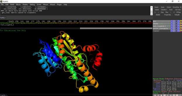
© 2021 JETIR May 2021, Volume 8, Issue 5                                                            www.jetir.org (ISSN-2349-5162)

2.4 Chemical Constituents of M. koenigii with tested pharmacological activities table (Harish et. al. 2012):

  JETIR2105436         Journal of Emerging Technologies and Innovative Research (JETIR) www.jetir.org                     d351

© 2021 JETIR May 2021, Volume 8, Issue 5                                       www.jetir.org (ISSN-2349-5162)

 JETIR2105436     Journal of Emerging Technologies and Innovative Research (JETIR) www.jetir.org     d352
© 2021 JETIR May 2021, Volume 8, Issue 5 www.jetir.org (ISSN-2349-5162)
3. OBJECTIVE
3.1 Tools and Materials used
In our present study we took help of different biological databases such as PubChem, ZINC, RCSB-PDB (Protein Data Bank) and
software’s like Autodock vina, Discovery studio visualizer, PyMOL, Open Babel GUI and Avogadro.The PDB (Protein Data Bank) is
the single worldwide archive of Structural data of Biological macromolecules, established in Brookhaven National Laboratories (BNL)
in 1971[15]. It contains Structural information of the macromolecules determined by X-ray crystallography, NMR methods, etc. and
provides access to 3D structure data for large biological molecules (proteins, DNA, and RNA). Autodock vina is an advanced version
of Autodock 4.0 which is quite efficient and provides good accuracy and performance in comparison to Autodock 4.0. These both
Autodock 4.0 and Autodock vina are currently maintained by The Scripps Research Institute, Florida, USA. Usage of AutoDock has
contributed to the discovery of several drugs. Discovery studio visualizer is a software for performing simulations of small molecule,
and analysing macromolecule systems. It includes tools for receptor-ligand docking. PyMOL is a software for visualization of
macromolecules such as proteins. Open Babel GUI is a software mainly built for interconverting chemical file formats.
3.2 Methodology
3.2.1 Retrieval of protein files from major database (Target selection)
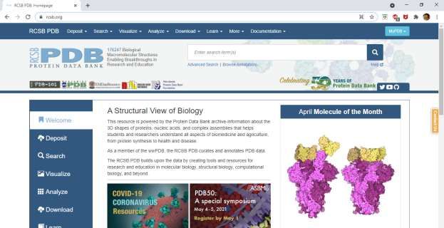
We retrieved protein.pdb files from major protein databases using following link.
https://www.rcsb.org/
Figure 3.1 Home page of RCSB-PDB
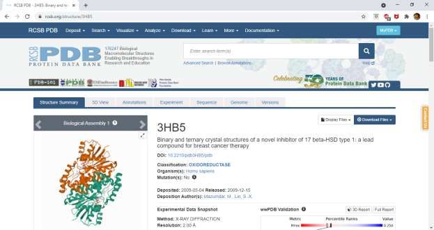
We then entered the name of protein that was docked (For example, Oestradiol or its pdb id:3HB5).
JETIR2105436 Journal of Emerging Technologies and Innovative Research (JETIR) www.jetir.org d353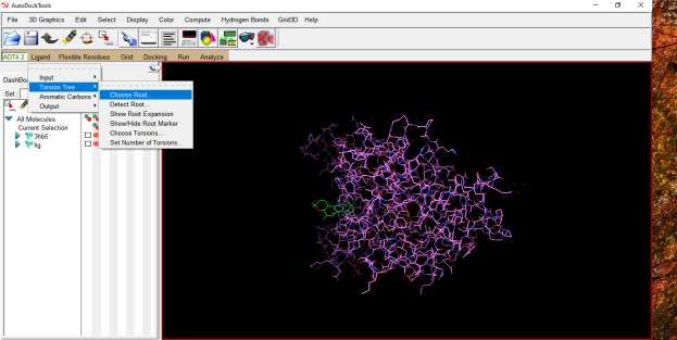
© 2021 JETIR May 2021, Volume 8, Issue 5                                                           www.jetir.org (ISSN-2349-5162)

                                                     Figure 3.2 Oestradiol protein

We selected ‘Download file from drop down list’.
Then we clicked PDB File (text) and downloaded it.
Then we opened this text file and deleted all the heteroatoms. Next step involved was deletion of X chains as all the chains are similar
and ligand would bind to either of those chains.

                      Figure 3.3 Prepared Oestradiol protein in Discovery Studio Visualizer

3.2.2 Retrieval of Ligand molecules from different databases https://zinc.docking.org/substances/home/ or
https://pubchem.ncbi.nlm.nih.gov/
The above links are available to download the desired ligand molecules. Also, analogous structures of the ligand can be drawn with the
help of ACD ChemSketch, Marvin Sketch, etc. However, it is more convenient to download the file from the database.

 JETIR2105436         Journal of Emerging Technologies and Innovative Research (JETIR) www.jetir.org                          d354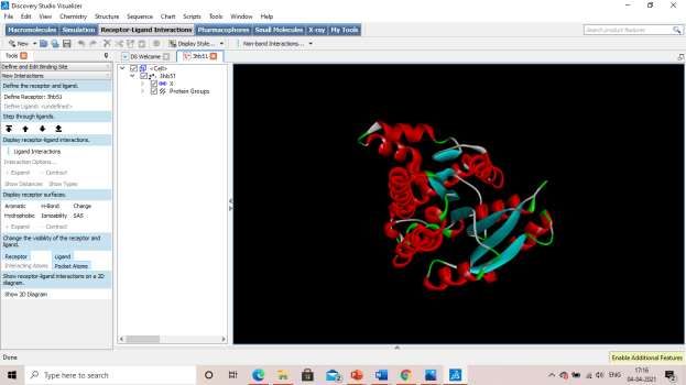
© 2021 JETIR May 2021, Volume 8, Issue 5 www.jetir.org (ISSN-2349-5162)
Figure 3.4 Ligand Mahanine in PubChem database
We clicked on “3D image and save Sdf”.
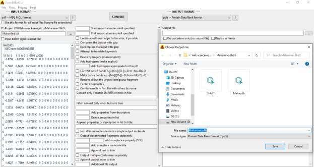
Then the file was converted from Sdf file to pdb file with the help of Open Babel GUI as autodock can read only pdb files.
Figure 3.5 Click on Save [SDF format] under 3D conformer section to
download ligand
JETIR2105436 Journal of Emerging Technologies and Innovative Research (JETIR) www.jetir.org d355© 2021 JETIR May 2021, Volume 8, Issue 5 www.jetir.org (ISSN-2349-5162)
Figure 3.6 shows the conversion of Mahanine from sdf format to pdb format in Open Babel GUI
application
3.2.3 Preparation of Pdbqt format for protein and ligand (protein.pdbqt and ligand.pdbqt)
The Pdbqt(s) of the protein and the ligand were prepared by using the Autodock tools software downloaded from MGL tools.
i) Preparation of Pdbqt file for Protein
• Remove the water molecule from the target receptor as it forms unwanted bond with other molecules (ligand) of interest.
• Add polar hydrogen group to the protein to stabilize it.
• Add the kollhman charges.
• Compute gasteiger charges to the protein.
• Save in pdbqt format.
• Know the X, Y, Z dimension of the active site for performing the site-specific docking.
Figure 3.7 shows the addition of charges to the oestradiol protein in Auto dock Tools application
JETIR2105436 Journal of Emerging Technologies and Innovative Research (JETIR) www.jetir.org d356© 2021 JETIR May 2021, Volume 8, Issue 5 www.jetir.org (ISSN-2349-5162)
Figure 3.8 shows SBD Site Sphere inside which docking has to be processed in Discovery Studio
Visualizer
Figure 3.9 shows the Attributes of the site where Ligand was attached to the protein oestradiol
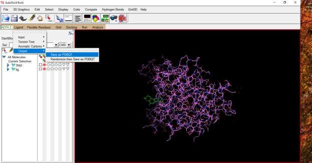
ii) Preparation of PDBQT file for Ligand
• Choose torsion root.
• Detect torsion root where docking has to be processed.
• Save in pdbqt format.
JETIR2105436 Journal of Emerging Technologies and Innovative Research (JETIR) www.jetir.org d357© 2021 JETIR May 2021, Volume 8, Issue 5 www.jetir.org (ISSN-2349-5162)
Figure 3.10 Choose and detect root where docking would be processed in the ligand
Figure 3.11 Then Save Ligand in pbdqt format shown above
Now we prepared the text file providing all the details about the pdbqt files of protein and its ligand and the attributes of SBD site
sphere.
JETIR2105436 Journal of Emerging Technologies and Innovative Research (JETIR) www.jetir.org d358© 2021 JETIR May 2021, Volume 8, Issue 5 www.jetir.org (ISSN-2349-5162)
Figure 3.12 shows the configuration file which will be compiled and run
3.2.4 Molecular Docking using Autodock vina
• Set the path for auto-dock compilation where we placed the prepared file in the following manner.
• Give command as follows…
• vina.exe –config config.txt –log log.txt.
vina_split.exe –input out.pdbqt
Figure 3.13 shows the Command Prompt dialog box
JETIR2105436 Journal of Emerging Technologies and Innovative Research (JETIR) www.jetir.org d359© 2021 JETIR May 2021, Volume 8, Issue 5 www.jetir.org (ISSN-2349-5162)
Figure 3.14 shows the Docking results of Ligand Mahanine and Protein oestradiol.
3.2.5 Analysis of molecular docking with Discovery Studio Visualizer to have information about the various conformations of
the ligand on the protein.
• See the different interactions between the protein and the ligand.
• Calculate the distance between the amino acid residues and the ligand.
• Find out the different bonds involved in the interaction between the ligand and the protein.
• Find the best pose of the ligand during the interactions between the ligand and the protein.
• See the 2-D interaction between the ligand and the protein.
• Look for the amino acids’ residues involved in the bond formation between ligand and the respective protein.
JETIR2105436 Journal of Emerging Technologies and Innovative Research (JETIR) www.jetir.org d360© 2021 JETIR May 2021, Volume 8, Issue 5 www.jetir.org (ISSN-2349-5162)
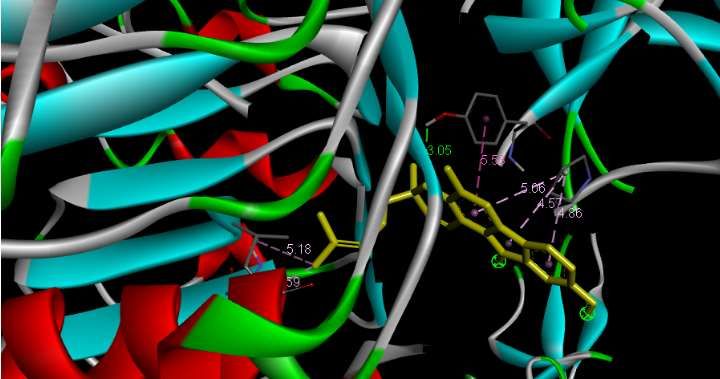
Fig 3.15 This figure shows the distance of the bond lengths between the mahanine molecule and
and amino acid residues of protein HER-2.
Fig 3.16. Shows the interaction of Mahanine and HER-2 protein.
JETIR2105436 Journal of Emerging Technologies and Innovative Research (JETIR) www.jetir.org d361© 2021 JETIR May 2021, Volume 8, Issue 5 www.jetir.org (ISSN-2349-5162)
Figure 3.17 shows the H-Bonds of the docked Girinimbine and Oestradiol.
4. RESULTS AND DISCUSSIONS
All of these compounds have been shown to be potent anti-cancerous properties. Docking studies were performed for breast cancerous
proteins with three marker compounds. The interaction of protein and ligands for the docked ligands with least binding energy was
calculated.
Figure 4.1 shows the Molecular docking between Oestradiol and Girinimbine
JETIR2105436 Journal of Emerging Technologies and Innovative Research (JETIR) www.jetir.org d362© 2021 JETIR May 2021, Volume 8, Issue 5 www.jetir.org (ISSN-2349-5162)
Figure 4.2 shows the 2-D
interaction of Ligand
Mahanine and HER2
protein
Figure 4.3 shows the molecular docking between the ligand Girinimbine and oestradiol protein
JETIR2105436 Journal of Emerging Technologies and Innovative Research (JETIR) www.jetir.org d363© 2021 JETIR May 2021, Volume 8, Issue 5 www.jetir.org (ISSN-2349-5162)
Table 1. This table depicts the binding energy between proteins and their respective ligands.
5. CONCLUSION
Mahanine and Pyrayafoline D showed least binding energy with the breast cancerous proteins. The present study concludes that the
Murraya koenigii may serve as a potential source of bioactive compounds in the prevention of cancer. The potential for developing
anticancerous drugs from higher plants appears rewarding as it leads to the development of new drugs which is required today.
In future research work the molecular modelling and molecular dynamic simulations of the protein-ligand complex can be performed
and the ADME/T (Absorption, Distribution, Metabolism, Excretion/Toxicity) properties of these compounds can be tested in wet lab
and research can be proceeded for clinical trials. In future, research work can be used further in clinical trials to test its effectiveness
and for social benefit thus reducing the time and cost in drug discovery process.
ACKNOWLEDGEMENT- I,am very thankful to Dr. Arun Bhatt who constantly encourage me and provided me the ambience
environment for my work.
REFERENCES
1) PS Shabnashmi and C Cynthia. In vitro and In-silico Studies of Murraya koenigii (L) against Streptococcus mutant. J.Chem. Pharm.
Res., 2017,9(11):67-72
2) Harish K Handral, Anup Pandith and Shruthi SD. A Review on Murraya koenigii: Multipotential Medicinal Plant. Asian J Pharm
Clin Res, Vol 5, Suppl 4, 2012, 5-14
3) C. Baskaran, M. Ramachandran. Computational molecular docking studies on anticancer drugs Asian Pacific Journal of Tropical
Disease (2012) S734-S738
4) Radhika Ramaswamy, J. Srikanth, C. Umamaheswara Reddy. Comparative Study of in silico and in vitro anticancer activity of
Traditional Indian Medicinal Plants-A reverse Pharmacological Approach Int J Curr Pharm Res, Vol 9, Issue 4, 42-46
5) Vikas Sharma, Prabodh Chander Sharma, and Vipin Kumar. In Silico Molecular Docking Analysis of Natural Pyridoacridines as
Anticancer Agents Hindawi Publishing Corporation Advances in Chemistry Volume 2016, Article ID 5409387, 9 pages
6) Santiago Vilar, Giorgio Cozza and Stefano Moro. Medicinal Chemistry and the Molecular Operating Environment (MOE):
Application of QSAR and Molecular Docking to Drug Discovery. Current Topics in Medicinal Chemistry, 2008, 8, 1555-1572
JETIR2105436 Journal of Emerging Technologies and Innovative Research (JETIR) www.jetir.org d364© 2021 JETIR May 2021, Volume 8, Issue 5 www.jetir.org (ISSN-2349-5162) 7) Pawel Sledz and Amedeo Caflisch. Protein structure-based design:from docking to molecular dynamics. Current Opinion in Structural Biology 2018, 48:93-102 8) Mohd. Ahmar Rauf, Swaleha Zubair and Asim Azhar. Ligand docking and binding site analysis with pymol and autodock/vina. International Journal of Basic and Applied Sciences, 4(2), (2015), 168-177 9) Daniel Seeliger, Bert L. de Groot. Ligand docking and binding site analysis with PyMOLand Autodock/Vina. J Comput Aided Mol Des (2010) 24:417-422 10) Mateusz Pikora and Artur Gieldon. RASMOL AB-New functionalities in the program for structure analysis. Communication Vol.62, No 3/2015 11)Harish Handral and Anup Pandith. A review on Murraya koenigii: Multipotential Medicinal Plant Asian J Pharm Clin Res, Vol 5, Suppl 4, 2012, 5-14 BIBLIOGRAPHY 1) Siti Aisyah, Ekowati Handharyani, Nurliani Bermawie, and Agus Setiyono. Potency of Murraya koenigii Leaves as Anti-Cancer Mammary in 7,12 dimethylbenz(α) anthracene (DMBA) induced-Sprague Dawley Rats E3S Web of Conferences 151, 01058 (2020) 2) Shao-Xing Dai, Wen-Xing Li, Fei-Fei Han, Yi-ChengGuo, Jun-Juan Zheng, Jia-Qian Liu, QianWang, Yue-DongGao, Gong-Hua Li & Jing-Fei Huang. In silico identification of anti-cancer compounds and plants from traditional Chinese medicine database Scientific Reports 6:25462 3) Jain M, Gilhotra R, Singh RP, et al. Curry leaf (Murraya Koenigii): a spice with medicinal property. MOJ Biol Med. 2017;2(3):236‒ 256. 4) Kok YY, Mooi LY, Ahmad K, Sukari MA, Mat N, Rahmani M et al. Anti-Tumour promoting activity and antioxidant properties of Girinimbine Isolated from the stem bark of Murraya koenigii S. Molecules 2012; 14(17):4651-4660. 5) Dheeraj K.Gahlawat, Savita Jakhar and Pushpa Dahiya. Murraya koenigii (L.) Spreng: an ethnobotanical, phytochemical and pharmacological review Journal of Pharmacognosy and Phytochemistry 2014; 3 (3): 109-119 6) Satish Chand Saini and Dr. Gopu Bala Show Reddy. A review on Curry Leaves (Murraya koenigii): Versatile Multi-Potential Medicinal Plant JETIR2105436 Journal of Emerging Technologies and Innovative Research (JETIR) www.jetir.org d365
You can also read