The effect of dietary supplementation with high- or low-dose omega-3 fatty acid on inflammatory pathology after traumatic brain injury in rats
←
→
Page content transcription
If your browser does not render page correctly, please read the page content below
Translational Neuroscience 2021; 12: 76–82
Research Article
Elise K. Black, Jack K. Phillips, Jack Seminetta, Julian Bailes, John M. Lee, John D. Finan*
The effect of dietary supplementation with high-
or low-dose omega-3 fatty acid on inflammatory
pathology after traumatic brain injury in rats
https://doi.org/10.1515/tnsci-2021-0010 are real. These findings are preliminary, but they justify
received July 15, 2020; accepted December 9, 2020 further study to determine the functional benefits asso-
Abstract: This study investigated dietary supplementa- ciated with improvements in histological outcomes and
tion as a prophylactic for neuroinflammation following understand associated dose-response curves.
traumatic brain injury (TBI) in a preclinical model. Adult Keywords: omega-3 fatty acid, docosahexanoic acid, eico-
male Sprague-Dawley rats received 30 days of supplemen- sapentaenoic acid, traumatic brain injury, dietary supple-
tation with either water or two dietary supplements. The mentation, neuroinflammation, neuroprotection
first consisted of high-dose omega-3 fatty acid (O3FA)
(supplement A) along with vitamin D3 and vitamin E.
The second had the same ingredients at different doses
with an addition of cannabidiol (supplement B). Rats 1 Introduction
were subjected to an impact TBI and then euthanized 7
days post-injury and neuroinflammation quantified by his- Traumatic brain injury (TBI) remains a pressing public
tological detection of glial fibrillary acidic protein (GFAP), health concern. Approximately 2.5 million people are
a marker of astrocyte activation, and CD68, a marker of diagnosed with TBI in U.S. emergency rooms every year
microglial activity. There was a trend toward increased [1]. Most of these injuries are classified as mild. However,
GFAP staining in injured, unsupplemented animals as recent findings suggest that even mild TBIs can have
compared to sham, unsupplemented animals, consistent severe long-term consequences [2], particularly in the
with increased activation of astrocytes in response to cases of individuals who sustain repeated mild TBI.
trauma which was reversed by supplement A but not by Mild TBI diagnosis can increase the incidence of Alzhei-
supplement B. The pattern of CD68 staining across groups mer’s disease and Parkinson’s disease and can also cause
was similar to that of GFAP staining. There was a trend a TBI-specific form of neurodegeneration called chronic
toward an increase in the injured unsupplemented group, traumatic encephalopathy in some patients [3–5]. In light
relative to sham which was reversed by supplement A but of these concerning trends, there is an urgent need for
not by supplement B. CD68 staining in injured animals interventions that can mitigate the morbidity associated
was concentrated in the perivascular domain. The consis- with mild TBI, particularly among populations at ele-
tency between trends across different measures of neuro- vated risk of mild TBI. These populations include military
inflammation showing benefits of high-dose O3FA supple- service members and athletes participating in contact
mentation following TBI suggests that the observed effects sports.
TBI remains without an approved therapy. One reason
for this is that the pathology of TBI proceeds rapidly after
the impact [6]. Preclinical studies find that candidate
* Corresponding author: John D. Finan, Department of Mechanical
and Industrial Engineering, University of Illinois at Chicago, Room therapies are most effective when applied immediately
2035 Engineering Research Facility, 842 W Taylor Street, Chicago, after or even before injury [7]. However, immediate treat-
IL 60607, United States of America, e-mail: jdfinan@uic.edu ment is clinically impractical because TBI occurs unex-
Elise K. Black, Jack K. Phillips, Jack Seminetta, Julian Bailes: pectedly, and delays associated with diagnosis, transport,
Department of Neurosurgery, NorthShore University HealthSystem,
and management of other trauma sequelae are common.
Evanston, IL, United States of America
John M. Lee: Department of Pathology and Laboratory Medicine,
The best strategy to avoid the detrimental short- or
NorthShore University HealthSystem, Evanston, IL, United States of long-term effects of all TBIs, mild-to-severe, is through
America avoidance of the injury or primary prevention. TBI is too
Open Access. © 2021 Elise K. Black et al., published by De Gruyter. This work is licensed under the Creative Commons Attribution 4.0
International License.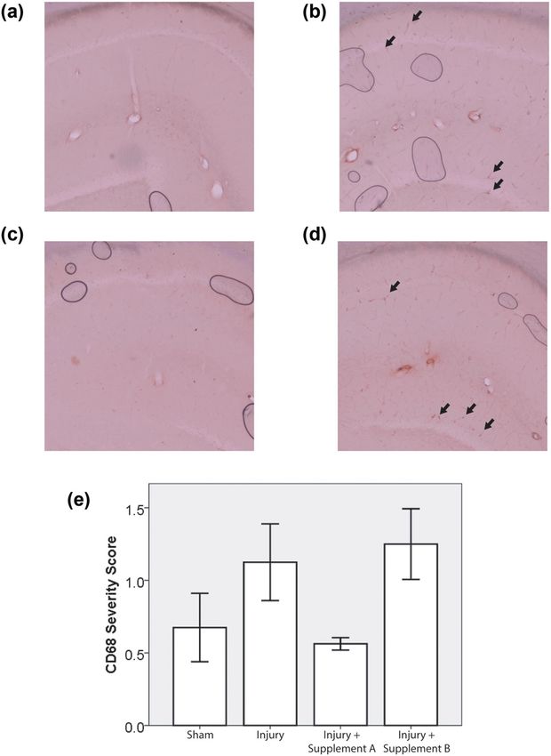
Effect of omega-3 fatty acid on brain inflammation 77
complicated for a narrow pharmaceutical approach. Table 1: Supplement composition and dosing of animals (DHA =
Approaches that target multiple aspects of TBI – secondary docosahexaenoic acid, EPA = eicosapentaenoic acid, CBD =
injury, repair, regeneration, and protection of the brain – are cannabidiol)
needed. For the latter approach, a nutraceutical is attractive.
Ingredient Amount per mL Amount per dose
Effective interventions should also treat persistent symptoms
associated with the long-term effects of TBI (post-concussive Supplement A
symptoms; e.g., memory disturbances, depression, head- DHA 250 mg 67.5 mg
EPA 125 mg 33.8 mg
ache) [8].
Vitamin D3 360 IU 97.2 IU
Studies have clearly shown that omega-3 polyunsa- Vitamin E 9 IU 2.43 IU
turated fatty acids (O3FA) such as eicosapentaenoic acid Supplement B
(EPA) and docosahexaenoic acid (DHA) are essential for DHA 50 mg 13.5 mg
proper brain development and function [9,10]. There EPA 25 mg 6.75 mg
have been numerous preclinical studies in rodent models Hemp 14 mg 3.78 mg
CBD 2.5 mg 0.675 mg
that have shown the benefits of O3FA in reducing the
Vitamin D3 100 IU 27 IU
pathology and negative outcomes associated with TBI, Vitamin E 7.5 IU 2.03 IU
stroke, and spinal cord injury [11–15]. However, work
on the underlying mechanisms is still unfolding [15,16].
Cannabidiol (CBD) has also shown the potential to miti- four experimental groups: sham injury without supple-
gate TBI pathology in preclinical models [17–19]. Accord- mentation, an injury without supplementation, injury with
ingly, this study tested two O3FA dietary treatments: one supplementation A and injury with supplementation B.
with high-dose DHA and one with low-dose DHA com-
bined with cannabidiol (CBD) to examine their efficacy as Ethical approval: The research related to animals’ use
prophylactic interventions against TBI. Vitamin D3 and complied with all the relevant national regulations and
vitamin E were also included at various doses in the hope institutional policies for the care and use of animals.
that they would synergize with O3FA and CBD to optimize All animal experiments were performed in accordance
outcome. Vitamin D mitigates lipid peroxidation [20], with The Guide for the Care and Use of Laboratory
which is an important pathology in neurotrauma [21]. Animals, and all protocols were approved by the
Vitamin E protects cells from free radicals, which play Institutional Animal Care and Use Committee at
an important role in neurotrauma pathology [22]. NorthShore University Health System [20] (Protocol
Number: EH18-191).
2 Method 2.2 The Marmarou impact acceleration
injury model
2.1 Animals and dietary supplement
information The Marmarou model is well established to produce brain
injuries as previously described [11,21]. Briefly, anesthesia
About 48 male Sprague-Dawley rats were obtained from was induced with inhaled 4% isoflurane and maintained
Charles River and received chow and water ad libitum. with 2.5% isoflurane. The surgical site was shaved, and
Their diet was supplemented with 270 µL of either water, 1% lidocaine at 3 µg/kg was administered intradermally
supplement A or supplement B every day for 30 days at the planned incision sites. A 3-cm midline incision in
before the injury. The 30-day treatment period was selected the scalp was made to expose the skull. A metal disk
based on successful neuroprotection in similar prior stu- 10 mm in diameter and 3 mm thick was cemented to the
dies from our group that used this treatment period and skull using dental acrylic on the midline centered between
also on evidence in the literature that it takes at least bregma and lambda. Animals were then disconnected from
12 days for dietary changes to be reflected as changes in the isoflurane and placed prone on a foam bed (Type E bed
the fatty acid composition of the brain [11–13,24]. manufactured by Foam to Size, Inc., Ashland, Virginia)
Table 1 details the composition of the supplements with the metal disk directly under a Plexiglas tube. A
(product supplied by Trident Brands Inc.). The animals 450 g brass weight was dropped through the tube from a
were evenly and randomly distributed across the following height of 2 m onto the disk attached to the skull. In some78 Elise K. Black et al.
cases, apnea occurred immediately after impact. In these a 4× air objective in an automated fashion by a
cases, animals were ventilated via a nosecone with 100% TissueGnostics Tissue Cytometer. One region of interest
oxygen using a TOPO Dual Mode Ventilator (Kent Scientific containing the hippocampus and adjacent cortex was
Corporation) until spontaneous respiration was observed then selected in each animal using the TissueFAXS soft-
[22]. The animals were subsequently sutured under iso- ware provided by the manufacturer. The hippocampus
fluorane anesthesia and returned to their housing. Bupre- was the focus of histological investigation because of
norphine was administered subcutaneously at 0.05 mg/kg its critical role in learning and memory, functions that
every 8–12 h for the first 24 h after surgery for analgesia. are often impaired after mild TBI. This region was
scanned again with a 10× air objective with a numerical
aperture of 0.5. GFAP fluorescence was captured with the
rhodamine filter, and DAPI fluorescence was captured
2.3 Histology with the DAPI filter. A custom written, semi-automated
Matlab (Mathworks Inc.) script was used to quantify the
Inflammatory pathology was quantified by histological extent of GFAP fluorescence. This script presented the
detection of glial fibrillary acidic protein (GFAP), a marker DAPI channel image to a blinded operator in a rando-
of astrocyte activation, and CD68, a marker of microglial mized sequence and allowed the operator to define the
activity. All animals were euthanized with a lethal dose boundary of the hippocampus. This bounded region was
injection of 120 mg/kg ketamine and 15 mg/kg xylazine then automatically thresholded in the GFAP channel
7 days after injury. The animals were immediately per- using Matlab’s graythresh function, which employs the
fused transcardially with cold phosphate-buffered saline Otsu method [24]. The extent of the GFAP staining was
(PBS) to clear the blood, followed by 4% paraformalde- then quantified as the ratio of the GFAP-positive area of
hyde in Millonig buffer until fixation tremors ceased. The the hippocampus to the total area of the hippocampus
brain was dissected out of the skull and placed in 4% (Figure 1a). The process was performed by two different
paraformaldehyde for 72 h, before being dehydrated in blinded operators, and their results were averaged.
30% sucrose until it lost buoyancy (3–5 days). It was then Staining was performed in four cohorts of animals dis-
blocked, and 50-µm thick coronal cryosections of the fore- tributed randomly across the experimental groups. The
brain was taken at locations 3.5–5 mm caudal to bregma. GFAP ratio in each animal was normalized to the mean
For CD68 labeling, cryosections were subjected to GFAP ratio for sham animals in the same cohort to protect
temperature-controlled microwave antigen retrieval using against the possibility of cohort-by-cohort variation in
a Pelco Biowave Microwave 34700 as previously described GFAP staining. The CD68 slides were microscopically
[23]. Sections were then incubated in primary mouse anti- inspected by an experienced, blinded clinical neuro-
CD68 monoclonal antibody (MA5-13324, Thermo Fisher pathologist and scored on a zero to two scale based on
Scientific) at a 1:150 dilution overnight in 0.25% Triton in the extent of the CD68 staining.
tris-buffered saline (TBS). Staining was visualized using a
Mouse Specific HRP/DAB Detection IHC Kit (ab64259,
Abcam). For GFAP labeling, the tissue was incubated over-
night with a primary unconjugated rabbit anti-GFAP poly- 2.5 Statistics
clonal antibody (Z033429-2, Agilent Dako) at a dilution of
1:750. This was followed by a 2 h incubation in Donkey The normality of the data was confirmed using a Shapiro–
anti-Rabbit secondary antibody Alexa Fluor 555 at a dilu- Wilks test. For normally distributed data, statistical sig-
tion of 1:500(A-31572, Thermo Fisher Scientific). In both nificance was assessed using a one-way ANOVA test.
cases, the slides were mounted in Vectashield mounting Otherwise, statistical significance was evaluated using
medium (Vector Laboratories) containing DAPI that labeled a Kruskal–Wallis test. Statistical tests were performed
the nuclei. using SPSS software.
2.4 Microscopy and image analysis 3 Results
For GFAP quantification, whole, fluorescently labeled, Although animals weighed between 350 and 450 g at the
coronal sections of the forebrain were scanned with start of the study, they continued to grow during theEffect of omega-3 fatty acid on brain inflammation 79
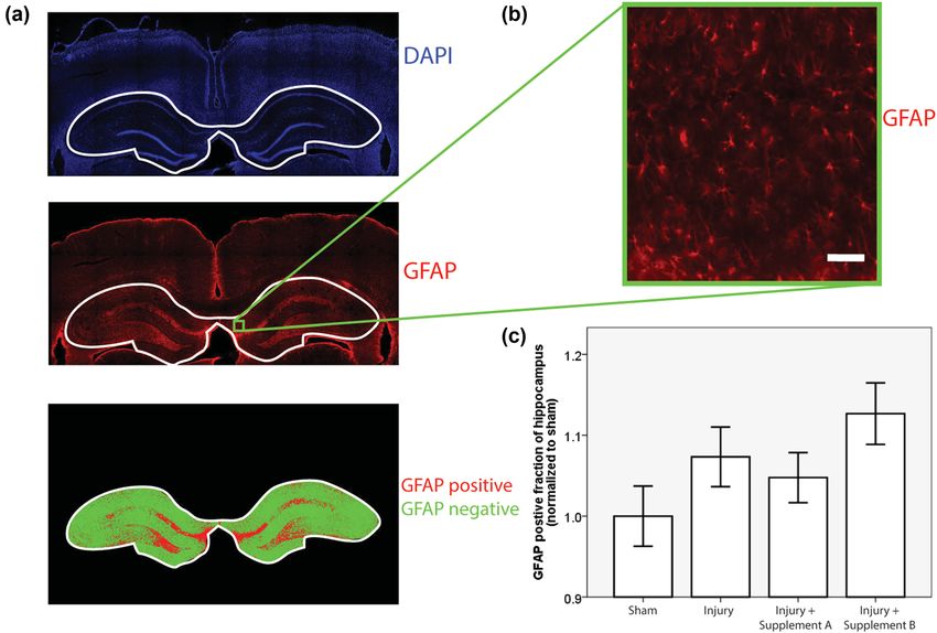
Figure 1: GFAP quantification. (a) The GFAP quantification workflow. A blinded operator outlined the hippocampus in the DAPI image. The
hippocampal region in the GFAP images was then automatically thresholded to identify the GFAP-positive (red) and GFAP-negative (green)
regions, (b) close-up view of GFAP fluorescence (scale bar = 50 µm), (c) mean values of GFAP positive fraction of the hippocampus by group,
and normalized to sham values within each cohort (n ≥ 11, error bars = standard error, ANOVA p = 0.104).
30-day supplementation period and had an average weight not by supplement B. The p-value of the associated
of 529 g (SD = 46 g) at the time of injury (Table 2). There was ANOVA test was 0.104 (Figure 1c).
no statistically significant correlation between the weight CD68 staining, where present, was concentrated in
at the time of injury and the experimental group (ANOVA, the perivascular domain (Figure 2b). The pattern of CD68
p = 0.238). staining across groups was similar to that of GFAP stain-
GFAP-positive cells were observed throughout the ing. The Shapiro–Wilks test returned p > 0.05 for three of
cortex and hippocampus and displayed the classic star- the four experimental groups but p was less than 0.05 for
shaped morphology expected in astrocytes (Figure 1b). one group, the group treated with supplement A. Since one
The Shapiro–Wilks test returned a p > 0.05 for all four of the groups was not normally distributed, a Kruskal–
experimental groups, indicating that the data were nor- Wallis test was used to determine the statistical signifi-
mally distributed. There was a trend toward increased cance of the trends. There was a trend toward an increase
GFAP staining in injured, un-supplemented animals as in the injured un-supplemented group, relative to sham.
compared to sham, un-supplemented animals, consistent This trend was reversed by supplement A but not by sup-
with increased activation of astrocytes in response to plement B. The p-value of the associated Kruskal–Wallis
trauma. This trend was reversed by supplement A but test was 0.057 (Figure 2c).
Table 2: Average animal weights on the day of injury 4 Discussion
Experimental group N Weight (g) ± SD This study observed trends suggesting an inflammatory
Sham 12 532 ± 48 response to mild TBI that was mitigated by pre-injury
Injury 12 548 ± 48 supplementation with supplement A but not with sup-
Injury with supplementation A 12 527 ± 55 plement B. The superior performance of supplement A
Injury with supplementation B 12 510 ± 26
compared to supplement B may arise from the fact that
Total 48 529 ± 46
supplement A contains a four-fold higher concentration80 Elise K. Black et al.
of these agents in this model system to optimize dose and
synergy effects.
The injury phenotype in this study was mild. The
most likely explanation of the mild phenotype lies in
the weight of the animals. Prior studies used rats
weighing between 350 and 400 g at the time of injury,
which corresponds roughly to the start of adulthood in
rats. In this study, the animals began their period of
dietary supplementation at a similar stage of maturity,
consistent with the projected application of these supple-
ments in young adults. Supplementation continued for
30 days, and the rats kept growing during this time,
reaching an average weight of 529 g at the time of injury
(Table 2). As rats grow larger, the skull becomes thicker
and the head becomes more resilient to impact. This
phenomenon is the most likely reason for the absence
of axonal damage and apoptotic biomarkers. However,
there was consistent evidence of neuroinflammation after
trauma (Figures 1 and 2). Two biomarkers were used to
measure neuroinflammation, GFAP and CD68. The pat-
tern of variation in these biomarkers across experimental
groups was strikingly consistent. In both cases, the levels
rose with injury, and this trend was mitigated by treat-
ment with supplement A but not with supplement B. This
similarity occurred in spite of the fact that the two out-
comes measured inflammatory pathology by staining dif-
Figure 2: CD68 Semi-quantitative Scoring. CD68 staining in hippo-
ferent markers in different cells and quantifying that
campal sections from animals close to the average for the following
groups: (a) Sham, (b) injury, (c) injury + supplement A, and staining in different ways (semi-automated image ana-
(d) injury + supplement B. Arrows indicate positive staining in the lysis in the case of GFAP and visual inspection by a
perivascular domain, (e) summary statistics for the semi-quantita- blinded neuropathologist in the case of CD68). ANOVA
tive scoring of CD68 pathology (n ≥ 5, error bars = standard error. testing was used to test the statistical significance of the
Kruskal–Wallis test p = 0.057).
GFAP results, which were normally distributed, while a
Kruskal–Wallis test was applied to the CD68 results,
which were not normally distributed.
of DHA and EPA than supplement B (Table 1). Previous The p-value was 0.104 in the former case and 0.057 in
reports in the literature by our group and others have the latter case. Neither score meets the conventional
shown that DHA supplementation mitigates head injury threshold for statistical significance (p < 0.05). In
pathology in preclinical rat models [11–13] in a dose- formal terms, the likelihood that the GFAP results
dependent manner [13]. However, it is worth noting reflect random variation is about 1 in 10 while the like-
that supplement A also contains more vitamin D3 than lihood that the CD68 results are random events is a
supplement B, and this may also influence therapeutic little more than 1 in 20. However, the likelihood of
trends. Vitamin D improved outcomes in prior TBI studies achieving this level of significance by chance simulta-
in rat models and humans [25,26]. Vitamin D also syner- neously in two different outcomes indicating the same
gized with progesterone treatment [26] and it may underlying pattern of pathology is much lower.
synergize with DHA treatment in this system. Further The mild injury phenotype observed is consistent
investigations will be required to address this possibility. with the goal of the study, which was to understand
In this study, CBD could not enhance the benefit of lower the capacity of nutraceutical approaches to mitigate the
dose O3FA to match the therapeutic trend observed with pathology of concussive and sub-concussive impact com-
the higher dose of O3FA or vitamin D3. These trends raise monly encountered by athletes. Neuroinflammation lies
intriguing questions about the potential benefits of nutra- at the heart of this process [27]. A drawback of this mild
ceutical interventions for TBI and support further study injury phenotype is that it hinders efforts to reach theEffect of omega-3 fatty acid on brain inflammation 81
commonly used statistical significance threshold of 0.05 [7] Abrahamson EE, Poloyac SM, Dixon CE, Dekosky ST,
and that threshold was not crossed for either of the Ikonomovic MD. Acute and chronic effects of single dose
neuroinflammatory outcomes reported. Nevertheless, the memantine after controlled cortical impact injury in adult rats.
Restor Neurol Neurosci. 2019;37(3):245–63.
consistency between the trends observed across different
[8] Petraglia AL, Maroon JC, Bailes JE. From the field of play to the
measures of neuroinflammation suggests that the observed field of combat: a review of the pharmacological management
effects are real. These findings are preliminary but they of concussion. Neurosurgery. 2012 Jun;70(6):1520–33.
justify further study to determine the functional benefits [9] Lauritzen L, Brambilla P, Mazzocchi A, Harsløf LB,
associated with improvements in histological outcomes Ciappolino V, Agostoni C. DHA effects in brain development
and function. Nutrients. 2016 Jan;8(1):E6.
and understand associated dose-response curves.
[10] Echeverría F, Valenzuela R, Catalina Hernandez-Rodas M,
Valenzuela A. Docosahexaenoic acid (DHA), a fundamental
Acknowledgments: The authors thank Vimal Patel, Ph.D., fatty acid for the brain: new dietary sources. Prostaglandins
for assistance with editing, preparing figures, formatting, Leukot Essent Fatty Acids. 2017 Sep;124:1–10.
and submitting the manuscript for publication. [11] Mills JD, Bailes JE, Sedney CL, Hutchins H, Sears B. Omega-3
fatty acid supplementation and reduction of traumatic axonal
injury in a rodent head injury model. J Neurosurg. 2011
Research funding: Funding for this study was provided
Jan;114(1):77–84.
by Trident Brands. [12] Bailes JE, Mills JD. Docosahexaenoic acid reduces traumatic
axonal injury in a rodent head injury model. J Neurotrauma.
Conflict of interest: Dr. Bailes is a consultant for Trident 2010 Sep;27(9):1617–24.
Brands Inc., a nutritional supplement company, and is a [13] Mills JD, Hadley K, Bailes JE. Dietary supplementation with the
omega-3 fatty acid docosahexaenoic acid in traumatic brain
consultant for and receives royalties from Mizuho, Inc.
injury. Neurosurgery. 2011 Feb;68(2):474–81.
The remaining authors state no conflict of interest. [14] Sun GY, Simonyi A, Fritsche KL, Chuang DY, Hannink M, Gu Z,
et al. Docosahexaenoic acid (DHA): an essential nutrient and a
Data availability statement: The datasets generated during nutraceutical for brain health and diseases. Prostaglandins
and/or analyzed during the current study are available from Leukot Essent Fatty Acids. 2018 Sep;136:3–13.
the corresponding author on reasonable request. [15] Samaddar S. Effect of docosahexaenoic acid (DHA) on spinal
cord injury. Adv Neurobiol. 2016;12:27–39.
[16] Calder PC. Omega-3 fatty acids and inflammatory processes:
from molecules to man. Biochem Soc Trans. 2017
Oct;45(5):1105–15.
[17] Elliott MB, Tuma RF, Amenta PS, Barbe MF, Jallo JI. Acute
References effects of a selective cannabinoid-2 receptor agonist on neuro-
inflammation in a model of traumatic brain injury.
[1] Faul M, Xu L, Wald MM, Coronado VG. Traumatic brain injury in J Neurotrauma. 2011 Jun;28(6):973–81.
the United States: emergency department visits, hospitaliza- [18] Amenta PS, Jallo JI, Tuma RF, Elliott MB. A cannabinoid type 2
tions and deaths 2002–2006. Atlanta (GA): Centes for Disease receptor agonist attenuates blood-brain barrier damage and
Control and Prevention, National Centres for Injury Prevention neurodegeneration in a murine model of traumatic brain
and Control; 2010. https://www.cdc.gov/traumaticbrainin- injury. J Neurosci Res. 2012 Dec;90(12):2293–305.
jury/pdf/blue_book.pdf [19] Arain M, Khan M, Craig L, Nakanishi ST. Cannabinoid agonist
[2] Barnes DE, Kaup A, Kirby KA, Byers AL, Diaz-Arrastia R, Yaffe K. rescues learning and memory after a traumatic brain injury.
Traumatic brain injury and risk of dementia in older veterans. Ann Clin Transl Neurol. 2015 Mar;2(3):289–94.
Neurology. 2014 Jul;83(4):312–9. [20] Animals CftUotGftCaUoL. Guide for the care and use of
[3] Fleminger S, Oliver DL, Lovestone S, Rabe-Hesketh S, Giora A. laboratory animals Washington (DC). US: National Research
Head injury as a risk factor for Alzheimer’s disease: the evi- Council; 2011.
dence 10 years on; a partial replication. J Neurol Neurosurg [21] Marmarou A, Foda MA, van den Brink W, Campbell J, Kita H,
Psychiatry. 2003 Jul;74(7):857–62. Demetriadou K. A new model of diffuse brain injury in rats.
[4] Gardner RC, Burke JF, Nettiksimmons J, Goldman S, Tanner CM, Part I: pathophysiology and biomechanics. J Neurosurg.
Yaffe K. Traumatic brain injury in later life increases risk for 1994 Feb;80(2):291–300.
Parkinson disease. Ann Neurol. 2015 Jun;77(6):987–95. [22] Rindfield T, McBrian S. Assisted ventilation without
[5] McKee AC, Cairns NJ, Dickson DW, Folkerth RD, Keene CD, endotracheal intubation in rats. J Invest Surg. 2012
Litvan I, et al. The first NINDS/NIBIB consensus meeting to Jun;25(3):197–9.
define neuropathological criteria for the diagnosis of chronic [23] Stone JR, Walker SA, Povlishock JT. The visualization of a new
traumatic encephalopathy. Acta Neuropathol. 2016 class of traumatically injured axons through the use of a
Jan;131(1):75–86. modified method of microwave antigen retrieval. Acta
[6] Barkhoudarian G, Hovda DA, Giza CC. The molecular patho- Neuropathol. 1999 Apr;97(4):335–45.
physiology of concussive brain injury – an update. Phys Med [24] Otsu N. A threshold selection method from gray-level histo-
Rehabil Clin N Am. 2016 May;27(2):373–93. grams. IEEE Trans Syst Man Cybern. 1979;9(1):62–6.82 Elise K. Black et al.
[25] Lawrence DW, Sharma B. A review of the neuroprotective role than monotherapy for nervous system injury and disease.
of vitamin D in traumatic brain injury with implications for Front Neuroendocrinol. 2009 Jul;30(2):158–72.
supplementation post-concussion. Brain Inj. [27] Simon DW, McGeachy MJ, Bayır H, Clark RS, Loane DJ,
2016;30(8):960–8. Kochanek PM. The far-reaching scope of neuroinflammation
[26] Cekic M, Sayeed I, Stein DG. Combination treatment with after traumatic brain injury. Nat Rev Neurol. 2017
progesterone and vitamin D hormone may be more effective Mar;13(3):171–91.You can also read


