Effects of lumican expression on the apoptosis of scleral fibroblasts: In vivo and in vitro experiments
←
→
Page content transcription
If your browser does not render page correctly, please read the page content below
EXPERIMENTAL AND THERAPEUTIC MEDICINE 21: 495, 2021

           Effects of lumican expression on the apoptosis of scleral
                 fibroblasts: In vivo and in vitro experiments
                   JINSONG WU1, YANZHI ZHAO2, YANMEI FU1, SHURONG LI1 and XU ZHANG3

1
 Department of Pediatric Ophthalmology, The Second Affiliated Hospital of Nanchang University, Nanchang, Jiangxi 330006;
2
 The First Clinical Medical College; 3The Affiliated Eye Hospital, Nanchang University, Nanchang, Jiangxi 330000, P.R. China

                                     Received February 12, 2020; Accepted September 15, 2020

                                                      DOI: 10.3892/etm.2021.9926

Abstract. Lumican serves an important role in the mainte‑               sclera during the development of myopia can induce scleral
nance of sclera biomechanical properties. However, whether              remodeling and lengthening of the ocular axis (3); with scleral
lumican expression is altered in myopia and the mechanisms              remodeling serving an important role in emmetropia and eye
of action involved are unknown. In the present study, the               development (4). Fibroblasts are the major cell type within the
expression of lumican in cultured scleral fibroblasts and in            sclera and the main cells affected in myopia (5). Structural
the scleral tissue of a rat model of form‑deprivation myopia            and functional abnormalities in scleral fibroblasts have been
was assessed. It was confirmed that diopter was decreased,              implicated in various pathologies (6).
whereas axial length was increased in modeled eyes relative                 The lumican gene encodes leucine glycan, a regulator of
to normal control eyes, indicating that the model of myopia             scleral development that is present at high levels in the extra‑
was successfully established. These pathologic changes                  cellular matrix. The protein core interacts with collagen
were accompanied by the upregulation of lumican and tissue              molecules to regulate the diameter of collagen fibers (7)
inhibitor of metalloproteinases (TIMP)‑2, as well as the                and the inter‑fiber space (8). Lumican has been shown to
downregulation of matrix metalloproteinase (MMP)‑2 and                  inhibit the lateral aggregation of collagen molecules and
MMP‑14. The same trends in TIMP‑2, MMP‑2 and MMP‑14                     limit the diameter of collagen fibers so as to maintain the
expression were observed when lumican was overexpressed                 biomechanical properties of the sclera, such as elasticity and
in cultured scleral fibroblasts. Additionally, cell proliferation       tension (9,10). Mutations to the lumican gene are thought to
decreased whereas apoptosis increased compared with those               be a cause of myopia, as demonstrated by loss‑of‑function
of control cells. Inhibiting lumican expression had no effect on        experiments in animal models (11). However, whether
cell proliferation or apoptosis, but stimulated the expression of       lumican expression is altered in myopia and the mechanisms
MMP‑2 and MMP‑14 while decreasing that of TIMP‑2. The                   of action involved are unclear. The present study aimed to
results suggested that lumican overexpression contributed to            address these areas using cultured scleral fibroblasts and a
myopia by promoting apoptosis in scleral fibroblasts via the            rat model of myopia.
modulation of TIMP‑2, MMP‑2 and MMP‑14 expression.
                                                                        Materials and methods
Introduction
                                                                        Animals. Male Sprague‑Dawley rats (21 days old; n=20;
Myopia is one of the most common eye diseases, affecting                120±5 g) were purchased from Shanghai Laboratory Animal
~27% of the population worldwide in 2010 (1). The sclera,               Research Center [license no. scxk (Lu) 2018‑0006] and
which contributes to the regulation of refractive status, is a          housed under specific pathogen‑free conditions at 23±2˚C, at a
critical structure involved in the development of myopia (2).           relative humidity of 45‑65% and under a 12 h light/dark cycle.
Clinical and experimental studies have demonstrated that                The animals were fed rodent pellets and water ad libitum. For
changes in the biochemical and mechanical properties of the             the cell culture, an additional 6 male newborn rats (5 g) were
                                                                        also purchased from Shanghai Laboratory Animal Research
                                                                        Center [license no. scxk (Lu) 2018‑0006] All experimental
                                                                        protocol was approved by the Ethics Committee of the Second
Correspondence to: Dr Jinsong Wu, Department of Pediatric               Affiliated Hospital of Nanchang University.
Ophthalmology, The Second Affiliated Hospital of Nanchang
University, 1 Minde Road, Nanchang, Jiangxi 330006, P.R. China          Establishment of the myopia model. The myopia model was
E‑mail: wujinsong628@163.com                                            established as previously described (12). Rats were anesthetized
                                                                        with isoflurane (5% for induction and 2% for maintenance)
Key    words: myopia,     lumican,    scleral   fibroblasts,   matrix   and immobilized on a surgical table in the prone position. The
metalloproteinase                                                       hair around the right eye was shaved with a razor and the skin
                                                                        sterilized with 1% iodophor. A 2‑mm long incision was made
                                                                        with ophthalmic scissors, 3 mm inside the right eye, outside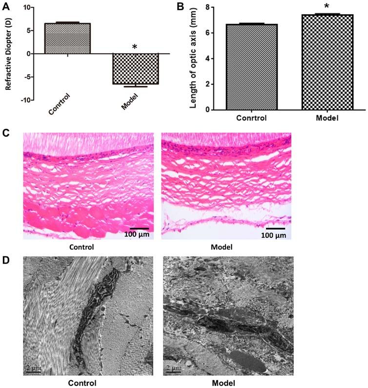
2                              WU et al: LUMICAN PROMOTES APOPTOSIS OF SCLERAL FIBROBLASTS

the outer canthus and sutured with a 6‑0 needle. The tip of the     Transmission electron microscopy. Scleral tissue samples
needle was inserted through the incision of the outer canthus,      were fixed with 2.5% glutaraldehyde at room temperature for
lower eyelid and inner canthus of the eye. The start and end        30 min and then dehydrated, embedded in epoxy resin at 60˚C
points of the suture were tightened until the eyelid was closed.    for 24 h and sectioned at a thickness of 100 nm. The sections
The uninjured left eye served as the control. Experiments           were stained with 3% uranyl acetate and lead citrate at room
using the rats were performed 8 weeks after surgery.                temperature for 5 min, and then imaged by transmission elec‑
                                                                    tron microscopy (magnification, x1,000; 80 kV; JEM‑1230;
Diopter detection. Tropicamide (0.5%, 5 µl, each eye) was           JEOL, Ltd.).
applied to the eyes as drops. After 3 min, the pupils were
sufficiently dilated, with a suitable working distance. The         Cell culture. The Scleral tissue was collected from six
3 hole/1 line was maintained with a strip light detector and        newborn rats and cut into 1x1 mm sections that were digested
diopter was determined while moving the light band back             in 0.1% type II collagenase at 37˚C for 30 min. After filtering
and forth. The final diopter was calculated by adding and           through a 70‑µm mesh, the cells were collected by centrifuga‑
subtracting the positive and negative diopters of different         tion (1,000 x g for 5 min at 4˚C), resuspended in RPMI‑1640
degrees for neutralization according to the following formula:      medium containing 20% FBS and cultured at 37˚C and
Final diopter = (degrees for examined eye) ‑ [working distance      5% CO2, to make primary scleral fibroblast cultures.
mirror (d)].
                                                                    Cell transfection. Lumican overexpression or silencing
Axial measurement. Rats were anesthetized with isoflu‑              constructs (2 µg/µl) and a negative control were produced
rane (5%) and then sacrificed by decapitation. The eyeball was      by Shanghai GenePharma Co., Ltd using pcDNA3.1 plasmid
dissected, placed on a sterile gauze moistened with normal          (Invitrogen; Thermo Fisher Scientific, Inc.). Scleral fibro‑
saline on ice and the axis was measured with Vernier calipers.      blasts were cultured until they reached 70% confluence, then
The average value of 3 measurements was reported.                   transfected at room temperature with 1.25 µg of pcDNA3.1
                                                                    plasmid (Invitrogen; Thermo Fisher Scientific, Inc.) using
Hematoxylin and eosin (H&E) staining. Sclera tissue samples         5 µl Lipofectamine ® 3000 (Invitrogen; Thermo Fisher
were fixed in 4% paraformaldehyde for 30 min at room                Scientific, Inc.). After 2 h, the culture medium was refreshed
temperature, then dehydrated in 70, 80 and 90% ethanol solu‑        and the cells were divided into the following five groups:
tions. The samples were then immersed in pure ethanol and           Untransfected (negative control), lumican overexpression
xylene for 15 min, xylene I for 15 min and xylene II for 15 min     construct (2 µg/µl, Shanghai GenePharma Co., Ltd), empty
at room temperature until the tissue became transparent. This       vector (negative control), small interfering (si)RNA construct,
was followed by immersion in a mixture of xylene and paraffin       and scrambled siRNA construct (negative control). After 48 h,
for 15 min and paraffin I and paraffin II for 50‑60 min. Finally,   the cells were used to evaluate cell proliferation, apoptosis
the samples were embedded in paraffin at room temperature           and protein expression. The siRNA sequences for lumican are
for 5 min and sectioned at a thickness of 10 µm. The sections       listed in Table I.
were collected on a slide and baked at 60˚C for 2 h, depa‑
raffinized and rehydrated, then stained with hematoxylin            Reverse transcription‑quantitative (q)PCR. RNA was
for 3 min and eosin for 3 min at room temperature. The              extracted from cells using TRIzol® (Thermo Fisher Scientific,
slides were sealed and at least four fields of each slide were      Inc.) and cDNA was synthesized at 30˚C for 10 min according
imaged using a light microscope (magnification, x200; BX51;         to the instructions of the reverse transcriptase kit (CoWin
Olympus Corporation).                                               Biosciences). cDNA was used as the template for qPCR, which
                                                                    was performed on a 7500 Fast Real-Time PCR System (Applied
Immunohistochemistry. Eyeball tissue sections were prepared         Biosystems). The level of β ‑actin was used as the internal
as aforementioned followed by blocking in 5% bovin serum            reference to calculate the mRNA expression levels of, MMP‑2,
albumin (Hyclone; GE Healthcare Life Science) at room               MMP‑14 and TIMP‑2. The qPCR reaction consisted of 9.5 µl
temperature for 2 h and incubated overnight at 4˚C with mono‑       RNase‑free dH2O, 1 µl cDNA/DNA, 2 µl primers and 12.5 µl
clonal antibodies against MMP2 (1:250; cat. no. AF0577),            2X UltraSYBR mix (cat. no. A25778; Thermo Fisher Scientific,
tissue inhibitor of metalloproteinases (TIMP)‑2 (1:200;             Inc.). The cycling protocol was as follows: Pre‑denaturation at
cat. no. AF7008; each from Affinity Biosciences),                   95˚C for 10 min, 40 cycles of 95˚C for 10 sec, 54.3˚C for 30 sec
MMP‑14 (1:100; cat. no. ab51074) and lumican (1:100,                and 72˚C for 30 sec. The target gene expression was normal‑
cat. no. ab168348; each purchased from Abcam), followed by          ized to β‑actin as previously described (13). Primer sequences
horseradish peroxidase (HRP)‑conjugated goat anti‑rabbit IgG        are listed in Table II.
(1:10,000; cat. no. A16104; Thermo Fisher Scientific, Inc.) or
Alexa Fluor 593 goat anti‑mouse IgG (1:200; cat. no. A‑11001;       Western blotting. Total protein was extracted from scleral tissues
Thermo Fisher Scientific, Inc.) for 30 min at room tempera‑         or cultured scleral fibroblasts. After determining the concentra‑
ture. Immunoreactivity was visualized by incubation with            tion using the BCA method, the proteins (20 µg) were separated
3,3'‑diaminobenzidine for 3 min at room temperature. At least       by 12% SDS‑PAGE and transferred to a PVDF membrane.
four fields were taken from each image using a light micro‑         Non‑specific antibody binding was blocked by incubating the
scope (magnification, x200; BX51, Olympus Corporation).             membrane in 5% non‑fat milk at room temperature for 2 h. The
The grey value of the staining was analyzed by Image‑Pro            membrane was then incubated overnight at 4˚C with antibodies
Plus software v6.0 (National Institutes of Health).                 against MMP‑2 (1:1,000), MMP‑14 (1:1,000), TIMP‑2 (1:1,000),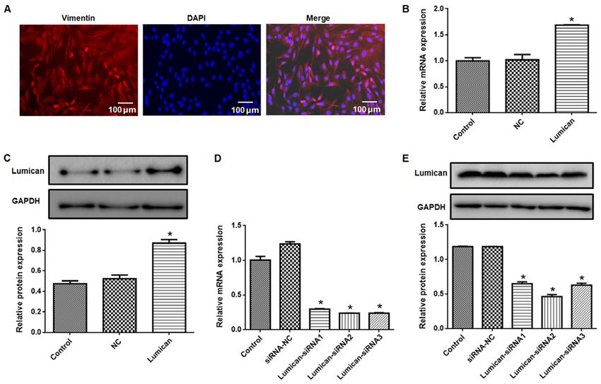
EXPERIMENTAL AND THERAPEUTIC MEDICINE 21: 495, 2021 3
Table I. siRNA sequences.
siRNA siRNA sequence (5'‑3')
Lumican‑siRNA1 ACAAUAAGCUCAAGAGUAUTTAUACUCUUGAGCUUAUUGUTT
Lumican‑siRNA2 UGAAGAAGCUGCAUAUAAATTUUUAUAUGCAGCUUCUUCATT
Lumican‑siRNA3 ACUCCAAGAUCAAAGGAAATTUUUCCUUUGAUCUUGGAGUTT
Scrambled siRNA (siRNA‑NC) UUCUCCGAACGUGUCACGUTTACGUGACACGUUCGGAGAATT
NC, negative control; Si, small interfering.
Table II. Primer sequences.
Primer length Product length Annealing temperature
Primer Sequence (5'‑3') (bp) (bp) (˚C)
Lumican F GCCTTTGAGAATGTAACGGAT 21 169 57.1
Lumican R CTTGTAGGGACTTTGGGAGC 20
MMP‑2 F AGGACACCCTCAAGAAGATGC 21 134 58.1
MMP‑2 R GCGGGGAAAGAAGTTGTAGTT 21
MMP‑14 F GCAGTATGGCTACCTACCTCC 21 118 58.4
MMP‑14 R CTTGCCTGTCACTTGTAAACC 21
TIMP‑2 F GCAACCCCATCAAGAGGA 18 212 56.6
TIMP‑2 R CCAGGGCACAATAAAGTCAC 20
β‑actin F ACGGTCAGGTCATCACTATC 20 90 56.5
β‑actin R TGCCACAGGATTCCATACC 19
bp, base pairs; F, forwards; R, reverse; TIMP, tissue inhibitor of metalloproteinases.
lumican (1:1,000) and GAPDH (1:3,000; cat. no. ab8245; Abcam), Apoptotic cells were detected by NovoCyte™ flow cytometer
followed by incubation with HRP‑conjugated anti‑rabbit IgG (NovoCyte 2060R; ACEA Bioscience, Inc.) and analyzed
(1:10,000; cat. no. A16104SAMPLE; Thermo Fisher Scientific, using FlowJo v10 (FlowJo, LLC).
Inc.) at room temperature for 2 h. An enhanced chemilumines‑
cence reagent kit (cat. no. WP20005; Thermo Fisher Scientific, Statistical analysis. Data are expressed as the mean ± SD
Inc.) was used to visualize immunoreactivity. The blots were and were analyzed using SPSS v19.0 software (IBM Corp.).
scanned with a ChemiDoc XRS imaging system (Bio‑Rad The control and modeled eyes were from the same animals;
Laboratories, Inc.) and signal intensity was analyzed with therefore, paired Student's t‑tests were used to analyze the
Quantity One v1.4.6 software (Bio‑Rad Laboratories, Inc.) as differences. When there were ≥3 groups, the differences were
previously described (14). evaluated using one‑way ANOVA followed by a post‑hoc
Tukey's test. P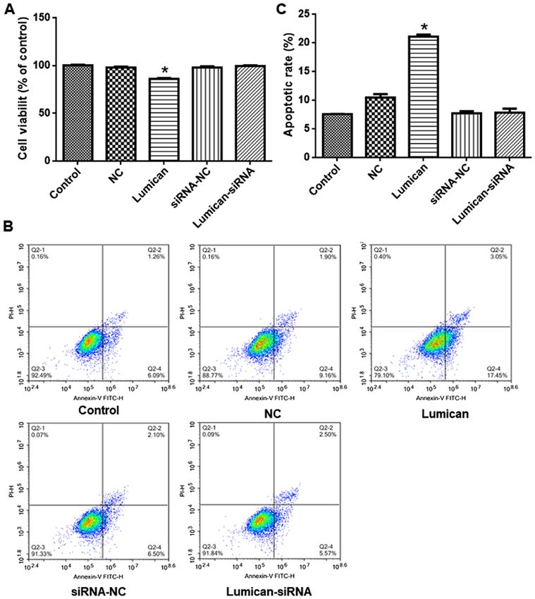
4                                   WU et al: LUMICAN PROMOTES APOPTOSIS OF SCLERAL FIBROBLASTS

Figure 1. Abnormal diopter and axial length, and pathologic changes in scleral tissue in a model of form‑deprivation myopia. (A) D value was reduced in
model eyes and (B) the optic axis was increased in the model eyes. (C) Pathologic changes in scleral tissue were observed by hematoxylin and eosin staining.
(D) Ultrastructural analysis of scleral tissue was performed via electron microscopy. *P
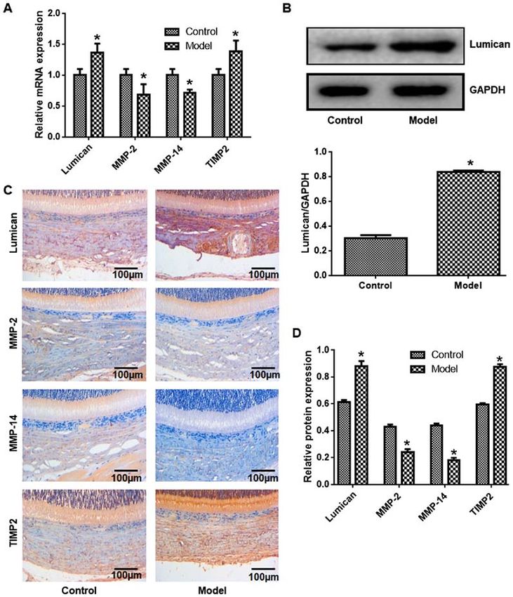
EXPERIMENTAL AND THERAPEUTIC MEDICINE 21: 495, 2021                                                             5

Figure 2. Expression of lumican, MMP‑2, MMP‑14 and TIMP‑2 in scleral tissue. Expression levels of lumican, MMP‑2, MMP‑14 and TIMP‑2 expression at the
(A) mRNA and (B) protein levels, detected by reverse transcription‑quantitative PCR and western blotting, respectively. (C) Immunohistochemical analysis of
lumican, MMP‑2, MMP‑14 and TIMP‑2 expression. (D) Quantification of results presented in panel C. *P
6                                    WU et al: LUMICAN PROMOTES APOPTOSIS OF SCLERAL FIBROBLASTS

Figure 3. Effects of lumican overexpression and knockdown. (A) Vimentin expression were observed in cultured primary scleral fibroblasts. (B) mRNA and
(C) protein expression levels of lumican in cells transfected with the lumican overexpression vector. (D) mRNA and (E) protein expression levels of lumican in
cells transfected with lumican siRNA. *P
EXPERIMENTAL AND THERAPEUTIC MEDICINE 21: 495, 2021                                                               7

Figure 4. Lumican overexpression reduces cell viability and promotes apoptosis in scleral fibroblasts. (A) Cell viability was evaluated using a Cell Counting
Kit‑8 assay. (B) Apoptotic cells were detected using flow cytometry and (C) representative scatterplots were presented. *P
8                                 WU et al: LUMICAN PROMOTES APOPTOSIS OF SCLERAL FIBROBLASTS

scleral fibroblasts and TIMP‑2 expression and reducing MMP‑2              11. Zhang P, Karani R, Turner RL, Dufresne C, Ferri S, Van Eyk JE
                                                                              and Semba RD: The proteome of normal human retrobulbar
and MMP‑14 levels. As such, therapeutic strategies targeting                  optic nerve and sclera. Proteomics 16: 2592‑2596, 2016.
lumican may be effective for the treatment of myopia.                     12. Xiao H, Fan ZY, Tian XD and Xu YC: Comparison of
                                                                              form‑deprived myopia and lens‑induced myopia in guinea pigs.
                                                                              Int J Ophthalmol 7: 245‑250, 2014.
Acknowledgements                                                          13. Livak KJ and Schmittgen TD: Analysis of relative gene expres‑
                                                                              sion data using real‑time quantitative PCR and the 2(‑Delta Delta
Not applicable.                                                               C(T)) method. Methods 25: 402‑408, 2001.
                                                                          14. Song ZJ, Yang SJ, Han L, Wang B and Zhu G: Postnatal calpeptin
                                                                              treatment causes hippocampal neurodevelopmental defects in
Funding                                                                       neonatal rats. Neural Regen Res 14: 834‑840, 2019.
                                                                          15. Guggenheim JA, Ghorbani Mojarrad N, Williams C and
                                                                              Flitcroft DI: Genetic prediction of myopia: Prospects and
No funding was received.                                                      challenges. Ophthalmic Physiol Opt 37: 549‑556, 2017.
                                                                          16. Steidl SM: How does visual acuity change over time in adults
Availability of data and materials                                            with high myopia? Br J Ophthalmol 90: 524, 2006.
                                                                          17. Stone RA, Lin T, Laties AM and Iuvone PM: Retinal dopamine
                                                                              and form‑deprivation myopia. Proc Natl Acad Sci USA 86:
The datasets used and/or analyzed during the current study are                704‑706, 1989.
available from the corresponding author on reasonable request.            18. Sun MS, Song YZ, Zhang FJ, Tao J and Liu YB: Changes of ocular
                                                                              biological parameters and Lumican expression in the monocularly
                                                                              deprivation myopic model of mutant Lumican transgenic mice.
Authors' contributions                                                        Zhonghua Yan Ke Za Zhi 52: 850‑855, 2016 (In Chinese).
                                                                          19. Song Y, Zhang F, Zhao Y, Sun M, Tao J, Liang Y, Ma L, Yu Y,
                                                                              Wang J and Hao J: Enlargement of the axial length and altered
JW, YZ, YF and SL performed the experiments and analyzed                      ultrastructural features of the sclera in a mutant lumican
the data. JW and XZ designed the study and wrote the                          transgenic mouse model. PLoS One 11: e0163165, 2016.
manuscript. All authors read and approved the final manuscript.           20. Lu P, Takai K, Weaver VM and Werb Z: Extracellular matrix
                                                                              degradation and remodeling in development and disease.
                                                                              Cold Spring Harb Perspect Biol 3: a005058, 2011.
Ethics approval and consent to participate                                21. Sternlicht MD and Werb Z: How matrix metalloproteinases
                                                                              regulate cell behavior. Annu Rev Cell Dev Biol 17: 463‑516,
                                                                              2001.
All experimental protocol was approved by the Ethics                      22. Vij N, Roberts L, Joyce S and Chakravarti S: Lumican suppresses
Committee of the Second Affiliated Hospital of Nanchang                       cell proliferation and aids Fas‑Fas ligand mediated apoptosis:
University.                                                                   Implications in the cornea. Exp Eye Res 78: 957‑971, 2004.
                                                                          23. Leung KH, Yiu WC, Yap MK, Ng PW, Fung WY, Sham PC and
                                                                              Yip SP: Systematic investigation of the relationship between
Patient consent for publication                                               high myopia and polymorphisms of the MMP2, TIMP2, and
                                                                              TIMP3 genes by a DNA pooling approach. Invest Ophthalmol
                                                                              Vis Sci 52: 3893‑3900, 2011.
Not applicable.                                                           24. Siegwart JT Jr and Norton TT: Steady state mRNA levels in tree
                                                                              shrew sclera with form‑deprivation myopia and during recovery.
Competing interests                                                           Invest Ophthalmol Vis Sci 42: 1153‑1159, 2001.
                                                                          25. Dai SZ, Zeng JW and Wang LY: Effect of pirenzepine on form
                                                                              deprivation myopia in chicks and its possible mechanism.
The authors declare that they have no competing interests.                    Zhonghua Yan Ke Za Zhi 42: 42‑47, 2006 (In Chinese).
                                                                          26. Pietraszek K, Chatron‑Colliet A, Brézillon S, Perreau C,
                                                                              Jakubiak‑Augustyn A, Krotkiewski H, Maquart F and
References                                                                    Wegrowsk i Y: Lum ican: A new in hibitor of matr ix
                                                                              metalloproteinase‑14 activity. FEBS Lett 588: 4319‑4324, 2014.
 1. Foster PJ and Jiang Y: Epidemiology of myopia. Eye (Lond) 28:         27. La m C, Ja merson M, Cabra l G, Ca rlesso A M a nd
    202‑208, 2014.                                                            Marciano‑Cabral F: Expression of matrix metalloproteinases in
 2. Hayashi M, Ito Y, Takahashi A, Kawano K and Terasaki H:                   Naegleria fowleri and their role in invasion of the central nervous
    Scleral thickness in highly myopic eyes measured by enhanced              system. Microbiology (Reading) 163: 1436‑1444, 2017.
    depth imaging optical coherence tomography. Eye (Lond) 27:            28. Murata K, Hirata A, Ohta K, Enaida H and Nakamura KI:
    410‑417, 2013.                                                            Morphometric analysis in mouse scleral fibroblasts using
 3. Metlapally R and Wildsoet CF: Scleral mechanisms underlying               focused ion beam/scanning electron microscopy. Sci Rep 9:
    ocular growth and myopia. Prog Mol Biol Transl Sci 134:                   6329, 2019.
    241‑248, 2015.                                                        29. Chen ZT, Wang IJ, Shih YF and Lin LL: The association of
 4. Harper AR and Summers JA: The dynamic sclera: Extracellular               haplotype at the lumican gene with high myopia susceptibility in
    matrix remodeling in normal ocular growth and myopia develop‑             Taiwanese patients. Ophthalmology 116: 1920‑1927, 2009.
    ment. Exp Eye Res 133: 100‑111, 2015.                                 30. Lu YP, Ishiwata T, Kawahara K, Watanabe M, Naito Z,
 5. Rada JA, Shelton S and Norton TT: The sclera and myopia.                  Moriyama Y, Sugisaki Y and Asano G: Expression of lumican
    Exp Eye Res 82: 185‑200, 2006.                                            in human colorectal cancer cells. Pathol Int 52: 519‑526, 2002.
 6. Zhan X, Zhu ZC, Sun SQ and Wen YC: Dynamic changes of acti‑           31. Niewiarowska J, Brézillon S, Sacewicz‑Hofman I, Bednarek R,
    vator protein 1 and collagen I expression in the sclera of myopia         Maquart F, Malinowski M, Wiktorska M, Wegrowski Y and
    guinea pigs. Int J Ophthalmol 12: 1272‑1276, 2019.                        Cierniewski CS: Lumican inhibits angiogenesis by interfering
 7. Chen L, Zhang Y, Zuo Y, Ma F and Song H: Lumican expression               with alpha2beta1 receptor activity and downregulating MMP‑14
    in gastric cancer and its association with biological behavior and        expression. Thromb Res 128: 452‑457, 2011.
    prognosis. Oncol Lett 14: 5235‑5240, 2017.                            32. Pietraszek K, Brézillon S, Perreau C, Malicka‑Błaszkiewicz M,
 8. Austin BA, Coulon C, Liu CY, Kao WW and Rada JA: Altered                  Maquart F and Wegrowski Y: Lumican‑derived peptides inhibit
    collagen fibril formation in the sclera of lumican‑deficient mice.        melanoma cell growth and migration. PLoS One 8: e76232, 2013.
    Invest Ophthalmol Vis Sci 43: 1695‑1701, 2002.
 9. Mouw JK, Ou G and Weaver VM: Extracellular matrix assembly: A                            This work is licensed under a Creative Commons
    multiscale deconstruction. Nat Rev Mol Cell Biol 15: 771‑785, 2014.                      Attribution-NonCommercial-NoDerivatives 4.0
10. Stuart K, Paderi J, Snyder PW, Freeman L and Panitch A:                                  International (CC BY-NC-ND 4.0) License.
    Collagen‑binding peptidoglycans inhibit MMP mediated collagen
    degradation and reduce dermal scarring. PLoS One 6: e22139, 2011.
You can also read