Bruton's tyrosine kinase (BTK) is a binding partner for hypoxia induced mitogenic factor (HIMF/FIZZ1) and mediates myeloid cell chemotaxis
←
→
Page content transcription
If your browser does not render page correctly, please read the page content below
The FASEB Journal • Research Communication

Bruton’s tyrosine kinase (BTK) is a binding partner for
  hypoxia induced mitogenic factor (HIMF/FIZZ1)
  and mediates myeloid cell chemotaxis
           Qingning Su, Yifu Zhou, and Roger A. Johns1
           Department of Anesthesiology and Critical Care Medicine, Johns Hopkins University
           School of Medicine, Baltimore, Maryland, USA

ABSTRACT        Hypoxia induced mitogenic factor (HIMF)      As an inflammatory and ischemic tissue marker, the
is a member of the FIZZ/resistin/RELM family of            physiological function of HIMF remains unclear. XCP1,
proteins that we have shown to have potent mitogenic,      another member of the FIZZ/resistin/RELM family,
angiogenic, and vasoconstrictive effects in the lung       has been reported as a secreted protein that is chemo-
vasculature. In the current report, we identified Bru-     tactic to myeloid cells from C/EBP-epsilon-null mice
ton’s tyrosine kinase (BTK) as a functional HIMF           and interacts with alpha-defensin (4). We therefore
binding partner through glutathione S-transferase          investigated whether HIMF is a targeting molecule for
(GST)-HIMF pull-down studies and mass spectrometry.        bone marrow cells and which molecule is the HIMF-
Using primary cultured HIMF-stimulated murine bone         binding partner. By using GST-pulldown and mass
marrow cells, we demonstrated that HIMF causes redis-      spectrometry techniques, we isolated a HIMF-binding
tribution of BTK to the leading edge of the cells. HIMF    molecule identified as BTK, a molecule known to be
stimulation induced BTK autophosphorylation, which         crucial in regulation of B-cell maturation and involved
peaked at 2.5 min. A transwell migration assay showed      in cell migration. Mutations in BTK are responsible for
that treatment with recombinant murine HIMF induced        X-linked agamma globulinemia (XLA) in humans and
migration of primary cultured bone marrow cells that       X-linked immunodeficiency (xid) in mice (5, 6).
was completely blocked by the BTK inhibitor, LFM-
A13. Our results demonstrate BTK as the first known
functional binding partner of the HIMF/FIZZ family of      MATERIALS AND METHODS
proteins and that HIMF acts as a chemotatic molecule
in stimulating the migration of myeloid cells through      Constructs and reagents
activation of the BTK pathway.—Su, Q., Zhou, Y., and
Johns, R. A. Bruton’s tyrosine kinase (BTK) is a binding   Flag-tagged HIMF was prepared as described previously (1).
partner for hypoxia induced mitogenic factor (HIMF/        For GST-HIMF construction, mouse HIMF cDNA was first
FIZZ1) and mediates myeloid cell chemotaxis. FASEB J.      amplified by polymerase chain reaction (PCR) from a mouse
21, 1377–1383 (2007)                                       lung cDNA library using primers 5⬘- GAATTCATGGCG-
                                                           TATAAAAGCATCTCA-3⬘ and 5⬘- CTCGAGTTAGGACAGTTG-
                                                           GCAGCAGCG-3⬘ and cloned into TA vector. Then the insert
Key Words: resistin 䡠 LFM-A13 䡠 migration                  was cut from TA vector by EcoR I and XhoI, and cloned into
                                                           pGEX-5X-1 with the sites of EcoRI and XhoI.
Hypoxia-induced mitogenic factor (HIMF), also
called FIZZ1 or RELM alpha, is a resistin-like molecule    Antibodies and inhibitor
that is up-regulated in the lungs in rodent models of
chronic hypoxia induced pulmonary hypertension (1,         Anti-actin and anti-Fyn rabbit polyclonal antibodies were
2). We have shown HIMF to be expressed in the              purchased from Sigma. Anti-BTK monoclonal antibody
                                                           (mAb), Rabbit anti-BTK phosphorylated (Y223), and phos-
remodeling hyperplastic vascular smooth muscle and
                                                           pho-Src family (Y416) polyclonal antibodies were purchased
endothelium and to stimulate pulmonary microvascu-         from Cell Signaling Technology, Inc. (Danvers, MA, USA)
lar mitogenesis. HIMF was found to have angiogenic         Anti-focal adhesion kinase F-actin (FAK) rabbit polyclonal
properties and to increase pulmonary artery pressure       antibody (pAb) was purchased from Upstate (Lake Placid,
and pulmonary vascular resistance more potently than       NY, USA). Monoclonal anti-fyn (Y528) phospho-specific anti-
endothelin, angiotensin (ANG), or serotonin (1). The       body was purchased from BD Transduction Laboratories
expression of HIMF was also markedly up-regulated in       (Franklin Lakes, NJ, USA). Antihis mAb and anti-GST mAb
hypertrophic, hyperplastic bronchial epithelium dur-
ing allergic pulmonary inflammation in mouse models          1
                                                              Correspondence: Department of Anesthesiology and Critical
of acute pulmonary inflammation (3) and in the lymph       Care Medicine, Johns Hopkins University, Ross 361, 720 Rutland
nodes (4), with the highest expression in B cells and      Ave., Baltimore, MD 21205, USA. E-mail: rajohns@jhmi.edu
macrophages.                                                 doi: 10.1096/fj.06-6527com

1376                                                                                     0892-6638/07/0021-1376 © FASEB
were purchased from Novagen (Madison, WI, USA). Anti- TOF-MS were analyzed by searching against an NCBInr
BTK rabbit pAb was purchased from Santa Cruz Biotechnol- database using MASCOT (Matrix Science, London, UK)
ogy (Santa Cruz, CA, USA). FITC and rhodamine-labeled search software.
secondary antibodies were purchased from Jackson Immu-
noResearch. BTK inhibitor LFM-A13 was purchased from Protein phosphorylation assays
Calbiochem (San Diego, CA, USA).
Bone marrow cells were cultured in 100 ⫻ 20 mm culture
Cell culture and transfections plates and treated with 50 nm BSA or HIMF for different time
serials, washed quickly by PBS, and lysed in TBST buffer
Mouse bone marrow cells were maintained in Dulbecco’s containing 1 mM sodium vanadate. The samples were incu-
modified Eagle’s high glucose medium (DMEM; GIBCO, bated on ice for 20 min, mixed several times during the
Gaithersburg, MD, USA) containing 10% FBS at 37°C and 5% incubation, and then centrifuged. The supernatants of the
CO2. Cells were transfected with plasmids, as mentioned, samples were quantified for protein concentration and sub-
using LipofectAMINE 2000 reagent (Invitrogen, Carlsbad, jected to electrophoresis on a 4 –15% SDS-polyacrylamide gel
CA, USA) according to the manufacturer’s protocols. The (Bio-Rad, Hercules, CA, USA). Rabbit anti-BTK phosphoryla-
transfected cells were then fixed in precooled methanol for tion (Y-223) pAb and anti-BTK mAb were used for immuno-
immunocytochemistry. blotting. The same samples were used for the detection of Fyn
phosphorylation. Rabbit anti-phospho-Src family (Y416) pAb,
monoclonal anti-fyn (Y528) phospho-specific antibody, and
GST and GST-HIMF fusion protein expression rabbit anti-Fyn poloyclonal antibody were used for the blot-
ting.
BL21 cells harboring GST or GST-HIMF constructs were
grown overnight in a 50 ml tube with LB medium containing Mouse bone marrow-derived mesenchymal stem cell
50 g/ml ampicillin and then transferred to a 500 ml flask preparation and culture
and grown until the optical density (OD) was 0.6 at 600 nm.
The cultures were then induced with isopropyl--d-thiogalac-
Six C57BL/6 mice (7 wk old) were anesthetized with intra-
topyranoside (IPTG) for an additional 4 h. Cell lysates were
muscularly injection of 1 mg ketamine plus 0.5 mg xylazine
prepared in TBST buffer (50 mM Tris, pH 7.5, 150 mM NaCl,
per animal. Tibiae and femurs were isolated using sterile
5 mM EDTA, 1% Triton X-100, 10 g/ml leupeptin, 10
techniques. The mouse bone marrow cells were prepared by
g/ml aprotinin, and 1 mM phenylmethylsulfonyl fluoride).
flushing the tibiae and femurs with serum-free DMEM (low
glucose, supplemented with 1⫻ penicillin-streptomycin and 1
GST pull-down assay mM EDTA) using 25 G needles. Pooled marrow from three
animals was first dispersed by gentle pipetting and then
Bone marrow cells were cultured in ten 150 ⫻ 25 mm plates separated by gradient centrifugation with lymphocyte separa-
until confluence and collected for lysate using TBST buffer. tion liquid (Sigma, density: 1.083 g/ml) as follows: 6 ml of the
Lysate from 10 plates of bone marrow cells was used for a medium containing the marrow cells was layered on top of 3
HIMF-binding partner screening assay. Hypoxia tissue ho- ml of separation liquid and centrifuged at room temperature
mogenate was prepared as described above. BL21 bacterial at 2800 rpm for 20 min. The mononuclear cells in the middle
lysates for GST and GST-HIMF were first incubated with layer were collected and then washed with serum-free DMEM
glutathione agarose in 0.1% TBST buffer for 3 h and then three times by centrifugation, first at 2000 rpm for 15 min and
washed three times by 0.1% TBST buffer. The GST and then two times at 700 rpm for 10 min. The cells collected after
GST-HIMF binding glutathione agaroses were incubated with the last wash (2–3⫻108) were resuspended in 10 ml DMEM
bone marrow cell lysate or hypoxia tissue homogenate for 3 h supplemented with 10% FBS and 1⫻ penicillin/streptomycin
and then washed three to five times before SDS-PAGE. and then cultured at 37°C with 5% CO2 in one 10-cm culture
dish (uncoated plastic). Three days later, nonadherent cells
Mass spectrometry were removed by changing medium and the adherent cells
were grown for 2 wk.
Glutathione-Sepharose beads that bind to GST-HIMF fusion Reverse transcriptase-polymerase chain reaction (RT-PCR)
protein and GST were incubated with bone marrow cell lysate
in TBST buffer at 4°C for 3 h. The beads were washed five
times with TBST, and loading buffer was added to the Fresh bone marrow cells from three mice were prepared as
samples. After electrophoresis, the gel was stained by Coomas- described above. Mouse RAW 309 Cr.1 monocyte cell line was
sie blue. The band that was pulled-down by GST-HIMF was cultured in a 60cm plate with 10% FBS D-MEM medium until
cut from the Coomassie blue stained gel and another band in confluent and collected by cell scraper. Both bone marrow
the unstained area was cut from the same gel as the control. cells and RAW cells were used for RNA purification using an
The bands were washed with 50% methanol twice and stored RNeasy mini kit (Qiagen, Valencia, CA, USA). Two micro-
in 1.7 ml ultraClear tubes with 50% methanol. Then, the grams of total RNA for each sample were used in the cDNA
samples were washed twice with deionized water and dehy- synthesis by Amersham first-strand cDNA synthesis kit. Prim-
drated in 80 l acetonitrile (ACN) twice. The samples were ers for mouse HIMF coding region (310 bp), -actin C-
then swollen in a digestion buffer containing 20 mmol/l terminal coding region (500 bp), and BTK C-terminal coding
NH4HCO3 and 12.5 ng/l trypsin at 4°C for 30 min and then region (800 bp) were used for the PCR (95°C 2 min, 35 cycles
37°C for 12 h. Peptides were then extracted twice using 0.1% of 95°C 30 s, 60°C 30 s and 72°C 90 s, final extension was set
TFA in 50% ACN at room temperature. The extracts were for 72°C 7 min).
dried under the protection of N2, and the peptides were
eluted onto the target with 0.7 l matrix solution (␣-cyano- Coimmunoprecipitation
4-hydroxy-cinnamic acid in 0.1% TFA, 50% ACN). Samples
were allowed to air-dry before being inserted into them into Three microliters of rabbit IgG and rabbit anti-BTK pAb were
the mass spectrometer. Mass spectrum data from MALDI- mixed with 300 l TBST solution and 20 l agarose-protein
HIMF BINDS TO BTK AND INITIATES CHEMOTAXIS 1377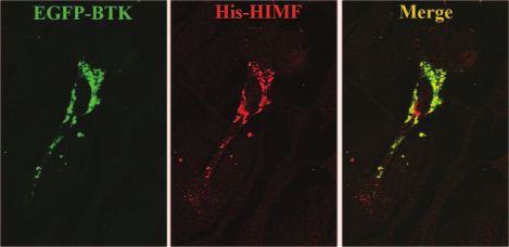
A/G mixture and incubated at 4°C for 3 h. After being lysate for GST and GST-HIMF protein from BL21. We
washed by TBST three times, 300 l of TBST and 30 l of then conducted an experiment to pull down the bind-
bone marrow cell lysate were mixed with the IgG bound ing protein from the lysate of bone marrow primary
agarose-protein A/G and incubated at 4°C for 3 h. The
samples were washed by TBST three times and run on 4 –20% cultured cells. As shown in Fig. 1, GST-HIMF pulled
SDS-PAGE gel. Rabbit anti-HIMF antibody was used for the down a protein of ⬃70 kDa. To identify the amino acid
blotting. sequence for the HIMF-binding protein, the candidate
band was cut from the gel and sent for mass spectrom-
Mouse hind limb ischemic model etry analysis. Peptide mass fingerprinting (PMF), and
tandem mass spectrum data from MALDI-TOF-MS were
Animals were subjected to left femoral artery ligation and analyzed by searching against an NCBInr database
excision to create unilateral hind limb ischemia. For each using MASCOT (Matrix Science, London, UK) search
animal, 25 mg/kg ketamine plus 10 mg/kg xylazine were software. With the use of this approach, a protein
injected subcutaneously. Skin incisions were performed at the
middle portion of the left hind limb overlying the femoral
corresponding to BTK that is involved in B cell matu-
artery. The femoral artery was gently isolated. First the ration was found.
proximal portion and then the distal portion of the femoral To confirm our finding, we conducted two additional
artery were ligated, and then other arterial branches as well as binding experiments. First, bone marrow lysate and
veins were dissected free and excised. The overlying skin was bacteria lysates of GST and GST-HIMF were used in
closed using two surgical staples. Tissue in the hypoxia area pull-down assays. GST-HIMF pulled down BTK from
was removed and homogenized in TBST buffer 2 wk after the bone marrow cell lysate but GST did not. When homog-
operation.
enate of mouse hind limb hypoxic tissue was used
Cell migration assay instead of bone marrow cell lysate in the GST-HIMF
pull-down assay, BTK was again shown to bind to
Bone marrow cells were detached with trypsin-EDTA, washed GST-HIMF but not to GST. These results further indi-
in serum-free medium, and then counted and adjusted to106 cate that BTK is a HIMF binding partner. Next, we used
cells/ml. Five hundred microliters of the cell suspension was BTK antibody to test whether HIMF can be coprecipi-
placed in the Transwell membranes and allowed to migrate to tated with BTK from endogenous proteins. Again, BTK
the underside for 6 h or overnight at 37°C in the presence of antibody can pull down HIMF from bone marrow cell
50 nM BSA, HIMF, or HIMF plus 25 M of the BTK inhibitor, lysate but rabbit IgG cannot.
LFM-A13. The cells were fixed in precooled methanol and
stained with Coomassie blue solution for 10 min. The cells on
the top chamber were removed with a cotton swab, and the Colocalization between BTK and HIMF
cells migrating to the underside of the filter were visualized
and photographed randomly using a Nikon Eclipse micro- To demonstrate that BTK acts as a HIMF binding
scope. Migration cells were counted under the microscope or
partner, we conducted experiments to show whether
from the pictures.
BTK and HIMF colocalize together in bone marrow
Immunofluorescence microscopy cells. Bone marrow cells were cultured on cover glasses
and cotransfected with enhanced GFP (EGFP)-BTK
Bone marrow cells were cultured on coverslips in DMEM and his-HIMF plasmids. The transfected cells were then
containing 10% FBS and fixed in precooled methanol for 5 fixed by methanol and used for immunofluorescence
min. The cells were then permeabilized with 0.2% Triton by anti-his mAb and rhodamine-labeled donkey anti-
X-100 in PBS and blocked with 0.5% BSA in PBS followed by mouse IgG. As shown in Fig. 2, BTK and HIMF colo-
incubation with the indicated antibodies. FITC-donkey anti- calized in transfected bone marrow cells.
rabbit IgG or FITC-donkey anti-rat IgG and rhodamine-
donkey anti-mouse IgG were used as second antibodies. For
transfection experiments, cells were cultured overnight and Translocation of BTK in bone marrow cells in
transfected with indicated constructs in serum-free medium response to the stimulation of HIMF
for 4 h and then changed into DMEM containing 10% FBS
overnight. Cells were fixed and stained as above. A 510 BTK family tyrosine kinases have been shown to regu-
confocal microscope was used for the imaging.
late actin cytoskeleton and to mediate cell mobility in
response to stimulation (7, 8). The involvement of BTK
Statistical analysis
in thrombin-stimulated platelets (9, 10) suggested that
All the results are expressed as mean ⫾ se. Differences BTK may be a mediator of cytoskeleton reorganization.
between groups were analyzed by the Student-Newman-Keul’s The activation of BTK family tyrosine kinases will result
method with P ⬍ 0.05 considered to be significant. in their stimulated translocation to membrane fractions
(11). As a partner of BTK, HIMF may be involved in
BTK signaling pathways and play a role in regulation of
RESULTS BTK activity. We therefore conducted an assay to
examine whether the distribution of BTK in bone
BTK was pulled down by GST-HIMF marrow cells was altered in response to the stimulation
of HIMF. Bone marrow cells were cultured on cover-
To search for HIMF binding partners, we first con- slips in 12-well plates for 2 days and then treated with
structed GST-HIMF plasmids and prepared bacterial HIMF (50 nM) or BSA (50 nM) for 5 min. The cells
1378 Vol. 21 May 2007 The FASEB Journal SU ET AL.BTK phosphorylation, we treated the cells with HIMF
in time series of 2.5, 5, 10, and 20 min. The phosphor-
ylation of BTK reached a peak at or before 2.5 min and
gradually decreased after 10 min with the treatment of
HIMF. To verify if HIMF specifically activates BTK, the
same cell lysates used in BTK phosphorylation experi-
ments were used for the phosphorylation study of Fyn.
As shown in Fig. 5C, phosphorylation sites of both the
activated and inactivated state of Fyn remained the
same in response to the stimulation of HIMF. There-
fore, HIMF is a specific stimulator for the activation of
BTK. Fyn was heavily phosphorylated in bone marrow
cells without any stimulation, suggesting nonspecificity.
BTK stimulated the migration of bone marrow cells
To test whether HIMF stimulates the migration of bone
marrow cells, bone marrow cells were cultured in
transwells in the presence of 50 M HIMF or BSA. The
number of migrated cells was significantly increased in
the membrane treated with HIMF for the overnight
culture (Fig. 6A). To demonstrate whether BTK is
involved in HIMF-stimulated cell migration, cells were
Figure 1. BTK is a HIMF-binding molecule. GST-HIMF fusion cultured in transwell as above in the presence of 50 M
protein and GST were incubated with glutathione-Sepharose BSA, 50 M HIMF, or 50 M HIMF plus 25 M BTK
beads and then incubated with bone marrow cell lysate, inhibitor LFM-A13 for 6 h. Significantly more bone
respectively, in TBST buffer at 4°C for 3 h. Beads were washed
5 times by TBST, and loading buffer was added to samples.
marrow cells were stimulated to migrate out of the
After electrophoresis, gel was stained by Coomassie blue. HIMF stimulated transwell than from the transwell
Arrow shows a HIMF-binding candidate protein (A). GST- treated with BSA (Fig. 6B). The migration stimulated by
HIMF and GST were first bonded to glutathione-Sepharose HIMF was blocked by the BTK inhibitor LFM-A13 (Fig.
and then incubated with lysate of bone marrow cells (left) or 6B). Statistical analysis using Student-Newman-Keuls
homogenate of ischemic tissue (right) at 4°C for 3 h and then method showed a significant change (P⬍0.001) in the
washed by TBST solution 3 times. After SDS-PAGE and number of migrated cells after treatment with HIMF
transfer to membrane, samples were detected by anti-BTK
and anti-GST antibodies (B). Agarose-protein A/G bound (Fig. 6C).
rabbit IgG and rabbit anti-BTK antibody were incubated with
bone marrow cell lysate respectively in TBST buffer at 4°C for
3 h and then washed by TBST 3 times before SDS-PAGE. DISCUSSION
Rabbit anti-HIMF antibody was used for immunoblot (C).
RT-PCR was conducted by using cDNA prepared from bone BTK has been shown to be crucial in the regulation of
marrow cells and from RAW cells as described in Materials B-cell maturation, and defects in BTK lead to X-linked
and Methods. -actin primers were used as a control (D).
agamma globulinemia in humans and X-linked immu-
nodeficiency defect in mice (5, 6). BTK family kinases
were fixed in precooled methanol at ⫺20°C for 10 min play diverse roles in various cellular processes including
and used for immunocytochemistry. As the results show
in Fig. 3 and Fig. 4, HIMF was found to induce a rapid
redistribution of BTK to cell processes, the leading
edge of cells (Fig. 3D–L), where BTK colocalizes with
actin (Fig. 4A) and FAK (Fig. 4B). Fyn, a binding
protein of BTK, was also found colocalized with BTK in
the cell processes.
BTK was autophosphorylated by HIMF stimulation
To address whether HIMF can activate BTK, we tested
the autophosphorylation of BTK in response to HIMF.
Bone marrow cells expanded from mouse were stimu- Figure 2. Colocalization between BTK and HIMF. EGFP-BTK
and His-HIMF plasmids were cotransfected into bone marrow
lated with HIMF (50 nM). HIMF induced BTK auto- cells. Cells were fixed by precooled methanol and stained by
phosphorylation at site Y223 (Fig. 5A). The tyrosine antihis mAb and rhodamine-labeled donkey anti-mouse IgG
phosphorylation of BTK returned to normal after 30 second antibody. EGFP-BTK and his-HIMF colocalized very
min of treatment. To find out the more exact process of well in transfected cells.
HIMF BINDS TO BTK AND INITIATES CHEMOTAXIS 1379example to demonstrate the physiological importance
of this kinase. In the regulation of differentiation of
bone marrow hematopoietic cells into B cells, the BTK
pathway is obviously necessary. Other members of the
BTK family kinase have been shown to be involved in
the signaling pathway of integrins that are key mole-
cules regulating actin cytoskeleton and cell mobility
(13).
By using bone marrow cells as the starting material in
our study, we demonstrated that BTK is a HIMF bind-
ing molecule. As an inflammatory marker molecule,
HIMF may likely be involved in the regulation of the
immune system in response to inflammatory stimula-
tion (3). A large number of studies have shown that
leukocytes promote angiogenesis in inflammatory tis-
sues by delivering vascular endothelial growth factor
(VEGF) to the target sites (14), where vascular remod-
eling is important for the tissue regeneration. The
activation of BTK will induce the differentiation and
migration of bone marrow-derived leukocytes that may
be involved in inflammatory responses in hypoxic tis-
sues (15). HIMF, acting as a chemotactic molecule, may
stimulate the migration of bone marrow derived cells to
targeted tissue in response to tissue inflammation or
hypoxia.
Pleckstrin homology domains (PH) are commonly
found in eukaryotic signaling proteins. They are often
involved in protein-protein interactions and target pro-
teins to the plasma membrane. Mutations in BTK
within its PH domain cause XLA in patients (5, 6).
Figure 3. BTK redistribution in response to the stimulation of
HIMF in primary cultured bone marrow. Bone marrow cells HIMF was up-regulated in inflammatory or hypoxic
were cultured on cover glass and then treated with 50 nM BSA tissues and stimulated the phosphorylation of AKT, a
(A–C) or HIMF (D–L) for 5 min. Cells were fixed by pre- kinase with a PH domain, in cultured cells (1). Inter-
cooled methanol before indirect immunofluorescence. BTK estingly, BTK is also a PH domain containing molecule.
or FYN, a binding partner for BTK, was redistributed in bone HIMF can also stimulate the autophosphorylation of
marrow cells after the treatment of HIMF. Outline of cell (D, BTK in bone marrow cells (Fig. 5), indicating that BTK
E) was shown in I. DIC approach was also used to show the
leading edge of cell (J–K).
is a HIMF-targeted molecule that is activated in re-
sponse to the stimulation of hypoxia or inflammatory
reactions. Fyn, another soluble tyrosine kinase of src
growth, differentiation, apoptosis, cytoskeletal reorga- family members, was not changed in activity by the
nization, and cell motility (7–10, 12). The mutation of stimulation of HIMF, although Fyn was reported as a
BTK resulting in immunodeficiency diseases is a good BTK-binding protein (16) and shared common distri-
Figure 4. BTK colocalizes with FAK and actin in
HIMF stimulating cells. Bone marrow cells were
cultured on cover glass in 12-well plates in
DMEM culture medium containing 1% FBS
and then treated with 50nM HIMF for 5 min.
Cells were fixed by precooled methanol at
⫺20°C for 5–10 min. Monoclonal anti-BTK
antibody and rabbit polyclonal anti-FAK (A) or
antiactin (B) antibody were used for indirect
immunofluorescence. Signals of BTK overlap
with those of FAK and actin in cells.
1380 Vol. 21 May 2007 The FASEB Journal SU ET AL.one of the physiological functions of HIMF. Recently,
we have found that intravenously tail vein injection of
HIMF in mice caused a marked increase of CD68-
Figure 5. BTK self-phosphorylation in response to treatment
of HIMF. Three plates of primary cultured bone marrow cells
that were passed from same plate of cells were cultured to
confluence and then treated with HIMF (5 and 30 min) or
without HIMF. Cell lysates were used for the Western blot.
Membrane was first probed by rabbit polyclonal anti-BTK
phosphorylation (Y223) antibody and then was probed by
anti-BTK mAb after stripping (A). Five plates of primary
cultured bone marrow cells that were passed from the same
plates of cells were cultured to confluence and then treated
with HIMF (2.5, 5, 10, and 20 min respectively) or without
HIMF. Cell lysates were used for the Western blot. The
membrane was first detected by Rabbit polyclonal anti-BTK
phosphorylation (Y223) antibody and then by anti-BTK mAb
after stripping (B). Same samples were used for the detection
of Fyn phosphorylation (C). Rabbit antiphospho-Src family
(Y416) pAb or monoclonal Anti-Fyn (Y528) phospho-specific
antibody were used for the first blot. Then membranes were
probed by rabbit anti-Fyn poloyclonal antibody to show equal
loading after stripping. Phosphorylation of Y416 up-regulates
enzyme activity. Phosphorylation of Y528 negatively regulates
enzyme activity.
bution with BTK in the cells (Fig. 3). But the redistri-
bution of Fyn and colocalization with BTK after treat-
ment with HIMF suggested that Fyn may be involved in
the BTK signaling pathway. BTK was found to colocal-
ize with actin and FAK at the cell process (Fig. 4). These
results indicate that BTK is active in the mediation of
cell migration in response to HIMF stimulation. The
heavy phosphorylation of Fyn with or without HIMF
simulation indicates that Fyn may be a common inter-
mediate regulator in the cell signaling process. Conse-
quently, BTK is a specific targeted molecule for HIMF
binding and interaction. Cells cotransfected with GFP-
BTK and His-HIMF plasmids also showed a clear colo- Figure 6. HIMF stimulated bone marrow cell migration; 5 ⫻
calization of BTK and HIMF (Fig. 2). 105 bone marrow cells were cultured in transwell plates in the
When bone marrow cells were treated with HIMF, presence of 50 nM BSA or HIMF. Cells were cultured
BTK was recruited to the leading edge of the cells (Figs. overnight, fixed in precooled methanol and stained by Coo-
3, 4). This result further indicated that HIMF stimulates massie blue solution. Cells growing on the surface of the
membrane were removed after staining (A). A Nikon Eclipse
the migration of bone marrow cells. By using the microscope was used for the imaging. 5 ⫻ 105 bone marrow
transwell migration assay, we found that HIMF mark- cells were cultured as above in the presence of 50 nM BSA,
edly stimulated bone marrow cell migration. The che- HIMF, or HIMF plus 25 M LFM-A13, respectively. Cells were
motactic characteristic of HIMF was shown to be de- cultured for 6 h, fixed in precooled methanol, and stained by
pendent on the activation of BTK because the BTK Coomasie Blue solution. Cells growing on the surface of
inhibitor completely inhibited the chemokine-like membrane were removed after staining. Bone marrow cell
function of HIMF in the bone marrow cell migration migration was induced by HIMF, and this induction was
inhibited by BTK inhibitor LFM-A13 (B). Migration cells as
assay. Hence, HIMF stimulates the migration of bone described in B were counted under a microscope and quan-
marrow cells through the activation of BTK and is a titated as in C (n⫽4). Differences between groups were
chemotatic factor for bone marrow derived cells. The analyzed by Student-Newman-Keul’s method with P ⬍ 0.001
recruitment of leukocytes to the target tissues may be (P⬍0.05 is considered to be significant).
HIMF BINDS TO BTK AND INITIATES CHEMOTAXIS 1381positive inflammatory cells in the lungs (17). This in 6. Thomas, J. D., Sideras, P., Smith, C. I., Vorechovsky, I., Chap-
man, V., and Paul, W. E. (1993) Colocalization of X-linked
vivo result is fully consistent with our hypothesis that agammaglobulinemia and X-linked immunodeficiency genes.
leukocytes may be recruited to hypoxia tissues in re- Science 261, 355–358
sponse to HIMF. 7. Mangla, A., Khare, A., Vineeth, V., Panday, N. N., Mukho-
HIMF is a secreted protein, while BTK is reported to padhyay, A., Ravindran, B., Bal, V., George, A., and Rath, S.
(2004) Pleiotropic consequences of Bruton tyrosine kinase
be a soluble cytoplasmic molecule. We know that HIMF deficiency in myeloid lineages lead to poor inflammatory re-
is excreted because we produce it in eukaryotic cells, sponses. Blood 104, 1191–1197
extracting it from the media in which they grow (1). 8. Abassi, Y. A., Rehn, M., Ekman, N., Alitalo, K., and Vuori, K.
BTK is a tyrosine kinase which makes it a potential (2003) p130Cas Couples the tyrosine kinase Bmx/Etk with
regulation of the actin cytoskeleton and cell migration. J. Biol.
receptor-like molecule. Although there is a region on Chem. 278, 35636 –35643
the molecule that is a possible transmembrane domain 9. Laffargue, M., Ragab-Thomas, J. M., Ragab, A., Tuech, J., Missy,
according to some programs (such as TMpred and DAS K., Monnereau, L., Blank, U., Plantavid, M., Payrastre, B.,
Raynal, P., et al. (1999) Phosphoinositide 3-kinase and integrin
program) for membrane prediction, BTK is mostly signalling are involved in activation of Bruton tyrosine kinase in
localized in cytoplasm. BTK indeed can localize to thrombin-stimulated platelets. FEBS Lett. 443, 66 –70
membrane as we and others have shown, but whether it 10. Hamazaki, Y., Kojima, H., Mano, H., Nagata, Y., Todokoro, K.,
becomes integrated with the membrane or just binds to Abe, T., and Nagasawa, T. (1998) Tec is involved in G protein-
coupled receptor- and integrin-mediated signalings in Human
the membrane is not known. HIMF is a small cysteine blood platelets. Oncogene 16, 2773–2779
rich protein. Whether it functions as a ligand extracel- 11. Varnai, P., Rother, K. I., and Balla, T. (1999) Phosphatidylino-
lularly or acts as a modulator intracellularly or both also sitol 3-kinase-dependent membrane association of the Bruton’s
tyrosine kinase pleckstrin homology domain visualized in single
remain a puzzle. Considering that many molecules like living cells. J. Biol. Chem. 274, 10983–10989
the thiorodoxin-like molecule (18), and EF hand mol- 12. Hamazaki, Y., Kojima, H., Mano, H., Nagata, Y., Todokoro, K.,
ecules (19) can work both inside and outside of the Abe, T., and Nagasawa, T. (1998) Tec is involved in G protein-
cells and function as chemokine-like proteins, HIMF coupled receptor- and integrin-mediated signalings in Human
blood platelets. Oncogene 16, 2773–2779
may function in a similar manner. 13. Chen, R., Kim, O., Li, M., Xiong, X., Guan, J. L., Kung, H. J.,
Chen, H., Shimizu, Y., and Qiu, Y. (2001) Regulation of the
PH-domain-containing tyrosine kinase Etk by focal adhesion
kinase through the FERM domain. Nat. Cell Biol. 3, 439 – 444
14. Scapini, P., Morini, M., Tecchio, C., Minghelli, S., Di Carlo, E.,
Tanghetti, E., Albini, A., Lowell, C., Berton, G., Noonan, D. M.,
REFERENCES et al. (2004) CXCL1/macrophage inflammatory protein-2-in-
duced angiogenesis in vivo is mediated by neutrophil-derived
vascular endothelial growth actor-A. J. Immunol. 172, 5034 –5040
1. Teng, X., Li, D., Champion, H. C., and Johns, R. A. (2003)
15. Jefferies, C. A., and O’Neill, L. A. (2004) Bruton’s tyrosine
FIZZ1/RELM␣, a novel hypoxia-induced mitogenic factor in
kinase (Btk)-the critical tyrosine kinase in LPS signalling?
lung with vasoconstrictive and angiogenic properties. Circ. Res.
Immunol. Lett. 92, 15–22. Review
92, 1065–1067
16. Alexandropoulos, K., Cheng, G., and Baltimore, D. (1995)
2. Wagner, K. F., Hellberg, A. K., Balenger, S., Depping, R.,
Proline-rich sequences that bind to Src homology 3 domains
Dodd-O, J., Johns, R. A., and Li, D. (2004) Hypoxia induced
with individual specificities. Proc. Natl. Acad. Sci. U. S. A. 92,
mitogenic factor has antiapoptotic action and is up-regulated in
3110 –3114
the developing lung: coexpression with hypoxia-inducible fac-
17. Yamaji-Kegan, K., Su, Q., Angelini, D. J, Champion, H. C., and
tor-2␣. Am. J. Respir. Cell Mol. Biol. 31, 276 –282
Johns, R. A. Hypoxia-induced mitogenic factor has pro-angio-
3. Holcomb, I. N., Kabakoff, R. C., Chan, B., Baker, T. W., Gurney,
genic and pro-inflammatory effects in the lung via VEGF and
A., Henzel, W., Nelson, C., Lowman, H. B., Wright, B. D.,
VEGF receptor-2. Am. J. Physiol. Lung Cell Mol Physiol. [Epub
Skelton, N. J., et al. (2000) FIZZ1, a novel cysteine-rich secreted
ahead of print] 2006 Aug 4
protein associated with pulmonary inflammation, defines a new
18. Bertini, R., Howard, OM., Dong, HF., Oppenheim, JJ., Bizzarri,
gene family. EMBO J. 19, 4046 – 4055
C., Sergi, R., Caselli, G., Pagliei, S., Romines, B., Wilshire, JA., et
4. Chumakov, A. M., Kubota, T., Walter, S., and Koeffler, H. P.
al. (1999) Thioredoxin, a redox enzyme released in infection
(2004) Identification of murine and human XCP1 genes as
and inflammation, is a unique chemoattractant for neutrophils,
C/EBP-epsilon-dependent members of FIZZ/Resistin gene fam-
monocytes, and T cells. J. Exp. Med. 189, 1783–1789
ily. Oncogene 23, 3414 –3425
19. Xu, K., and Geczy, C. L. (2000) IFN-gamma and TNF regulate
5. Vetrie, D., Vorechovsky, I., Sideras, P., Holland, J., Davies, A.,
macrophage expression of the chemotactic S100 protein
Flinter, F., Hammarstrom, L., Kinnon, C., Levinsky, R., Bobrow,
S100A8. J. Immunol. 164, 4916 – 4923
M., et al. (1993) The gene involved in X-linked agammaglobu-
linaemia is a member of the src family of protein-tyrosine Received for publication May 26, 2006.
kinases. Nature 361, 226 –233 Accepted for publication December 6, 2006.
1382 Vol. 21 May 2007 The FASEB Journal SU ET AL.You can also read