HYDROXYCHLOROQUINE AND AZITHROMYCIN MOLECULAR ACTION AGAINST SARS-COV-2 VIRAL PROTEIN: A MOLECULAR DYNAMIC STUDY
←
→
Page content transcription
If your browser does not render page correctly, please read the page content below
Open Access

Austin Journal of Nanomedicine &
Nanotechnology

Research Article

Hydroxychloroquine and Azithromycin Molecular Action
against SARS-CoV-2 Viral Protein: A Molecular Dynamic
Study
Picaud F* and Herlem G
                                                                  Abstract
Nanomedicine Lab EA4662, University of Franche-
Comté, France                                                          For the past few months, the world has gone through hell with the
*Corresponding author: Fabien Picaud,                             emergence of the SARS-CoV-2 virus and the resulting pandemic. Faced with
Nanomedicine Lab EA4662, University of Franche-                   this disease, various therapeutic strategies have been developed to understand
Comté, UFR Sciences & Techniques, 16 Route de Gray,               how to eradicate this virus. Here we present a molecular dynamics simulation
25030 Besançon Cedex, France                                      study on the effect of a dual therapy (hydroxychloroquine and azithromycin)
                                                                  on the open and closed forms of a viral protein. We show in particular that
Received: December 03, 2020; Accepted: December                   hydroxychloroquine has no significant interaction with the viral receptor-binding
28, 2020; Published: January 04, 2021                             domain RBD when it interacts with its host receptor. However, this molecule can,
                                                                  in the closed form of the virus, block the movement of these receptors and thus
                                                                  prevent the attachment of the virus to the host cell. The azithromycin molecule
                                                                  interacts very well with the open receptor but can also be inserted into the S2
                                                                  domain of the protein. It therefore presents two potential mechanisms of action
                                                                  against the virus, mainly on the closed state of the viral protein.
                                                                     Keywords: Hydroxychloroquine; Azythromicin; SARS-CoV-2 structure;
                                                                  MD-Simulation; Docking

Introduction                                                                   these figures lead to a mortality rate close to 2.1%, i.e. 4 times the
                                                                               rate of the seasonal flu. While the peak of the pandemic seems to be
     In 2002, the first emergence of a pathogenic coronavirus revealed         behind us, the second wave forces us to find a therapeutic method to
to the world the possibility of a pandemic. The severe acute respiratory       treat this virus.
syndrome coronavirus, or SARS-CoV, was responsible for very
important breathing syndromes [1-4] with, however, a small amount                  Many drugs have been clinically tested in numerous clinical
of death around the world (8000 persons) while the mortality rate              projects (“discovery” for France, “recovery” for England”) but no one
reached 10%. More recently (2012), a severe pneumonia appeared in              has determined a truly effective treatment against the SARS-CoV-2
Saudi Arabia due to a novel coronavirus [5,6]. This one, called MERS-          virus. In parallel, a protocol, confirmed by many other studies [14-
CoV for Middle East Respiratory Syndrome Coronovirus, still exists             17], seems to be the most appropriate for combating the virus and
but concerns only the Arabic peninsula. While very localized on a              reducing the degree of contagiousness. This protocol, that should be
small area, this MERS-Cov is highly dangerous since its mortality rate         administered as soon as the first symptoms appear, combines both
is about 35% (1 over 3 patients). These two cases are not the only             the antimalarial drug Hydroxychloroquine (HCQ) and the antibiotic
ones since periodically, other coronaviruses, while less virulent are          Azithromycin (AZM). The results of the various studies lead to a
appearing [7,8,9].                                                             sharp decrease of the mortality rate (under 0.5%). However, many
                                                                               other analyzes question the results of the therapies. In order to better
    These first viral apparitions should have alerted us to the                understand the role of this double drug treatment against this new
possibility, in the long term, of the emergence of a more virulent             virus, an analysis of its action is necessary at the molecular level [18].
coronavirus clearly difficult to manage. At the end of 2019, a new
coronavirus called severe acute respiratory syndrome coronavirus                    With regard to its genome sequence, SARS-CoV-2 belongs to
2 (SARS-CoV-2), or COVID- 19 disease, developed as a human                     the same beta-coronavirus family as SARS-CoV and MERS-CoV.
pathogen in a Chinese city (Wuhan). Despite strong resolutions in              These coronaviruses have a spherical envelope with a diameter close
China and over several states (locking up millions of people), global          to 100 nm. The latter is composed of a N nucleoprotein surrounded
economic and touristic development is leading to a general spread of           by a lipid bilayer originating from the host cell. Three other proteins
the disease. Although SARS-CoV-2 has many points in common with                are then found on this surface, protein S (or spike protein), protein
SARS-CoV and MERS-CoV, it appears to be transmitted more easily                M (or membrane protein), and protein E (or envelope protein).
and much faster than SARS-CoV [10,11] and MERS-CoV [12,13]. To                 Homotrimerization of S proteins [11] on the surface of the virion
date, more than 64 million cases of COVID-19 have been confirmed               is the key step in viral infection. To decrease the viral progression,
and at least 1.400,000 deaths have been recorded worldwide by the              the drug must therefore target the S protein. However, the latter is
World Health Organization which declared the first real pandemic               separated into two domains which have a very specific role [19]. The
of March 21, 2020. Although all the cases have not been identified,            S1 domain, mainly formed by a Receptor Binding Domain (RBD),

Austin J Nanomed Nanotechnol - Volume 9 Issue 1 - 2021     Citation: Picaud F and Herlem G. Hydroxychloroquine and Azithromycin Molecular Action against SARS-CoV-2
ISSN : 2381-8956 | www.austinpublishinggroup.com           Viral Protein: A Molecular Dynamic Study. Austin J Nanomed Nanotechnol. 2021; 9(1): 1061.
Picaud et al. © All rights are reserved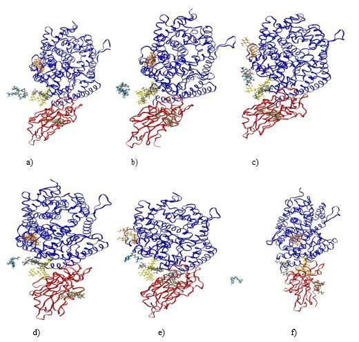
Picaud F                                                                                                                    Austin Publishing Group

binds to the host cell receptor [20] (called angiotensin converting
enzyme 2 (ACE2) [21,22] while that the S2 domain is at the origin of
the fusion of protein E with the host cell [19].
    Therefore, the main objective of any viral treatment is to block the
binding of the RBD domain to the ACE2 receptor in order to avoid
the fusion of the virus with the host cell. Note that the drugs could
prevent protein fusion with the host cell through structural changes.
     Here we propose to determine what are the main sites of
interaction of the HCQ and the AZM drug molecules on structural S
protein. Our work, based on several molecular dynamics simulations
will be separated into two parts. We will first determine the binding of
each drug when RBD is associated with the ACE2 receptor. Then, in a
second step, we will show that these molecules can also have another
target to on the state close to the S protein.
Method                                                                      Figure 1: Molecular structure of a) hydroxychloroquine, b) azithromycin, c)
                                                                            RBD (red)+ACE2 (blue) complex and d) 6VXX protein (note that the RBD,
    Hydroxychloroquine (HCQ) is (RS)-2-[[4-[(7-chloroquinoline-             NTD and S2 parts 11of the protein were depicted in van der Waals, licorice
4-yl)amino]pentyl](ethyl)amino] ethanol. Its 3D structure has been          and ribbon+lines modes for the three monomers shown in different colors).
obtained through the Pub-Chem CID: 3652 file. Azithromycin
(AZM) is (2R, 3S, 4R, 5 100 R, 8R, 10R, 11R, 12S, 13S, 14R)-11-            finally solvated in a water box large enough to prevent interaction of
[(2S, 3R, 4S, 6R)-4-(dimethylamino)-3-hydroxy-6-methyloxan-2-yl]           each entity with its neighbor in adjacent cells when periodic boundary
oxy-2-ethyl-3, 4, 10-trihydroxy-13- [(2R, 4R, 5S, 6S)-5-hydroxyl-          conditions are used. To mimic a salt medium, NaCl ions (at a
4-methoxy-4,6-dimethyloxan-2-yl] oxy-3, 5, 6, 8, 10, 12, 14 -              concentration of 0.15M) were added to the water model (transferable
heptamethyl-1-oxa-6-azacyclopentadecan-15-one. Its 3D structure            intramolecular potential with 3 points, TIP3P). CHARMM36 force-
has been obtained through the Pub-Chem CID: 447043 file.
                                                                           field optimization parameters are used in all simulations [29]. The
    Our strategy was organized in two different steps. First of all, we    complete systems contained 245805, 245757, 301911 and 301896
focused on the simulation of the Receptor Binding Domain (RBD)             atoms, for HCQ/RBD, AZM/RBD, HCQ/6VXX and AZM/6VXX
bound to the ACE2 receptor of the host cell with the HCQ or the            systems, respectively.
AZM molecules. The goal of these first calculations is to observe
                                                                               All simulations were performed under constant temperature and
whether the molecules of the drug can interact directly with the viral
                                                                           pressure with a 13Å cutoff for unbonded forces. The temperature is
protein when they are attached to its host cell. To simulate such
                                                                           fixed at 310K (Langevin dynamics) [30] and the pressure at 1atm
arrangement of proteins, we use the 6M0J pdb structure. The relaxed
                                                                           (Langevin piston), [31] respectively. Long range electrostatic forces
crystal structures approaching the living organism as well as the effect
                                                                           are evaluated using the classical Particle Mesh Ewald (PME) method
of glycosylation on the stability of the structure were studied.
                                                                           [32] with a grid spacing of 1.2Å, and fourth-order spline interpolation.
    Then, in a second step, the full conformation of the spike             The simulations were carried out in three stages: first, a minimization
glycoprotein trimer SARS-CoV-2 was simulated in presence of HCQ            stage (steepest descent) of 5000 steps was carried out to remove the
or AZM molecules. Its structure was obtained from pdb file #6VXX.          strongest atomic hindrance from the system. Then, an equilibration
It has a resolution of 2.80Å as determined from electron microscopy.       phase was imposed on the system where the movement of the protein
It is composed of 3 chains intercalated with different respective          were constrained in order to stabilize the drug, the solvent and the
domains such as the NTD (N-terminal domain) and the RBD                    ions around the protein. The harmonic force constants on the protein
(receptor binding domain) which belong to the S1 part of the protein.      backbone and the sidechain were chosen to be respectively equal
                                                                           to 400kJ mol-1 nm-2 and 40kJ mol-1nm-2, for a total duration of 1ns.
     Classical MD simulations were performed by constructing the
                                                                           Finally, the production step was performed without position restrains
full molecular force field for HCQ and AZM using the SwissParam
                                                                           for a total of at least 60ns (time step of 1fs). Each simulation uses
Force Field Toolkit package [23,24].
                                                                           periodic boundary conditions in the three directions of space and
     For the protein, the molecular force field was constructed            the list of neighbors is refreshed every 12-time steps. No constraint is
according to the CHARMM-GUI procedure in order to appropriately            imposed during the production phase of MD simulations. Note that
relax the different parts of the protein [25,26]. N-glycosylation of       for system equilibration, all bond lengths involving hydrogen atoms
the proteins, when necessary, was achieved using the CHARMM-               were fixed using the SHAKE algorithm [33]. Consequently, our
GUI Glycolipid Modeler.[27] The structure of these proteins was            results are obtained with all the atoms left free in the simulation box.
first minimized and then progressively equilibrated (1ns) and run
(40ns) using MD simulations (NAMD 2.12 package) for a total of 41              To determine the sites of interaction in such huge systems,
ns in saline solution media [28]. All the structures of proteins and       several simulations were run with different starting configurations.
molecules are shown in Figure 1a-d.                                        These configurations were obtained through a docking procedure
                                                                           between the viral protein and the drugs. AutoDock Vina (a fast and
    The systems (protein or protein+drug) were then combined and           accurate evolution of AutoDock) was used as the molecular docking

Submit your Manuscript | www.austinpublishinggroup.com                                      Austin J Nanomed Nanotechnol 9(1): id1061 (2021) - Page - 02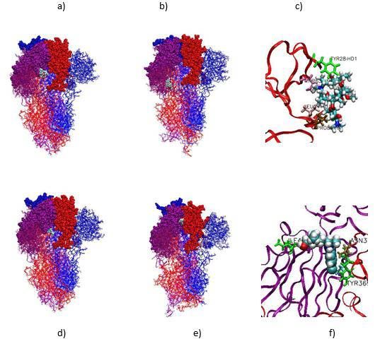
Picaud F                                                                                                                       Austin Publishing Group

engine, being able to process a massive number of ligand positions
in a relatively short time. The different starting configurations used
in the molecular dynamics simulations were thus chosen with regard
to the best scoring functions obtained thanks to the optimization
algorithm. When the drug interacted with the 6M0J, at least 7 different
simulations were run for each drug. For the 6VXX system, only 3
simulations were performed due to the huge number of atoms in the
system. Note that for each system, we also performed an additional
simulation with a drug position located outside the protein surface in
order to study the path to the active site of the protein.
Results
Relaxations of the proteins and the molecules in saline
solution
     The 6M0J and 6VXX structures were relaxed in their respective
solvated plus ionized solvent box via MD simulations. As with half of        Figure 2: a) RMSDs of 6M0J structures (with non-glycosylated sites (black
the proteins, the glycosylation of the protein should also be studied        curve) and with glycosylated ones (red curve)). b) Same but for 6VXX
[34]. Indeed, this could have an importance on how the virus adapted         structures. c-d) Superimposed crystallized (blue) and relaxed (red) structures
                                                                             for 6M0J. (c) and 6VXX (d) proteins, respectively. e-f) Superimposed
during the infection of the host cell [35]. Its impact on S protein          crystallized (blue) and relaxed (red) structures for glycolyzed 6M0J. (e) and
has been examined for Sars-CoV with mainly N- glycosylation type             6VXX (f) ) Proteins, respectively.
versus O-glycosylation [36]. Different mutagenesis analyzes [37] have
identified N-glycosylation sites. For 6M0J, the latter were determined
but only four sites (3 on ACE2 and 1 on RBD chain) were accessible
by GlyProt [38] (90, 322, 546 for ACE2 and 343 for RBD). For 6VXX,
we can find 22 possible N-linked glycosylation sites per monomer and
only 13 are conserved in the pdb structure. In this context and amongst
all the possibilities of glycosylation (sialylation, fucosylation, …), we
have limited our studies to branches of N-glycosylation formed by
association of N-acetyl-glycosamine (BGLC) and Mannose (BMAN
and AMAN) groups such as --ASN-BGLC-BGLC-BMAN-(AMAN)2.
    For each protein the progressive modification of the structure
was followed by calculations of its root mean square deviation. The
results are shown in Figure 2a-b and the superimposition of relaxed
glycosylated and non-glycosylated proteins are shown in Figure 2c-f.
    As shown in Figure 2a-b, the simulations ended as soon as the
RMSD and the total energy converged for a reasonable simulation
time thanks to the system size. The change in structure is similar,
although slightly higher for glycosylated proteins compared to non-          Figure 3: The three best stable configurations obtained through molecular
                                                                             dynamics simulations. a,b,c) for AZM; d,e,f) for HCQ. For each configuration,
glycosylated proteins. These slight differences mainly come from             the mean interaction energy and the protein residues interaction with the
the glycan groups which are free to move around in the solvent and           molecule are given to precise the differences between each system.
can assume random positions during their interactions with water
molecules. The 6M0J RMSDs converge towards a value close to                 Interactions of          the    RBD-ACE2           complex        with     the
2.5Å while the 6VXX ones tend to 4.0Å. The difference in molecular          therapeutics
weight between the two respective proteins could explain this                   Our initial investigation was devoted to the action of drugs on
RMSD difference. To compare the final structures obtained after the         the Receptor Binding Domain (RBD) bound to the ACE2 receptor
relaxation of the proteins, we describe the superimposed structure          exhibiting N-glycosylation. Indeed, this part of the Sars-Cov-V2
between the crystallized and relaxed structures in Figure 2c-f. As          virus is dedicated to binding the virus to the host cell. Most studies
expected, the proteins did not show strong changes in their backbone        aim to develop specific treatments the goal of which is to block the
and no significant folding or unfolding.                                    interaction between the virus (RBD) and the host cell (ACE2).
    HCQ and AZM were also relaxed under the same conditions                      Due to the large size of the system, docking simulations were
of solvation in order to start the simulations using both drugs and         first performed in order to determine the most relevant interaction
proteins with optimized structures. According to the analyzes of their      sites between the drugs and the protein. Seven configurations were
RMSD, the modification of their structure did not undergo a value           then carried out in the case of the HCQ/RBD systems with scoring
greater than 2.0Å, which is very low. We can use these structures to        functions varying from -7.78 to -6.73. For AZM/RBD systems, nine
study their interaction with the different proteins.                        systems were obtained with stable scoring functions, i.e. ranging from

Submit your Manuscript | www.austinpublishinggroup.com                                        Austin J Nanomed Nanotechnol 9(1): id1061 (2021) - Page - 03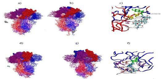
Picaud F Austin Publishing Group
Figure 5: a) Pair interaction (smooth data in line representation), and b)
RMSD of the RBD+ACE2 protein in presence of AZM (black) or HCQ (red)
drugs.
Figure 4: Figure 4a-c) Sketch of AZM positions during the simulation (at t=0
ns, t=4 ns and t=19 ns, respectively). d-f) Sketch of HCQ positions during
the simulation (at t=0 ns, t=7.4 ns and t=19 ns, respectively). The RBD is
depicted using red ribbon while ACE2 is in blue ribbon The chains showed in
color correspond to the N-glycan parts of the protein.
-9.94 to -7.52. Based on these different configurations, molecular
dynamics simulations in full solvent were performed in order to
test the stability of the drug under biological conditions. For these
simulations, a 1ns equilibration phase was performed before running
Figure 6: a-c) Initial and final AZM positions during the simulation (at t=0
some 10ns production simulations. The energy between the drug ns and t=42 ns, respectively), and protein residues in interaction with AZM
and the protein was then determined to obtain the most stable site of at the final stage of the simulation. d-f) Initial and final HCQ positions during
interaction by molecular dynamics simulation. the simulation (at t=0 ns and t=42 ns, respectively) and protein residues in
interaction with HCQ at the final stage of the simulation. The RBD, NTD
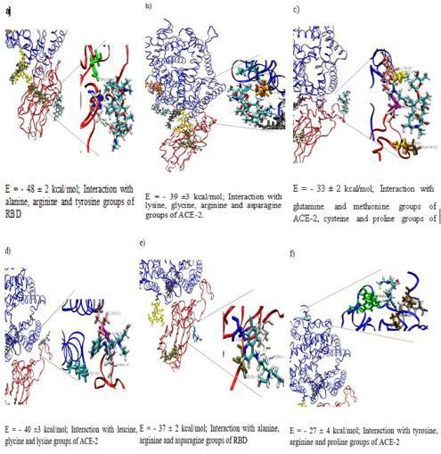
As shown in Figure 3, the different sites of interaction determined and S2 parts of the protein were depicted in van der Waals, licorice and
ribbon+lines modes for the three monomers shown in different colors.
by simulations can belong to several parts of the protein. RBD (red
part of 6M0J) is however at the origin of the best pair interaction find a better adsorption site on the protein, HCQ quickly desorbs and
energies for each molecule. We can also observe, from these data, that return to the solvent bath (Figure 4f), where it is confirmed by a pair
the AZM molecules tend to interact strongly with the 6M0J protein interaction equal to 0 with the protein (Figure 5a). On the contrary,
compared to HCQ. We did not observe any specific residue in the during the simulations, AZM never desorbed from the protein and
adsorption configurations. found a stable site which is more stable than the first one (Figure 4c)
We finally carried out a final simulation where the drugs were and of the same order as those obtained in Figure 3.
placed far from the adsorption sites of the protein (15Å). The The RMSDs of the protein were also plotted during these two
complete systems were gradually relaxed in order to study the production phases to see the impact of the drug on the stabilized
diffusion of the drug to this part of the protein. The two molecules protein structure (Figure 5b). Regardless of the drug considered, the
(protein and drug) being relaxed in independent simulations, RMSD followed the same trend when approaching the drug. It should
the equilibration phase here was equal to 1ns, and the system was be noted however that a series of upward peaks is observed around
running for a production phase for 38ns. We extracted during this t=14.4 ns, which corresponds to the arrivals of the HCQ on the
phase the pair interaction between drug and protein over time. Figure protein. These small successive deformations could be at the origin
4 shows different simulation times where we can observe the position of the HCQ desorption since for AZM case, we rather observed some
of the drug and also the state of the protein. At the beginning of the small downward peaks. It should be noted that we have not observed
simulation, the molecule is left 15.0Å from the junction between the any direct influence of the N-glycosylation functions toward the
RBD and ACE2. The drugs diffuse in the solvent due to different pair adsorption or the desorption of the HCQ and AZM molecules,
interactions and thermal agitation and come closer to the protein. although they are very labile when they are subjected to thermal
As can be seen in the middle of the simulation, each drug has found agitation. They could however interfere with the accessible sites
a site where it can be stabilized. Figure 5a, which exclusively shows determined by the previous calculations by steric hindrance.
the pairwise interaction of AZM (in black) and HCQ (in red) with
the protein, clearly demonstrates that the drug could be adsorbed on Interactions of the 6VXX trimer with the drugs
the protein. However, while the AZM molecule can arrange itself to HCQ and AZM were then relaxed close to optimized 6VXX
Submit your Manuscript | www.austinpublishinggroup.com Austin J Nanomed Nanotechnol 9(1): id1061 (2021) - Page - 04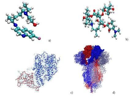
Picaud F                                                                                                                             Austin Publishing Group

 Figure 7: a) Pair interaction (smooth data in line representation), and b)
 RMSD of the 6VXX protein in presence of AZM (black) or HCQ (red) drugs.              Figure 9: a) Pair interaction (smooth data in line representation), and b)
                                                                                      RMSD of the 6VXX protein in presence of AZM (black) or HCQ (red) drugs.

                                                                                     The modification of the protein structure is directly impacted as soon
                                                                                     as the drugs fall into their potential valley. Indeed, although slightly
                                                                                     modified during the first ten ns of simulations, it sharply increases to
                                                                                     reach, after 42 ns, a value of 4Å for AZM (6Å for HCQ) which is not
                                                                                     stabilized yet (Figure 7b). We observed that the main deformation of
                                                                                     the 6VXX is located at the bottom of the protein, near the intrusion
                                                                                     zone. The progressive unfolding of this zone can be observed in the
                                                                                     sketches shown in figure 6. Although very stable when placed alone
                                                                                     in the solvent, this zone is particularly sensitive to the presence of the
                                                                                     drug during the simulations. The estimate of the RMSF (root mean
                                                                                     square fluctuations) of the protein confirms these observations since
                                                                                     the RBD (NTD) zones have a maximum RMSF equal to 3.2Å (4.7Å)
 Figure 8: a-c) Initial and final AZM positions during the simulation (at t=0 ns,    while the intrusion zone RMSF can reach a value of 8.3 Å.
 t=42 ns) and protein residues in interaction with AZM at the final stage of the
 simulation. d-f) Initial and final of HCQ positions during the simulation (at t=0        When the drugs are now placed far from the 6VXX structure
 ns, and t=42 ns, respectively) and protein residues in interaction with HCQ at      (close to 15Å) (Figure 8a-f), the pair interactions with the protein
 the final stage of the simulation. The RBD, NTD and S2 parts of the protein
 were depicted in van der Waals, licorice and ribbon+lines modes for the three
                                                                                     are at first zero then gradually become attractive, ending up with a
 monomers shown in different colors.                                                 large stabilized plateau in the last 15ns (Figure 9a). For AZM, this
                                                                                     well is located at the bottom of the trimer shown in ribbon+line
protein. Due to the large size of the protein, we only put the drug in               representations in Figure 8. The approach of the AZM is quite slow.
two different situations. First, the drugs were placed in a cavity of the            For 15ns, it did not interact with the protein. Then it finds an attractive
protein where the molecule could take place. This cavity is located                  interaction site and reaches its final stable state translating along
between two RBDs of the trimer and was determined by docking                         the surface of the protein (as shown in Figure 6a-c). The mean pair
calculations implemented in QuikVina-w [39]. The purpose of these                    interaction energy obtained after this simulation is equal to -30+/-5
first calculations is to determine if the drug could accommodate on                  kcal/mol. On the other hand, the HCQ molecule finds more rapidly its
the viral protein. Then, in order to simulate a more realistic diffusion             lower potential well (-40+/- 3kcal/mol) by diffusing in the solvent for
of the drugs, we let them freely approach the molecules. Due to the size             8ns then adsorbed on the protein in its lowest energy state from t=20
of the protein, and the time simulations, no additional configuration                ns until the end of the simulations (Figure 8d-e). The final adsorption
was performed as the production runs were longer than with RBD-                      state of the HCQ is between the RBD and the NTD. The modification
ACE2 alone to converge.                                                              of the protein structure is of the same order as previously. However,
                                                                                     we can notice that the RMSDs tend to the same stable value after 42ns
     When the drugs are placed in the cavity near the two RBD sites                  of simulations for AZM and for HCQ (around 6Å) as shown in Figure
(Figure 6a-f), the interaction with the protein is initially slightly                9b. The main deformation of the protein subjected to AZM is again at
attractive (Figure 7a). Then, progressively the molecules tend to                    the bottom of the protein, close to the intrusion zone. Furthermore,
translate and find the way to fall into a well of significant attractive             AZM is directly responsible for this deformation since it adsorbs
potential well. For AZM, this well is located near a N-Terminal                      precisely in this zone at the end of the simulation and progressively
Domain (NTD) shown in CPK representation in Figure 6. Two states                     unfold the protein. On the other hand, we observed that the bottom
of equivalent energies were explored by AZM which are around the                     of protein remains stable during the interaction with HCQ while the
same area as the final configuration observed in Figure 6c. The mean                 RSMD exhibits the same feature. The S2 zone (purple ribbon) of the
pair interaction energy obtained during the simulation time at this site             protein is here impacted by the presence of the drug HCQ.
is equal to -32±5 kcal/mol. In contrast, the HCQ molecule falls into
its lower potential well (-37±3 kcal/mol) by translating between the
                                                                                     Discussion
two RBDs (Figure 6d-f). Then it eventually moves into an area where                     Recently, it was well shown, using combined docking and MD
its pair interaction energy fluctuates slightly around its average value.            simulations, that both HCQ and AZM could block the binding of the

Submit your Manuscript | www.austinpublishinggroup.com                                                Austin J Nanomed Nanotechnol 9(1): id1061 (2021) - Page - 05
Picaud F                                                                                                                        Austin Publishing Group

protein virus to its cell receptor and more particularly, to gangliosides   the disease since our data suggest that its interaction is only effective
as soon as they are placed in the corresponding cavities [40]. AZM          on the closed state of viral protein. It may also explain why not all
was found to occupy the binding domain of the spike protein and             treatments using HCQ alone were as relevant as biotherapy [14,15].
thus could limit virus binding to the cell receptor. HCQ was also           Indeed, the AZM does not have the same action and could therefore
responsible for blocking the virus binding through attachment to            serve to eradicate the virus before its fusion with the host cell, either
ganglioside and RBD, with the same mechanism as AZM but with a              by blocking the process of binding or by limiting the dissociation of
different targeted site [41]. It has also been pointed out that AZM and     the S1 and S2 part of the protein. This would help lower the viral load
HCQ can act synergistically to improve therapy against viral disease.       in the infected body and limite the degree of transmission of SARS-
Their results thus confirm the strong binding of these pharmaceutical       Cov-2, as also demonstrated in the study by Fantini et al., [41]. In this
molecules against the viral protein [41,42].                                latest study, it was suggested that the two drugs act together in specific
                                                                            targets of the SARS-Cov-2 protein, namely the RBD and cofactors of
     Our MD were carried out simultaneously with first the influence
                                                                            cell attachment. Taking at the earlier stage of the disease the double
of drugs (AZM or HCQ) on RBD linked to its cell receptor (ACE2).
                                                                            cocktail of HCQ+AZM would be an interesting solution to prevent
Our results, performed under physiological conditions, suggest that
                                                                            the development of this virus.
AZM is strongly docked to RBD when it is attached to ACE2 domain
while HCQ is less bound to this protein conformation and even               Conclusion
returned to the solvent. Our simulations showed that AZM could
                                                                                 Our molecular dynamics simulations aimed to study the
bind to the 6M0J while, for HCQ, diffusion to the stable adsorption
                                                                            interaction of hydroxychloroquine and azithromycin molecules with
site seemed more difficult to achieve. The modification of the protein
                                                                            the SARS-Cov-2 protein. To do this, we first simulated the interaction
is of the same order for the two different drugs. On the contrary,
                                                                            of these molecules with the receptor binding domain and its host
when the full S1/S2 parts of the protein are simulated in presence of
                                                                            receptor in order to understand the role of each molecule on this
AZM or HCQ, the deformation of the protein is greater regardless of
                                                                            important site. We demonstrated that AZM was strongly attracted to
the conditions of the simulations. Indeed, we observed a progressive
                                                                            RBD in each simulation while HCQ had to be close to its adsorption
unfolding of the lower part of the protein or of its S2 domain. The pair
                                                                            site to fall inside. When the viral protein is in closed state, HCQ
interaction of HCQ with the RBD domain in the whole protein is of
                                                                            interacts with two RBDs in closed state and one NTD while AZM only
the same order as the AZM molecule with the bottom of the protein.
                                                                            adsorbes to the bottom of the viral protein, near the fusion domain.
In this particular case, the 6VXX presents a RBD domain in a closed
                                                                            The deformations of the viral protein are always on the same order
state whereas one could estimate that, in the 6M0J case, the RBD is in
                                                                            suggesting the equivalent role of drugs toward the virus. According
its open state. The position and orientation of the RBD thus appear to
                                                                            to our data, HCQ can still interact with the closed state of the virus
be a fundamental means of understanding the key interaction of the
                                                                            while AZM can act in both the closed and open states, explaining why
HCQ and AZM with the protein.
                                                                            biotherapy can be used to be effective.
    Binding to virus receptor like SARS coronavirus (SARS-CoV),
MERS coronavirus (MERS-CoV) and SARS-Cov-2 aims to recognize
                                                                            Acknowledgement
the receptor in the host cell. This RBD constantly evolves between two           Calculations were performed at the supercomputer regional
conformations, i. e. standing up (open state) and lying down (closed        facility Mesocentre of the University of Franche-Comté with the
state). In the open state, the interaction between the S1 and S2 parts      assistance of K. Mazouzi. This work was granted access to the HPC
of viral proteins are weakened. These parts can thus easily dissociate      resources of IDRIS, Jean Zay supercomputer, under the allocation
and allow the fusion of the virus inside the host cell [19,21,43,44].       2020 - DARI AP010711661 made by GENCI. We would like to
RBD thus plays a fundamental role in the attachment of the virus            express our gratitude to the IDRIS team (S Requena, PF Lavallée, R
inside the host cell. NTD, located on the trimer side showed no             Lacroix and S Van Crienkingen), which was able to be very reactive
conformational changes during the fusion process but may play a role        to our request in a very tense pandemic climate, without whom this
in viral attachment.                                                        work would not have been possible.
     Based on our results, we can suggest that AZM has two potential        References
targets in SARS-Cov-2. First, it can adsorb at the bottom of the closed     1. Zaki SR, Goldsmith CS. SARS coronavirus infection: pathology and
state of the viral protein (near the fusion zone) or, second, (nicely          pathogenesis of an emerging virus disease, Coronaviruses with Special
                                                                               Emphasis on First Insights Concerning SARS. 2005; 87-99.
reported in literature [41,42]) be adsorbed close to the binding
domain between the open state of RBD and the cell receptor. In              2. Drosten C, Günther S, Preiser W, van der Werf S, Brodt HR, Becker S, et al.
                                                                               Identification of a novel coronavirus in patients with severe acute respiratory
contrast, we found only one sensitive adsorption site for HCQ in the
                                                                               syndrome, The New England journal of medicine. 2003; 348: 1967-1976.
closed state of the protein. Its interaction with two RBDs and one
NTD could prevent the evolution of the viral protein from evolving          3. Zhong NS, Zheng BJ, Li Poon YM, Xie ZH, Chan KH, Li PH, et al.
                                                                               Epidemiology and cause of Severe Acute Respiratory Syndrome (SARS) in
from its closed to its open form. The literature concerning the use            Guangdong, People’s Republic of China, in February. 2003. Lancet (London,
of HCQ in the Sars-COV2 disease shows some controversy over the                England). 2003; 362; 1353-1358.
efficacy of the molecules. [45,46] However, when HCQ is given early         4. Peiris JS, Lai ST, Poon LL, Guan Y, Yam LY, Lim W, et al. Coronavirus
in the infection, the majority of clinical studies have reported positive      as a possible cause of severe acute respiratory syndrome, Lancet (London,
effects [47,48]. Our results, although made only from numerical data,          England). 2003; 36: 1319-1325.
may explain why HCQ might be more effective in the early stages of          5. Zaki AM, van Boheemen S, Bestebroer TM, Osterhaus AD, Fouchier RA.

Submit your Manuscript | www.austinpublishinggroup.com                                         Austin J Nanomed Nanotechnol 9(1): id1061 (2021) - Page - 06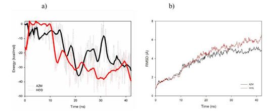
Picaud F                                                                                                                                    Austin Publishing Group

   Isolation of a novel coronavirus from a man with pneumonia in Saudi Arabia.          Chem. 2013; 34: 2757-2770.
   The New England journal of medicine. 2012; 367; 1814-1820.
                                                                                     25. Jo S, Cheng X, Islam SM, Huang L, Rui H, Zhu A, et al. CHARMM-GUI PDB
6. de Groot RJ, Baker SC, Baric RS, Brown CS, Drosten C, Enjuanes L, et al.              Manipulator for Advanced Modeling and Simulations of Proteins Containing
   Middle East respiratory syndrome coronavirus (MERS-CoV): announcement                 Nonstandard Residues. Karabencheva-Christova T, editors. In: Advances in
   of the Coronavirus Study Group, J Virol. 2013; 87: 7790-7792.                         Protein Chemistry and Structural Biology, Academic Press. 2014; 235-265.

7. van der Hoek L, Pyrc K, Jebbink MF, Vermeulen-Oost W, Berkhout RJ,                26. Jo S, Kim T, Iyer VG, Im W. CHARMM-GUI: A web-based graphical user
   Wolthers KC, et al. Identification of a new human coronavirus, Nature                 interface for CHARMM, Journal of Computational Chemistry. 2008; 29: 1859-
   medicine. 2004; 10: 368-373.                                                          1865.

8. Woo PC, Lau SK, Chu CM, Chan KH, Tsoi HW, Huang Y, et al. Characterization        27. Lee J, Patel DS, Ståhle J, Park SJ, Kern NR, Kim S, et al. CHARMM-GUI
   and complete genome sequence of a novel coronavirus, coronavirus HKU1,                Membrane Builder for Complex Biological Membrane Simulations with
   from patients with pneumonia. J Virol. 2005; 79; 884-895.                             Glycolipids and Lipoglycans, J Chem Theory Comput. 2019; 15: 775-786.

9. Woo PC, Lau SK, Yip CC, Huang Y, Yuen KY. More and More Coronaviruses:            28. Phillips JC, Braun R, Wang W, Gumbart J, Tajkhorshid E, Villa E, et al.
   Human Coronavirus HKU1, Viruses. 2009; 1: 57-71.                                      Schulten, Scalable molecular dynamics with NAMD, Journal of Computational
                                                                                         Chemistry. 2005; 26: 1781-1802.
10. Li W, Moore MJ, Vasilieva N, Sui J, Wong SK, Berne MA, et al. Angiotensin-
    converting enzyme 2 is a functional receptor for the SARS coronavirus,           29. Best R, Zhu X, Shim J, Lopes P, Mittal J, Feig M, et al. Optimization of
    Nature. 2003; 426: 450-454.                                                          the Additive CHARMM All-Atom Protein Force Field Targeting Improved
                                                                                         Sampling of the Backbone ϕ, ψ and Side- Chain χ1 and χ2 Dihedral Angles,
11. Wrapp D, Wang N, Corbett KS, Goldsmith JA, Hsieh CL, Abiona O, et al.                Journal of Chemical Theory and Computation. 2012; 8; 3257-3273.
    Cryo-EM structure of the 2019-nCoV spike in the prefusion conformation,
    Science (New York, NY). 2020; 367: 1260-1263.                                    30. Jambrina PG, Aldegunde J. Computational Tools for the Study of
                                                                                         Biomolecules. Martín M, Eden MR, Chemmangattuvalappil NG, editors. in:
12. Zhou P, Yang XL, Wang XG, Hu B, Zhang L, Zhang W, et al. A pneumonia                 Computer Aided Chemical Engineering, Elsevier. 2016. 583-648.
    outbreak associated with a new coronavirus of probable bat origin, Nature.
    2020; 579: 270-273.                                                              31. Feller SE, Zhang Y, Pastor RW, Brooks BR. Constant pressure molecular
                                                                                         dynamics simulation: The Langevin piston method. The Journal of Chemical
13. Paraskevis D, Kostaki EG, Magiorkinis G, Panayiotakopoulos G, Sourvinos              Physics. 1995; 103: 4613-4621.
    G, Tsiodras S. Full- genome evolutionary analysis of the novel corona virus
    (2019-nCoV) rejects the hypothesis of emergence as a result of a recent          32. Darden T, York D, Pedersen L. Particle mesh Ewald: An Nlog (N) method for
    recombination event, Infection, genetics and evolution : journal of molecular        Ewald sums in large systems. The Journal of Chemical Physics. 1993; 98:
    epidemiology and evolutionary genetics in infectious diseases. 2020; 79:             10089-10092.
    104212.
                                                                                     33. Ryckaert J, Ciccotti G, Berendsen H. Numerical-Integration of Cartesian
14. Gautret P, Lagier JC, Parola P, Hoang VT, Meddeb L, Mailhe M, et al.                 Equations of Motion of a System with Constraints - Molecular-Dynamics of
    Hydroxychloroquine and azithromycin as a treatment of COVID-19: results              N-Alkanes, Journal of Computational Physics. 1977; 23: 327-341.
    of an open-label non-randomized clinical trial. International journal of
    antimicrobial agents. 2020; 382: 105949-105949.                                  34. Fung TS, Liu DX. Post-translational modifications of coronavirus proteins:
                                                                                         roles and function, Future Virology. 2018; 13; 405-430.
15. Das S, Bhowmick S, Tiwari S, Sen S. An Updated Systematic Review of
    the Therapeutic Role of Hydroxychloroquine in Coronavirus Disease-19             35. Sugrue RJ. Viruses and Glycosylation: an overview. Sugrue RJ, editors. In:
    (COVID-19), Clin Drug Investig. 2020; 1-11.                                          Glycovirology Protocols, Humana Press, Totowa, NJ. 2007; 1-13.

16. Erickson TB, Chai PR, Boyer EW. Chloroquine, hydroxychloroquine and              36. Goettig P. Effects of Glycosylation on the Enzymatic Activity and Mechanisms
    COVID-19, Toxicol Commun. 2020; 4; 40-42.                                            of Proteases, International Journal of Molecular Sciences. 2016; 17: 1969.

17. Rodrigo C, Fernando SD, Rajapakse S. Clinical evidence for repurposing           37. Zheng J, Yamada Y, Fung TS, Huang M, Chia R, Liu DX. Identification of
    chloroquine and hydroxychloroquine as antiviral agents: a systematic review,         N-linked glycosylation sites in the spike protein and their functional impact on
    Clin Microbiol Infect. 2020; S1198-1743X (1120): 30293-30297.                        the replication and infectivity of coronavirus infectious bronchitis virus in cell
                                                                                         culture, Virology. 2018; 513: 65-74.
18. Baildya N, Ghosh NN, Chattopadhyay AP. Inhibitory activity of
    hydroxychloroquine on COVID-19 main protease: An insight from MD-                38. Böhm M, Bohne-Lang A, Frank M, Loss A, Rojas-Macias MA, et al.
    simulation studies, Journal of Molecular Structure. 2020; 1219: 128595.              Glycosciences. DB: an annotated data collection linking glycomics and
                                                                                         proteomics data (2018 update). Nucleic Acids Research. 2018; 47:
19. Gui M, Song W, Zhou H, Xu J, Chen S, Xiang Y, et al. Cryo-electron                   D1195-D1201.
    microscopy structures of the SARS-CoV spike glycoprotein reveal a
    prerequisite conformational state for receptor binding, Cell Research. 27;       39. Hassan NM, Alhossary AA, Mu Y, Kwoh CK. Protein-Ligand Blind Docking
    2017: 119-129.                                                                       Using QuickVina-W With Inter-Process Spatio-Temporal Integration.
                                                                                         Scientific Reports. 2017; 7; 15451.
20. Walls AC, Tortorici MA, Frenz B, Snijder J, Li W, Rey FA, et al. Glycan shield
    and epitope masking of a coronavirus spike protein observed by cryo-electron     40. Matrosovich M, Herrler G, Klenk HD. Sialic Acid Receptors of Viruses.
    microscopy, Nature structural & molecular biology. 2016; 23: 899-905.                Gerardy-Schahn R, Delannoy P, von Itzstein M, editors. In: Top current
                                                                                         Chemistry, Springer International Publishing. Cham. 2015; 1-28.
21. Song W, Gui M, Wang X, Xiang Y. Cryo-EM structure of the SARS
    coronavirus spike glycoprotein in complex with its host cell receptor ACE2.      41. Fantini J, Chahinian H, Yahi N. Synergistic antiviral effect of hydroxychloroquine
    2018; 14: e1007236.                                                                  and azithromycin in combination against SARS-CoV-2: What molecular
                                                                                         dynamics studies of virus-host interactions reveal. Int J Antimicrob Agents.
22. Li W, Moore MJ, Vasilieva N, Sui J, Wong SK, Berne MA, et al. Angiotensin-           2020; 106020.
    converting enzyme 2 is a functional receptor for the SARS coronavirus,
    Nature. 2003; 426: 450-454.                                                      42. Fantini J, Di Scala C, Chahinian H, Yahi N. Structural and molecular
                                                                                         modelling studies reveal a new mechanism of action of chloroquine and
23. Zoete V, Cuendet MA, Grosdidier A, Michielin O. SwissParam: A fast force             hydroxychloroquine against SARS-CoV-2 infection, International Journal of
    field generation tool for small organic molecules, Journal of Computational          Antimicrobial Agents. 2020; 55: 105960.
    Chemistry. 2011; 32: 2359-2368.
                                                                                     43. Yuan Y, Cao D, Zhang Y, Ma J, Qi J, Wang Q, et al. Cryo- EM structures of
24. Mayne CG, Saam J, Schulten K, Tajkhorshid E, Gumbart JC. Rapid                       MERS-CoV and SARS-CoV spike glycoproteins reveal the dynamic receptor
    parameterization of small molecules using the Force Field Toolkit, J Comput          binding domains. Nat Commun. 2017; 8: 15092.

Submit your Manuscript | www.austinpublishinggroup.com                                                   Austin J Nanomed Nanotechnol 9(1): id1061 (2021) - Page - 07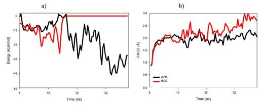
Picaud F                                                                                                                          Austin Publishing Group

44. Shang J, Wan Y, Liu C. Structure of mouse coronavirus spike protein          47. Sulaiman T, Mohana A, Alawdah L, Mahmoud N, Hassanein M, Wani T,
    complexed with receptor reveals mechanism for viral entry. PLOS Pathogens.       et al. The Effect of Early Hydroxychloroquine-based Therapy in COVID-19
    2020; 16: e1008392.                                                              Patients in Ambulatory Care Settings: A Nationwide Prospective Cohort
                                                                                     Study, medRxiv. 2020; 41-43.
45. Alexander PE, Debono VB, Mammen MJ, Iorio A, Aryal K, Deng D, et al.
    COVID-19 coronavirus research has overall low methodological quality thus    48. Soto-Becerra P, Culquichicón C, Hurtado-Roca Y, Araujo-Castillo RV. Real-
    far: case in point for chloroquine/hydroxychloroquine, Journal of Clinical       world effectiveness of hydroxychloroquine, azithromycin, and ivermectin
    Epidemiology. 2020; 123: 120-126.                                                among hospitalized COVID-19 patients: results of a target trial emulation
                                                                                     using observational data from a nationwide healthcare system in Peru.
46. White NJ, Watson JA, Hoglund RM, Chan XHS, Cheah PY, Tarning J.                  medRxiv. 2020.
    COVID-19 prevention and treatment: A critical analysis of chloroquine
    and hydroxychloroquine clinical pharmacology, PLOS Medicine. 2020; 17:
    e1003252.

Submit your Manuscript | www.austinpublishinggroup.com                                             Austin J Nanomed Nanotechnol 9(1): id1061 (2021) - Page - 08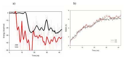
You can also read