Combination Analysis of Metatranscriptome and Metagenome Reveal the Composition and Functional Response of Coral Symbionts to Bleaching During an ...
←
→
Page content transcription
If your browser does not render page correctly, please read the page content below
ORIGINAL RESEARCH
                                                                                                                                          published: 20 March 2020
                                                                                                                                    doi: 10.3389/fmicb.2020.00448

                                             Combination Analysis of
                                             Metatranscriptome and Metagenome
                                             Reveal the Composition and
                                             Functional Response of Coral
                                             Symbionts to Bleaching During an El
                                             Niño Event
                                             Fulin Sun 1,3,4,6 , Hongqiang Yang 1,2,5,6* , Guan Wang 1,2,6 and Qi Shi 1,2,6
                                             1
                                              South China Sea Institute of Oceanology, Institute of South China Sea Ecology and Environmental Engineering, Chinese
                                             Academy of Sciences, Guangzhou, China, 2 Key Laboratory of Ocean and Marginal Sea Geology, South China Sea Institute
                                             of Oceanology, Chinese Academy of Sciences, Guangzhou, China, 3 State Key Laboratory of Tropical Oceanography, South
                                             China Sea Institute of Oceanology, Chinese Academy of Sciences, Guangzhou, China, 4 Daya Bay Marine Biology Research
                                             Station, Chinese Academy of Sciences, Shenzhen, China, 5 Nansha Marine Ecological and Environmental Research Station,
                                             Chinese Academy of Sciences, Sansha, China, 6 Southern Marine Science and Engineering Guangdong Laboratory
                                             (Guangzhou), Guangzhou, China

                                             With the abnormal rise in ocean temperatures globally in recent years, coral bleaching
                           Edited by:        is becoming common and serious. However, the response mechanisms and processes
                       Alison Buchan,
         The University of Tennessee,
                                             of coral symbionts to bleaching are not well understood. In this study, metagenomics
             Knoxville, United States        and metatranscriptomics were used to explore the composition of coral symbionts and
                         Reviewed by:        their functions in response to coral bleaching. All four bleaching coral species displayed
                               Jin Zhou,
                                             a significant reduction of the abundance and function of Dinophyceae-like eukaryotes at
             Tsinghua University, China
                           Akira Iguchi,     the DNA and RNA levels. However, different species of bleaching coral have their own
        National Institute of Advanced       characteristic symbiotic components. Bleaching Acropora tenuis and Goniastrea minuta
    Industrial Science and Technology
                          (AIST), Japan
                                             corals exhibited a very high abundance of prokaryotes and associated gene functions,
                  *Correspondence:
                                             especially for opportunistic bacteria. In contrast, algae and fungi were identified as
                     Hongqiang Yang          the main microbial associate components and had relatively high RNA abundance in
                  hqyang@scsio.ac.cn
                                             bleaching Pocillopora verrucosa and Pocillopora meandrina. Different coral species,
                   Specialty section:        whether unbleached or bleaching, have the same symbiotic taxa that perform the same
         This article was submitted to       biological functions in vivo. Different stages of bleaching, or transitional states, were
                 Aquatic Microbiology,
                                             identified by different genome content and functional gene abundance among bleaching
               a section of the journal
             Frontiers in Microbiology       corals. These stages should be considered in future coral bleaching studies to accurately
            Received: 31 July 2019           determine symbiont structure and function. An implicit hypothesis is that there is a causal
          Accepted: 02 March 2020            relationship between the stability of eukaryotic communities and coral bleaching.
          Published: 20 March 2020
                                             Keywords: coral symbionts, bleaching, metagenome, metatranscriptome, composition response, functional
                             Citation:
                                             response
   Sun F, Yang H, Wang G and Shi Q
          (2020) Combination Analysis
                 of Metatranscriptome
             and Metagenome Reveal
                                            INTRODUCTION
      the Composition and Functional
        Response of Coral Symbionts
                                            El Nino events have a significant impact on global climate, most notably causing warming
to Bleaching During an El Niño Event.       events, which affect the stability of marine ecosystems (Heron et al., 2016; Hughes et al., 2018a).
              Front. Microbiol. 11:448.     Widespread bleaching of coral reefs, resulting in high levels of coral mortality due to heat stress
      doi: 10.3389/fmicb.2020.00448         (Hoegh-Guldberg, 1999; Heron et al., 2016), is now recognized as a global threat to coral. 2015–2017

Frontiers in Microbiology | www.frontiersin.org                                   1                                            March 2020 | Volume 11 | Article 448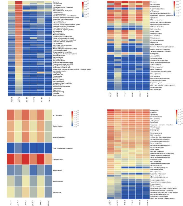
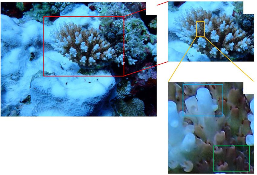
Sun et al.                                                                                                  Response of Coral Symbionts to Bleaching

were the three warmest years in the instrumental record period              acute stress (Fine and Loya, 2002; Fine et al., 2006; del Campo
since 18801 and record high temperatures triggered a pan-tropical           et al., 2016). Despite the fact that other eukaryotes are ubiquitous
coral bleaching episode (Hughes et al., 2018b). The world’s largest         in corals, little is known about their diversity and ecological
coral reef ecosystem, the Great Barrier Reef, has experienced the           function during coral bleaching events.
highest temperatures ever recorded and lost nearly 30% of coral                 Until now, fundamental gaps have existed in our
cover (Hughes et al., 2018a).                                               understanding of the coral microbe and its functional
   Coral is closely related to a complex group of microorganisms,           contribution to coral. In 2016, a bleaching event also affected
including Symbiodiniaceae, fungi, bacteria, archaea, endolithic             a large area of coral in the South China Sea, where mass
algae, and viruses, in a relationship known as coral symbiosis              coral bleaching had not previously been recorded. In this
(Reshef et al., 2006). Coral microbes play an important role                study, four different coral species were studied to provide an
in nutrient cycling and antimicrobial protection in coral reefs             overview of the metagenomic and metatranscriptome response
(Ritchie and Smith, 2004; Zhang et al., 2015; Bourne et al., 2016).         of coral symbionts (prokaryotes, Symbiodiniaceae, other
Therefore, it is important to investigate the effects of bleaching          endolithic eukaryotes and the coral itself) to bleaching and to
events on the function of coral microbial communities. Recently,            highlight differences in their functional performance. Coral
metagenomics has been used to investigate the taxonomic                     species were collected at the same location to eliminate any
diversity and metabolic capabilities of coral-associated microbes           potential external environmental influences. In order to study
under thermal stress or bleaching (Thurber et al., 2009; Littman            the different components of coral symbionts separately, each
et al., 2011; Lee et al., 2015; Ziegler et al., 2017). These                symbiotic component was separated based on the NCBI non-
studies suggested that microbes can undergo major shifts, from              redundant (NR) protein database. Combined with KEGG (Kyoto
symbionts to opportunistic microbes or potential disease-causing            Encyclopedia of Genes and Genomes database) annotation,
bacteria, during heat stress or bleaching (Bourne et al., 2007;             the corresponding functions of DNA and RNA in different
Thurber et al., 2009; Littman et al., 2011). In addition to this,           components were explored.
the metabolism of the microbial community can shift from
autotrophy to heterotrophy (Littman et al., 2011), which involves
sulfur and nitrogen metabolism, fatty acid and lipid utilization,           MATERIALS AND METHODS
and secondary metabolism.
   Traditionally, research into coral bleaching has mainly                  Sample Collection
focused on studying photosynthetic symbiotic algae known as                 Coral samples were collected at Xiane reef, Nansha Islands,
Symbiodiniaceae. Physiological damage and expulsion of algal                South China Sea, in June 2016 (Figure 1). Four coral species
symbionts are thought to be the result of the host immune                   (unbleached coral and bleached coral collected from the same
response triggered by reactive oxygen species produced by                   coral colony), Acropora tenuis (AC-H, AC-W), Goniastrea
coral hosts, algal symbionts, or both (Weis et al., 2008).                  minuta (GM-H, GM-W), Pocillopora verrucosa (PV-H, PV-W)
Research on Symbiodiniaceae has been focused on changes                     and Pocillopora meandrina (PM-H, PM-W) were sampled. The
in diversity and density of Symbiodiniaceae (Fine and Loya,                 field sampling process of coral samples was showed in Figure 2.
2002; Cunning and Baker, 2012; Howells et al., 2013; Kemp                   Water temperatures in the sampling area ranged from 30.5 to
et al., 2014), Photosystem II damage in symbiotic dinoflagellates           31◦ C. Three replicate samples of unbleached and bleached parts
(Warner et al., 1999), thermal tolerance of Symbiodiniaceae (Ruiz           of coral (including tissue, mucus and skeleton) were collected
Sebastián et al., 2009; Tong et al., 2017), and functional changes in       using a hammer and chisel. Once collected, the coral samples
Symbiodiniaceae (Loram et al., 2007). However, few studies have             (1 cm × 1 cm) were washed with sterile seawater three times
reported the functional response of Symbiodiniaceae to bleaching            to remove any surface attachments. Each sample was divided
(Gierz et al., 2017).                                                       into two parts and placed in sterile centrifuge tubes with Sample
   Although previous studies had not focused on eukaryotes, they            Protector (Takara, Japan) for DNA and RNA extraction. DNA
have hinted that eukaryotes are the most abundant component                 and RNA isolation of replicate samples was mixed and conducted
of coral symbionts (Wegley et al., 2007; Littman et al., 2011).             using DNeasy and RNeasy plant mini kit (Qiagen, Germany)
Microeukaryotes have been most widely associated with coral                 following the manufacturer’s instructions. Coral tissues were
diseases and mortality (Ainsworth et al., 2017). To date, studies           removed with an airbrush for the identification of each species.
on other microeukaryotes associated with coral have mainly                  All coral samples were identified according to their ecological and
focused on several key populations, including fungi, endolithic             morphological characteristics.
microalgae and protists. The potential diversity of coral related
fungi suggests a broader role beyond pathogenicity (Raghukumar              Library Preparation and Metagenome
and Ravindran, 2012). Metagenomic analysis has revealed that                Sequencing
endolithic algae can play a key role in the microbial community             For each coral sample, 200 ng of total DNA was randomly
by driving important chemical processes (Littman et al., 2011).             fragmented to
Sun et al.                                                                                                                       Response of Coral Symbionts to Bleaching

  FIGURE 1 | Location map of sampling sites in the South China Sea during an El Nino period.

  FIGURE 2 | The field sampling process of Acropora tenuis. (A) Acropora tenuis is in bleaching during our sampling time, the lower left part of its growth base is
  bleached Favites sp.; (B) This is the red frame part of the (A), Acropora tenuis in the bleaching; (C) This is the yellow frame of the part (B); the upper blue frame of
  part (C) is the collection part of the bleaching coral sample, and the lower part of the green frame is the collection part of unbleached coral samples.

Mag PCR Clean-up (Axygen), and fragments of ∼410 bp                                       instrument (Illumina, United States). Sequencing was carried
were recovered. Each sample was then amplified by PCR                                     out using a 2 × 150 paired-end (PE) configuration; image
for eight cycles using P5 (50 -AATGATACGGCGACCACCGA)                                      analysis and base calling were conducted by the HiSeq Control
and P7 (50 -CAAGCAGAAGACGGCATACGA) primers. Then                                          Software (HCS) + OLB + GAPipeline-1.6 (Illumina) on the
libraries were multiplexed and loaded on an Illumina HiSeq                                HiSeq instrument.

Frontiers in Microbiology | www.frontiersin.org                                       3                                                 March 2020 | Volume 11 | Article 448
Sun et al.                                                                                                    Response of Coral Symbionts to Bleaching

Metagenomic Data Analysis                                               Clean-up (Axygen, China), and fragments of ∼410 bp were
Raw shotgun sequencing reads were trimmed using cutadapt                recovered. The dUTP-marked second strand was digested
(v1.9.1). Low-quality reads, N-rich reads and adapter-polluted          with Uracil-Specific Excision Reagent enzyme (New England
reads were removed. Each samples was assembled de novo to               Biolabs). Each sample was then amplified by PCR for 14
obtain separate assemblies. Whole genome de novo assemblies             cycles using P5 and P7 primers. Sequencing was the same as
were performed using MEGAHIT (v1.13) (Li et al., 2015) with             macrogenome operation.
different k-mer (39, 59, 79, and 119). The best assembly result
of Scaffold, which has the largest N50, was selected for the gene       Data Analysis of Metatranscriptome
prediction analysis.                                                    Raw shotgun sequencing reads were trimmed using cutadapt
   Genes of each sample were predicted using Prodigal (v3.02)           (v1.9.1). Low-quality reads, N-rich reads and adapter-polluted
(Hyatt et al., 2010). CD-HIT (Fu et al., 2012) was used to              reads were removed. The PE reads were assembled using
cluster genes derived from all samples with a default identity          Trinity v2.4.0. Trinity uses the de Bruijn graph strategy
of 0.95 and coverage of 0.9. In order to analyze the relative           to assemble the transcriptome. Open reading frames
abundance of unigenes in each sample, paired-end clean reads            were identified using the TransDecoder program3 , with
were mapped to unigenes using SOAPAligner (version 2.2.1)               default parameters.
to generate read coverage information for unigenes. Based on                The sequence reads from six samples were individually
the number of mapped reads and the length of gene, the                  aligned to the assembled transcriptome, using the aligner
abundance information of each gene was calculated in each               bowtie2. The resulting files were then quantified using RSEM
sample. The formula is as follows, r represents the number              (v1.2.4) which default parameters to get gene based raw hit-
of reads mapped to the genes and L represents gene’s length.            count data. After adjustment with Benjamini and Hochberg’s
                                                                        approach for controlling the false discovery rate, P-value of
                                       Tk     1                         genes were set < 0.05 to detect differentially expressed genes.
                               Gk =       · n
                                       Lk P     ri                      The metatranscriptome used FPKM (Fragment Per Kilo bases
                                                   Li                   per Million reads) to calculate gene expression abundance. The
                                             i=1
                                                                        FPKM formula is as follows: AFPKM = 109 C/NL, where C
   The unigene sequences were blasted against the constructed           is the fragment number to compare gene A, N is the total
microbial database. And the lowest common ancestor was                  fragment number to compare all genes, L is the base length
determined using Metagenome Analyzer (MEGAN, v6.4.4).                   of gene A. BLASTX was used to search the protein sequences
The abundance of a species in one sample equaled the sum                of the predicted genes with the NCBI non-redundant (NR)
of the gene abundance annotated for the species. Diamond                protein database, CAZy database, eggNOG database and KEGG
(version v0.8.15.77) was used to search the protein sequences           database with E –value cut off of 1e−5 . Scripts were used in
of the unigenes with the NR database and KEGG database2                 house to enrich the significant differential expression of genes
with E < 1e-5. The statistical significance threshold of the            in KEGG pathways.
sequence alignment was set at 1e−5 and the sequence alignment
length was set as no less than 60% of the reference gene
protein length. The matched result with best scores was                 RESULTS
selected for annotation.
                                                                        Overview of the Metagenome and
Library Preparation and                                                 Metatranscriptome
Metatranscriptome Sequencing                                            A summary of the metagenome and metatranscriptome results
Total RNA of each sample was quantified and qualified by Agilent        are shown in Supplementary Tables S1, S2. Figure 3A shows the
2100 Bioanalyzer (Agilent Technologies, United States) and 1 µg         taxonomy annotations of metagenome and metatranscriptome
total RNA with a RIN value above 7 was used for following               based on different classification levels. At the DNA level, the
library preparation. The rRNA was depleted from total RNA               abundance of unbleached coral genomes accounted for more
using Ribo-Zero rRNA Removal Kit (Illumina, United States).             than 80% of the total number of sequences, but the DNA
The ribosomal depleted mRNA was then fragmented, reverse-               content had low abundance in AC-W (48.55%) and GM-W
transcribed, and used to synthesize first strand cDNA with              (7.90%) corals. In comparison with unbleached corals, bacterial
random primers and Actinomycin D. The second-strand cDNA                abundance increased sharply in AC-W (32.53%) and GM-W
was synthesized using Second Strand Synthesis Enzyme Mix                (78.63%) corals, as did the abundance of eukaryotes in AC-
(include dACG-TP/Dutp). The purified double-stranded cDNA               W. At the RNA level, AC-W coral had a reduced abundance
by AxyPrep Mag PCR Clean-up (Axygen, China) was then                    of Symbiodiniaceae compared with AC-H coral. In contrast,
treated with End Prep Enzyme Mix to repair both ends and                AC-W coral had a higher abundance of prokaryotes and
add a dA-tailing in one reaction, followed by a T-A ligation            eukaryotes than AC-H. Unlike AC corals, the abundance of
to add adaptors to both ends. Size selection of Adaptor-                eukaryotes was higher in PV-W and PM-W in comparison to
ligated DNA was then performed using AxyPrep Mag PCR                    unbleached corals. Symbiodiniaceae abundance was significantly
2                                                                       3
    https://www.kegg.jp/                                                    https://github.com/TransDecoder/TransDecoder/wiki

Frontiers in Microbiology | www.frontiersin.org                     4                                              March 2020 | Volume 11 | Article 448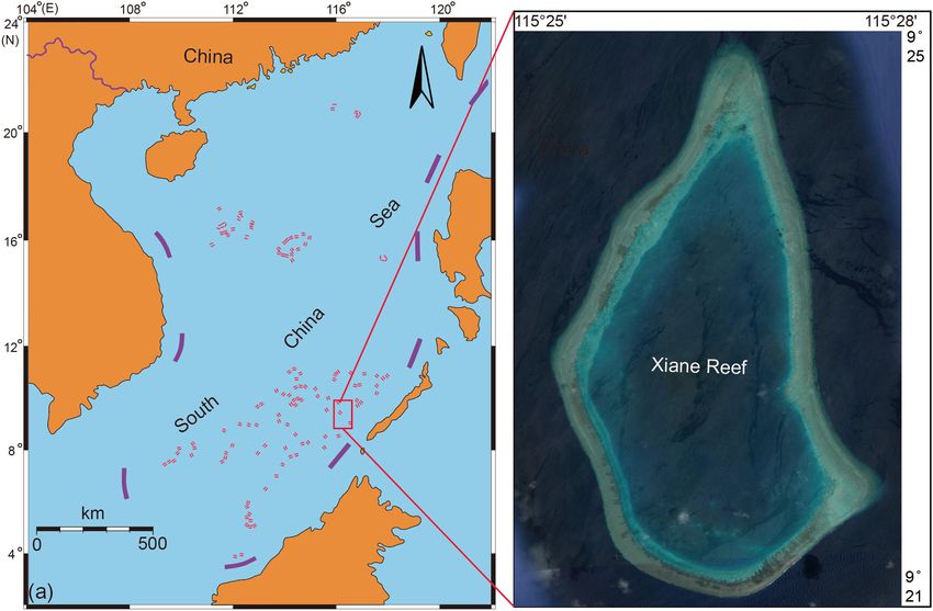
Sun et al.                                                                                                                Response of Coral Symbionts to Bleaching

  FIGURE 3 | Taxonomic distribution of corals through metagenomic(-G) and metatranscriptomic(-T) profile analysis. (A) Coral symbiosis components; (B) taxa
  abundance at the phylum level; (C) taxa abundance at the class level; and (D) genus abundance with homogenized data.

lower in PV-W and PM-W corals compared with PV-H                                     of Symbiodiniaceae and bacterial taxa. Bleaching corals
and PM-H corals.                                                                     exhibited a high diversity and abundance of bacterial taxa at
                                                                                     the DNA and RNA level. These taxa included Chlorobiales,
Composition and Transcriptional Active                                               Rhodobacterales, Alteromonadales, Oceanospirillales, and
of Symbiosis Community Between                                                       Vibrionales. As seen in Figure 4A, more bacterial orders were
                                                                                     recorded from AC and GM than PV and PM at the DNA
Unbleached Corals and Bleaching Corals                                               and RNA levels. A higher amount of Campylobacterales,
The results showed that there were distinct differences in the                       Alteromonadales and Streptomycetale were revealed at the
composition of symbionts among different corals at RNA and                           RNA level, in contrast to the results for Rhodobacterales
DNA levels (p < 0.01, Fisher’s exact test). For AC and GM                            and Vibrionales, which were highly abundant at both the
corals, metagenomic analysis showed that the most obvious                            RNA and DNA levels.
response to bleaching was the increase in abundance of bacterial                         For PV and PM corals, the symbiotic component was mainly
taxa, affiliated with Alphaproteobacteria, Deltaproteobacteria,                      composed of microeukaryotes, and there was a distinct difference
Epsilonproteobacteria, Gammaproteobacteria (Proteobacteria),                         in the abundance and composition for unbleached and bleaching
Cytophagia (Bacteroidetes) and Chlorobia (Chlorobi), in                              corals (t-test, p < 0.01). Most notably, Dinophyceae-like genera
addition to a significant decrease in Dinophyceae (Dinoflagellata)                   had a significantly lower abundance in bleaching corals, including
abundance (Figures 3B,C).                                                            Symbiodiniaceae, Crypthecodinium, Amphidinium, Karlodinium,
   Furthermore, distinct differences were observed at the class                      Heterocapsa, Pfiesteria, and Prorocentrum. The Puccinia genus
level between unbleached and bleaching corals (Figure 3C). The                       (Basidiomycota) was dominant at the RNA level, and was
dominant class of symbionts in PV and PM were Dinophyceae,                           more abundant in bleaching than unbleached corals. In
Eurotiomycetes, Pucciniomycetes (Ascomycota), and Bacilli                            unbleached corals, Gymnodiniales, Cantharellales, Peridiniales,
(Figure 3B). In the case of both unbleached and bleaching                            Prorocentrales and Gonyaulacales, all of which are affiliated
PV and PM corals, there was a very low abundance of                                  with Dinophyceae, were observed to be more abundant at
prokaryotes compared with other symbiont components.                                 the RNA and DNA levels in comparison with bleaching
Apart from the decreased abundance of Dinophyceae,                                   corals (Figure 4C).
another feature of bleaching PV and PM corals was the
higher abundance of eukaryotes, especially Pucciniomycetes,
when compared with unbleached corals. The major genera                               Functional Changes of Symbiotic
identified at the RNA and DNA levels varied with samples.                            Prokaryotes in Coral
Figure 3D shows changes in the structure and abundance                               For AC and GM corals, gene composition and transcriptional
of dominant genera between unbleached and bleaching                                  abundance increased significantly (t-test, p < 0.01) in
corals. For AC and GM corals, the most obvious response                              bleaching corals in comparison with unbleached corals
to bleaching in coral symbionts was the shift in abundance                           (Figures 5A, 6A), including ribosomal, carbon fixation,

Frontiers in Microbiology | www.frontiersin.org                                  5                                              March 2020 | Volume 11 | Article 448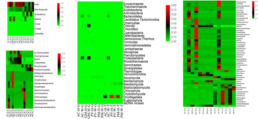
Sun et al.                                                                                                                     Response of Coral Symbionts to Bleaching

  FIGURE 4 | Comparison of 16 major orders and related functions of coral symbionts based on RNA and DNA sequences achieved by metagenomic and
  metatranscriptomic analysis. (A) Prokaryotic orders of coral symbionts; (B) function of prokaryotic orders; (C) eukaryotic orders of coral symbionts; and (D) function
  of eukaryotic orders.

cofactor and vitamin biosynthesis, and ATP synthesis. Both                              pathways, assimilatory sulfate reduction and dissimilatory
the metagenomic and metatranscriptome results of the current                            sulfate reduction, were detected in AC and GM corals only.
study indicated that the main contributing prokaryotes for                              Dominant bacteria, such as Campylobacterales (Arcobacter
these functions were Proteobacteria, Bacteroidetes, Chlorobi                            and Sulfurimonas) mainly contributed to assimilatory
and Actinobacteria (Supplementary Figure S1), and the                                   sulfate reduction.
abundance of bacterial orders was significantly correlated with
function, especially at the RNA level (t-test, R2 > 0.8, p < 0.01).
As seen in Figure 4B, Rhodobacterales, Campylobacterales,                               Functional Changes of Symbiotic
Vibrionales and Alteromonadales contributed more to                                     Symbiodiniaceae in Coral
gene function at the RNA level, in contrast to the finding                              The results of metagenomic and metatranscriptomic analysis
that dominant of Flavobacteriales, Oceanospirillales and                                showed that 24 Symbiodiniaceae species were detected
Cellvibrionales had a slightly higher contribution to gene                              in corals (Supplementary Figure S2). According to the
function at the DNA level. For PV and PM corals, the                                    results, all Symbiodiniaceae species decreased in abundance
bacterial contribution to gene function was very low in                                 in bleaching corals in comparison with unbleached corals,
both bleaching and unbleached corals (Figure 4B and                                     especially for dominant species Cladocopium and Symbiodinium
Supplementary Figure S1).                                                               sp. ex Scolymia sp.
   The abundance of prokaryotes was significantly correlated                               Of all Symbiodinium types, Cladocopium and Symbiodinium
with functional abundance at the RNA and DNA levels (t-                                 ex Scolymia sp. were the two genera with the highest abundance
test, R2 > 0.9, p < 0.01). For AC and GM corals, carbon                                 of gene composition and transcription. Among the functions
fixation pathways, including the reductive citrate cycle                                performed by Symbiodiniaceae (Figures 5B, 6B), photosynthesis
(Arnon-Buchanan cycle), the reductive pentose phosphate                                 and ATP synthesis were the most important functions. The
cycle and the 3-hydroxypropionate bi-cycle, were contributed                            abundance of Symbiodiniaceae was significantly correlated with
by Proteobacteria and Bacteroidetes. However, the gene                                  functional abundance at the RNA and DNA levels (t-test,
abundance of carbon fixation was very low in unbleached and                             R2 > 0.8, p < 0.05). In comparison with unbleached corals,
bleaching PV and PM corals. Dissimilatory nitrate reduction                             Symbiodiniaceae were less abundant in almost all functional
was the main pathway of nitrogen metabolism identified.                                 genes in bleaching corals. As seen in Supplementary Figure
Proteobacteria, Bacteroidetes, Chlorobi and Tectomicrobia                               S3, Cladocopium and Symbiodinium sp. ex Scolymia sp. were
mainly contributed to sulfur metabolism in AC and GM                                    the main contributors to photosynthesis. Cladocopium and
corals. Two of three high abundance sulfur metabolism                                   Symbiodinium minutum were the main contributors to ATP

Frontiers in Microbiology | www.frontiersin.org                                     6                                                March 2020 | Volume 11 | Article 448
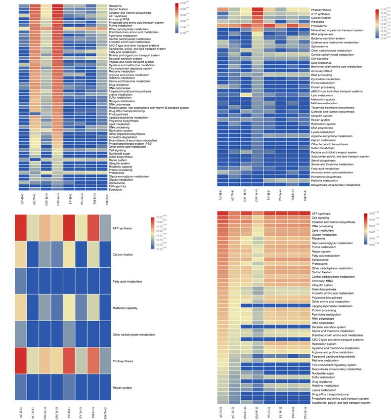
Sun et al.                                                                                                               Response of Coral Symbionts to Bleaching

  FIGURE 5 | Functional analysis of coral symbiont components through metagenomic analysis. (A) prokaryotic function; (B) Symbiodinium function; (C) eukaryotic
  function; and (D) coral function.

synthesis. At the RNA level, Cladocopium was the main                                in the Symbiodiniaceae. It had a high abundance in all
contributor to ATP synthesis in AC corals, while Cladocopium                         unbleached corals, and exhibited a reduced gene abundance in
and Symbiodinium minutum were the main contributors for                              bleaching corals.
PV and PM corals. Cladocopium was the only performer for
RNA processing and Spliceosome at the RNA level. Spliceosome
genes in unbleached corals had a higher expression of abundance                      Functional Changes of Other Eukaryotes
than bleaching corals, the majority of which belonged to                             (Fungi and Algae) in Corals
genes involving the heat shock gene (HSPA1_8). There was                             The results indicated that most eukaryotes displayed reduced
one carbon fixation pathway (Calvin-Benson cycle) detected                           function in bleaching corals compared with unbleached

Frontiers in Microbiology | www.frontiersin.org                                  7                                             March 2020 | Volume 11 | Article 448
Sun et al.                                                                                                                Response of Coral Symbionts to Bleaching

  FIGURE 6 | Functional analysis of coral symbiont components through metatranscriptomic analysis. (A) Prokaryotic function; (B) Symbiodinium function; (C)
  eukaryotic function; and (D) coral function.

corals. This decline in function resulted in decreased                               DNA level, where Eurotiales mostly contributed to the function
proteasome function, carbon fixation, ATP synthesis and                              (Figure 4D). It was unexpectedly found that Dinophyceae were
central carbohydrate metabolism. However, photosynthetic                             the main contributors to all functions except photosynthesis
activity was found to increase in all bleaching corals, in particular                (Supplementary Figure S4). Interestingly, there was an obvious
AC coral (Figures 5C, 6C). As seen in Supplementary Figure S4,                       increase in the abundance of genes involved in photosynthesis
the main contributors to function are the dominant Dinophyceae                       in bleaching coral, mainly attributed to Bacillariophyceae,
(including Gymnodiniales, Peridiniales, Prorocentrales,                              Florideophyceae, and Trebouxiophyceae. The Calvin cycle is the
and Gonyaulacales) and Eurotiales. A higher number of                                main pathway for carbon fixation of these eukaryotes. There
Gymnodiniales, Peridiniales, Prorocentrales and Gonyaulacales                        was a lower abundance of genes involved in carbon fixation in
contributed to function at the RNA level, in contrast to the                         bleaching corals compared with unbleached corals. Dinophyceae

Frontiers in Microbiology | www.frontiersin.org                                  8                                              March 2020 | Volume 11 | Article 448
Sun et al.                                                                                                 Response of Coral Symbionts to Bleaching

were the main contributor to Spliceosomes, and the abundance               Bleaching Significantly Reduced
of the heat shock 70 gene was significantly reduced in bleaching           Functional Gene Abundance of
corals compared with unbleached corals.
                                                                           Symbiotic Symbiodiniaceae
                                                                           According to the results of the current study, the abundance of
Functional Comparison of Unbleached                                        all Symbiodiniaceae obviously decreased in all bleaching corals in
and Bleaching Corals                                                       comparison with unbleached corals, indicating that the nutrition
Almost all of the functions of AC-W coral were reduced                     supplied to coral by Symbiodiniaceae decreased. Exocytosis or
compared with unbleached corals, in particular cell signaling,             in situ symbiotic degradation during bleaching seemed to be less
ribosomal activity, spliceosome activity, glycan metabolism,               invasive and cost effective than host cell degradation (Oakley
RNA processing and ATP synthesis (Figures 5D, 6D).                         and Davy, 2018). This study identified that the four coral species
In contrast to AC coral, almost all functions of PV-W                      could simultaneously host a very high diversity of genotypic
and PM-W were increased in comparison with unbleached                      Symbiodiniaceae phylotypes, more than described by other
corals (Figure 6D).                                                        studies (Toller et al., 2001; Kemp et al., 2008). Cladocopium is
                                                                           often regarded as a sensitive species to temperature or bleaching
                                                                           (Kemp et al., 2014), and is dominant in scleractinian corals in
DISCUSSION                                                                 the South China Sea. Cladocopium were the main contributors to
                                                                           photosynthesis and ATP synthesis in coral.
Bleaching Significantly Altered Coral                                          This result provided direct evidence that bleaching may have
Bacterial Community and Increased Its                                      important effects on photosynthesis via the inhibition of the
Functional Gene Abundance                                                  Calvin cycle, limiting carbon fixation in Symbiodiniaceae, as
An obvious response to bleaching displayed by coral symbionts              described in previous studies (Jones et al., 1998; Wooldridge,
was the shift in abundance of bacteria. Bleaching corals exhibited         2016). It had been found that carbon fixation via the Calvin cycle
a higher diversity and abundance of bacterial taxa at the DNA              is sensitive to heat stress (Murata et al., 2007). Previous studies
and RNA level than unbleached corals, indicating that bacteria             reported that the maximum quantum yield of photosystem
were easily affected by coral bleaching. Many studies have shown           II was significantly lower and highly variable in bleaching
that potentially opportunistic microbes can sharply increase in            corals in comparison with healthy corals (Kemp et al., 2014).
abundance and become dominant in bleaching corals (Mouchka                 This occurred with a corresponding loss of electron flow
et al., 2010; Kumar et al., 2016), causing the coral to move               supporting carbon fixation (Slavov et al., 2016). ATP synthesis
toward an unstable state (Thurber et al., 2009; Littman et al.,            was essential for repair in photosystem II (Murata et al., 2007),
2011; Zaneveld et al., 2016; McDevitt-Irwin et al., 2017), even            therefore the repair rate that this study suggested was decreased
inducing coral disease (Rosenberg et al., 2007; Kimes et al., 2012;        through the inhibition of ATP synthesis in bleaching coral. In
Maynard et al., 2015). Coral bleaching can alter the chemical              addition, a low abundance of the heat shock gene indicated that
composition of coral mucus, increase organic matter and mucus              bleaching inhibited the activity of heat-inducible genes and heat
production (Niggl et al., 2009), which induces a shift in the              acclimation of Symbiodiniaceae (Rosic et al., 2011) resulting in
coral-associated microbial community (Raina et al., 2013; Lee              the reduced ability of Symbiodiniaceae to resist thermal stresses.
et al., 2016). The results of the current study indicated that these
opportunistic microbes were highly abundant in bleaching coral,            Importance of Other Eukaryotes (Fungi
and may have resulted in an elevation of bacterial-organic matter          and Algae) in Bleaching Corals
coupling (Figure 4A).                                                      Another finding of the current study was that, when corals
    Almost all functional genes of prokaryotes were improved               are bleaching, they not only expel Symbiodiniaceae, but all of
in bleaching AC and GM corals. Previous studies demonstrated               the identified Dinophyceae genera, which indicates that these
that the metabolism of the microbial community could shift                 algae have the same response mechanism to coral bleaching.
from autotrophy to heterotrophy under stress, resulting in an              Other eukaryotic algae also showed distinct changes in bleaching
increase in the abundance of microbial genes involved in sulfur            corals compared to unbleached corals. It has been reported that
and nitrogen metabolism, and secondary metabolism (Thurber                 when Symbiodiniaceae are absent, these algae could provide an
et al., 2009; Littman et al., 2011). As the microbial community            alternative source of photoassimilates, and provide nutrients that
shifts from autotrophy to heterotrophy, bacterial consumption of           increase coral survival during stress (Fine et al., 2006; del Campo
organic matter becomes greatly enhanced, and the contribution              et al., 2016). The current study also suggested that fungi displayed
of fixed N2 and photosynthesis for nitrogen and carbon                     high abundance of RNA in bleaching PV and PM corals, and
budgets became less obvious. High abundance sulfur metabolism              bleaching stimulated fungal growth. Some studies have suggested
pathways (assimilatory sulfate reduction and dissimilatory sulfate         that fungi were also thought to be opportunistic pathogens, and
reduction) were detected in bleaching AC and GM corals,                    their abundance depended on coral health (Yarden et al., 2007;
increasing the possibility of producing sulfide. High abundance            Toledo-Hernández et al., 2008).
of these heterotrophic bacteria could deplete nutrients in coral,              The functional genes of other eukaryote algae increased in
and deteriorate the microenvironment, ultimately making coral              abundance during bleaching, indicating that the photosystems of
bleaching irreversible.                                                    other eukaryotes (such as Bacillariophyta, Chlorophyta) used a

Frontiers in Microbiology | www.frontiersin.org                        9                                        March 2020 | Volume 11 | Article 448
Sun et al. Response of Coral Symbionts to Bleaching
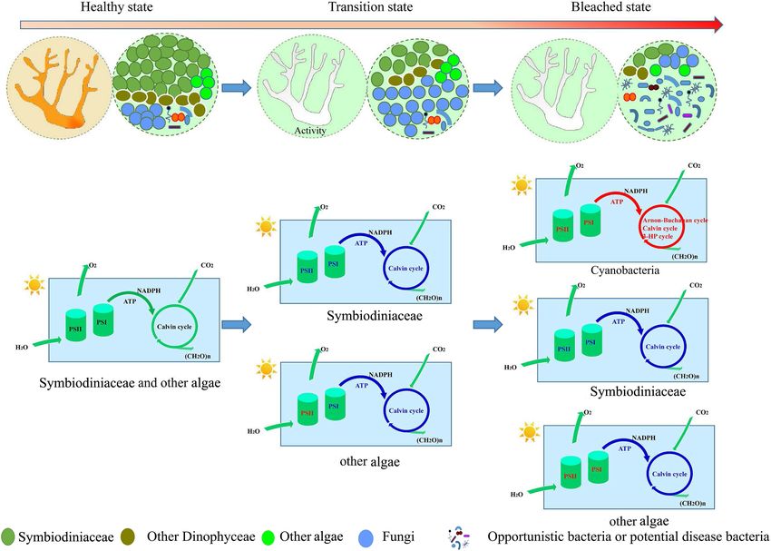
FIGURE 7 | A hypothetical conceptual map summarizing the shift of coral symbionts during bleaching, illustrating the functional changes in photosynthesis and
carbon fixation in different coral symbiont components. Blue represent down regulation of function; Red represent up regulation of function.
different mechanism and had higher levels of thermal tolerance inhibited the ability of the host to survive or recover from thermal
compared to Dinophyceae algae. It was also thought that other stress (Oakley and Davy, 2018). This suggested that bleaching
endosymbiotic algae benefit the host coral during periods of AC and GM corals lost most of their physiological metabolic
stress (Gutiérrez-Isaza et al., 2015; del Campo et al., 2016). activity and function.
This may be because during bleaching, the shading effect of However, almost all functions of bleaching PV and PM were
Symbiodiniaceae was lost, allowing increased light to penetrate increased in comparison with unbleached corals. This indicated
the coral skeleton, possibly resulting in increased photosynthetic that coral was in a temporary stage of transformation from an
activity of these algae. Given the ability of microalgae to unbleached to a seriously bleached state. At this stage, corals
adapt rapidly to heat stress, these populations might become exhibit symptoms of bleaching as they have expelled most
important as “secondary” symbionts (Fine and Loya, 2002; symbiotic algae; however, the symbiotic structure in bleaching
Fine et al., 2005), and continue to provide nutrients for coral corals has certain similarities in composition to unbleached
through photosynthesis. corals. The remaining Symbiodiniaceae and other algae play an
important role in bleaching coral, providing nutrition to the host
Functional Gene Change in Coral at and maintaining coral activities under stress.
Different Stages of Coral Bleaching
The results of the current study indicated that the coral symbionts
under investigation were at different stages of bleaching. Almost CONCLUSION
all of the functions of bleaching AC and GM coral were reduced
compared with unbleached corals (Figures 5D, 6D), suggesting For both unbleached and bleaching coral, it was found that
that the nucleic acid of bleaching corals was being degraded and different coral species have common symbiotic taxa that perform
was in an apoptotic state. A reduction in mRNA abundance of biological functions in vivo. Overall, different coral species
cytochrome c and ATP synthase, which were central components were found to have common characteristics when bleached:
of the respiratory electron transport chain (Dunn et al., 2012), a decreased abundance of Symbiodiniaceae and associated
Frontiers in Microbiology | www.frontiersin.org 10 March 2020 | Volume 11 | Article 448Sun et al. Response of Coral Symbionts to Bleaching
function and the exclusion of Dinophyceae-like eukaryotes. FUNDING
Furthermore, this study might reflect the different stages of the
coral bleaching process. In the early stages of coral bleaching, The present study was supported financially by the Strategic
algae such as Symbiodiniaceae and other Dinophyceae were Priority Research Program of the Chinese Academy of
expelled from corals. If the coral microbe could maintain a level Sciences (Grant No. XDA13010103), the National Natural
of stability similar to that of an unbleached coral, the coral Sciences Foundation of China (Grant Nos. 41506061 and
itself could retain its functional activity. Otherwise, the coral 41406130), the Project of Guangdong Science and Technology
itself gradually decreased in activity due to the lack of nutrients Department (Grant No. 2017A020216008), and the Project
usually provided by algae. Opportunistic bacteria then multiply of Innovation Academy of South China Sea Ecology and
in large numbers, and result in the deterioration of the coral Environmental Engineering, Chinese Academy of Sciences
microenvironment (Figure 7). In fact, symbionts are more often (Grant No. ISEE2018PY01).
in a transient state of disturbance than one of stability. This study
suggested that when coral microbes are studied during bleaching,
the activity of the coral itself should also be considered. SUPPLEMENTARY MATERIAL
The Supplementary Material for this article can be found
online at: https://www.frontiersin.org/articles/10.3389/fmicb.
DATA AVAILABILITY STATEMENT 2020.00448/full#supplementary-material
The raw sequence data was deposited in the NCBI Sequence Read FIGURE S1 | Main contributing prokaryote for functions through metagenomic
and metatranscriptome analysis. (A) DNA level; (B) RNA level.
Archive under the BioProject PRJNA525179.
FIGURE S2 | Abundance of Symbiodinium species in different corals through
metagenomic and metatranscriptome sequencing.
FIGURE S3 | Main contributing Symbiodinium for functions through metagenomic
AUTHOR CONTRIBUTIONS and metatranscriptome analysis. (A) DNA level; (B) RNA level.
FIGURE S4 | Main contributing other eukaryote for functions through
FS and HY conceived the research and performed all the metagenomic and metatranscriptome analysis. (A) DNA level; (B) RNA level.
experiments. FS, HY, GW, and QS performed data analysis and
TABLE S1 | Information summary of the metagenome data.
revised the manuscript. FS wrote the manuscript. HY and GW
contributed the materials and identified coral species. TABLE S2 | Information summary of the metatranscriptome data.
REFERENCES Fu, L., Niu, B., Zhu, Z., Wu, S., and Li, W. (2012). CD-HIT: accelerated for
clustering the next-generation sequencing data. Bioinformatics 28, 3150–3152.
Ainsworth, T. D., Fordyce, A. J., and Camp, E. F. (2017). The other doi: 10.1093/bioinformatics/bts565
microeukaryotes of the coral reef microbiome. Trends Microbiol. 25, 980–991. Gierz, S. L., Forêt, S., and Leggat, W. (2017). Transcriptomic analysis of thermally
doi: 10.1016/j.tim.2017.06.007 stressed symbiodinium reveals differential expression of stress and metabolism
Bourne, D., Iida, Y., Uthicke, S., and Smith-Keune, C. (2007). Changes in coral- genes. Front. Plant Sci. 8:271. doi: 10.3389/fpls.2017.00271
associated microbial communities during a bleaching event. ISME J. 2, 350–363. Gutiérrez-Isaza, N., Espinoza-Avalos, J., León-Tejera, H. P., and González-Solís, D.
doi: 10.1038/ismej.2007.112 (2015). Endolithic community composition of Orbicella faveolata (Scleractinia)
Bourne, D. G., Morrow, K. M., and Webster, N. S. (2016). Insights into the coral underneath the interface between coral tissue and turf algae. Coral Reefs 34,
microbiome: underpinning the health and resilience of reef ecosystems. Annu. 625–630. doi: 10.1007/s00338-015-1276-0
Rev. Microbiol. 70, 317–340. doi: 10.1146/annurev-micro-102215-095440 Heron, S., Johnston, L., Liu, G., Geiger, E., Maynard, J., De La Cour, J., et al. (2016).
Cunning, R., and Baker, A. C. (2012). Excess algal symbionts increase the Validation of reef-scale thermal stress satellite products for coral bleaching
susceptibility of reef corals to bleaching. Nat. Clim. Change 3:259. doi: 10.1038/ monitoring. Remote Sens. 8:59. doi: 10.3390/rs8010059
nclimate1711 Hoegh-Guldberg, O. (1999). Climate change, coral bleaching and the future of the
del Campo, J., Pombert, J.-F., Šlapeta, J., Larkum, A., and Keeling, P. J. (2016). The world’s coral reefs. Mar. Freshw. Res. 50, 839–866. doi: 10.1071/MF99078
‘other’ coral symbiont: ostreobium diversity and distribution. ISME J. 11:296. Howells, E. J., Willis, B. L., Bay, L. K., and Oppen, M. J. H. (2013). Spatial and
doi: 10.1038/ismej.2016.101 temporal genetic structure of Symbiodinium populations within a common
Dunn, S. R., Pernice, M., Green, K., Hoegh-Guldberg, O., and Dove, S. G. reef-building coral on the Great Barrier Reef. Mol. Ecol. 22, 3693–3708. doi:
(2012). Thermal stress promotes host mitochondrial degradation in symbiotic 10.1111/mec.12342
cnidarians: are the batteries of the reef going to run out? PLoS ONE 7:e39024. Hughes, T. P., Kerry, J. T., Baird, A. H., Connolly, S. R., Dietzel, A., Eakin, C. M.,
doi: 10.1371/journal.pone.0039024 et al. (2018a). Global warming transforms coral reef assemblages. Nature 556,
Fine, M., and Loya, Y. (2002). Endolithic algae: an alternative source of 492–496. doi: 10.1038/s41586-018-0041-2
photoassimilates during coral bleaching. P. R. Soc. B- Biol. Sci. 269, 1205–1210. Hughes, T. P., Kerry, J. T., and Simpson, T. (2018b). Large-scale bleaching of corals
doi: 10.1098/rspb.2002.1983 on the Great Barrier Reef. Ecology 99, 501–501. doi: 10.1002/ecy.2092
Fine, M., Meroz-Fine, E., and Hoegh-Guldberg, O. (2005). Tolerance of endolithic Hyatt, D., Chen, G.-L., LoCascio, P. F., Land, M. L., Larimer, F. W., and Hauser,
algae to elevated temperature and light in the coral Montipora monasteriata L. J. (2010). Prodigal: prokaryotic gene recognition and translation initiation
from the southern Great Barrier Reef. J. Exp. Biol. 208, 75–81. doi: 10.1242/jeb. site identification. BMC Bioinformatics 11:119. doi: 10.1186/1471-2105-11-119
01381 Jones, R. J., Hoegh-Guldberg, O., Larkum, A. W. D., and Schreiber, U. (1998).
Fine, M., Roff, G., Ainsworth, T. D., and Hoegh-Guldberg, O. (2006). Phototrophic Temperature-induced bleaching of corals begins with impairment of the CO2
microendoliths bloom during coral “white syndrome”. Coral Reefs 25, 577–581. fixation mechanism in zooxanthellae. Plant Cell Environ. 21, 1219–1230. doi:
doi: 10.1007/s00338-006-0143-4 10.1046/j.1365-3040.1998.00345.x
Frontiers in Microbiology | www.frontiersin.org 11 March 2020 | Volume 11 | Article 448Sun et al. Response of Coral Symbionts to Bleaching
Kemp, D. W., Fitt, W. K., and Schmidt, G. W. (2008). A microsampling method Rosic, N. N., Pernice, M., Dove, S., Dunn, S., and Hoegh-Guldberg, O. (2011). Gene
for genotyping coral symbionts. Coral Reefs 27, 289–293. doi: 10.1007/s00338- expression profiles of cytosolic heat shock proteins Hsp70 and Hsp90 from
007-0333-8 symbiotic dinoflagellates in response to thermal stress: possible implications
Kemp, D. W., Hernandez-Pech, X., Iglesias-Prieto, R., Fitt, W. K., and Schmidt, for coral bleaching. Cell Stress Chaperones 16, 69–80. doi: 10.1007/s12192-010-
G. W. (2014). Community dynamics and physiology of Symbiodinium spp. 0222-x
before, during, and after a coral bleaching event. Limnol. Oceanogr. 59, 788–797. Ruiz Sebastián, C., Sink, K. J., McClanahan, T. R., and Cowan, D. A. (2009).
doi: 10.4319/lo.2014.59.3.0788 Bleaching response of corals and their Symbiodinium communities in southern
Kimes, N. E., Grim, C. J., Johnson, W. R., Hasan, N. A., Tall, B. D., Kothary, M. H., Africa. Mar. Biol. 156, 2049–2062. doi: 10.1007/s00227-009-1236-8
et al. (2012). Temperature regulation of virulence factors in the pathogen Vibrio Slavov, C., Schrameyer, V., Reus, M., Ralph, P. J., Hill, R., Büchel, C., et al.
coralliilyticus. ISME J. 6, 835–846. doi: 10.1038/ismej.2011.154 (2016). “Super-quenching” state protects Symbiodinium from thermal stress —
Kumar, V., Zozaya-Valdes, E., Kjelleberg, S., Thomas, T., and Egan, S. (2016). Implications for coral bleaching. Biochim. Biophys. Acta (BBA) – Bioenerget.
Multiple opportunistic pathogens can cause a bleaching disease in the red 1857, 840–847. doi: 10.1016/j.bbabio.2016.02.002
seaweed Delisea pulchra. Environ. Microbiol. 18, 3962–3975. doi: 10.1111/1462- Thurber, R. V., Willner-Hall, D., Rodriguez-Mueller, B., Desnues, C., Edwards,
2920.13403 R. A., Angly, F., et al. (2009). Metagenomic analysis of stressed coral
Lee, S. T. M., Davy, S. K., Tang, S.-L., Fan, T.-Y., and Kench, P. S. (2015). Successive holobionts. Environ. Microbiol. 11, 2148–2163. doi: 10.1111/j.1462-2920.2009.
shifts in the microbial community of the surface mucus layer and tissues of the 01935.x
coral Acropora muricata under thermal stress. FEMS Microbiol. Ecol. 91:142. Toledo-Hernández, C., Zuluaga-Montero, A., Bones-González, A., Rodríguez, J. A.,
doi: 10.1093/femsec/fiv142 Sabat, A. M., and Bayman, P. (2008). Fungi in healthy and diseased sea fans
Lee, S. T. M., Davy, S. K., Tang, S.-L., and Kench, P. S. (2016). Mucus sugar content (Gorgonia ventalina): is Aspergillus sydowii always the pathogen? Coral Reefs
shapes the bacterial community structure in thermally stressed Acropora 27, 707–714. doi: 10.1007/s00338-008-0387-2
muricata. Front. Microbiol. 7:371. doi: 10.3389/fmicb.2016.00371 Toller, W. W., Rowan, R., and Knowlton, N. (2001). Repopulation of zooxanthellae
Li, D., Liu, C.-M., Luo, R., Sadakane, K., and Lam, T.-W. (2015). MEGAHIT: an in the caribbean corals Montastraea annularis and M. faveolata following
ultra-fast single-node solution for large and complex metagenomics assembly experimental and disease-associated bleaching. Biol. Bull. 201, 360–373. doi:
via succinct de Bruijn graph. Bioinformatics 31, 1674–1676. doi: 10.1093/ 10.2307/1543614
bioinformatics/btv033 Tong, H., Cai, L., Zhou, G., Yuan, T., Zhang, W., Tian, R., et al. (2017).
Littman, R., Willis, B. L., and Bourne, D. G. (2011). Metagenomic analysis of the Temperature shapes coral-algal symbiosis in the South China Sea. Sci. Rep.
coral holobiont during a natural bleaching event on the Great Barrier Reef. 7:40118. doi: 10.1038/srep40118
Environ. Microbiol. Rep. 3, 651–660. doi: 10.1111/j.1758-2229.2010.00234.x Warner, M. E., Fitt, W. K., and Schmidt, G. W. (1999). Damage to photosystem II
Loram, J. E., Trapido-Rosenthal, H. G., and Douglas, A. E. (2007). Functional in symbiotic dinoflagellates: a determinant of coral bleaching. Proc. Natl. Acad.
significance of genetically different symbiotic algae Symbiodinium in a coral reef Sci. U.S.A. 96, 8007–8012. doi: 10.1073/pnas.96.14.8007
symbiosis. Mol. Ecol. 16, 4849–4857. doi: 10.1111/j.1365-294X.2007.03491.x Wegley, L., Edwards, R., Rodriguez-Brito, B., Liu, H., and Rohwer, F. (2007).
Maynard, J., van Hooidonk, R., Eakin, C. M., Puotinen, M., Garren, M., Williams, Metagenomic analysis of the microbial community associated with the coral
G., et al. (2015). Projections of climate conditions that increase coral disease Porites astreoides. Environ. Microbiol. 9, 2707–2719. doi: 10.1111/j.1462-2920.
susceptibility and pathogen abundance and virulence. Nat. Clim. Change 5:688. 2007.01383.x
doi: 10.1038/nclimate2625 Weis, V. M., Davy, S. K., Hoegh-Guldberg, O., Rodriguez-Lanetty, M., and Pringle,
McDevitt-Irwin, J. M., Baum, J. K., Garren, M., and Vega Thurber, R. L. J. R. (2008). Cell biology in model systems as the key to understanding corals.
(2017). Responses of coral-associated bacterial communities to local and global Trends Ecol. Evol. 23, 369–376. doi: 10.1016/j.tree.2008.03.004
stressors. Front. Mar. Sci. 4:262. doi: 10.3389/fmars.2017.00262 Wooldridge, S. A. (2016). Excess seawater nutrients, enlarged algal symbiont
Mouchka, M. E., Hewson, I., and Harvell, C. D. (2010). Coral-associated bacterial densities and bleaching sensitive reef locations: 1. Identifying thresholds of
assemblages: current knowledge and the potential for climate-driven impacts. concern for the Great Barrier Reef, Australia. Mar. Pollut. Bull. doi: 10.1016/
Integr. Compar. Biol. 50, 662–674. doi: 10.1093/icb/icq061 j.marpolbul.2016.04.054 [Epub ahead of print].
Murata, N., Takahashi, S., Nishiyama, Y., and Allakhverdiev, S. I. (2007). Yarden, O., Ainsworth, T. D., Roff, G., Leggat, W., Fine, M., and Hoegh-Guldberg,
Photoinhibition of photosystem II under environmental stress. Biochim. O. (2007). Increased prevalence of ubiquitous ascomycetes in an acropoid coral
Biophys. Acta (BBA) – Bioenerget. 1767, 414–421. doi: 10.1016/j.bbabio.2006. (Acropora formosa) exhibiting symptoms of brown band syndrome and skeletal
11.019 eroding band disease. Appl. Environ. Microbiol. 73, 2755–2757. doi: 10.1128/
Niggl, W., Glas, M., Laforsch, C., Mayr, C., and Wild, C. (2009). “First evidence of aem.02738-06
coral bleaching stimulating organic matter release by reef corals,” in Proceedings Zaneveld, J. R., Burkepile, D. E., Shantz, A. A., Pritchard, C. E., McMinds, R., Payet,
of the International Coral Reef Symposium, Ft Lauderdale, FL, 7–11. J. P., et al. (2016). Overfishing and nutrient pollution interact with temperature
Oakley, C. A., and Davy, S. K. (2018). “Cell biology of coral bleaching,” in Coral to disrupt coral reefs down to microbial scales. Nat. Commun. 7:11833. doi:
Bleaching: Patterns, Processes, Causes and Consequences, eds M. J. H. van Oppen 10.1038/ncomms11833
and J.M. Lough (Cham: Springer International Publishing), 189–211. doi: 10. Zhang, Y. Y., Ling, J., Yang, Q. S., Wen, C. Q., Yan, Q. Y., Sun, H. Y., et al. (2015).
1007/978-3-319-75393-5_8 The functional gene composition and metabolic potential of coral-associated
Raghukumar, C., and Ravindran, J. (2012). “Fungi and their role in corals and microbial communities. Sci. Rep. 5:11. doi: 10.1038/srep16191
coral reef ecosystems,” in Biology of Marine Fungi, eds C. Raghukumar (Berlin: Ziegler, M., Seneca, F. O., Yum, L. K., Palumbi, S. R., and Voolstra, C. R. (2017).
Springer), 89–113. doi: 10.1007/978-3-642-23342-5_5 Bacterial community dynamics are linked to patterns of coral heat tolerance.
Raina, J.-B., Tapiolas, D. M., Forêt, S., Lutz, A., Abrego, D., Ceh, J., et al. (2013). Nat. Commun. 8:14213. doi: 10.1038/ncomms14213
DMSP biosynthesis by an animal and its role in coral thermal stress response.
Nature 502, 677–680. doi: 10.1038/nature12677 Conflict of Interest: The authors declare that the research was conducted in the
Reshef, L., Koren, O., Loya, Y., Zilber-Rosenberg, I., and Rosenberg, E. (2006). absence of any commercial or financial relationships that could be construed as a
The coral probiotic hypothesis. Environ. Microbiol. 8, 2068–2073. doi: 10.1111/ potential conflict of interest.
j.1462-2920.2006.01148.x
Ritchie, K. B., and Smith, G. W. (2004). “Microbial communities of coral surface Copyright © 2020 Sun, Yang, Wang and Shi. This is an open-access article distributed
mucopolysaccharide layers,” in Coral Health and Disease, eds E. Rosenberg and under the terms of the Creative Commons Attribution License (CC BY). The use,
Y. Loya (Berlin: Springer), 259–264. doi: 10.1007/978-3-662-06414-6_13 distribution or reproduction in other forums is permitted, provided the original
Rosenberg, E., Koren, O., Reshef, L., Efrony, R., and Zilber-Rosenberg, I. (2007). author(s) and the copyright owner(s) are credited and that the original publication
The role of microorganisms in coral health, disease and evolution. Nat. Rev. in this journal is cited, in accordance with accepted academic practice. No use,
Microbiol. 5, 355–362. doi: 10.1038/nrmicro1635 distribution or reproduction is permitted which does not comply with these terms.
Frontiers in Microbiology | www.frontiersin.org 12 March 2020 | Volume 11 | Article 448You can also read