Management of Thyroid Nodules Detected at US: Society of Radiologists in Ultrasound Consensus Conference Statement1
←
→
Page content transcription
If your browser does not render page correctly, please read the page content below
Editorials
Radiology

             Mary C. Frates, MD                           Management of Thyroid
             Carol B. Benson, MD
             J. William Charboneau, MD                    Nodules Detected at US:
             Edmund S. Cibas, MD
             Orlo H. Clark, MD                            Society of Radiologists in
             Beverly G. Coleman, MD
             John J. Cronan, MD
             Peter M. Doubilet, MD,
                                                          Ultrasound Consensus
                PhD
             Douglas B. Evans, MD
                                                          Conference Statement1
             John R. Goellner, MD
             Ian D. Hay, MD, PhD                             The Society of Radiologists in Ultrasound convened a panel of specialists from a
             Barbara S. Hertzberg, MD                        variety of medical disciplines to come to a consensus on the management of thyroid
             Charles M. Intenzo, MD                          nodules identified with thyroid ultrasonography (US), with particular focus on which
             R. Brooke Jeffrey, MD                           nodules should be subjected to US-guided fine needle aspiration and which thyroid
             Jill E. Langer, MD                              nodules need not be subjected to fine-needle aspiration. The panel met in Wash-
                                                             ington, DC, October 26 –27, 2004, and created this consensus statement. The
             P. Reed Larsen, MD                              recommendations in this consensus statement, which are based on analysis of the
             Susan J. Mandel, MD                             current literature and common practice strategies, are thought to represent a
             William D. Middleton, MD                        reasonable approach to thyroid nodular disease.
                                                             ©
             Carl C. Reading, MD                                 RSNA, 2005
             Steven I. Sherman, MD
             Franklin N. Tessler, MD
                                                          The Society of Radiologists in Ultrasound convened a panel of specialists from a variety of
             Published online                             medical disciplines to come to a consensus about management of thyroid nodules iden-
               10.1148/radiol.2373050220
                                                          tified at thyroid ultrasonography (US). The panel met in Washington, DC, October 26 –27,
             Radiology 2005; 237:794 – 800
                                                          2004, and created this consensus statement. While many facets of the management of
             Abbreviation:                                thyroid nodular disease could have been considered by such a panel, this conference was
             FNA ⫽ fine-needle aspiration                 convened to determine which thyroid nodules should undergo US-guided fine-needle
                                                          aspiration (FNA) and which need not undergo FNA.
             1                                               Several US characteristics have been studied as potential predictors of thyroid malig-
               From the Depts of Radiology (M.C.F.)
             and Pathology (E.S.C.) and Thyroid Unit,     nancy (1– 6). Although there are certain trends in the US distinction of benign and
             Div of Endocrinology, Diabetes and Hy-       malignant thyroid nodules, there is also overlap in their appearances. Because of the
             pertension, Dept of Medicine (P.R.L.),
             Brigham and Women’s Hosp, Harvard            inconsistent predictive value of US features, most agree that FNA and cytopathologic
             Medical School, 75 Francis St, Boston,       evaluation of a thyroid nodule are usually required before a patient undergoes surgical
             MA 02115; Depts of Radiology (J.W.C.,
             C.C.R.) and Pathology (J.R.G.) and Divi-     resection for a possible thyroid malignancy. The widespread use of FNA and cytopatho-
             sion of Endocrinology (I.D.H.), Mayo         logic analysis has improved the detection of thyroid cancer and has led to a decreased
             Clinic, Rochester, Minn; Dept of Surgery,
             Univ of California at San Francisco          frequency of thyroid surgery and increased cancer rates at thyroidectomy (7–10). However,
             (O.H.C.); Dept of Radiology (B.G.C.,         the importance of early diagnosis of thyroid cancer in patients at low risk remains
             J.E.L.) and Div of Endocrinology, Diabe-
             tes and Metabolism (S.J.M.), Hosp of the     uncertain because thyroid cancers are typically slow growing and are associated with low
             Univ of Pennsylvania, Pa; Dept of Radiol-    morbidity and mortality.
             ogy, Rhode Island Hosp, Brown Univ
             Medical School, Providence (J.J.C.);            This consensus panel attempted to define recommendations based on nodule size and
             Depts of Surgery (D.B.E.) and Endocrine      US characteristics for those thyroid nodules that should undergo US-guided FNA and for
             Neoplasia and Hormonal Disorders
             (S.I.S.), Univ of Texas M.D. Anderson        those nodules that need not undergo FNA. In this statement, we not only present the
             Cancer Ctr, Houston; Dept of Radiology,      recommendations of the consensus panel along with background information and expla-
             Duke Univ Medical School, Durham, NC
             (B.S.H.); Dept of Radiology, Thomas Jef-     nations but also suggest topics for future research.
             ferson Univ, Philadelphia, Pa (C.M.I.);
             Dept of Radiology, Stanford Univ, Palo
             Alto, Calif (R.B.J.); Mallinckrodt Inst of   METHODS AND CONFERENCE PREPARATIONS
             Radiology, Washington Univ School of
             Medicine, St Louis, Mo (W.D.M.); and
             Dept of Radiology, Univ of Alabama, Bir-
             mingham (F.N.T.). Received Feb 8, 2005;      The panel consisted of the director (M.C.F.), codirector (C.B.B.), and 19 panelists, all of
             revision requested Mar 29; revision re-      whom have specialty experience in thyroid nodule evaluation and/or treatment. The
             ceived Apr 26; accepted May 11. Ad-
             dress correspondence to M.C.F. (e-           panel members were from several medical disciplines, including radiology, endocrinology,
             mail: mfrates@partners.org).                 cytopathology, and surgery. Prior to the conference, 14 recent articles related to thyroid
             © RSNA, 2005
                                                          nodular disease and US evaluation of thyroid nodules were selected by the conference
                                                          director and codirector and were sent to conference participants (1– 4,6,9,11–18). In

            794
addition, a summary of several studies
            and an abstract that assessed US features      TABLE 1
                                                           US Features Associated with Thyroid Cancer
            associated with thyroid cancer and pro-
            vided adequate data to determine sensi-                                         Sensitivity   Specificity   Positive Predictive   Negative Predictive
            tivity, specificity, and positive and nega-             US Feature*                (%)           (%)            Value (%)            Value (%)
            tive predictive values were compiled and
Radiology

                                                           Microcalcifications (1–5)        26.1–59.1     85.8–95.0         24.3–70.7             41.8–94.2
            provided to participants (1– 6) (Table 1).     Hypoechogenicity (2–5)           26.5–87.1     43.4–94.3         11.4–68.4             73.5–93.8
               The consensus conference took place         Irregular margins or no halo
            on October 26 –27, 2004, in Washington,           (2–5)                         17.4–77.5     38.9–85.0          9.3–60.0             38.9–97.8
                                                           Solid (4–6)                      69.0–75.0     52.5–55.9         15.6–27.0             88.0–92.1
            DC. An audience consisting of invited          Intranodule vascularity (3, 6)   54.3–74.2     78.6–80.8         24.0–41.9             85.7–97.4
            representatives from medical specialty         More tall than wide (2)            32.7          92.5              66.7                  74.8
            societies and industry observed the pro-
                                                           * Numbers in parentheses are reference numbers.
            ceedings. The 1st day of the conference
            was devoted to presentations and discus-
            sion on the epidemiology of thyroid nod-
            ules and cancer, US characteristics of thy-
            roid cancer, cytopathology issues related
            to thyroid FNA, and medical and surgical
            management of nodular thyroid disease.
            At the end of the 1st day, a subset of
            panelists spent the evening drafting a
            consensus statement. This statement was
            discussed by the entire group the follow-
            ing morning until the group arrived at a
            consensus.

            BACKGROUND AND SUMMARY
            OF THE LITERATURE
            Clinical Epidemiology of Thyroid
                                                          Figure 1. Punctate echogenicities in thyroid nodules. (a) Sagittal US image of nodule (arrow-
            Nodules and Cancer                            heads) containing multiple fine echogenicities (arrow) with no comet-tail artifact. These are
               Thyroid nodules are very common:           highly suggestive of malignancy. FNA and surgery confirmed papillary carcinoma. (b) Transverse
                                                          US image of nodule (arrowheads) containing cystic areas with punctate echogenicities and
            They are found in 4%– 8% of adults by
                                                          comet-tail artifact (arrow) consistent with colloid crystals in a benign nodule.
            means of palpation, in 10%– 41% by
            means of US (19 –23), and in 50% by
            means of pathologic examination at au-
            topsy (24). The prevalence of thyroid
                                                          multiple nodules decreases the likelihood             palpable thyroid nodules is the same as
            nodules increases with age. The likeli-
                                                          of thyroid cancer (29). In patients with              that in patients with palpable nodules.
            hood that a nodule is malignant is af-
                                                          multiple nodules, the cancer rate per                    Compared with the very high preva-
            fected by a variety of risk factors. Malig-
                                                          nodule decreases, but the decrease is ap-             lence of nodular thyroid disease, thyroid
            nancy is more common in nodules found
                                                          proximately proportional to the number                cancer is not common. On the basis of
            in patients who are younger than 20 or
                                                          of nodules so that the overall rate of can-           American Cancer Society estimates, in
            older than 60 years of age than in pa-
                                                          cer per patient, 10%–13%, is the same as              2005 25 690 new cases of thyroid cancer
            tients between 20 and 60 years of age
            (25). Physical examination factors associ-    that in patients with a solitary nodule               will be diagnosed, and 1460 patients will
            ated with increased likelihood of malig-      (27,28). While the thyroid cancers found              die of thyroid malignancy in the United
            nancy include firmness of the nodule,         in patients with multiple nodules are of-             States (30).
            rapid growth, fixation to adjacent struc-     ten in the dominant or largest nodule, in                The majority (75%– 80%) of new cases
            tures, vocal cord paralysis, and enlarged     approximately one-third of cases the can-             of thyroid cancer diagnosed in the
            regional lymph nodes (25). In addition, a     cer is in a nondominant nodule. Thus,                 United States in 2005 will be papillary
            history of neck irradiation or a family       FNA interrogation only of the dominant                thyroid cancer. Other histologic types of
            history of thyroid cancer increases the       nodule will result in detection only of               thyroid cancer include follicular (10%–
            risk that a thyroid nodule is malignant       approximately two-thirds of thyroid can-              20%), medullary (3%–5%), and anaplas-
            (26).                                         cers in these patients (27).                          tic (1%–2%) cancers (30,31). The morbid-
               The overall incidence of cancer in pa-       Many patients present for US for eval-              ity and mortality rates of thyroid cancer
            tients with thyroid nodules selected for      uation of a suspected thyroid nodule                  are low compared with the rates for many
            FNA is approximately 9.2%–13.0%, no           found incidentally with other imaging                 other cancers, but both increase with ad-
            matter how many nodules are present at        tests, such as carotid US or cervical mag-            vancing age of the patient and stage of
            US (3,25,27,28). This recent finding,         netic resonance imaging. In many such                 the disease (32). The most common fol-
            based on the evaluation of large groups       cases, the nodules are not palpable. Sev-             licular cell– derived cancer is papillary
            of patients undergoing thyroid US and         eral investigators (2,3,14,28) have dem-              thyroid carcinoma, and it is generally ac-
            US-guided FNA, contradicts the com-           onstrated that the incidence of thyroid               cepted that the 30-year survival rate for
            monly held belief that the presence of        cancer in incidentally identified or non-             this malignancy is approximately 95%

            Volume 237   䡠   Number 3                                                                Management of Thyroid Nodules Detected at US             䡠   795
tive of malignancy. The results of these
TABLE 2 studies are mixed, with some reporting
Recommendations for Thyroid Nodules 1 cm or Larger in Maximum Diameter
that Doppler US is helpful (3,12,42,43)
US Feature Recommendation and others reporting that Doppler US
did not improve diagnostic accuracy
Solitary nodule
(13,41,44,45). In one study (6) in partic-
Radiology
Microcalcifications Strongly consider US-guided FNA if ⱖ1 cm
Solid (or almost entirely solid) or coarse Strongly consider US-guided FNA if ⱖ1.5 cm ular, central flow was seen in a higher
calcifications percentage of malignant nodules than
Mixed solid and cystic or almost entirely Consider US-guided FNA if ⱖ2 cm benign nodules (42% vs 14%). However,
cystic with solid mural component
None of the above but substantial growth Consider US-guided FNA like other US features, color Doppler US
since prior US examination cannot be used to diagnose or exclude
Almost entirely cystic and none of the above US-guided FNA probably unnecessary malignancy with a high degree of confi-
and no substantial growth (or no prior US) dence; rather, the color Doppler US find-
Multiple nodules Consider US-guided FNA of one or more
nodules, with selection prioritized on basis ing of predominantly internal or central
of criteria (in order listed) for solitary blood flow appears to increase the chance
nodule* that a nodule is malignant.
Note.—FNA is likely unnecessary in diffusively enlarged gland with multiple nodules of similar US
appearance without intervening parenchyma. Presence of abnormal lymph nodes overrides US Cytopathologic Evaluation
features of thyroid nodule(s) and should prompt US-guided FNA or biopsy of lymph node and/or
ipsilateral nodule. FNA with cytologic evaluation has be-
* Panel had two opinions regarding selection of nodules for FNA. The majority opinion is stated come the accepted method for screening
here. a thyroid nodule for cancer, and, in the
hands of an experienced cytologist, FNA
has a high accuracy rate (7). Cytologic
specimens are typically classified as neg-
(33). Most patients with papillary cancer associated with an increased risk of thy- ative (or benign), suspicious for cancer or
(80%– 85%) are considered to be low risk, roid cancer (Table 1), including presence follicular neoplasm, positive (or diagnos-
with 99% survival at 20 years after sur- of calcifications, hypoechogenicity, irreg- tic for cancer), or nondiagnostic. In gen-
gery (34). Adjuvant radioiodine treat- ular margins, absence of a halo, predom- eral, the false-positive rate for aspirates
ment after complete tumor removal at inantly solid composition, and intra- classified as positive for cancer is less
thyroidectomy improves disease-free sur- nodule vascularity. However, the sensi- than 1%. Of the aspirates read as suspi-
vival in high-risk patients, with no clear tivities, specificities, and negative and cious for cancer, 30%– 65% will prove to
survival benefit in low-risk patients positive predictive values for these crite- be cancer at surgery (7). Samples that are
(32,35,36). Suppression of thyroid-stimu- ria are extremely variable from study to not suspicious or diagnostic for malig-
lating hormone with exogenous thyroid study, and no US feature has both a high nancy and that contain a smaller number
hormone is a routine postoperative prac- sensitivity and a high positive predictive of cells than required for diagnosis of a
tice that may further improve both over- value for thyroid cancer. The feature with benign nodule must be considered non-
all and disease-free survival in patients the highest sensitivity, in the range of diagnostic. Even in centers with substan-
with papillary or follicular cancer (37, 69.0%–75.0%, is solid composition; how- tial experience, the nondiagnostic rate
38). ever, this feature has a fairly low positive may be as high as 15%–20% (46). The
predictive value in that a solid nodule rate of cancer in surgically resected nod-
US Features of Thyroid Cancer has only a 15.6%–27.0% chance of being ules with nondiagnostic FNA results is
A thyroid nodule is a discrete lesion malignant. The feature with the highest 5%–9% (47– 49).
within the thyroid gland that is sono- positive predictive value, 41.8%–94.2%, FNA is safe, accurate, and inexpensive.
graphically distinguishable from the ad- is the presence of microcalcifications; Complications of the procedure, such as
jacent parenchyma (Fig 1). For each thy- however, microcalcifications are only hematoma or pain, are rare and usually
roid nodule, gray-scale and color Doppler found in 26.1%–59.1% of cancers (low minor. The use of US guidance ensures
US are used to evaluate the US features, sensitivity). The combination of factors that the sample is obtained from the nod-
which include size, echogenicity (hypo- improves the positive predictive value of ule in question and permits direction of
echoic or hyperechoic), and composition US to some extent (3,4). In particular, a the needle into the solid portions of par-
(cystic, solid, or mixed), as well as pres- predominantly solid nodule (⬍25% cys- tially cystic nodules, which will improve
ence or absence of coarse or fine calcifi- tic) with microcalcifications has a 31.6% the diagnostic yield (10,17,50).
cations, a halo, irregular margins, and in- likelihood of being cancer, as compared
ternal blood flow. with a predominantly cystic nodule CONSENSUS DISCUSSION AND
Many studies have been published in (⬎75% cystic) with no calcification, STATEMENT
which the ability to predict whether a which has a 1.0% likelihood of being
Discussion
thyroid nodule is benign or malignant on cancer (5).
the basis of US findings was assessed (1– Color Doppler US has also been evalu- The consensus statement was devel-
5,12,13,28,39 – 41). Nodule size is not ated as a diagnostic tool for predicting oped to assist physicians in deciding
predictive of malignancy, because the thyroid cancer, with the hypothesis that which thyroid nodules should undergo
likelihood of cancer in a thyroid nodule flow that is predominantly at the periph- US-guided FNA and which nodules need
has been shown to be the same regardless ery of a nodule is suggestive of a benign not undergo FNA. The statement was de-
of the size measured at US (2,3,5,28). Sev- nodule, while flow predominantly in the veloped on the basis of the state of
eral US features have been found to be central portion of the nodule is sugges- knowledge and available data at the time
796 䡠 Radiology 䡠 December 2005 Frates et alof the conference, and it is understood
that as research continues and more in-
formation is obtained, recommendations
regarding US-guided FNA of thyroid nod-
ules may change. The recommendations
Radiology
allow physicians some flexibility in the
selection of which nodules require FNA.
The decision to perform or defer US-
guided FNA for a particular thyroid nod-
ule in a given patient should be made by
the physician according to the individual
circumstances.
The goal in evaluating a thyroid nod-
ule is to determine whether it is benign
or malignant so that patients with thy-
roid cancer can receive a diagnosis and
undergo treatment at an earlier stage to
reduce possible morbidity and mortality
due to the disease, while avoiding unnec-
essary tests and surgery in patients with
benign nodules. The panelists aimed to
develop recommendations to achieve
this goal, taking into account the fact
that there are insufficient data to answer
a number of related questions: Does di-
agnosis of microcarcinomas (⬍1.0 cm) or
even of cancers smaller than 2.0 cm im-
prove life expectancy in view of the fact
Figure 2. US images of thyroid nodules of
that thyroid cancer tends to grow slowly
varying parenchymal composition (solid to
and most of these patients have an excel- cystic). (a) Sagittal image of solid nodule (ar-
lent prognosis? Do the benefits of remov- rowheads), which proved to be papillary car-
ing papillary thyroid cancers smaller cinoma. (b) Sagittal image of predominantly
than 1 cm outweigh the risks of more solid nodule (arrowheads), which proved to
patients undergoing thyroid surgical pro- be benign at cytologic examination. (c) Trans-
verse image of mixed solid and cystic nodule
cedures? If recommendations lead to an
(calipers), which proved to be benign at cyto-
increased number of FNAs of thyroid logic examination. (d) Sagittal image of pre-
nodules and subsequent thyroid surgery, dominantly cystic nodule (calipers), which
what are the cost-benefit consequences, proved to be benign at cytologic examination.
and how (if at all) should cost consider- (e) Sagittal image of cystic nodule (arrow-
ations be taken into account? The panel- heads). FNA of this presumed benign lesion
was not performed because the nodule ap-
ists considered these issues as they cre-
pears entirely cystic.
ated the consensus statement.
For the purposes of these recommen-
dations, a thyroid nodule is defined as
any discrete lesion that is sonographi-
ule size than for nodules without such Solitary nodule.—Strongly consider FNA
cally distinguishable from the adjacent
features. for (a) a nodule 1.0 cm or more in largest
thyroid parenchyma. These recommen-
diameter if microcalcifications are present
dations apply to nodules 1.0 cm in size or
Consensus Statement and (b) a nodule 1.5 cm or more in largest
larger because of the uncertainty as to
diameter if any of the following apply:
whether or not diagnosis of smaller can- The consensus statement is summa-
cers improves life expectancy, as well as rized in Table 2. (i) nodule is solid or almost entirely solid,
concern that inclusion of smaller nod- Preamble.—These are general recom- or (ii) there are coarse calcifications within
ules would lead to an excessive number mendations for adult patients who have the nodule.
of biopsies. The size criteria for nodule a thyroid nodule on US images, regard- Consider FNA for (a) a nodule 2.0 cm
selection were chosen on the basis of the less of how the nodule was initially de- or more in largest diameter if any of the
risk of cancer associated with the US fea- tected. The recommendations may not following apply: (i) the nodule is mixed
tures. For nodules with US features asso- apply to all patients, including those who solid and cystic, or (ii) the nodule is al-
ciated with a higher risk of cancer, the have historical, physical, or any other most entirely cystic with a solid mural
size cutoff is smaller than that for nod- features suggesting they are at increased component; or (b) the nodule has shown
ules with features associated with benign risk for cancer or who have a history of substantial growth since prior US exami-
cytologic findings. In particular, the pres- thyroid cancer. nation.
ence of features most suggestive of malig- Part I.—The following are general rec- FNA is likely unnecessary if the nodule
nancy (eg, microcalcifications) should ommendations for nodules 1.0 cm or is almost entirely cystic, in the absence of
prompt US-guided FNA at a smaller nod- greater in largest diameter: the above-listed features.
Volume 237 䡠 Number 3 Management of Thyroid Nodules Detected at US 䡠 797Radiology
Figure 3. Role of color Doppler US. (a) Transverse gray-scale image of predominantly solid
thyroid nodule (calipers). (b) Addition of color Doppler mode shows marked internal vascularity,
indicating increased likelihood that nodule is malignant. This was a papillary carcinoma.
Figure 5. Abnormal cervical lymph nodes.
(a) Sagittal US image of enlarged node (cali-
pers) with central punctate echogenicities,
consistent with microcalcifications, shows
mass effect on internal jugular vein (V). Node
was proved to be metastatic papillary carci-
noma. (b) Sagittal US image of enlarged node
(calipers) with cystic component. Node was
proved to be metastatic papillary carcinoma.
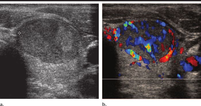
Figure 4. Transverse US images of mostly cystic thyroid nodule with a mural component
containing flow. (a) Gray-scale image shows predominantly cystic nodule (calipers) with small
solid-appearing mural component (arrowheads). (b) Addition of color Doppler mode demon-
strates flow within mural component (arrowheads), confirming that it is tissue and not debris.
solid nodule (Fig 2a) are associated with
US-guided FNA can be directed into this area. The lesion was benign at cytologic examination.
an approximately threefold increase in
cancer risk and coarse calcifications are
associated with a twofold increase, as
Multiple nodules.—Consider FNA of one ules meeting criteria for FNA of solitary compared with predominantly solid nod-
or more nodules, with selection priori- nodules, as outlined above. ules without calcifications (5). Microcal-
tized on the basis of the previously stated Part III.—The presence of abnormal cifications likely represent multiple calci-
criteria in the order listed above. FNA is lymph nodes overrides the US features fied psammoma bodies, which are typical
likely unnecessary in diffusely enlarged criteria and should prompt biopsy of the of papillary thyroid cancer (51). Care
glands with multiple nodules of similar lymph node and/or (if necessary) of an must be taken to differentiate these fine
US appearance without intervening nor- ipsilateral thyroid nodule. punctate calcifications, which are indi-
mal parenchyma. vidually too small to induce posterior
Note that these recommendations are acoustic shadowing, from echogenic foci
Explanations
not absolute or inflexible. In certain cir- with posterior comet-tail artifacts, which
cumstances, the physician’s clinical judg- Measurements.—Nodules should be are commonly seen in benign cystic or
ment may lead him or her to determine measured with the calipers placed out- partially cystic nodules (40) (Fig 2b). In
that FNA need not be performed for nod- side of any visible halo. The maximum the absence of comet-tail artifacts, tiny
ules that meet the recommendations diameter should be used when consider- echogenicities must be assumed to be cal-
above. In others, FNA may be appropriate ing whether or not US-guided FNA cifications when considering the risk of
for nodules that do not meet the criteria should be performed. cancer. There are insufficient data to
listed above. Calcification.—The presence of any cal- know whether intense rim calcification,
Part II.—The recommendation for non- cification within the nodule raises the as opposed to calcifications within the
diagnostic aspirates from initial FNA is as likelihood of malignancy. In particular, nodule, is associated with malignancy.
follows: Consider a second FNA of nod- microcalcifications in a predominantly Composition.—Each nodule should be
798 䡠 Radiology 䡠 December 2005 Frates et alevaluated with regard to the fraction of for the consensus statement, nor on how calculations should be used to monitor
the nodule that is solid versus the frac- to monitor growth. growth: maximum diameter, average di-
tion that is cystic. Nodules can be classi- Multiple nodules.—In many patients, ameter, or volume?
fied semiquantitatively, according to the more than one nodule is identified or the 2. In a patient with multiple nodules,
estimated percentage of solid or cystic gland appears diffusely enlarged with which and how many nodules should
composition or in descriptive terms multiple nodules of similar US appear- undergo US-guided FNA? Strategies for
Radiology
based on the predominant composition ance without intervening normal paren- follow-up in patients with multiple nod-
(eg, solid, predominantly solid, mixed chyma. The panel agreed that FNA is ules should be devised.
solid and cystic, predominantly cystic, likely not necessary in the latter setting. 3. Are there other US characteristics of
and cystic) (Fig 2). Solid or predomi- In patients with multiple discrete nod- a nodule that might be used to prove a
nantly solid nodules have a higher risk of ules, the panel had two opinions regard- nodule is benign, thus precluding FNA in
malignancy than do mixed or predomi- ing selection of nodules for FNA. The ma- some other patients besides those with
nantly cystic nodules. Cystic and almost jority opinion was that the selection almost entirely cystic nodules? Are there
completely cystic nodules have a very should be based primarily on US charac- combinations of US characteristics that
low likelihood of being malignant. Nod- teristics other than nodule size (5). Thus, might be used to help direct manage-
ules with mixed composition have an av- a solid nodule with microcalcifications ment?
erage risk of malignancy. For this reason, should be selected for FNA before a larger 4. What is the cost-effectiveness of var-
the recommended minimal size for US- mixed cystic and solid nodule without ious approaches to the diagnosis of soli-
guided FNA is lower for solid or predom- calcifications. The minority opinion was tary and multiple nodules?
inantly solid nodules than the recom- that the largest nodule should undergo
mended minimal size for mixed solid and US-guided FNA, and the selection of
other nodules for US-guided FNA should References
cystic nodules. 1. Khoo ML, Asa SL, Witterick IJ, Freeman JL.
Color Doppler US.—When color Dopp- be based on US characteristics.
Thyroid calcification and its association
ler US is included in the evaluation of a Abnormal cervical lymph nodes (exclud- with thyroid carcinoma. Head Neck 2002;
thyroid nodule, available research indi- ing submandibular nodes).—The presence 24:651– 655.
cates that marked internal flow suggests of abnormal cervical lymph nodes over- 2. Kim EK, Park CS, Chung WY, et al. New
rides the recommendations in parts I and sonographic criteria for recommending
an increased likelihood of malignancy, as fine-needle aspiration biopsy of nonpal-
II of the statement and should prompt
compared with the absence of marked pable solid nodules of the thyroid. AJR
biopsy of the abnormal lymph nodes Am J Roentgenol 2002;178:687– 691.
internal flow. Marked internal flow is de-
and/or an ipsilateral thyroid nodule of 3. Papini E, Guglielmi R, Bianchini A, et al.
fined as more flow in the nodule than in Risk of malignancy in nonpalpable thyroid
any size. On occasion, a patient has an
the surrounding thyroid gland and more nodules: predictive value of ultrasound
abnormal lymph node representing met-
flow in the central part of the nodule and color Doppler features. J Clin Endocri-
astatic thyroid cancer and a sonographi- nol Metab 2002;87:1941–1946.
than at the periphery (Fig 3). Appropriate
cally normal gland, because the primary 4. Peccin S, de Castro JA, Furlanetto TW, Fur-
Doppler US technique is imperative for tado AP, Brasil BA, Czepielewski MA. Ultra-
tumor is not visible at US. US diagnosis of
accurate assessment of nodule vascular- sonography: is it useful in the diagnosis of
an abnormal lymph node depends on
ity, with color Doppler gain settings max- cancer in thyroid nodules? J Endocrinol
size, shape, vascularity, and internal ar- Invest 2002;25:39 – 43.
imized for slow flow. Color Doppler US is
chitecture (53,54). The US features asso- 5. Frates MC, Benson CB, Doubilet PM, et al.
also useful for evaluating mixed cystic ciated with the highest risk of cancer in- Likelihood of thyroid cancer based on
and solid nodules and predominantly clude heterogeneous echotexture, calcifi- sonographic assessment of nodule size and
cystic nodules with a focal area that ap- composition [abstr]. In: Radiological Soci-
cations, and cystic areas within the ety of North America Scientific Assembly
pears solid. This will help differentiate lymph node (Fig 5). A rounded lymph and Annual Meeting Program. Oak Brook,
solid tissue, which will have blood flow, node or one causing a mass effect is also Ill: Radiological Society of North America,
from an avascular blood clot or debris at elevated risk of being malignant. In 2004; 395.
(Fig 4). When US-guided FNA is per- 6. Frates MC, Benson CB, Doubilet PM, Cibas
general, size is a less reliable criterion for ES, Marqusee E. Can color Doppler sonog-
formed on such nodules, the needle malignancy in a lymph node than are raphy aid in the prediction of malignancy
should be directed toward the regions shape and architecture, although the of thyroid nodules? J Ultrasound Med
with visible flow, to increase the likeli- chance of malignancy increases as the 2003;22:127–131.
hood of a diagnostic aspirate. While oc- 7. Gharib H, Goellner JR. Fine-needle aspira-
size of the lymph node increases. Thus,
casionally useful in selecting nodules for tion biopsy of the thyroid: an appraisal.
lymph nodes should be considered suspi- Ann Intern Med 1993;118:282–289.
US-guided FNA, color Doppler US should cious if they measure more than 7 mm in 8. Hamberger B, Gharib H, Melton LJ, Goell-
not be considered a requirement for the the short axis (54). ner JR, Zinsmeister AR. Fine-needle aspira-
selection of nodules for sampling. tion biopsy of thyroid nodules: impact on
thyroid practice and cost of care. Am J Med
Interval growth.—The panelists agreed
Research Topics 1982;73:381–384.
that US-guided FNA should be consid- 9. Mittendorf EA, Tamarkin SW, McHenry
ered for nodules demonstrating substan- The panel identified several important CR. The results of ultrasound guided fine-
tial growth on serial US studies, even if a unanswered questions that merit future needle aspiration biopsy for evaluation of
nodular thyroid disease. Surgery 2002;132:
prior FNA result was benign. Although research. 648 – 654.
the natural history for both benign and 1. How should substantial growth be de- 10. Danese D, Sciacchitano S, Farsetti A, An-
malignant nodules is growth over time fined? In particular, if a nodule has a dreoli M, Pontecorvi A. Diagnostic accu-
(16), rapid growth of a nodule indicates prior FNA diagnosis of being benign, how racy of conventional versus sonography-
guided fine-needle aspiration biopsy of
an increased risk for malignancy (15,52). much growth over what period of time thyroid nodules. Thyroid 1998;8:15–21.
The panelists did not come to a consen- should prompt consideration for repeat 11. Hegedus L. The thyroid nodule. N Engl
sus on how to define substantial growth US-guided FNA? What measurements or J Med 2004;351:1764 –1771.
Volume 237 䡠 Number 3 Management of Thyroid Nodules Detected at US 䡠 79912. Chan BK, Desser TS, McDougall IR, Weigel 27. Kunreuther E, Orcutt J, Benson CB, et al. fact in thyroid ultrasound. J Clin Ultra-
RJ, Jeffrey RB. Common and uncommon Prevalence and distribution of carcinoma sound 1996;24:129 –133.
sonographic features of papillary thyroid in the uninodular and multinodular goi- 41. Iannuccilli JD, Cronan JJ, Monchik JM.
carcinoma. J Ultrasound Med 2003;22: ter. Presented at the 76th Annual Meeting Risk for malignancy of thyroid nodules as
1083–1090. of the American Thyroid Association, Van- assessed by sonographic criteria. J Ultra-
13. Wienke JR, Chong WK, Fielding JR, Zou couver, British Columbia, Canada, Sep- sound Med 2004;23:1455–1464.
KH, Mittelstaedt CA. Sonographic features tember 29 –October 3, 2004. 42. Pacella CM, Guglielmi R, Fabbrini R, et al.
Radiology
of benign thyroid nodules: interobserver 28. Nam-Goong IS, Kim HY, Gong G, et al. Papillary carcinoma in small hypoechoic
reliability and overlap with malignancy. J Ultrasonography-guided fine-needle aspi- thyroid nodules: predictive value of echo
Ultrasound Med 2003;22:1027–1031. ration of thyroid incidentaloma: correla- color Doppler evaluation—preliminary re-
14. Hagag P, Strauss S, Weiss M. Role of ultra- tion with pathological findings. Clin En- sults. J Exp Clin Cancer Res 1998;17:127–
sound-guided fine-needle aspiration bi- docrinol (Oxf) 2004;60:21–28. 128.
opsy in evaluation of nonpalpable thyroid 29. Hermus AR. Clinical manifestations and 43. Holden A. The role of colour and duplex
nodules. Thyroid 1998;8:989 –995. treatment of nontoxic diffuse and nodular Doppler ultrasound in the assessment of
15. Alexander EK, Hurwitz S, Heering JP, et al. goiter. In: Braverman LE, Utiger RD, Ing- thyroid nodules. Australas Radiol 1995;39:
Natural history of benign solid and cystic bar SH, Werner SC, eds. Werner and Ing- 343–349.
thyroid nodules. Ann Intern Med 2003; bar’s the thyroid: a fundamental and clin- 44. Rago T, Vitti P, Chiovato L, et al. Role of
138:315–318. ical text. Philadelphia, Pa: Lippincott Wil- conventional ultrasonography and color
16. Quadbeck B, Pruellage J, Roggenbuck U, et liams & Wilkins, 2000; 867. flow-Doppler sonography in predicting
al. Long-term follow-up of thyroid nodule 30. Jemal A, Murray T, Ward E, Tiwari RC, malignancy in ‘cold’ thyroid nodules. Eur J
growth. Exp Clin Endocrinol Diabetes Feuer EF, Thun MJ. Cancer statistics, 2005. Endocrinol 1998;138:41– 46.
2002;110:348 –354. CA Cancer J Clin 2005;55:10 –30. 45. Shimamoto K, Endo T, Ishigaki T, Sakuma
17. Court-Payen M, Nygaard B, Horn T, et al. 31. Gilliland FD, Hunt WC, Morris DM, Key S, Makino N. Thyroid nodules: evaluation
US-Guided fine-needle aspiration biopsy CR. Prognostic factors for thyroid carci- with color Doppler ultrasonography. J Ul-
of thyroid nodules. Acta Radiol 2002;43: noma: a population-based study of 15,698 trasound Med 1993;12:673– 678.
131–140. cases from the Surveillance, Epidemiology 46. Fraker DL. Thyroid tumors. In: DeVita V Jr,
18. Marqusee E, Benson CB, Frates MC, et al. and End Results (SEER) program 1973– Hellman S, Rosenberg S, eds. Cancer: princi-
Usefulness of ultrasonography in the man- 1991. Cancer 1997;79:564 –573. ples and practice of oncology. Philadelphia,
Pa: Lippincott-Raven, 1997; 1629 –1652.
agement of nodular thyroid disease. Ann 32. Mazzaferri EL, Jhiang SM. Long-term im-
47. Alexander EK, Heering JP, Benson CB, et
Intern Med 2000;133:696 –700. pact of initial surgical and medical therapy
al. Assessment of nondiagnostic ultra-
19. Wiest PW, Hartshorne MF, Inskip PD, et al. on papillary and follicular thyroid cancer.
sound-guided fine needle aspirations of
Thyroid palpation versus high-resolution Am J Med 1994;97:418 – 428.
thyroid nodules. J Clin Endocrinol Metab
thyroid ultrasonography in the detection 33. Hay ID. Papillary thyroid carcinoma. En-
2002;87:4924 – 4927.
of nodules. J Ultrasound Med 1998;17: docrinol Metab Clin North Am 1990;19:
48. Chow LS, Gharib H, Goellner JR, van Heer-
487– 496. 545–576.
den JA. Nondiagnostic thyroid fine-needle
20. Carroll BA. Asymptomatic thyroid nod- 34. Hay ID, Bergstralh EJ, Goellner JR, Eber- aspiration cytology: management dilem-
ules: incidental sonographic detection. sold JR, Grant CS. Predicting outcome in mas. Thyroid 2001;11:1147–1151.
AJR Am J Roentgenol 1982;138:499 –501. papillary thyroid carcinoma. Surgery 49. McHenry CR, Walfish PG, Rosen IB. Non-
21. Brander A, Viikinkoski P, Nickels J, 1993;114:1050 –1058. diagnostic fine needle aspiration biopsy: a
Kivisaari L. Thyroid gland: US screening in 35. Wong JB, Kaplan MM, Meyer KB, Pauker dilemma in management of nodular thy-
a random adult population. Radiology SG. Ablative radioactive iodine therapy for roid disease. Am Surg 1993;59:415– 419.
1991;181:683– 687. apparently localized thyroid carcinoma: a 50. Bellantone R, Lombardi CP, Raffaelli M, et
22. Bruneton JN, Balu-Maestro C, Marcy PY, decision analytic perspective. Endocrinol al. Management of cystic or predomi-
Melia P, Mourou MY. Very high frequency Metab Clin North Am 1990;19:741–760. nantly cystic thyroid nodules: the role of
(13 MHz) ultrasonographic examination 36. DeGroot LJ, Kaplan EL, McCormick M, ultrasound-guided fine-needle aspiration
of the normal neck: detection of normal Straus FH. Natural history, treatment, and biopsy. Thyroid 2004;14:43– 47.
lymph nodes and thyroid nodules. J Ultra- course of papillary thyroid carcinoma. 51. Maitra A, Abbas AK. The endocrine system.
sound Med 1994;13:87–90. J Clin Endocrinol Metab 1990;71:414 – In: Kumar V, Abbas A, Fausto N, eds. Rob-
23. Horlocker TT, Hay ID. Prevalence of inci- 424. bins and Cotran’s pathologic basis of dis-
dental nodular thyroid disease detected 37. Cooper DS, Specker B, Ho M, et al. Thyro- ease. Philadelphia, Pa: Saunders, 1994;
during high-resolution parathyroid sonog- tropin suppression and disease progression 1155–1226.
raphy. In: Medeiros-Neto G, Gaitan E, eds. in patients with differentiated thyroid 52. Grant CS, Hay ID, Gough IR, McCarthy
Frontiers in thyroidology. Vol 2. New cancer: results from the National Thyroid PM, Goellner JR. Long-term follow-up of
York, NY: Plenum, 1985; 1309 –1312. Cancer Treatment Cooperative Registry. patients with benign thyroid fine-needle
24. Mortensen JD, Woolner LB, Bennett WA. Thyroid 1998;8:737–744. aspiration cytologic diagnoses. Surgery
Gross and microscopic findings in clini- 38. Pujol P, Daures JP, Nsakala N, Baldet L, 1989;106:980 –986.
cally normal thyroid glands. J Clin Endo- Bringer J, Jaffiol C. Degree of thyrotropin 53. Vassallo P, Wernecke K, Roos N, Peters PE.
crinol Metab 1955;15:1270 –1280. suppression as a prognostic determinant Differentiation of benign from malignant
25. Hegedus L, Bonnema SJ, Bennedbaek FN. in differentiated thyroid cancer. J Clin En- superficial lymphadenopathy: the role of
Management of simple nodular goiter: docrinol Metab 1996;81:4318 – 4323. high-resolution US. Radiology 1992;183:
current status and future perspectives. En- 39. Alexander EK, Marqusee E, Orcutt J, et al. 215–220.
docr Rev 2003;24:102–132. Thyroid nodule shape and prediction of 54. Ying M, Ahuja A, Metreweli C. Diagnostic
26. Sherman SI, Angelos P, Ball D, et al. Thy- malignancy. Thyroid 2004;14:953–958. accuracy of sonographic criteria for evalu-
roid carcinoma. J Natl Compr Canc Netw 40. Ahuja A, Chick W, King W, Metreweli C. ation of cervical lymphadenopathy. J Ul-
2005;3:404 – 457. Clinical significance of the comet-tail arti- trasound Med 1998;17:437– 445.
800 䡠 Radiology 䡠 December 2005 Frates et alYou can also read