Metabolic changes in pomegranate fruit skin following cold storage promote chilling injury of the peel
←
→
Page content transcription
If your browser does not render page correctly, please read the page content below
www.nature.com/scientificreports
OPEN Metabolic changes in pomegranate
fruit skin following cold storage
promote chilling injury of the peel
Ravi Singh Baghel1,2, Alexandra Keren‑Keiserman1 & Idit Ginzberg1*
Pomegranate cv. ‘Wonderful’ fruit are susceptible to chilling injuries of the peel (CIp) when stored at
7 °C in modified-atmosphere bags for more than 3 months. The damage, manifested as superficial
browning, is restricted to the fruit skin, i.e., the outer colored layer of the peel. To characterize
possible causes of CIp development, fruit were collected at early harvest—when the premature fruit
are poorly colored and susceptible to CIp development, and at late harvest—when mature fruit have
fully red skin and less susceptibility to CIp. Skin samples were collected on day of harvest and at
different time points during storage. Anatomical study of skin with CIp disorder showed a broken
cuticle layer with underlying degenerated cells. A high total phenol content, which is associated with
high antioxidant capacity, was not sufficient to prevent the development of CIp in the premature fruit.
The concentration of punicalagin was the same for premature and mature skin at harvest and during
storage, and therefore not associated with CIp development in the premature fruit skin. Furthermore,
the expression of antioxidant-related genes CAT2, SOD and GR2 was similar for both premature and
mature fruit skin. Poor pigmentation of the premature fruit skin and chilling-induced downregulation
of key anthocyanin-biosynthesis genes were associated with CIp development. High total phenol
concentration combined with high expression of the gene encoding PPO was also associated with
CIp; however, high expression ratio of PAL to PPO was found in mature skin, and may be associated
with reduced CIp disorder. The results presented suggest future possibilities for controlling the CIp
phenomenon.
Pomegranate (Punica granatum L.) fruit is well known for its health-beneficial m etabolites1–4, and there is exten-
sive demand for it in global markets. The main cultivar in Israel is ‘Wonderful’. Harvesting lasts 3 weeks, and then
the fruit is sorted, packed and stored. High premium pomegranate is intended for export, which necessitates
prolonged storage of the fruit. For that purpose, the fruit is packed in modified-atmosphere (MA) bags and stored
at 7 °C until shipment. Up to 3 months of storage, the fruit maintains its visual, organoleptic and nutritive quality.
However, when storage period is extended up to 5 months, fruit appearance deteriorates even if maintained in
MA bags at 7 °C. The response of the pomegranate peel to reduced temperatures is usually referred to as ‘husk
scald’ (HS), however, later on in this work we show that this term refers to two different symptoms that develop
on the surface of the fruit during the cold storage.
In the literature, HS has been studied in pomegranate accession lines and under different storage c onditions5–8.
HS is superficial browning that is restricted to the peel, and has been suggested to result from the oxidation of
o-dihydroxyphenols9. However, its causes are not yet clear. The HS phenomenon differs from chilling damage
to the inner fruit tissues that is mostly manifested as internal browning of the membranous walls and the white
spongy tissue surrounding the arils10,11. Chilling injury (CI) of inner tissues is induced after a short period of
storage (2 weeks) at 1 °C, and browning becomes apparent only after transfer of the fruit to shelf-life conditions
at 20 °C.
The pomegranate fruit peel contains a high concentration of two groups of polyphenols, anthocyanins (AT)—
the main pigments in pomegranate, and hydrolyzable tannins (HT), both are synthesized from an intermediate of
the shikimate p athway12,13. The AT and HT confer strong antioxidant activity and are considered to be potential
protectants from chilling damage7,14,15.
The biochemical pathway and genetic control of AT production have been well characterized in numer-
ous plant species and are considered to be highly conserved among different species in the plant kingdom16.
1
Institute of Plant Sciences, Agricultural Research Organization, Volcani Center, 68 HaMacabim Road,
P.O. Box 15159, 7505101 Rishon LeZion, Israel. 2Present address: Biological Oceanography Division, CSIR-National
Institute of Oceanography, Dona Paula, Goa 403004, India. *email: iditgin@volcani.agri.gov.il
Scientific Reports | (2021) 11:9141 | https://doi.org/10.1038/s41598-021-88457-4 1
Vol.:(0123456789)
www.nature.com/scientificreports/
Common flavonoid precursors are catalyzed by chalcone synthase (CHS), chalcone isomerase (CHI), flavanone
3-hydroxylase, dihydroflavonol 4-reductase (DFR), and leucoanthocyanidin dioxygenase. The expression of DFR
is correlated with pomegranate color and the timing of color appearance during fruit d evelopment17.
Six AT pigments have been identified in pomegranate fruit: mono- and diglucosides of cyanidin (red pig-
ments), delphinidin (purple pigments) and pelargonidin (orange pigments)18–21. The transcription factor PgWD40
has been shown to regulate the expression of downstream structural genes involved in AT biosynthesis in pome-
granate fruit. Its expression level was correlated with the expression level of the structural enzyme DFR and with
total cyanidin level in the s kin17.
Only traces of the HT-specific pathway are known. It branches from the shikimate pathway, with shikimate
dehydrogenase as the first committed step catalyzing gallic acid12. This is followed by production of β-glucogallin
and then pentagalloyl g lucose22. Further oxidative modifications of pentagalloyl glucose lead to the formation
of gallotannins and ellagitannins (e.g., punicalagin in pomegranate)22–24.
The correlation between antioxidant capacity, AT and HT levels, and the resistance of the pomegranate peel
to HS development is not yet c onclusive7,8. This could be due to variations in the response among different acces-
sion lines, or to metabolite analyses performed with the whole peel. In the present study, we used cv. ‘Wonderful’
which bears large, late-season fruit that ripen in October and is “sweet–sour” in taste2. Only the colored outer
layer of the peel, the skin, was analyzed, as the development of chilling-induced HS is limited to that layer. The
effect of skin color/maturation on tolerance to chilling conditions was tested in early- and late-harvested fruit,
differing in pigment intensity. The fruit were stored in Xtend bags, the current commercial practice for extended
storage time25. The major AT and HT metabolites in the pomegranate skin were profiled during storage, along
with a determination of total polyphenol content (TPC), skin antioxidant capacity, and related gene expression.
Materials and methods
Plant material and experimental design. Pomegranate cv. ‘Wonderful’ fruit were collected from a
commercial orchard at Kibbutz Tsor’a, located in the central region of Israel (31° 45′ 37″ N 34° 57′ 48″ E). The
use of pomegranate was carried out in accordance with relevant guidelines and regulation. Within 24 h of har-
vest, fruit were washed with the fungicide Scholar (Syngenta, USA) and stored at 7 °C in Xtend MA bags for
pomegranates (http://www.stepac.com/products.asp?siteid=971&pageid=1497; StePac L.A. Ltd., Israel)25.
Two experiments were conducted. In experiment 1, fruit harvested from commercial plots was sorted at the
packing house for export quality, and was stored for 4 months at 7 °C in Xtend bags (bin bags for ~ 400 kg, code:
RIM2.5), until CI of the peel were apparent. At that stage, fruit peel was sampled for the analyses detailed below,
at three biological replicates, four fruit per replicate.
Experiment 2 was aimed to monitor the changes occur in fruit peel during storage time, prior to CI develop-
ment. Fruit were picked manually at two time points: early harvest, on 8 Oct 2018, prior to full fruit maturity
[premature (PM) fruit] when the fruit color has a greenish-reddish tint, and at maturity, on 25 Oct 2018 [mature
(M) fruit] when the fruit fully exhibits the characteristic red color (demonstrated in Fig. 1a, HQ fruit).
PM and M fruit of similar size were collected from inner branches, avoiding the southern side of the tree
that is exposed to high daily radiation. The fruits were brought directly from the orchard to the Volcani Center.
Fruit of each type were placed in Xtend bags (code: 815-PG28/m for 5 kg), and were stored at 7 °C. For each fruit
type, a total of 60 fruit were divided in 15 Xtend bags, four fruit per a bag. One bag was considered one replicate.
Fruit were sampled at specific time points: day of harvest, time 0 (t0), after 1 week of storage (w1), and after 1, 2
and 3 months of storage (m1, m2, m3, respectively). At each time point, three bags, each with four fruits, were
sampled for each fruit type.
The peel of pomegranate fruit consists of an inner thick spongy white tissue, and an outer thin red skin made
up of epidermis cells covered by cuticle. All of the analyses, except for the anatomical study, were performed
with the colored skin tissue that was carefully removed from the spongy tissue, from all sides of the fruit, using a
peeler. Skin samples were collected for each biological replicate were pooled from all of the four fruits in each bag.
Skin anatomical study. Samples of the pomegranate surface (blocks of 4 × 3 × 3 mm) were fixed in FAA
(50% ethanol, 5% acetic acid and 3.7% formaldehyde, v/v, in water), dehydrated in an ethanol/xylene series and
embedded in paraplast (Surgipath Paraplast Plus, Leica Biosystems Richmond Inc., USA) according to standard
methods26. Tissue Sects. (20 µm) were stained with Safranin-O/Fast green (Sigma Chemicals, Israel) for exami-
orphology27. Sections were observed under a light microscope (Leica DMLB, Germany) and
nation of tissue m
images were displayed on a monitor through a CCD camera (Leica DC2000) using the Leica IM1000 program.
For each treatment, three independent fruit were examined, each was sectioned at three locations along the
affected area.
Determination of AT and phenol content in the skin. Total AT content was determined based on
Markham and Ofman (1993)28. Skin samples, 200–300 mg, were ground in liquid nitrogen, followed by extrac-
tion in 1 mL of water:methanol:acetic acid (5:11:1, v/v/v) solution for 1 h at 4 °C. The mixture was vortexed and
separated by centrifugation for 10 min at 14,500 g and 4 °C, and the pellet was re-extracted in the same man-
ner. The supernatants containing AT were combined and read in a spectrophotometer (UV-2401PC Shimadzu,
Japan) at 520 nm. Analysis was performed with three biological replicates, each with three technical replicates.
TPC was determined in skin extract consisting of 50 mg vacuum-dried powdered samples homogenized in
500 µL d dH2O. The homogenates were centrifuged at 5000 g for 15 min to separate the liquid extract from the
solids, and the supernatants were held at − 20 °C until use. The peel extract was diluted 1:100 and 10 µL was
added to the well of a 96-well plate with 50 µL of 10% Folin–Ciocalteu reagent (#F9252, Sigma Chemicals) (w/v
in water). After 3 min incubation at room temperature, 40 µL of 7.5% Na2CO3 (w/v in water) was added to each
Scientific Reports | (2021) 11:9141 | https://doi.org/10.1038/s41598-021-88457-4 2
Vol:.(1234567890)
www.nature.com/scientificreports/
Figure 1. Physiological blemishes on pomegranate skin following 3–4 months of storage at 7 °C in Xtend bags
with modified atmosphere. Fruit were collected at maturity (a), and at prematuration stage of the skin (b).
Anatomy of the skin physiological blemishes taken from the mature fruit after 2 or 4 months in storage (c). The
epidermal cells are underlined with a dashed line. The cuticle layer overlying the epidermal cells is marked with
arrows. Each frame demonstrates skin of one representative fruit out of three independent fruit. Bar = 100 µm.
HQ smooth, shiny and fully colored skin of high quality, CIp chilling injuries of the skin, appearing as dark
brown regions, HS husk scald, appearing as loss of red pigment intensity and development of brownish tint,
together with peel hardening.
well, mixed gently and incubated for 40 min at 37 °C. Analysis was performed with three biological replicates,
each with three technical replicates. Reactions were read at 760 nm using the Synergy H1 Hybrid Multi-Mode
Microplate Reader (Biotek, Fisher Scientific, USA). TPC values were calculated using a standard curve of 0–0.5 g
L−1 gallic acid (#G7384, Sigma Chemicals), and were expressed as g k g−1 gallic acid equivalents (GAE) on a dry
weight basis.
Profiling of individual HT and AT metabolites was done in the laboratory of Rachel Amir, Migal, Israel12.
Identification and quantification of punicalagin isomers, gallic acid and ellagic acid were achieved by comparing
Scientific Reports | (2021) 11:9141 | https://doi.org/10.1038/s41598-021-88457-4 3
Vol.:(0123456789)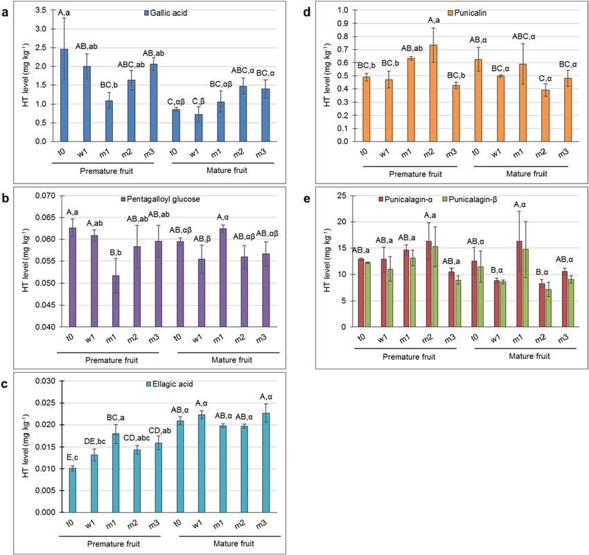
www.nature.com/scientificreports/
the retention time and standard curves of the authentic standards (Sigma-Aldrich, Rehovot, Israel). Unidentified
HTs were quantified based on the corresponding chromatogram peak area. Level of the mono- and diglucoside
(G and dG, respectively) forms of the major pomegranate ATs was quantified as follow: authentic standards were
used for delphinidin-3,5-dG (D-3,5-dG), and cyaniding-3,5-dG (C-3,5-dG) (Sigma Aldrich, St. Louis, MO).
D-3-G, C-3-G and pelargonidin glucosides (P-3-G and P-3,5-dG) were quantified based on the corresponding
chromatogram peak area.
Antioxidant capacity of the skin. Two methods were used to analyze the antioxidant capacity of pome-
granate skin: ferric reducing ability of plasma (FRAP) and 2,2-diphenyl-1-picrylhydrazyl (DPPH) radical-scav-
enging activity15,29. The FRAP method was performed according to30. It measures the ferric-reducing ability of
plasma at low pH. An intense blue color is formed when the ferric–tripyridyltriazine ( Fe3+–TPTZ) complex is
reduced to the ferrous ( Fe2+) form, which is read at 593 nm. The reaction was performed in a 96-well plate and
included 15 µL skin extract (prepared as described for TPC determination) diluted 1:150, and 285 µL reagent
solution [25 mL of 300 mM pH 3.6 acetate buffer, 2.5 mL of 10 mM F e3+–TPTZ (#T1253, Sigma Chemicals) and
2.5 mL of 20 mM FeCl3 (#157740, Sigma Chemicals)], and was read following 4 min incubation at 25 °C in a
microplate reader as above, at 595 nm.
DPPH radical-scavenging activity was determined according to Wissam et al. (2012)31 in a 96-well plate.
The reaction consisted of 15 µL skin extract (prepared as above) diluted 1:100 with water, and 285 µL of 100 µM
DPPH (#D9132, Sigma Chemicals) in methanol. After incubation with shaking at 25 °C for 15 min, recordings
were performed at 517 nm at 3-min intervals in the microplate reader.
For both FRAP and DPPH methods, the blank reaction was performed with 15 µL methanol. Analyses
were run on three biological replicates and three technical replicates. The skin antioxidant capacity was calcu-
lated using a standard curve of 0.1–0.7 mM 6-hydroxy-2,5,7,8-tetramethylchroman-2-carboxylic acid (Trolox,
#238813, Sigma Chemicals) in methanol. Units were expressed as mmol k g-1 Trolox equivalents (TE) on a dry
weight basis.
RNA extraction and quantitative reverse transcription PCR (RT‑qPCR). Total RNA was extracted
according to Ginzberg et al. (2009)32; cDNA was synthesized using the QuantiTect Reverse Transcription Kit
(QIAGEN GmbH, Germany), and ABsolute™ Blue QPCR SYBR Green ROX Mix (Thermo Scientific, USA) was
used for qPCR according to the manufacturer’s protocol with specific primers (Supplementary Table S1). Gene
expression data was quantified using a standard curve—A mixture of the tested cDNA samples at equal amounts
was diluted 1:30, 1:240, and 1:1920 and used in each qPCR with the specific primers. Each RT-qPCR was per-
formed with three biological replicates, each with three technical replicates. Values in each sample were normal-
ized to the expression levels of PHOSPHOGLYCERATE KINASE (PGK) as the reference g ene23.
Statistical analysis. Data were analyzed for statistical significance by Student’s t-test using JMP software
(http://www.jmp.com). Values presented are averages of three biological replicates ± SE. Different letters indicate
significant differences among means by Student’s t-test (P < 0.05). When multiple comparisons were made on the
same chart, different fonts—uppercase, lowercase or Greek letters—were used for each comparison.
Results
Chilling injury of the peel (CIp). The skin of pomegranate fruit that were collected from the packing
house following 4 months’ storage in Xtend bags at 7 °C exhibited imperfections. These were superficial physi-
ological defects of two types—dark brown regions covering up to 40% of the fruit surface and were defined as
chilling injuries of the peel, CIp (Fig. 1a), and regions that appeared as loss of red pigment intensity together
with the development of a brownish tint and peel hardening, and were defined as HS (Fig. 1a). Both CIp and HS
were limited to a few upper layers of the peel, and were not evident in the spongy tissue, the inner membranes or
the aril compartments (Supplementary Figure S1). Fruit with skin of high quality (HQ fruit) appeared smooth
and shiny, with an intense color (Fig. 1a). Overall, 52% of the fruit were of the HQ-skin type, and 27% and 21%
were of CIp- and HS-skin types, respectively. Note, CIp and HS were mutually exclusive, and were not displayed
on the same fruit. These values demonstrate the severity of the phenomenon of deterioration in pomegranate
appearance under prolonged storage (4 months), especially in light of the fact that all fruit were sorted for high
export quality prior to storage.
Anatomical analysis of the fruit surface was performed to better describe the phenomenon. Peel samples
were taken from 4-month-stored fruit, from regions exhibiting HQ, CIp and HS skin types (Fig. 1c). These were
compared to peel samples from 2-month-stored fruit that exhibited an early stage of CIp development; at this
time, less than 10% of the fruit were affected and the CIp appeared as patches of low severity and limited size.
The HQ skin demonstrated one layer of compact organized epidermis cells with an overlying cuticle, and an
intact and compact subepidermal tissue, after both 2 months and 4 months of storage. CIp disorder exhibited
a broken cuticle layer with underlying degenerated cells; the damage was limited to the outermost layers after
2 months’ storage (Fig. 1c, skin type CIp, upper panel), whereas after 4 months’ storage, its severity increased
and the underlying tissues were affected as well (Fig. 1c, skin type CIp, lower panel). HS skin disorder developed
only after prolonged storage, and showed breaks in the epidermis and cuticle, with underlying degenerated cells;
however it was limited to the very outer layers of the skin, and the inner layers seemed unaffected (Fig. 1c, skin
type HS, lower right panel), in contrast to the CIp disorder.
Antioxidant activity in HQ‑, CIp‑, and HS‑skin types. As phenolics are known to contribute to the
antioxidant capacity of pomegranate peel and juice7,29, and may enhance skin tolerance to storage conditions,
Scientific Reports | (2021) 11:9141 | https://doi.org/10.1038/s41598-021-88457-4 4
Vol:.(1234567890)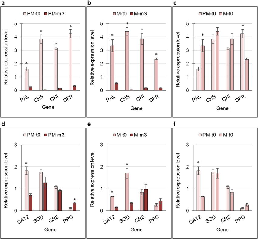
www.nature.com/scientificreports/

                                  Figure 2.  Total polyphenol content (TPC) (a), antioxidant capacity (b), and the expression of antioxidant-
                                  related genes and key anthocyanin-biosynthesis genes (c) of skin types shown in Fig. 1. TPC was determined
                                  using the Folin–Ciocalteu reagent, and is given as gallic acid equivalents (GAE). Antioxidant capacity was
                                  determined using the FRAP method, and is given as Trolox equivalents (TE). Values are averages of three
                                  biological replicates ± SE. Data were analyzed for statistical significance among means by Student’s t-test;
                                  different letters indicate significantly different values (P < 0.05).

                                  TPC was monitored in HQ-, CIp-, and HS-skin types. Results indicated a significant reduction in TPC in the
                                  damaged skin types compared to the HQ skin (Fig. 2a). Accordingly, antioxidant capacity was reduced in CIp
                                  and HS skin compared to the HQ skin (Fig. 2b, and Supplementary Figure S2).
                                     To evaluate additional factors that may be involved with skin antioxidant capacity, the expression of genes
                                  known to counteract oxidative stress was monitored. Relative transcript levels of GLUTATHIONE REDUCTASE
                                  2 (GR2) and POLYPHENOL OXIDASE (PPO) were upregulated in CIp and HS skin compared to the HQ skin,
                                  albeit not always significantly (Fig. 2c). CATALASE 2 (CAT2) transcript level was upregulated only in the HS
                                  skin, whereas the reverse expression profile was found for SUPEROXIDE DISMUTASE (SOD): low in HS skin
                                  and high in HQ skin (Fig. 2c). Expression of key genes in phenylpropanoids biosynthesis pathway—CHS, CHI
                                  and in ATs pathway DFR—which could also contribute to the antioxidant capacity of the skin, was strongly
                                  downregulated in CIp and HS skin compared to the HQ skin, whereas the expression level of PHENYLALANINE
                                  AMMONIA-LYASE (PAL) was similar for all skin types (Fig. 2c).

                                  Antioxidant capacity of the skin of early‑ and late‑harvested stored fruit. The overall data
                                  described above demonstrated the differences and similarities between storage-induced CIp and HS skin imper-
                                  fections and HQ skin. However, these were monitored when the damage was already noticeable. To understand
                                  the events that occur in the skin prior to its visible breakdown, a controlled experiment was conducted to char-
                                  acterize skin antioxidant capacity during storage. Pomegranate fruit were collected at two time points: early
                                  harvest, prior to fruit maturity (PM fruit), when fruit color had a greenish-reddish tint, and at maturity (M fruit)
                                  when fruit fully exhibited the characteristic red color. The PM fruit were expected to exhibit higher sensitivity

Scientific Reports |   (2021) 11:9141 |                https://doi.org/10.1038/s41598-021-88457-4                                                   5

                                                                                                                                               Vol.:(0123456789)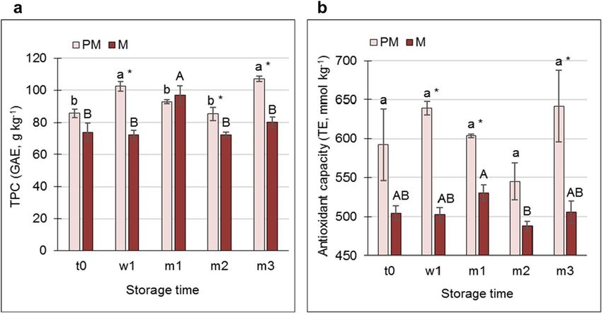
www.nature.com/scientificreports/
Figure 3. Total polyphenol content (TPC) (a) and antioxidant capacity (b) in the skin of premature (PM) and
mature (M) fruit at harvest (t0), after 1 week in storage (w1), and after 1, 2 and 3 months in storage (m1, m2,
m3, respectively). TPC was determined using Folin–Ciocalteu reagent, and is given as gallic acid equivalents
(GAE). Antioxidant capacity was determined using the FRAP method, and is given as Trolox equivalents (TE).
Values are averages of three biological replicates ± SE. Statistical analysis was performed by Student’s t-test
(P < 0.05). Significant difference among means between time points is indicated by different lowercase letters for
PM samples, and uppercase letters for M samples. Asterisks indicate significant differences between PM and M
values for the same time point.
to the storage conditions than the M fruit11. Fruit skin was sampled at time of harvest (t0), and the remaining
fruit were then stored in Xtend bags at 7 °C, and their skin was sampled after 1 week in storage (w1), and after 1,
2 and 3 months in storage (m1, m2, m3, respectively). At the m3 sampling, the PM fruit skin exhibited CIp that
appeared as brown regions on a yellow-tinted background (Fig. 1b).
A transient increase in TPC in the PM skin was recorded at w1, and then at m3, whereas for the M fruit, this
occurred at m1. However, interestingly, TPC values were significantly higher in the PM skin vs. M skin at the
longest storage points, m2 and m3 (Fig. 3a). Similarly, the antioxidant capacity in skin from PM fruit was mostly
significantly higher than that in the M skin (Fig. 3b, and Supplementary Figure S3).
Profiling HT in the skin of early‑ and late‑harvested stored fruit. Punicalagin isomers are the phe-
nolic metabolites involved in peel antioxidant a ctivity15,29. Accordingly, HT profiling was performed (Supple-
mentary Figure S4) to determine the association of punicalagin and its precursors to the differential response
of PM- and M-fruit skin to storage conditions. The level of gallic acid, the first metabolite in the HT-specific
pathway, was significantly higher in PM skin than in M skin at t0 (Fig. 4a). Its level decreased slightly (but not
significantly) in the PM skin during storage (t0 compared to m3), whereas its level increased slightly (but not
significantly) in M skin during storage. Nevertheless, overall, the level of gallic acid was maintained at a higher
level in the skin of PM fruit during storage compared to M skin, albeit not significantly (Fig. 4a). The reverse
trend was observed for ellagic acid level: significantly lower in PM fruit compared to M fruit at t0, and although
its level significantly increased in the former during storage, overall, ellagic acid level was significantly higher in
M fruit during storage (Fig. 4c). No differences between skin samples or time points were detected for pentagal-
loyl glucose (Fig. 4b), an intermediate in the pathway between gallic acid and ellagic acid. Note that the levels, in
terms of mg kg-1, were very low for ellagic acid and pentagalloyl glucose compared to the other HT metabolites.
The level of punicalin (Fig. 4d) increased significantly in PM skin up to m2 and then dropped to its initial
values as storage progressed. In M skin, punicalin levels remained similar throughout storage. Punicalagin was
the main HT in the pomegranate skin (Fig. 4e); its level was the same in PM and M skin, with no change during
the storage. This was true for both punicalagin isomers.
In addition to the known HT metabolites (Fig. 4), some unidentified HTs were detected (Supplementary
Figures. S4 and S5). Most of them exhibited higher levels in PM skin than in M skin, both at harvest and during
storage.
Summing the HT values, it could be concluded that PM skin contains slightly higher levels of HT than M
skin, and that the HT values for each skin type were similarly maintained during storage.
Total AT and profiling of AT metabolites in the skin of early‑ and late‑harvested stored
fruit. Skin AT accumulate largely toward harvest. This was nicely demonstrated by the skin of PM fruit, which
was harvested 2 weeks before the optimal time, and contained a significantly lower level of total AT than the skin
of M fruit (Supplementary Figure S6, t0). During storage, AT level in the skin of M fruit increased significantly
(66%, compare t0 to t3), whereas in PM skin, there was only a slight, nonsignificant increase.
Scientific Reports | (2021) 11:9141 | https://doi.org/10.1038/s41598-021-88457-4 6
Vol:.(1234567890)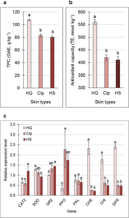
www.nature.com/scientificreports/

                                  Figure 4.  Contents of six members of the hydrolyzable tannins (HT) in the skin of premature and mature fruit
                                  at harvest (t0), after 1 week in storage (w1), and after 1, 2 and 3 months in storage (m1, m2, m3, respectively).
                                  (a) Gallic acid, (b) Pentagalloyl glucose, (c) Ellagic acid, (d) Punicalin, (e) Alpha- and beta-Punicalagin. Values
                                  are averages of three biological replicates ± SE. Different letters indicate significant differences among means
                                  by Student’s t-test (P < 0.05): uppercase letters for both peel types and all time points, lowercase letters for
                                  premature fruit at different time points, and Greek letters for mature fruit at different time points.

                                      Levels of the mono- and diglucoside (G and dG, respectively) forms of the major AT delphinidin (D-3-G and
                                  D-3,5-dG), cyanidin (C-3-G and C-3,5-dG), and pelargonidin (P-3-G and P-3,5-dG) in the fruit skin followed
                                  a similar pattern (Fig. 5). The levels of AT glucosides were significantly lower in the skin of PM fruit compared
                                  that of M fruit, both at harvest and during storage; pelargonidin was totally absent in PM skin. In the PM skin,
                                  there was a slight but not significant increase in delphinidin and cyanidin levels at the beginning of the storage
                                  period (w1 and m1), but then their levels dropped (Fig. 5a,b). No significant change was detected in the skin of
                                  M fruit during storage.

                                  Gene expression in the skin of early‑ and late‑harvested stored fruit. To complement the metabo-
                                  lite data, a gene-expression study was conducted, monitoring key genes in AT biosynthesis and those coding
                                  for antioxidant activity. Gene expression was compared between samples at harvest (t0) and after prolonged
                                  storage (m3) for PM- and M-fruit skin separately, and between PM and M fruit at harvest. There was a dramatic
                                  downregulation of AT-related genes—PAL, CHS, CHI, and DFR—during storage, in both skin types (Fig. 6a,b).
                                  Comparing the t0 data of the two skin types indicated reduced PAL and increased DFR expression in PM skin
                                  compared to M skin (Fig. 6c).
                                      The expression of antioxidant-related genes that are commonly associated with tolerance to low storage
                                  temperatures was monitored in the same skin samples. At harvest (t0), CAT2 expression level was significantly
                                  higher than after prolonged storage (m3), in both PM- and M-fruit skin (Fig. 6d,e), and its expression was sig-
                                  nificantly higher in the PM skin vs. M skin at t0 (Fig. 6f). The expression level of SOD was similar in both skin
                                  types at t0 (Fig. 6f), but was strongly downregulated following prolonged storage in the skin of M fruit (Fig. 6e),

Scientific Reports |   (2021) 11:9141 |                https://doi.org/10.1038/s41598-021-88457-4                                                       7

                                                                                                                                               Vol.:(0123456789)
www.nature.com/scientificreports/
Figure 5. The mono- and diglucoside (dG) forms of the major anthocyanins: (a) delphinidin (D-3-G and
D-3,5-dG), (b) cyanidin (C-3-G and C-3,5-dG), and (c) pelargonidin (P-3-G and P-3,5-dG), in the skin of
premature and mature fruit. See Fig. 4 for abbreviations and letters of significance; bolded letters refer to the dG
form.
whereas no change was observed in the skin of PM fruit (Fig. 6d). The expression level of GR2 was similar for
both skin types at all time points tested. The expression level of PPO was upregulated in the skin of PM fruit fol-
lowing prolonged storage (m3) (Fig. 6d), and only slightly, and nonsignificantly increased in the skin of M fruit.
Discussion
Prolonged cold storage (> 3 months, 7 °C) promoted the occurrence of brown patches on the skin of pomegranate
cv. ‘Wonderful’, although the fruit was held in MA Xtend bags (Fig. 1a). This CIp was manifested as physiological
necrosis of the epidermis and hypodermal cortical tissues (Fig. 1c). It was suggested that browning disorder may
Scientific Reports | (2021) 11:9141 | https://doi.org/10.1038/s41598-021-88457-4 8
Vol:.(1234567890)www.nature.com/scientificreports/
Figure 6. Expression of key anthocyanin-biosynthesis genes (a–c) and antioxidant-related genes (d–f) in the
skin of premature (PM) and mature (M) fruit at harvest (t0), and after 3 months in storage (m3). Comparisons
were made between t0 and m3 for PM (a,d) and for M fruit (b,e), and between t0 of PM and M fruit (c,f). Values
are averages of three biological replicates ± SE. Asterisks indicate significant difference among means for each
gene by Student’s t-test (P < 0.05).
be due to the enzymatic oxidation of o-dihydroxyphenols to quinone compounds9; however the primary cause
for the oxidation remained unclear.
The HQ-skin type was resistant during prolonged storage, with no visible anatomical changes after 4 months
(Fig. 1c). This raises the questions: Can we identify fruit with HQ-skin type prior to storage? If so, it would enable
development of a fruit-sorting system and storage protocol in which fruit with HQ skin could be stored for
prolonged periods, if required, while fruit suspected of being sensitive would be marketed immediately. Highly
pigmented mature skin is known to be tolerant to storage c onditions7,11. This is partially true, as PM fruit with
weakly pigmented skin was susceptible to the development of CIp (Fig. 1b); however fully mature red skin was
not totally immune (Fig. 1a), and 48% of the fruit were affected by CIp or HS. Hence, pigment intensity is not
the sole criterion for storage-resistant HQ skin.
The effect of harvest time on the fruit’s chilling response is evident in the internal tissues (the spongy tissue
of the peel and the inner membranes that line the aril compartments)33. Early-harvested fruit have been found
to be extremely chilling sensitive, whereas late-harvested are relatively tolerant33. Hence some maturity-related
factor determines fruit longevity in storage. To elucidate the underlying molecular mechanism, an RNA-Seq
analysis was performed, comparing inner membrane tissues from early- and late-harvested fruit on harvest day
and after 2-week exposure to a cold quarantine treatment at 1 °C11. The specific functions affected by chilling
were: ’starch degradation’, ’raffinose’, ’ethylene’, ’jasmonate’, ’abiotic heat stress’ and ’RNA regulation of transcrip-
tion’, including AP2/EREBP, bHLH, homeobox, HSFs, MYBs and WARKYs, all transcription factors known to be
involved in controlling plant stress responses. In particular, functions related to chilling tolerance of fully mature
(late-harvested) fruit included upregulation of jasmonate- and ethylene-signaling members, α- and β-amylase
gene members, and several galactinol synthase and stachyose synthase transcripts, which may all be involved
Scientific Reports | (2021) 11:9141 | https://doi.org/10.1038/s41598-021-88457-4 9
Vol.:(0123456789)www.nature.com/scientificreports/
in the increase in soluble solutes content that contributes to cold tolerance; finally, small heat-shock proteins
related to stress were upregulated as well. These were reviewed in34. However these important data refer to the
inner membranes of the fruit, and it is not clear how they can be used to acquire pomegranate with increased
resistance to CIp.
Do high TPC and PPO upregulation promote the development of CIp? Below critical tem-
peratures, membrane microviscosity increases, with a consequent loss of its semi‐permeability and cellular
compartmentalization35,36. To counteract this, free phenolic compounds scavenge the free radicals released by the
oxidation of lipids in cell membranes during the cold s tress37. On the other hand, the same phenol compounds
can be oxidized by PPO, resulting in brown c oloration38. This is most probably caused by the interaction between
PPO, which accumulates in the chloroplast, and phenols stored in the vacuole, following the loss of membrane
integrity35. Overall, these and similar reports suggest that under cold conditions, a combination of high TPC and
high PPO expression increase CIp incidence. This was evident in the skin of 4-month-stored pomegranate. HQ
skin was associated with significantly higher TPC compared to CIp- and HS-skin types (Fig. 2a), however PPO
expression in the former was significantly lower than in the other two (Fig. 2c), and the skin was devoid of CI.
This pattern was reversed in the two latter skin types: PPO expression was significantly higher in CIp and HS
skin than in HQ skin, and therefore associated with the development of chilling-related peel browning (Figs. 1a
and 2c). Similarly, increased PPO expression in the skin of early-harvested fruit that was stored for 3 months
(Fig. 6d, compare m3 to t0) was associated with CIp development on PM skin (Fig. 1b), whereas in M skin that is
devoid of CIp, PPO expression did not increase (Fig. 6e, compare m3 to t0). Furthermore, TPC level was signifi-
cantly lower in M skin compared to PM skin at m3 (Fig. 3a). These data are supported by a previous publication
in which PPO activity and total phenolic and total tannin contents were associated with the development of HS,
although this study used whole peel, not just the skin39.
In addition, lower peel browning was associated with a high ratio of PAL-to-PPO activity, obtained follow-
ing immersion of the pomegranate in arginine solution38. This high ratio signifies a high level of phenols and
AT, and a low level of PPO activity. Using our data, the calculated PAL-to-PPO ratio at the transcript level was
high for the HQ-skin type compared to a low value for CIp- and HS-skin types (2.18, 0.32 and 0.3, respectively);
similarly, the PAL-to-PPO transcript ratio following 3 months in storage was higher for M skin than for the CIp-
susceptible PM skin (1.2 and 0.7, respectively).
Overall, it is suggested that high TPC combined with high PPO expression are associated with CIp devel-
opment in pomegranate cv. ‘Wonderful’ (Fig. 7). Downregulation of PPO expression in the fruit skin during
prolonged storage may reduce CIp and HS disorders.
Antioxidant capacity and related gene expression are not associated with the prevention of
CIp development. High TPC has positive and negative aspects. On the one hand, in combination with
PPO, high TPC may promote skin browning. On the plus side however, high TPC is associated with high anti-
oxidant capacity of the tissue15. This was evident for the skin of HQ fruit, where high TPC was associated with a
significant increase in antioxidant capacity, and inversely so for CIp- and HS-skin types (Fig. 2a,b). Similarly, a
positive correlation between high TPC and high antioxidant capacity was detected in the skin of stored PM fruit
(Fig. 3); however, unexpectedly, the skin of PM fruit was sensitive to CIp development (Fig. 1b). In contrast, the
skin of M fruit had lower TPC levels and lower antioxidant capacity than the PM fruit, and was devoid of CIp
(Fig. 3). It is suggested that high TPC that is associated with high antioxidant capacity is not sufficient to prevent
the development of CIp (Fig. 7). This conclusion is supported by previous analysis of seven accessions indicating
that fruit with high antioxidant capacity, high TPC, and high levels of punicalin and AT in their peels are not
necessarily resistant to CIp7. The only reservation here is that the analyses were performed with whole fruit peel,
rather than just the skin.
The expression of genes related to antioxidant activity which are also known to be involved in C I38 provided
an additional aspect in our examination of the effect of antioxidative potential of the skin on resistance to CIp
development. CAT2 and GR2 were significantly upregulated in HS skin compared to HQ skin, whereas SOD
expression showed the reverse pattern (Fig. 2c). However, since these skin samples were already damaged by
the cold conditions, it is not clear whether the observed gene expression was in response to the cold stress or to
skin degradation. When gene expression was compared between PM and M skin at harvest (t0) and following
3 months of storage (m3), CAT2 expression level was significantly higher in the PM-skin type, and significantly
higher in both skin types at t0 compared to m3 (Fig. 6d–f). GR2 expression was the same for both skin types and
at the two time points; SOD expression showed a similar pattern, except for its significantly lower expression in
M skin following the prolonged storage (Fig. 6e). Results suggest that storage-induced downregulation of CAT2 is
not associated with CIp development, as its expression level was also reduced in M skin, where no CIp developed.
It is probable that SOD and GR2 also are not “players” in CIp development (Fig. 7).
HT and AT metabolites in PM and M skin and their association with skin tolerance to storage
conditions. Data on HT metabolites indicated no significant difference between PM- and M-skin types at
t0 or during storage, for either isomer of punicalagin, the major metabolites in pomegranate skin (Fig. 4). The
punicalagin remained constantly high in both tissues, and during storage. This in contrast to previous report
of a decline in punicalagin level in the peels of several tested accessions7,8. The discrepancy could result from a
different storage protocol and peel sampling method—our fruit were stored in Xtend bags and we sampled only
the skin, not the whole peel.
The level of gallic acid was lower in M skin than in PM skin at t0, and generally lower during storage, albeit not
significantly so; the same was found for some unidentified HT (Supplementary Figure S5). As the concentrations
Scientific Reports | (2021) 11:9141 | https://doi.org/10.1038/s41598-021-88457-4 10
Vol:.(1234567890)www.nature.com/scientificreports/
Figure 7. Schematic presentation summarizing the factors involved in promoting chilling injury of the
pomegranate peel (CIp). The content of total polyphenols, anthocyanins, punicalin and punicalagin, and
antioxidative capacity were compared between premature (PM) skin type that is susceptible for superficial
browning and mature (M) skin (Figs. 3, 4, 5 and supplementary Figure S3). Up- and downregulation of genes
expression was determined by comparing their level at harvest and following 3 month of storage in PM and M
skin types (Fig. 6). Red background represents reduced level; green, induced level; blue, no significant change.
Data suggests that high total polyphenol content and low anthocyanins level together with low value for
PAL/PPO ratio promote CIp.
of the various HT types were negligible compared to that of the punicalagin isomers, it is suggested that total
HT, and in particular punicalagin level, are not associated with CIp occurrence or prevention in the presently
tested pomegranate skin. However, although the physiological significance of the unidentified HTs is not yet
clear, the outstanding difference between PM and M skins in the levels of the HTs B, C, D and E (Supplementary
Figure S5) might be useful as a harvest marker for potential CI sensitivity.
In addition to HT, the AT metabolites are also known to contribute to the antioxidant capacity of a tissue.
They also serve as an indicator of peel maturity, and poorly colored fruit/accessions are more sensitive to CI
development7,33. In the skin of PM fruit, total AT concentration was significantly lower than in M skin (Sup-
plementary Figure S6). This was expected as the PM-skin type was collected from early-harvested fruit when
pigment accumulation has not yet peaked. Moreover, total AT concentration increased during storage only in
the skin of the late-harvested (M) fruit, whereas in the PM skin, total AT remained unchanged and was at a very
low level (Supplementary Figure S6). It is hypothesized that in PM skin, the AT-biosynthesis pathway is not fully
activated; once the fruit is detached from the tree, AT biosynthesis “freezes”; in M skin, the pathway is active at
harvest, and AT biosynthesis can proceed during storage. In this context, it is notable that the expression of PAL
was significantly lower in the skin of PM fruit at harvest compared to the skin of M fruit (Fig. 6c).
Interestingly, despite the above discussion, a study of key AT genes’ expression showed strong reduction of
CHS, CHI and DFR transcripts in both tissues and during storage. Chilling-induced downregulation of CHS and
DFR was also detected in the inner membrane transcriptome of ‘Wonderful’, and it was suggested that under
cold storage (2 weeks at 1 °C), the phenylpropanoid pathway is induced and there is diversion toward the phenol
metabolism pathway rather than toward the flavonoid biosynthesis p athway40. In the skin, cold regulation of
AT and phenylpropanoids may differ, as PAL expression is downregulated in this tissue (Fig. 6a,b), whereas it is
upregulated in the inner m embranes40.
Although its biosynthetic pathway is downregulated, AT continued to accumulate in the skin of M fruit during
storage (Supplementary Figure S6). It is assumed that in the M skin, enzyme activity was maintained, and AT
pigments were stabilized in the vacuole16, resulting in the maintenance of a high level of AT during storage. Low
AT levels and the inability of PM skin to upregulate the AT-biosynthesis pathway during storage may contribute
to the sensitivity of that skin type to CIp development (Fig. 7).
Scientific Reports | (2021) 11:9141 | https://doi.org/10.1038/s41598-021-88457-4 11
Vol.:(0123456789)www.nature.com/scientificreports/
HPLC analysis of the main AT in the skin indicated delphinidin glucosides as the major metabolites, followed
by cyanidin and pelargonidin glucosides (Fig. 5). The level of all of these metabolites in the PM skin at harvest
and during storage was significantly lower than the corresponding values in M skin. This is in accordance with
the data obtained from the determination of total AT.
HT- and AT-specific biosynthetic pathways divert from the shikimate pathway. It has been argued that the two
former pathways compete for the same precursor metabolites, and thus exhibit opposite trends of accumulation12.
A positive correlation between AT and total punicalagin contents in the peel of red pomegranate cv. P.G.116–17
which contains a high level of AT was s hown13. It was suggested that high activity of the related pathways,
including the shikimate pathway, provides sufficient precursors for AT and HT synthesis. However, a negative
correlation between ATs and HTs in the outer peel of 33 pomegranate accessions was s hown12. It suggested a
competition for precursors between the HT- and AT-biosynthesis pathways, with 3-dehydroshikimate being
the common precursor, partitioning the HT pathway from the shikimate pathway at the stage of shikimate
dehydrogenase activity, while the AT pathway is diverted from the downstream pathways of shikimate and the
phenylpropanoids12.
In the present study, total AT and punicalagin levels were monitored during skin maturation (PM and M
skin were compared at t0) and during 3 months in storage. In all samples tested, the levels of the punicalagin
isomers—the major HT in the skin—remained the same (Fig. 4e), even when AT level increased significantly,
by several fold (Fig. 5 and Supplementary Figure S6). This suggests that here, accumulation of AT did not inter-
fere with maintaining an even level of punicalagin. Yet, it is noted that the levels of gallic acid (Fig. 4a) and of
additional unidentified HTs (Supplementary Figure S5) were reduced, although the total change may have been
quantitatively insignificant.
Conclusion
It is well known that pomegranate fruit should be harvested at their mature stage with dark-pigmented skin,
because early-harvested, poorly colored fruit are more susceptible to CIp development. However, pigment inten-
sity is not the sole criterion for CIp resistance, nor are high HT concentration, or the high antioxidant capacity
associated with high TPC. Results suggest that high TPC combined with high PPO activity are associated with
CIp development in pomegranate. Hence, downregulation of PPO expression in the skin of pomegranate during
prolonged storage may reduce CIp disorder.
Received: 10 December 2020; Accepted: 13 April 2021
References
1. Viuda-Martos, M., Fernández-López, J. & Pérez-Álvarez, J. A. Pomegranate and its many functional components as related to
human health: A review. Compr. Rev. Food Sci. Food Saf. 9, 635–654 (2010).
2. Holland, D., Hatib, K. & Bar-Ya’akov, I. 2 Pomegranate: Botany, horticulture breeding. Horticult. Rev. 35, 127–191 (2009).
3. Mphahlele, R. R., Fawole, O. A., Stander, M. A. & Opara, U. L. Preharvest and postharvest factors influencing bioactive compounds
in pomegranate (Punica granatum L.)—A review. Sci. Horticult. 178, 114–123 (2014).
4. Singh, B., Singh, J. P., Kaur, A. & Singh, N. Antimicrobial potential of pomegranate peel: A review. Int. J. Food Sci. Technol. 54,
959–965 (2019).
5. Caleb, O. J., Opara, U. L. & Witthuhn, C. R. Modified atmosphere packaging of pomegranate fruit and arils: A review. Food Bio-
process Technol. 5, 15–30 (2012).
6. Defilippi, B. G., Whitaker, B. D., Hess-Pierce, B. M. & Kader, A. A. Development and control of scald on wonderful pomegranates
during long-term storage. Postharvest Biol. Technol. 41, 234–243 (2006).
7. Matityahu, I. et al. Total antioxidative capacity and total phenolic levels in pomegranate husks correlate to several postharvest fruit
quality parameters. Food Bioprocess Technol. 7, 1938–1949 (2014).
8. Matityahu, I., Marciano, P., Holland, D., Ben-Arie, R. & Amir, R. Differential effects of regular and controlled atmosphere storage
on the quality of three cultivars of pomegranate (Punica granatum L.). Postharv. Biol. Technol. 115, 132–141 (2016).
9. Ben-Arie, R. & Or, E. The development and control of husk scald on “Wonderful” pomegranate fruit during storage. J. Am. Soc.
Hortic. Sci. 111, 395–399 (1986).
10. Kashash, Y., Doron-Faigenboim, A., Holland, D. & Porat, R. Effects of low-temperature conditioning and cold storage on develop-
ment of chilling injuries and the transcriptome of ‘Wonderful’ pomegranate fruit. Int. J. Food Sci. Technol. 53, 2064–2076 (2018).
11. Kashash, Y., Doron-Faigenboim, A., Holland, D. & Porat, R. Effects of harvest time on chilling tolerance and the transcriptome of
“Wonderful” pomegranate fruit. Postharv. Biol. Technol. 147, 10–19 (2019).
12. Habashi, R. et al. Elucidating the role of shikimate dehydrogenase in controlling the production of anthocyanins and hydrolysable
tannins in the outer peels of pomegranate. BMC Plant Biol. 19, 476. https://doi.org/10.1186/s12870-019-2042-1 (2019).
13. Harel-Beja, R. et al. Gene expression and metabolite profiling analyses of developing pomegranate fruit peel reveal interactions
between anthocyanin and punicalagin production. Tree Genet. Genomes 15, 22. https://d oi.o
rg/1 0.1 007/s 11295-0 19-1 329-6 (2019).
14. Amir, R., Borochov-Neori, H., Tian, L. & Holland, D. The biodiversity of different traits of pomegranate fruit peels from a broad
collection of diverse cultivars. Sci. Hortic. 246, 842–848 (2019).
15. Tzulker, R. et al. Antioxidant activity, polyphenol content, and related compounds in different fruit juices and homogenates pre-
pared from 29 different pomegranate accessions. J. Agric. Food Chem. 55, 9559–9570 (2007).
16. Winkel-Shirley, B. Flavonoid biosynthesis. A colorful model for genetics, biochemistry, cell biology, and biotechnology. Plant
Physiol. 126, 485–493 (2001).
17. Ben-Simhon, Z. et al. A pomegranate (Punica granatum L.) WD40-repeat gene is a functional homologue of Arabidopsis TTG1
and is involved in the regulation of anthocyanin biosynthesis during pomegranate fruit development. Planta 234, 865–881 (2011).
18. Du, C., Wang, P. & Francis, F. Anthocyanins of pomegranate, Punica granatum. J. Food Sci. 40, 417–418 (1975).
19. Gil, M. I., García-Viguera, C., Artés, F. & Tomás-Barberán, F. A. Changes in pomegranate juice pigmentation during ripening. J.
Sci. Food Agric. 68, 77–81 (1995).
20. Fanali, C. et al. Antioxidant activity evaluation and HPLC-photodiode array/MS polyphenols analysis of pomegranate juice from
selected italian cultivars: A comparative study. Electrophoresis 37, 1947–1955 (2016).
Scientific Reports | (2021) 11:9141 | https://doi.org/10.1038/s41598-021-88457-4 12
Vol:.(1234567890)www.nature.com/scientificreports/
21. García-Pastor, M. E. et al. The effects of salicylic acid and its derivatives on increasing pomegranate fruit quality and bioactive
compounds at harvest and during storage. Front. Plant Sci. https://doi.org/10.3389/fpls.2020.00668 (2020).
22. Niemetz, R. & Gross, G. G. Enzymology of gallotannin and ellagitannin biosynthesis. Phytochemistry 66, 2001–2011 (2005).
23. Ono, N. N., Qin, X., Wilson, A. E., Li, G. & Tian, L. Two UGT84 family glycosyltransferases catalyze a critical reaction of hydro-
lyzable tannin biosynthesis in pomegranate (Punica granatum). PLoS ONE 11, e0156319. https://doi.org/10.1371/journal.pone.
0156319 (2016).
24. Yuan, Z. et al. The pomegranate (Punica granatum L.) genome provides insights into fruit quality and ovule developmental biology.
Plant Biotechnol. J. 16, 1363–1374 (2018).
25. Porat, R., Kosto, I. & Daus, A. Bulk storage of “Wonderful” pomegranate fruit using modified atmosphere bags. Israel J. Plant Sci.
63, 45–50 (2016).
26. Ruzin, S. E. Plant Microtechnique and Microscopy (Oxford University Press, 1999).
27. Johansen, D. A. Plant Microtechniques (McGraw-Hill Book Company Inc, 1940).
28. Markham, K. R. & Ofman, D. J. Lisianthus flavonoid pigments and factors influencing their expression in flower colour. Phyto-
chemistry 34, 679–685 (1993).
29. Gil, M. I., Tomás-Barberán, F. A., Hess-Pierce, B., Holcroft, D. M. & Kader, A. A. Antioxidant activity of pomegranate juice and
its relationship with phenolic composition and processing. J. Agric. Food Chem. 48, 4581–4589 (2000).
30. Benzie, I. F. & Strain, J. J. The ferric reducing ability of plasma (FRAP) as a measure of “antioxidant power”: The FRAP assay. Anal.
Biochem. 239, 70–76 (1996).
31. Wissam, Z., Ghada, B., Wassim, A. & Warid, K. Effective extraction of polyphenols and proanthocyanidins from pomegranate’s
peel. Int. J. Pharm. Pharm. Sci. 4, 675–682 (2012).
32. Ginzberg, I. et al. Transcriptomic profiling of heat-stress response in potato periderm. J. Exp. Bot. 60, 4411–4421 (2009).
33. Kashash, Y., Mayuoni-Kirshenbaum, L., Goldenberg, L., Choi, H. J. & Porat, R. Effects of harvest date and low-temperature con-
ditioning on chilling tolerance of ‘Wonderful’ pomegranate fruit. Sci. Hortic. 209, 286–292 (2016).
34. Kashash, Y., Holland, D. & Porat, R. Molecular mechanisms involved in postharvest chilling tolerance of pomegranate fruit. J. Sci.
Food Agric. 99, 5617–5623 (2019).
35. Busatto, N. et al. Apple fruit superficial scald resistance mediated by ethylene inhibition is associated with diverse metabolic
processes. Plant J. 93, 270–285 (2018).
36. Cantre, D. et al. Tissue breakdown of mango (Mangifera indica L. cv. Carabao) due to chilling injury. Postharv. Biol. Technol. 125,
99–111 (2017).
37. Di Meo, F. et al. Free radical scavenging by natural polyphenols: Atom versus electron transfer. J. Phys. Chem. A 117, 2082–2092
(2013).
38. Babalar, M., Pirzad, F., Sarcheshmeh, M. A. A., Talaei, A. & Lessani, H. Arginine treatment attenuates chilling injury of pomegran-
ate fruit during cold storage by enhancing antioxidant system activity. Postharv. Biol. Technol. 137, 31–37 (2018).
39. Arendse, E., Fawole, O. A., Magwaza, L. S., Nieuwoudt, H. & Opara, U. L. Evaluation of biochemical markers associated with the
development of husk scald and the use of diffuse reflectance NIR spectroscopy to predict husk scald in pomegranate fruit. Sci.
Hortic. 232, 240–249 (2018).
40. Kashash, Y. et al. Diversity among pomegranate varieties in chilling tolerance and transcriptome responses to cold storage. J. Agric.
Food Chem. 67, 760–771 (2019).
Acknowledgements
The authors thank Assaf Zur from Tsor’a for providing the pomegranate fruit, Ron Porat from the ARO, Volcani
Center, for providing the acclimated room for storing the fruit, and Uri Yaritz from the laboratory of Rachel
Amir, Migal, for HPLC analysis of HT and AT metabolites. The research was funded by Miriam Shoham Ltd.,
Ramot, Israel, and is a contribution of ARO, the Volcani Center.
Author contributions
R.S.B. and A.K.-K. collected the samples and performed the analyses; I.G. initiated the research project, planned
the experiments, carried out the literature review, and wrote most of the manuscript.
Competing interests
The authors declare no competing interests.
Additional information
Supplementary Information The online version contains supplementary material available at https://doi.org/
10.1038/s41598-021-88457-4.
Correspondence and requests for materials should be addressed to I.G.
Reprints and permissions information is available at www.nature.com/reprints.
Publisher’s note Springer Nature remains neutral with regard to jurisdictional claims in published maps and
institutional affiliations.
Open Access This article is licensed under a Creative Commons Attribution 4.0 International
License, which permits use, sharing, adaptation, distribution and reproduction in any medium or
format, as long as you give appropriate credit to the original author(s) and the source, provide a link to the
Creative Commons licence, and indicate if changes were made. The images or other third party material in this
article are included in the article’s Creative Commons licence, unless indicated otherwise in a credit line to the
material. If material is not included in the article’s Creative Commons licence and your intended use is not
permitted by statutory regulation or exceeds the permitted use, you will need to obtain permission directly from
the copyright holder. To view a copy of this licence, visit http://creativecommons.org/licenses/by/4.0/.
© The Author(s) 2021
Scientific Reports | (2021) 11:9141 | https://doi.org/10.1038/s41598-021-88457-4 13
Vol.:(0123456789)You can also read