Analysis of cranial neural crest migratory pathways in axolotl using cell markers and transplantation
←
→
Page content transcription
If your browser does not render page correctly, please read the page content below
Development 127, 2751-2761 (2000)                                                                                                  2751
Printed in Great Britain © The Company of Biologists Limited 2000
DEV4304

Analysis of cranial neural crest migratory pathways in axolotl using cell
markers and transplantation

Hans-Henning Epperlein1, Daniel Meulemans2, Marianne Bronner-Fraser2, Herbert Steinbeisser3 and
Mark A. J. Selleck4,*
1Institut für Anatomie, Techn.Universität, Fetscherstrasse 74, D-01307 Dresden, Germany
2Division of Biology and Beckman Institute, 139-74 California Institute of Technology, Pasadena, CA 91125, USA
3Max-Planck-Institut für Entwicklungsbiologie, Spemannstrasse 35, D-72076 Tübingen, Germany
4Department of Cell and Neurobiology, University of Southern California Keck School of Medicine, 1333 San Pablo         Street,
BMT 401, Los Angeles, CA 90089, USA
*Author for correspondence (e-mail: mselleck@hsc.usc.edu)

Accepted 3 April; published on WWW 23 May 2000

SUMMARY

We have examined the ability of normal and heterotopically           cells are displaced far from their original location, they
transplanted neural crest cells to migrate along cranial             appear unable to respond appropriately to their new milieu
neural crest pathways in the axolotl using focal DiI                 such that they fail to migrate or appear to migrate
injections and in situ hybridization with the neural crest           randomly. When trunk neural folds are grafted
marker, AP-2. DiI labeling demonstrates that cranial                 heterotopically into the head, trunk neural crest cells
neural crest cells migrate as distinct streams along                 migrate in a highly disorganized fashion and fail to follow
prescribed pathways to populate the maxillary and                    normal cranial neural crest pathways. Importantly, we find
mandibular processes of the first branchial arch, the hyoid          incorporation of some trunk cells into branchial arch
arch and gill arches 1-4, following migratory pathways               cartilage despite the random nature of their migration.
similar to those observed in other vertebrates. Another              This is the first demonstration that trunk neural crest cells
neural crest marker, the transcription factor AP-2, is               can form cartilage when transplanted to the head. Our
expressed by premigratory neural crest cells within the              results indicate that, although cranial and trunk neural
neural folds and migrating neural crest cells en route to and        crest cells have inherent differences in ability to recognize
within the branchial arches. Rotations of the cranial neural         migratory pathways, trunk neural crest can differentiate
folds suggest that premigratory neural crest cells are not           into cranial cartilage when given proper instructive cues.
committed to a specific branchial arch fate, but can
compensate when displaced short distances from their                 Key words: DiI, AP-2, Cell movement, Cartilage differentiation,
targets by migrating to a new target arch. In contrast, when         Branchial arches, Axolotl, Neural crest

INTRODUCTION                                                         One possibility is that the premigratory neural crest may be a
                                                                     heterogeneous population, containing single cells that are
The neural crest has long attracted the attention of                 committed to a specific lineage or to a subset of neural crest
developmental biologists because these cells migrate over            derivatives. In this model, neural crest cells might choose their
extensive distances to reach a variety of stereotyped target sites   migratory pathways in accordance with specific environmental
and subsequently give rise to a plethora of different cell types,    cues, as is the case during pigment pattern formation of certain
including sensory neurons, autonomic neurons, glial cells,           amphibian embryos (Epperlein and Löfberg, 1990). According
pigment cells, endocrine cells and connective and supportive         to this model, rostrocaudal differences in neural crest
tissue (Le Douarin, 1982; Hall and Hörstadius, 1988). It is well     derivatives would be due to the presence or absence of specific
established that some derivatives arise only from restricted         neural crest precursors in a region-specific manner.
positions along the rostrocaudal axis. For instance, sympathetic        An alternative possibility, supported by various data, is that
neurons are derived solely from trunk and upper lumbar neural        neural crest cells are initially multipotent and become
crest, whereas parasympathetic neurons are generated from            progressively committed to their fates as they migrate to, or
hindbrain and sacral neural crest cells. Additionally, only          after they reach, their destinations. Evidence comes from
cranial neural crest progenitors will form supportive tissues of     experiments in the trunk of chick embryos where individual
the head, such as cartilage and bone (Le Douarin, 1982).             neural crest precursors were shown to contribute to multiple
   How does the neural crest generate such diverse derivatives       neural crest derivatives (Bronner-Fraser and Fraser, 1988;
that vary according to their rostrocaudal position of origin?        1989; Selleck and Bronner-Fraser, 1995). Furthermore, clonal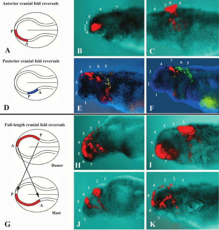
2752 H.-H. Epperlein and others

analyses in tissue culture support the idea that single cranial       of AP-2, a transcription factor expressed by this population
neural crest cells can form derivatives as diverse as neurons         (Mitchell et al., 1991; Shen et al., 1997).
and cartilage (Baroffio et al., 1991). It appears that the fate of       In addition to confirming most aspects of the fate map of
such multipotent precursors can be influenced by a variety of         Hörstadius and Sellman (1946), we provide evidence for a
growth factors, including BMPs and neuregulins, which induce          segmental origin and migration of neural crest cells from the
adrenergic (Varley et al, 1995; Shah et al., 1996; Reissman et        hindbrain. Grafting experiments within the rhombencephalic
al., 1996) and glial derivatives (Shah et al., 1994), respectively.   (posterior cranial) neural folds demonstrate that a small
   Possible explanations for the rostrocaudal difference in           transposition of neural crest leads to cells migrating to a new
cartilage-forming ability of the neural crest include (i) region-     branchial arch, suggesting that premigratory neural crest are
specific differences in pathway/target-derived instructive cues,      not committed to a specific target site. In other experiments,
and/or (ii) premigratory differences between cranial and trunk        we rotated entire cranial neural folds and found that when cells
neural crest precursors with respect to their chondrogenic            are displaced far from their original location, they appear
potential. Thus, neural crest cells in the head may differentiate     unable to respond appropriately to their new milieu. Most
into cartilage because the developing head contains cartilage-        notably, when trunk neural folds are heterotopically grafted
inducing signals not present at other levels of the developing        into the head, they migrate in a highly disorganized fashion and
embryo. Indeed, the importance of pharyngeal endoderm for             fail to follow normal cranial crest pathways. Despite the
the differentiation of visceral cartilage from cranial neural crest   random nature of their migration, we find incorporation of
cells has been shown in urodele embryos in vivo (Hörstadius           some trunk cells into branchial arch cartilage. These results
and Sellman, 1946) and in vitro (Epperlein and Lehmann,               suggest that, despite inherent differences in migratory ability
1975; Graveson and Armstrong, 1987). There appears to be no           between head and trunk neural crest cells, trunk neural crest
such cartilage-inducing signal in the trunk.                          can differentiate into cranial cartilage when given proper
   While these results suggest that pathway- or target-specific       instructive cues.
cues influence neural crest differentiation into cartilage, there
is also evidence for differences in chondrogenic potential
between cranial and trunk neural crest. In one classic study          MATERIALS AND METHODS
on urodele embryos (Hörstadius and Sellman, 1946), a
preliminary fate map of the cranial crest was made using vital        Embryos
dyes to follow the normal patterns of cranial neural crest cell       Wild-type (dark, D/−), white mutant (dd), and albino (aa) embryos of
migration (see Fig. 1). Hörstadius and Sellman’s main finding         the Mexican axolotl (Ambystoma mexicanum) were obtained from the
was that neural crest cells destined for different branchial          axolotl colony in Bloomington, Indiana, USA. The embryos were kept
arches (the mandibular arch, the hyoid arch and four gill             in tap water at room temperature or at 7-8°C and were staged
arches) arise from specific rostrocaudal levels of the neural         according to the normal table of Bordzilovskaya et al. (1989).
folds. Using heterotopic grafting of head and trunk neural fold
                                                                      DiI injections into neural folds
fragments, they challenged the fate of different neural crest
populations and found that trunk neural crest grafted into the        CellTracker CM-DiI (C-7000; Molecular Probes) and CellTracker
                                                                      Green CMFDA (C-7025; Molecular Probes) were dissolved in
head fails to migrate in an orderly fashion and was not observed      absolute ethanol to a concentration of 1 mg/ml and further diluted in
to form cartilage, confirming the results of Raven (1931).            4 or 9 parts of 10% sucrose in water just before starting the injections.
Furthermore, pharyngeal endoderm was not able to induce               Glass micropipettes were backfilled with the DiI solution and attached
cartilage formation from neural crest cells when grafted to the       to a Parker Hannifin Corporation Picospritzer II assembly. DiI was
trunk (Hörstadius and Sellman, 1946). Together, these data            injected into (A) selected parts of the head neural fold of dark neurulae
suggest that cranial neural crest differentiates into cartilage,      (stage 15/16) or (B) grafted head or trunk neural fold fragments of
but trunk crest will not, because of intrinsic differences in         dark neurulae (stages 15-17). CellTracker Green was used only in
chondrogenic potential between the two, in addition to specific       posterior head neural fold reversals (see below).
pathway-derived cues present only in the head.                        A. Cranial neural crest fate mapping experiments
   Because of the wealth of experimental embryology
                                                                      Embryos (stage 15/16) were dipped briefly in 70% ethanol, washed
performed on the neural crest in urodeles, this system bears          thoroughly with sterile Steinberg solution (Steinberg, 1957),
revisiting using improved vital dyes and newly available              decapsulated mechanically and placed into the hollow of an agar dish
molecular markers. The axolotl embryo offers many                     filled with Steinberg saline.
advantages over other vertebrate embryos for examining the
potential and commitment of neural crest cells, because at early         Labeling of the entire cranial neural folds with DiI (6 cases)
stages of development the entire cranial neural plate and neural      Zones 1-7 (Figs 1 and 5) of the left or right head neural fold were
folds are distinct and amenable to extensive microsurgical            labeled with DiI to examine the movement of the entire neural crest
manipulation. In this study, we have constructed a new fate           population. DiI injections were monitored with a Leica MZFLIII
map of the cranial neural crest in the axolotl using two              epifluorescence stereomicroscope. 1-2.5 days after injection, living
complementary methods for cell labeling. In some                      embryos were observed with an epifluorescence microscope.
                                                                      Fluorescent and bright-field images were captured and processed as
experiments, neural fold cells were labeled with DiI, a               described below.
lipophilic fluorescent dye that has been used extensively
(Honig and Hume, 1989) in a number of species to follow                  Focal injection of DiI into three distinct sites in a single
neural crest migration and derivatives (Serbedzija et al, 1989;          embryo (6 cases)
1992; Collazo et al., 1993). As a second marker, we have              DiI was focally injected at three separate sites (zones 3, 4 and 6) of
examined cranial neural crest cell migration by the expression        the cranial neural fold in order to investigate migration and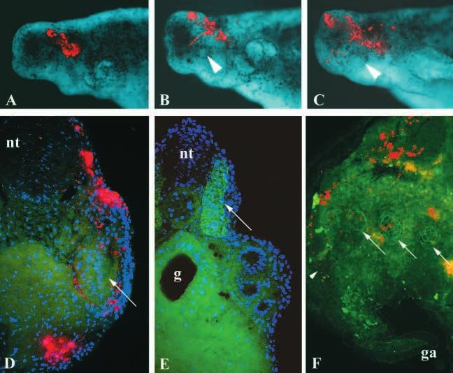
Axolotl cranial neural crest migration 2753

pathfinding of small groups of neural crest cells in living embryos.          embryos at stages 15-37) as described by Henrique et al. (1995) with
Images of fluorescently labeled cells were obtained and processed as          the addition of an extra wash in MAB-T overnight at 4°C (100 mM
described below.                                                              maleic acid, 150 mM NaCl, pH 7.5, 0.1% Tween 20). Hybridization
                                                                              was performed at 65°C.
    Focal injections of DiI into a single site (12 cases)
DiI was injected into one or three adjacent spots at zones 4-7 of the         Histology/immunostaining
head neural fold in order to study internal migration and localization        Transverse 100 µm sections were cut through DiI-labeled/
of neural crest cells in transverse sections. 1 (stage 25), 2 (stage 30/31)   formaldehyde-fixed embryos and through in situ hybridized (AP-2)
and 3 days (stage 35/36) after the injection, embryos were fixed in 4%        embryos, using a Vibratome Series 1000 sectioning system (Ted Pella,
paraformaldehyde (PFA) in 0.1 M PBS overnight. After washing in               Inc.). Sections found to contain DiI-labeled cells were, in most cases,
PBS, vibratome sections were made, stained as indicated below                 counterstained with an anti-fibronectin antibody to visualize tissue
(‘histology’) and examined with an epifluorescence microscope.                borders. Sections were incubated first with a polyclonal anti-
                                                                              fibronectin antibody (Dako, Hamburg), then with an FITC-conjugated
B. Injections into grafted tissue                                             goat-anti-rabbit secondary antibody (Dianova, Hamburg). These
For grafting experiments, neurulae were sterilized as indicated above.        sections were also stained with DAPI (0.1-1 µg/ml in PBS) to mark
Head or trunk neural fold fragments were removed under sterile                cell nuclei. To determine whether any mesodermal cells were
conditions using tungsten needles and grafted to the appropriate              inadvertently included in trunk neural fold fragments grafted to the
location. After a short time, to permit healing of the operated region,       head, vibratome sections (100 µm) were stained with the 12/101
DiI was expelled onto the surface of the graft while viewed under             monoclonal antibody (a skeletal muscle marker; DSHB, Iowa, USA),
epifluorescence optics.                                                       followed by an FITC-conjugated goat anti-mouse antibody. In some
                                                                              instances, DiI-labeled cells were found within cartilaginous
Heterotopic grafting experiments                                              condensations and a number of such sections were immunostained
A. Head neural fold reversals                                                 with an anti-6-chondroitin sulfate proteoglycan antibody (anti 6-
Three types of head neural fold reversals were carried out at the             CSPG; ICN, Eschwege). Vibratome sections were pre-treated with
neurula stage (stage 15) of dark embryos. (a) Anterior head neural            chondroitinase ABC (2 units/ml) for 45 minutes, followed by a 1 hour
fold reversals (5 cases): fragments comprising zones 1-4 were                 incubation in anti 6-CSPG antibody. Sections were then incubated in
explanted and inserted in the reversed orientation (4➝1; 4 anterior, 1        a FITC-conjugated goat anti-mouse IgG secondary antibody.
posterior); (b) posterior head neural fold reversals (16 cases):
fragments comprising zones 5-7 were excised and inserted in the               Image analysis
reversed orientation (7➝5; 7 anterior, 5 posterior); (c) unilateral full      Whole embryos and sections were analyzed with an epifluorescence
length cranial fold reversals (8 cases): the right cranial neural fold        microscope and images were recorded with a Spot camera. Using
(zones 1-7) was excised and regrafted in a reversed orientation to the        Adobe Photoshop software, the contrast and brightness of captured
left side of a host embryo and vice versa (7➝1; 7 anterior, 1 posterior).     images were optimized, and bright-field images of whole embryos
In all experiments, the graft was subsequently labeled with DiI. In           were combined with the corresponding fluorescence (DiI) images.
most cases, the entire length of the grafted fold was labeled, while in       Separate images were captured from sectioned material for DiI
other experiments, two spots of DiI were made into the transplanted           labeling (neural crest cells), anti-fibronectin staining (tissue borders),
fold. In some of the posterior cranial neural fold reversals, DiI             anti-6-CSPG staining (cartilaginous condensations), 12/101 staining
injections were made into the new anterior end of the graft and Cell-         (trunk mesoderm) and DAPI staining (cell nuclei), before being
Tracker Green was injected at the new posterior end.                          superimposed to produce a single image.

B. Trunk neural fold grafts to head (about 8 cases)
Mid-trunk neural fold fragments from donor neurulae (stages 15-18)
were grafted heterotopically into zones 5-7 of host embryos from              RESULTS
which these cranial areas had been extirpated. The grafts were injected
with DiI shortly after the operation. Embryos were grown to desired           Migratory pathways of cranial neural crest in the
stages (up to stage 34 or 38) and the distribution of DiI-labeled cells       Mexican axolotl
was examined both in wholemounts and transverse vibratome                     We performed a series of experiments to confirm the accuracy
sections. In some cases, sections were also labeled with a mesodermal         of the Hörstadius and Sellman (1946) cranial neural crest fate
marker (12/101) or an anti-6-chondroitin sulfate proteoglycan                 maps (Fig. 1), using modern dye-labeling techniques. Cranial
antibody.
                                                                              neural fold cells were marked with DiI, either by labeling the
Cloning and sequence analysis of Ambystoma AP-2                               entire neural folds, or by making small focal injections of DiI
cDNA was synthesized from stage 22-35 Ambystoma embryo total                  into small groups of cells.
RNA by poly-T-primed reverse transcription and used as a template
for PCR. The following degenerate primers were designed against               Whole neural fold labeling with DiI
conserved regions of vertebrate and Drosophila AP-2 genes and used            In six different dark neurulae (stage 15/16), the entire left head
to amplify a 522 bp fragment of the Ambystoma AP-2 homolog:                   neural fold (corresponding to zones 1-7 in Fig. 1) was labeled
   AP-25′1= 5′GTRTTCTGYKCAGKYCCYGGICG 3′ and                                  with a contiguous line of DiI (Fig. 2A). Approximately 1, 1.5,
   AP-23′1=5′GWKATVAGGKWGAAGTGSGTCA 3′.                                       2 and 2.5 days after dye injection, the distribution of DiI in the
   The fragment was TA cloned using the pGEM-T easy kit                       developing embryos was examined and recorded.
(Promega), sequenced, and compared to known AP-2 genes using the
Megalign program.
                                                                                 At various time points after labeling it was possible to
                                                                              distinguish (i) a relatively wide, longitudinal band of labeled
In situ hybridization                                                         cells on the dorsum of the embryo and (ii) a number of distinct
Sense and antisense riboprobes were sythesized from the cloned                narrow streams of labeled cells emanating from the dorsal
fragment (DIG RNA labeling kit, Roche diagnostics). In situ                   neural tube. The dorsal band of labeled cells comprised of
hybridization was performed on albino axolotl embryos (about 40               derivatives of the labeled neural fold, including epidermal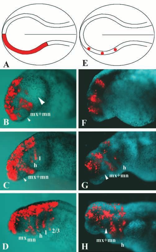
2754 H.-H. Epperlein and others

Fig. 1. Fate map of the neural crest cells in axolotl
(modified from Hörstadius and Sellman, 1946).
(A) A drawing of a stage 16 embryo describing the
sites of vital dye labeling. Cranial and anterior
trunk neural folds were marked unilaterally with
alternating spots of vital dyes, Nile Blue sulfate and
Neutral Red. (B) Stage 33; the prospective fate of
neural crest cells is indicated and divided into
arbitrary zones. Zones 4-7 give rise to the six
branchial arches of the visceral skeleton: mandibular arch (mn), hyoid arch, (hy), and four gill arches (1-4). Zones 1, 2 and 8 are non-
skeletogenic, while 3 gives rise to anterior trabeculae (Hall, 1999).

cells, neural tube cells and premigratory neural crest                      distal parts of their migratory pathway, we were unable to
precursors. As development proceeded, the rostrocaudal                      determine whether mixing of neural crest cells occurs
extent of labeled cells in this band appeared to increase,                  immediately after emigration from the neural folds, in the
presumably as a consequence of convergence and extension                    proximal part of the pathway. One reason for this is that labeled
movements.                                                                  neural folds give rise to epidermal cells, which migrate over
   Neural crest cells appeared as four dorsoventrally oriented              the dorsal part of the embryo, making it difficult to visualize
streams of cells. Each was distinct and separated from the                  underlying labeled neural crest cells.
others, at least along the distal two-thirds of their migratory
pathway, on the ventral aspect of the embryo. The streams                   Focal injection of DiI into the cranial neural folds
appeared in a temporal sequence, with anterior-most neural                  While DiI labeling of the entire cranial neural fold provides a
crest cells migrating prior to posterior-most cells.                        good over-view of neural crest migration in the head, it gives
   About 1 day after dye injection, neural crest cells were
observed migrating around the anterior and posterior margins
of the eye (Fig. 2B). Whereas the anterior-most cells probably
contribute to tissues of the eye itself and adjacent anterior head
mesenchyme, the posterior stream contains putative first
branchial arch (maxillary and mandibular) neural crest cells.
1.5 days after dye injection, first branchial arch neural crest
remained as a coherent stream en route to a peri-stomodeal
location. A second, putative hyoid neural crest stream could be
seen posterior to the first branchial arch stream (Fig. 2C),
followed by a third (1st gill arch) stream. 2 days after dye
injection (Fig. 2D), the first branchial arch stream appeared to
diverge into two tongues in the vicinity of the developing
mouth (presumably into separate maxillary and mandibular
arch neural crest cells). In addition to the hyoid stream (which
now extends to the ventral portion of the embryo), 2-3 streams
of neural crest could be seen advancing into the gill bulges.
After 2.5 days (stage 35), the maxillary and mandibular stream
became still more distinct, but the general pattern remained
unchanged (data not shown).
   Although our whole neural fold labeling experiments
provided evidence that neural crest cells do not mix along the

Fig. 2. Continuous and focal labeling of the head neural fold of
axolotl neurulae (stage 15/16) with DiI. (A) For labeling the entire
neural folds, DiI was injected throughout zones 1-7 (see Fig. 1) on
the left side. B-D illustrate the distribution of labeled cells
approximately 1 day, stage 24 (B), 1.5 days, stage 28 (C), and 2 days,
stage 31 (D) after dye injection. (E) For focal labeling, zones 3, 4 and
6 were injected with small amounts of DiI. F-H illustrate the
distribution after focal labeling approximately 1 day, stage 22 (F),
1.5 days, stage 30 (G) and 2.5 days, stage 35 (H) after dye injection.
The small arrowhead in B indicates the optic vesicle and the large
arrowhead is the gill bulge. In C, G, and H, the optic vesicle is
indicated by a small arrowhead. In B-D different embryos are shown,
whereas in F-H a single embryo is recorded at the different time
points. mx, maxillary arch; mn, mandibular arch; h, hyoid arch; 1, 2,
3: gill arches 1, 2, 3.
Axolotl cranial neural crest migration 2755
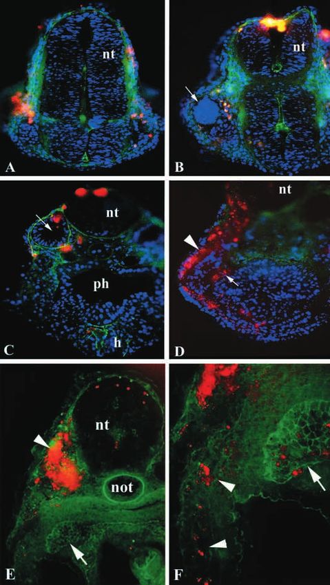
Fig. 3. Distribution of DiI-labeled crest cells in the head of axolotl
embryos following focal injections of DiI into zones 4-7 of an
axolotl neurula. (A) At forebrain levels, labeled crest cells (red) are
visible between epidermis and neural tube and at the eye vesicle by 2
days after dye injection. (B) At the level of the optic vesicles (arrow),
the injection site is visible in the dorsal neural tube (nt); labeled crest
cells spread ventrally and distribute in front of and behind the eye
vesicle at 2 days after dye injection. (C) Neural crest cells at the level
of the otic vesicle (arrow) are visible adjacent to it and also in the
prospective heart region (h) at 2 days post-injection. (D) Dorsal to
the pharynx at 3 days after dye injection, we find that the migrating
neural crest cell stream becomes divided into a subepidermal stream
(presumably pigment cells; arrowhead) and an internal stream
(presumably contributing to mesenchymal derivatives; arrow). In A-
D, transverse vibratome sections (100 µm) were counterstained with
anti-fibronectin antibodies (to identify tissue borders; green) and
DAPI (to identify cell nuclei; blue). In E and F, vibratome sections
were counterstained with an antibody to 6-chondroitin sulfate
proteoglycan to identify cartilaginous condensations. (E) Although
the DiI labeling in this embryo was extensive and had labeled the
mesoderm (arrowhead), labeled neural crest cells could be observed
migrating ventrally into the gill arches. F shows a higher
magnification of the ventral region of the same embryo and
illustrates that cells migrate into the gills (arrowheads) and are also
found within cartilage (arrow). The same cartilaginous condensation
is indicated by an arrow in both E and F. nt, neural tube; ph, pharynx;
h, heart; not, notochord.
observed that neural crest cells close to the neural tube do not
undergo extensive mixing.
Transverse sections through the head of focally injected
embryos
Neurulae that had received a single or three adjacent injections
into zones 4-7 of the head neural fold were fixed after 1, 2 and
3 days of further development. In vibratome sections through
the head region, DiI labeled cells were found at various sites
between fore- to hindbrain levels (Fig. 3), for example at the
eye and ear vesicles, in the prospective pharynx and heart
region and in regions of visceral cartilage. One of the most
important findings from these experiments was that migrating
cranial neural crest streams split into two divisions, one of
which migrates into the branchial arches adjacent to the
ectoderm (presumably giving rise to pigment cells), the other
migrating along the pharyngeal endoderm (Fig. 3D). Since
no indication of the precise origin of neural crest cells destined pharyngeal endoderm is known to be important for cartilage
for different branchial arches with respect to the 7 cranial zones formation (Hörstadius and Sellman, 1946; Epperlein and
(Fig. 1). For this reason, we performed a series of focal Lehmann, 1975; Graveson and Armstrong, 1987), these
injections of DiI into three neural fold sites (corresponding deeper neural crest cells most likely form cartilage of the
approximately to zones 3, 4 and 6; Fig. 2E) of early, stage visceral arches.
15/16 neurulae. Embryos were grown for a further 2.5 days
(Fig. 2F-H). In these labeling experiments, we tested whether Migration and differentiation of cranial neural crest
small regions of neural fold give rise to neural crest cells cells in whole mounts using the AP-2 transcription
populating single or multiple branchial arches. factor as a neural crest cell marker
Neural crest cells lying within zone 3 became distributed As an independent confirmation of our DiI fate mapping
mainly around the eye. In contrast, cells derived from zone 4 experiments, we analyzed the migration of neural crest cells
migrated into the position of the prospective maxillary/ using expression of AP-2 as a marker. AP-2 is a transcription
mandibular arch and those from zone 6 into the hyoid arch. factor expressed in neural crest cells in mice, frogs and chicks
These results indicate that the first and second branchial arches (Mitchell et al., 1991; Shen et al., 1997).
each receives neural crest cells from small, distinct regions of
the neural folds. Since these experiments employed focal Cloning and sequence analysis of Ambystoma AP-2
injections of DiI, fewer epidermal cells were labeled, allowing We cloned an Ambystoma AP-2 fragment with highest
us to observe the underlying neural crest cells more clearly. We homology to mouse AP-2 and human AP-2 alpha, indicating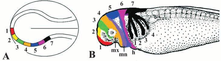
2756 H.-H. Epperlein and others

                                                                     Fig. 4. In situ hybridization illustrating the distribution pattern of the
                                                                     transcription factor, AP-2. (A,B). Whole-mount and vibratome
                                                                     section, respectively, through a stage 18 embryo after AP-2 staining.
                                                                     AP-2 staining is distributed in the dorsal and medial parts of head
                                                                     and anterior trunk neural folds (A, and asterisk in B) and in a lateral
                                                                     area of the prospective head region (A, and arrow in B). (C-J) Whole
                                                                     mounts and sections illustrating AP-2 staining as a function of time.
                                                                     C (stage 22), D (stage 25), E (stage 31), G (stage 33) and I (stage 35)
                                                                     demonstrate that there is a continuous decrease of AP-2 staining in
                                                                     the neural folds. By stage 22 (C), unlabeled areas become visible in
                                                                     front of rhombomere 2 (r2) and between rhombomeres 2 and 4 (r4)
                                                                     and 4 and 6 (r6). Staining from the dorsal neural tube continues into
                                                                     the neural crest stream with r2 being continuous with the maxillary
                                                                     and mandibular arch streams, r4 with the hyoid arch stream and r6
                                                                     with a migratory stream common for three gill arches. Concomitant
                                                                     with the decrease of AP-2 staining in the neural folds and branchial
                                                                     arches is the appearance and gradual increase of label in a group of
                                                                     cells in the lateral wall of the head and anterior trunk neural tube,
                                                                     seen most prominently in I and J (arrows). Transverse sections F,H
                                                                     and J are overlays of bright-field images of AP-2 staining (blue) with
                                                                     fluorescent images of fibronectin staining (green). The white arrow in
                                                                     F points to the core of a gill arch and two black arrows indicate AP-
                                                                     2-labeled crest cells surrounding it. mx, maxillary arch; mn,
                                                                     mandibular arch; e, otic vesicle; white dots in G, maxillary and
                                                                     mandibular arch neural crest stream; green dots in G, hyoid arch
                                                                     neural crest stream.

                                                                     continuous with a maxillary/mandibular stream, a more caudal
                                                                     hyoid stream and (at stage 25) a gill-arch stream. On the basis
                                                                     of this staining pattern, we propose that AP-2 may be expressed
                                                                     at higher levels in even (r2, r4, r6) than odd-numbered
                                                                     rhombomeres at this stage. Rhombomere 2 (corresponding to
                                                                     zone 4, Fig. 5) gives rise to first branchial arch neural crest,
                                                                     rhombomere 4 (zone 6, Fig. 5) contributes to the hyoid arch
                                                                     and rhombomere 6 (zone 7, Fig. 5) contributes to the gill
                                                                     arches. Although AP-2 expression appears segmental in the
                                                                     hindbrain, we find continuity in staining in the dorsal midline
                                                                     and on the lateral aspect of the embryo, proximal to the
                                                                     individualized neural crest streams.
                                                                        Between stages 25 to 33, the overall intensity of AP-2
                                                                     expression in the branchial arches increases, such that distinct
                                                                     maxillary/mandibular, hyoid and three gill arches can be
                                                                     distinguished in later stages (Fig. 4G). By stage 35 (Fig. 4I),
it is the axolotl AP-2 alpha (GenBank accession number               AP-2 appears somewhat down-regulated in all the branchial
AF209775). The published Xenopus AP-2 sequence (Winning              arches. Concomitant with the diminishing AP-2 staining in the
et al., 1991) also has high homology with these genes.               neural crest and branchial arches at these stages is an increase
                                                                     of label in the outer lateral wall of the neural tube in head and
In situ hybridizations with AP-2 riboprobe                           anterior trunk regions (Fig. 4E,G,I). Furthermore, transverse
In stage 18 embryos (late neurulae), we find expression of AP-       sections indicate that neural tube staining may be in the Rohon-
2 transcripts in dorsomedial parts of the head and anterior trunk    Beard cells (Fig. 4J). Between stages 31 and 35, AP-2 staining
neural folds. Although the neural folds are labeled along the        in the dorsal rhombencephalon diminishes until no staining can
entire rostrocaudal axis, we find higher levels of staining in the   be observed.
cranial versus trunk region (Fig. 4A,B). In addition to the             Taking the results of our DiI fate mapping experiments and
neural fold staining, a second domain of weaker staining is          the AP-2 expression analysis together, we propose a fate map,
found in a stripe of lateral head ectoderm, possibly coinciding      illustrated in Fig. 5.
with the region of prospective placodal ectoderm.
   At stage 22 (Fig. 4C) and, even more clearly at stage 25 (Fig.    Heterotopic grafting experiments
4D), staining of the neural folds is no longer uniform. Along        To investigate whether premigratory neural crest cells within
the rostrocaudal extent of the hindbrain, broader regions of AP-     the head neural folds are committed to a particular branchial
2 expression abut regions of more limited AP-2 expression in         arch cell fate, we have performed a series of heterotopic
an alternating pattern that is reminiscent of the even and odd       grafting experiments in which small lengths of cranial neural
rhombomeres observed in higher vertebrates. In the transverse        fold or entire cranial neural folds were reversed
plane, AP-2 staining in the dorsal cranial neural tube is            anteroposteriorly. In addition, we grafted trunk neural folds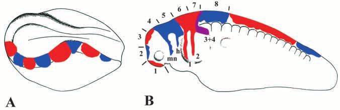
Axolotl cranial neural crest migration 2757

Fig. 5. Fate map of cranial neural crest cells
of an axolotl neurula based on labeling with
DiI and AP-2 staining. (A) The head neural
fold are divided into seven arbitrary zones
which give rise to the neural crest streams
(same colors) shown in (B) of a stage 33
embryo. The seven zones in B correspond
to the seven zones originally defined by
Hörstadius and Sellman in 1946 (see Fig.
1B). mx, maxillary arch; mn, mandibular
arch; h, hyoid arch; 1-4, four gill arches.

Fig. 6. Heterotopic grafting experiments
of cranial neural folds. (A) A schematic
diagram illustrating the procedure for
anterior cranial neural fold reversals at
stage 15. Anterior neural folds (zones
1 4) are excised, grafted into the same
embryo after rotation by 180° and
labeled with DiI. (B) After anterior
cranial neural fold reversal, neither
zone 1 nor 4 gave rise to a
maxillary/mandibular neural crest
stream. (C-F) After posterior neural fold
displacement, neural crest cells changed
their fate and populated new, instead of
original, target tissues. (D) For posterior
neural fold reversals, zones 5-7 were
excised and implanted in a reversed
orientation before injection with DiI
and/ or CellTracker Green. (C,F) Cells
from zone 5 change their branchial arch
fate and migrate into gill arches.
Similarly, zone 7 cells are able to
contribute to 1st and 2nd branchial arches
(E and F). (G) Schematic diagram
illustrating the methods for full cranial
fold reversals. At stage 15, head neural
fold fragments comprising zones 1-7
were excised in one neurula and
implanted after reversal into the
contralateral side of a host neurula.
Zones 1 and 7 came to lie far from their
original targets while zones 3-5 became
displaced only minor distances.
(H,J) After such reversals, labeled cells
from the center of the graft migrate to
new targets. In contrast, those at the rostral and caudal margins of the graft appeared less able to compensate for their displacement. (I,K) In one
case (K) zone 2/3 cells migrated towards the eye and appeared to move rostrally to their original target.

(which normally do not give rise to cartilage) into cranial                 trabeculae, only zone 4 normally gives rise to neural crest
locations to examine trunk neural crest migratory ability and               which has extensively migratory ability.
chondrogenic potential.                                                        In these operated embryos DiI-labeled neural crest cells
                                                                            from segment 4, which would normally be fated to form
Anterior head neural fold reversals                                         mandibular arch, did not migrate towards the developing
Zones 1-4 of the head neural fold of an axolotl neurula were                mandibular arch as would be appropriate for their level of
excised, rotated 180° anteroposteriorly and implanted into the              origin. Thus, the normally migratory segment 4 cells appeared
same embryo (Fig. 6A) so that zone 1 was transposed to                      to lose their migratory ability in this new environment. Neural
position 4 and vice versa (Fig. 6B). The entire length of neural            fold cells from segment 1, which normally exhibit little
fold was labeled with DiI prior to rotation. Since zones 1 and              migratory ability (see Figs 1, 5), failed to give rise to cells for
2 are non-skeletogenic and zone 3 gives rise to anterior                    the mandibular stream (Fig. 6B).
2758 H.-H. Epperlein and others
Fig. 7. Heterotopic grafting experiments of trunk
neural fold into cranial regions. Midtrunk neural fold
fragments labeled with DiI were grafted in place of
head neural folds (zones 4-5), which normally
contribute to the mandibular/hyoid arches. (A-C) As
a function of time after grafting, labeled neural crest
cells can be observed emigrating from the graft site;
they distribute in a random fashion around the head,
including a region above the eye vesicle
(arrowheads). Even after 5 days (C) these cells did
not align themselves with any of the visceral arch
neural crest streams. (D) Transverse sections through
head region containing DiI-labeled trunk neural fold
graft reveal that some DiI-labeled neural crest cells
(red) had migrated in front of and behind the eye
vesicle (white arrow), or in areas of developing
supportive tissue (bottom of picture). This particular
vibratome section was counterstained with DAPI
(blue) and immunolabeled with the mesodermal
marker 12/101. An absence of 12/101
immunoreactive cells (the green color is background
fluorescence) reveals that no trunk mesodermal cells
had been inadvertently included in the grafted neural
fold fragments, indicating that all DiI-labeled cells
were derived from the grafted neural folds. (E) A
control section through the trunk of an axolotl
embryo at stage 34 stained with a FITC-conjugated secondary antibody against anti-12/101 reveals specific labeling within the somites (arrow).
This section was counterstained with DAPI (blue). (F) While it is clear that trunk neural crest cells would not associate with specific cranial
neural crest cell streams, they were found within structures derived from the neural crest, such as cartilaginous elements. In this case, labeled
cells (arrowhead) were observed within a gill arch (ga) and also within three cartilaginous nodules (arrows). This section was immunolabeled
with an antibody recognizing 6-CSPG to reveal the cartilage. nt, neural tube; ga, gill arch.
Posterior head neural fold reversals consequence, cells within zones 1 and 7 come to lie far from
Head neural fold areas 5-7 of axolotl neurulae were excised their original target site, while neural crest progenitors within
and implanted in a reversed orientation (Fig. 6D), as discussed zones 3-5 became displaced only minor distances.
above, so that zone 5 fold lay at position 7 and zone 7 fold cells Our results indicate that cells at the center of the graft were
at position 5 (Fig. 6C,E,F). DiI was used to mark the entire able to migrate to new targets, consistent with the results above
graft in some cases (Fig. 6C), while in others, the borders of (Fig. 6H,J). In contrast, cells at the rostral and caudal margins
the rotated fragment were labeled with DiI and CellTracker of the graft appeared less able to compensate for their
Green. displacement and in most cases failed to migrate altogether
At the time of analysis, we discovered that the graft had (Fig. 6I). In one case (Fig. 6K), we found that zone 2/3 cells
shrunk to varying degrees and had come to lie at slightly migrated in close proximity to the eye and, after reaching its
different rostrocaudal positions. Nevertheless, we found that dorsal aspect, migrated rostrally towards their original target.
displaced neural crest cell progenitors could populate tissues Taken together with the results of small neural fold rotations,
appropriate for their new location, and found no evidence of our results suggest that premigratory cranial neural crest cells
cells migrating towards their original target tissues. For within the neural folds are not committed to a specific
instance, after rotation, cells within zone 5 migrated into the branchial arch fate, but can compensate to some extent when
gill arches (Fig. 6C,E,F) which is a tissue that they do not displaced short distances from their origin by migrating to a
normally encounter. Similarly, zone 7 cells changed their fate new target tissue. In contrast, when cells are displaced far from
and populated the 1st and 2nd branchial arches after neural fold their original location, they appear unable to respond
rotations (Fig. 6E,F). As expected, cells from zone 6 migrated appropriately to their new milieu and consequently either fail
to their normal targets as a result of their minimal to migrate or migrate in a disoriented fashion which
displacement. In comparing anterior and posterior neural fold occasionally brings them to appropriate target sites for their
reversals, these results suggest that neural crest cells from point of origin.
regions 4-7 compensate after transplantation to a new
environment, whereas anterior non-skeletogenic neural folds Trunk neural fold fragments
(regions 1-2) appear unable to acquire migratory ability after Head neural fold areas 5-7 of wild-type neurulae (stages 15-
displacement. 18) were replaced by midtrunk neural fold fragments in order
to study the migratory and differentiative potential of trunk
Entire head neural fold reversals neural crest cells in the head.
Head neural fold fragments comprising regions 1-7 were In some living embryos, we found that labeled crest cells left
excised in one neurula and implanted after reversal into the the graft caudo-laterally and also became distributed randomly
contralateral side of a host neurula (Fig. 6G). As a above and behind the eye vesicle (Fig. 7A-C). Even 5 days afterAxolotl cranial neural crest migration 2759
grafting, these cells did not associate with any of the visceral of cranial neural crest migration (see Fig. 1). Agar pieces
arch neural crest streams, as would the endogenous neural crest stained with Nile Blue sulfate and Neutral Red were placed
cells. Thus, in the axolotl embryo, trunk crest cells can migrate directly onto the neural folds of neurulae in order to label
out in the head region but fail to recognize distinct routes into presumptive neural crest. The chief findings from these studies
the visceral arches. are that neural crest cells migrate from the neural folds as
Surprisingly, a few trunk neural crest cells did appear to distinct streams en route to the branchial arches.
migrate ventrally towards the branchial arches, as seen clearly In the present study, focal injections of DiI into the neural
in sections through grafted embryos (Fig. 7D,F). At stage 34, folds confirmed these earlier findings by demonstrating a
labeled neural crest cells were observed in front of and behind maxillary/mandibular stream, a hyoid stream and 2-3 streams
the eye vesicle and in areas of developing visceral arches (Fig. for the gill arches. Furthermore, using focal injections, we
7D). Explantation of trunk neural fold material was carried out show that neural crest cells do not undergo extensive mixing
without obvious contamination by underlying mesoderm, on their initial route of migration (Fig. 2E-H). Furthermore, we
which can be recognized easily by its white color and larger show that neural crest cells associate with epithelium as they
cells. To rule out the possibility that a few mesodermal cells migrate into the branchial arches. We propose that those neural
might occasionally have been included in the graft and crest cells associated with the superficial ectoderm develop into
subsequently migrated in the head, transverse sections through pigment cells, while those in contact with the pharyngeal
the head region bearing the graft were stained with a endoderm differentiate into cartilage.
monoclonal antibody against mesodermal cells (12/101; Fig. By cloning axolotl AP-2, we have established an excellent
7E). As demonstrated in Fig. 7D, the mesodermal marker could molecular marker for the cranial neural crest. In situ
neither be observed on DiI-labeled cells nor on unlabeled cells. hybridization with AP-2 shows the pattern of the premigratory
In some experiments (n=4; data not shown), mesoderm was neural crest cells and their early routes of migration more
deliberately included in trunk neural fold grafts, but subsequent clearly than can be observed with DiI labeling. AP-2 is
analysis revealed that such mesoderm fails to migrate away expressed in the head crest in a number of genera, including
from the cranial grafts site towards the branchial arches. We amphibian, chick and mouse (Mitchell et al., 1991; Shen et al.,
conclude that the DiI-labeled cells observed adjacent to the eye 1997). This transcription factor is important for the regulation
and in the gill arches are trunk neural crest cells that have of facial structures and also for other genes expressed by neural
reached these sites by random migration. crest cells including Hoxa2 (Maconochie et al., 1999).
Strikingly, trunk neural folds grafted into the head In the axolotl, AP-2 is expressed within the premigratory
demonstrated the ability to form cartilage after heterotopic and migrating neural crest cells as well as in postmigratory
grafting. By stage 38, cartilage condensations have formed in cells within the branchial arches. In the hindbrain, AP-2
host embryos and can be recognized using antibodies to 6- expression in the neural tube is segmental, appearing more
chondroitin sulfate proteoglycan (6-CSPG; n=9). DiI-labeled prominently in even numbered rhombomeres (r2, r4, r6).
trunk cells were found within the gill branch cartilaginous There is apparent continuity in the cell streams emanating
condensations (Fig. 7F). DiI-labeled cells derived from trunk from the even numbered rhombomeres and giving rise to
neural folds became incorporated into these structures and individual branchial arch crest streams. Interestingly,
expressed CSPG. Approximately 5-10% of the cells in these scanning electron microscope analysis of axolotl embryos has
cartilage condensations were DiI labeled. shown that neural crest cells exist as a continuous flattened
epithelium along the length of the rhombencephalic neural
tube at these stages (Epperlein and Löfberg, 1993),
DISCUSSION suggesting that AP-2 is not expressed in all premigratory
neural crest populations.
In this study, we have revisited, using modern cell markers, the The combination of the AP-2 staining pattern and DiI
issue of the contribution of the neural crest to the visceral labeling would suggest the following fate map of neural crest
skeleton of urodeles, a topic originally examined more than 60 cells (Fig. 5).
years ago by Hörstadius. Most of the questions asked by (i) First branchial arch neural crest arises from a distinct
Hörstadius and Sellman (1946) examined environmental region of the neural folds, designated zone 4 (after Hörstadius
versus autonomous control of cartilage determination as well and Sellman, 1946). These neural crest cells express AP-2 as
as the effects of heterotopic transplantation of the neural folds they migrate ventrally, where they split into maxillary and
on head development. Below, we compare the present results mandibular streams.
to those of Hörstadius, to construct a more accurate map of (ii) Second branchial arch neural crest arises from about the
neural crest fate and potential. anterior three quarters of zone 6 and migrates as a distinct AP-
2-positive stream towards the hyoid arch.
Fate map of the cranial neural crest (iii) Third to sixth branchial arch neural crest cells arise as
The studies of Hörstadius and Sellman (1946) on the a single stream from about the posterior three quarters of zone
‘determination of the cartilaginous visceral skeleton of 7 before separating into streams corresponding to each of the
urodeles’ started in 1936/37 in Triturus and Ambystoma. They gill arches.
were based on earlier findings from Landacre (1921), Stone (iv) Neural crest cells do not seem to migrate laterally from
(1922, 1932), Raven (1931), Ichikawa (1933) and later refined either zone 5 or a region lying between zones 6 and 7.
by Chibon (1966) in Pleurodeles, another urodele. To establish Interestingly, both regions express AP-2 at very low levels,
a preliminary fate map, Hörstadius and Sellman used vital raising the possibility that these two areas do not contribute to
dyes, as devised by Vogt (1925), to follow the normal patterns the branchial arches.2760 H.-H. Epperlein and others
Regulatory ability of head neural crest segments transverse sections through the head region bearing the graft
We carried out heterotopic grafting experiments with different (Fig. 7D,F), DiI-labeled cells were found in various places
head neural fold fragments and labeled them with DiI or, in under the epidermis. Importantly, they also were observed
some cases, both with DiI and CellTracker Green, in order to within cartilaginous elements of the head skeleton (Fig. 7F).
examine the degree of plasticity versus prepattern in cranial Thus, most of the grafted trunk neural crest probably gives rise
neural crest cells. We found that cranial neural crest cells to pigment cells (subepidermal location) and neurons (not
can compensate for small displacements by migrating identified) in addition to skeletogenic derivatives. One possible
appropriately for their new environment (Fig. 6). However, reason why previous studies failed to find incorporation of
when cells are displaced far from their premigratory location, trunk neural crest into cartilage is that neural crest cells were
they do not migrate normally for their new environment, labeled with vital dyes that rapidly dilute to sub-detectable
appearing to undergo random movements and only rarely levels in dividing cells. If chondrogenic neural crest cells reach
finding their original destinations. These data indicate that their targets by migrating deep within the branchial arches, it
cranial neural crest cells have some intrinsic properties would be difficult to observe them in whole mounted embryos,
regarding their rostrocaudal position, but also have some or even in relatively thick transverse sections.
degree of plasticity in adapting to new surroundings. Our Various differences exist between trunk and head neural
results suggest that the anterior-most neural folds (regions 1- crest such as their cellular shape (elongated versus polygonal),
3) do not contribute to the neural crest even after heterotopic migratory pathways (between somites and epidermis or
transplantation. somites and neural tube/notochord in the trunk and between
Our neural fold transpositions are supported by recent results epidermis and endoderm in the head), mode of migration
in other systems. For example, rhombomere rotations in the (single cells versus cellular tongues) and the derivatives
chick show that the neural crest can partially compensate for generated. The production of cartilage has classically been
small changes by migrating to branchial arches appropriate for thought to be a property of the cranial neural crest only, since
their new location (Sechrist et al., 1994; Saldivar et al., 1997). trunk neural crest did not produce cartilage when transplanted
Similarly, Hunt et al. (1998) show that changing the spatial into the cranial region (Raven, 1931; Hörstadius and Sellman,
organization of the entire chick rhombencephalic neural tube 1946; Noden, 1975; Nakamura and Ayer-Le Lièvre, 1982;
leads to neural crest cells migrating towards branchial arches Graveson et al., 1995). For instance, in the axolotl, previous
closest to their new location. This type of operation is studies have failed to show that trunk neural crest cells form
analogous to the posterior cranial neural fold reversals cartilage when transplanted to the head (Raven, 1931;
performed in the present study. Graveson et al., 1995). However, trunk neural crest cells in
In general, the results of tissue grafts and ablations have amphibia do appear to contribute to the mesenchyme of the
demonstrated that some aspects of neural crest cell migration dorsal fin (DuShane, 1935; Collazo et al., 1993), though most
and differentiation are plastic, whereas others are somewhat mesenchyme is mesodermally derived. The formation of head
fixed. Neural crest adjacent or contralateral to the operation cartilage depends upon various environmental factors,
site, or the neural tube itself might be involved in regulatory specifically on an interaction with endodermal and ectodermal
events. For example, removal of the neural crest by the ablation epithelia (Tyler and Hall, 1977; Bee and Thorogood, 1980;
of a small portion of the dorsal neural tube at midbrain and Hall, 1981, 1987; Hall and Hörstadius, 1988).
trunk levels results in a normal embryo (Yntema and The results of our heterotopic grafting experiments suggest
Hammond, 1945, 1947; McKee and Ferguson, 1984). This has that trunk neural crest cells do not migrate along normal cranial
been taken as evidence that neighboring neural crest cells, both neural crest pathways, but move in a disoriented fashion. As a
rostral and caudal to the ablated region, repopulate the consequence, the cells appear to distribute randomly. Despite
extirpated segments. Ablation of midbrain neural folds in chick this random migration, they appear to encounter specific cues
embryos leads to normal development of neural crest-derived that allow them to form cartilage and contribute to elements of
structures (McKee and Ferguson, 1984). Interestingly, the the visceral skeleton. Our results provide the first evidence that
neural crest cells that ‘fill-in’ from ectopic regions appear to cartilage can develop from trunk neural crest cells in vivo.
readjust their Hox code after such an operation (Hunt et al., Thus, it seems that the migratory ability of trunk neural crest
1995). In addition to compensation by neighboring cells is uncoupled from their developmental potential.
populations, the neural tube itself can regulate to reform neural
crest after such ablations, but only for a limited period of time This work was supported by a Howard Hughes Medical Institute
Research Resources Grant and a James H. Zumberge Research and
in development (Scherson et al., 1993; Sechrist et al., 1995). Innovation Fund (M. A. J. S.), by NS36585 and NS34671 to M. B. F.
The potential of the trunk crest in the head and the Deutsche Forschungsgemeinschaft (Ep8/7-1) to H.-H. E. The
12/101 monoclonal antibody, developed by Jeremy Brockes, was
We and other authors (Raven, 1931; Hörstadius and Sellman, obtained from the Developmental Studies Hybridoma Bank
1946) grafted trunk neural crest cells into the head region of maintained by The University of Iowa, Department of Biological
axolotl neurulae in order to investigate their migratory and Sciences, Iowa City, IA 52242, USA.
differentiative potential in a new environment. Whereas
previous studies observed no indications of migration and
cartilage formation, we found evidence for both events. DiI-
REFERENCES
labeled trunk crest cells migrated out in a random fashion and
became distributed above and behind the eye vesicle. They did Baroffio, A., Dupin, E., and LeDouarin, N. M. (1991). Common precursors
not, however, associate with any of the neural crest streams as for neural and mesectodermal derivatives in the cephalic neural crest.
could be demonstrated in whole mounts (Fig. 7A-C). In Development 112, 301-305.Axolotl cranial neural crest migration 2761 Bee, J. and Thorogood, P. (1980). The role of tissue interactions in the (1991). Transcription factor AP-2 is expressed in neural crest cell lineages skeletogenic differentiation of avian neural crest cells. Dev. Biol. 78, 47- during mouse embryogenesis. Genes Dev. 5, 105-119. 62. Nakamura, H. and Ayer-Le Lièvre, C. (1982). Mesectodermal capabilities Bordzilovskaya, N. P., Dettlaff, T. A., Duhon, S. T., and Malacinski, G. M. of the trunk neural crest of birds. J. Embryol. exp. Morphol. 70, 1-18. (1989). Developmental-stage series of axolotl embryos. In The Noden, D. M. (1975). An analysis of the migratory behavior of avian cephalic Developmental Biology of the Axolotl (ed. J. B. Armstrong and G. M. neural crest cells. Dev. Biol. 42, 106-130. Malacinski.), pp. 201-219. New York: Oxford University Press. Raven, C. P. (1931). Zur Entwicklung der Ganglienleiste. I. Die Kinematik Bronner-Fraser, M. and Fraser, S. E. (1988). Cell lineage analysis reveals der Ganglienleistenentwicklung bei den Urodelen. Wilhelm Roux Arch. multipotency of some avian neural crest cells. Nature 335, 161-164. EntwMech.Org. 125, 210-293. Bronner-Fraser, M. and Fraser, S. (1989). Developmental potential of avian Reissmann, E., Ernsberger, U., Francis-West, P. H., Rueger, D., Brickell, trunk neural crest cells in situ. Neuron 3, 755-766. P. M. and Rohrer, H. (1996). Involvement of bone morphogenetic protein- Chibon, P. (1966). Analyse expérimentale de la régionalisation et des 4 and bone morphogenetic protein-7 in the differentiation of the adrenergic capacités morphogénètiques de la crête neurale chez l’ amphibien urodéle phenotype in developing sympathetic neurons. Development 122, 2079- Pleurodeles waltlii. Michah. Mem. Soc. Zool. Fr. 36, 1-107. 2088. Collazo, A., Bronner-Fraser, M. and Fraser, S. E. (1993). Vital dye labeling Saldivar, J. R., Sechrist, J. W., Krull, C. E., Ruffins, S. and Bronner- of Xenopus laevis neural crest cells reveals multipotentiality and novel Fraser, M. (1997). Dorsal hindbrain ablation results in rerouting of neural pathways of migration. Development 118, 363-376. crest migration and changes in gene expression, but normal hyoid DuShane, G. P. (1935). An experimental study of the origin of pigment cells development. Development 124, 2729-2739. in Amphibia. J. Exp. Zool. 19, 1-31. Scherson, T., Serbedzija, G. N., Fraser, S. and Bronner-Fraser, M. (1993). Epperlein, H. H. and Lehmann, R. (1975). The ectomesenchymal- Regulative capacity of the cranial neural tube to form neural crest. endodermal interaction system (EEIS) of Triturus alpestris in tissue culture. Development 118, 1049-1062. 2. Observations on the differentiation of visceral cartilage. Differentiation Sechrist, J. Scherson, T. and Bronner-Fraser, M. (1994). Rhombomere 4, 159-174. rotation reveals that multiple mechanisms contribute to the segmental Epperlein, H. H. and Löfberg, J. (1990). The development of the larval pattern of hindbrain neural crest migration. Development 120, 1777-1790. pigment patterns in Triturus alpestris and Ambystoma mexicanum. Adv. Sechrist, J., Nieto, M. A., Zamanian, R. T. and Bronner-Fraser, M. (1995). Anat. Embryol. Cell Biol. 118, 1-101. Regulative response of the cranial neural tube after neural fold ablation: Epperlein, H. H. and Löfberg, J. (1993). The development of the neural crest spatiotemporal nature of neural crest regeneration and up-regulation of Slug. in amphibians. Ann. Anat. 175, 483-499. Development 121, 4103-4115. Graveson, A. C. and Armstrong, J. B. (1987). Differentiation of cartilage Selleck, M. A. J. and Bronner-Fraser, M. (1995). Origins of the avian neural from cranial neural crest in the axolotl (Ambystoma mexicanum). crest: the role of neural plate-epidermal interactions. Development 121, 525- Differentiation 35, 16-20. 538. Graveson, A. C., Hall, B. K. and Armstrong, J. B. (1995). The relationship Serbedzija, G. N., Bronner-Fraser, M. and Fraser, S. E. (1989). A vital dye between migration and chondrogenic potential of trunk neural crest cells in analysis of the timing and pathways of avian trunk neural crest cell Ambystoma mexicanum. Wilhelm Roux’s Arch. Dev. Biol. 204, 477-254. migration. Development 106, 809-816. Hall, B. K. (1981). The induction of neural crest-derived cartilage and bone Serbedzija, G., Bronner-Fraser, M. and Fraser, S. E. (1992). Vital dye by embryonic epithelia: an analysis of the mode of action of an epithelial- analysis of cranial neural crest cell migration in the mouse embryo. mesenchymal interaction. J. Embryol. Exp. Morphol. 64, 305-320. Development 116, 297-307. Hall, B. K. (1987). Development of the mandibular skeleton in the embryonic Shah, N. M., Marchionni, M. A., Isaacs, I., Stroobant, P. and Anderson, chick as evaluated using the DNA-inhibiting agent 5-fluoro-2′-deoxyuridine. D. J. (1994). Glial growth factor restricts mammalian neural crest stem cells J. Craniofac. Genet. Dev. Biol. 7, 145-159. to a glial fate. Cell. 77, 349-60. Hall, B. K. (1999). The Neural Crest in Development and Evolution. New Shah, N. M., Groves, A. K. and Anderson, D. J. (1996). Alternative neural York: Springer-Verlag New York Inc. crest cell fates are instructively promoted by TGFbeta superfamily members. Hall, B. K. and Hörstsadius, S. (1988). The Neural Crest. London: Oxford Cell 85, 331-343. University Press. Shen, H., Wilke, T., Ashique, A. M., Narvey, M., Zerucha, T., Savino, E., Henrique, D., Adam, J., Myat, A., Chitnis, A., Lewis, J. and Ish-Horowicz, Williams, T. and Richman, J. M. (1997). Chicken transcription factor AP- D. (1995). Expression of a Delta homologue in prospective neurons in the 2: cloning, expression and its role in outgrowth of facial prominences and chick. Nature 375, 787-790. limb buds. Dev. Biol. 188, 248-266. Honig, M. G. and Hume, R. I. (1989). Carbocyanine dyes. Novel markers Steinberg, M. S. (1957). A nonnutrient culture medium for amphibian for labelling neurons. Trends Neurosci. 12, 336-338. embryonic tissue. Carnegie Inst. Wash. Year b. 56, 347-348. Hörstadius, S. and Sellman, S. (1946). Experimentelle Untersuchungen über Stone, L. S. (1922). Experiments on the development of the cranial ganglia die Determination des knorpeligen Kopfskelettes bei Urodelen. Nov. Act. and the lateral line sense organs in Amblystoma punctatum. J. Exp. Zool. 35, Reg. Soc. Scient. Ups. Ser. IV 13, 1-170. 421-496. Hunt, P., Ferretti, P., Krumlauf, K., and Thorogood, P. (1995). Restoration Stone, L. S. (1932). Transplantation of hyobranchial mesentoderm, including of normal Hox code and branchial arch morphogenesis after extensive the right lateral anlage of the second basibranchium in Amblystoma deletion of hindbrain neural crest. Dev. Biol. 168, 584-597 punctatum. J. Exp. Zool. 62, 109-123. Hunt, P., Clarke, J. D. W., Buxton, P., Ferretti, P. & Thorogood, P. (1998). Tyler, M. S. and Hall, B. K. (1977). Epithelial influences on skeletogenesis Stability and plasticity of neural crest patterning and branchial arch Hox in the mandible of the embryonic chick. Anat. Rec. 188, 229-239. code after extensive cephalic crest rotation. Dev. Biol. 198, 82-104. Varley, J. E., Wehby, R. G., Rueger, D. C. and Maxwell, G. D. (1995). Ichikawa, M (1933). Experiments on the gill formation in the urodelan Number of adrenergic and islet-1 immunoreactive cells is increased in avian Triturus. Mem. Coll. Sci. Kyoto Imp.Univ. Ser. B, 9, 47. trunk neural crest cultures in the presence of human recombinant osteogenic Landacre, F. L. (1921). The fate of the neural crest in the head of the urodeles. protein-1. Dev. Dyn. 203, 434-447. J. Comp. Neurol. 33, 1-43. Vogt, W. (1925). Gestaltungsanalyse am Amphibienkeim mit örtlicher Le Douarin, N. M. (1982). The Neural Crest. Cambridge: Cambridge Vitalfärbung. I. Methodik und Wirkungsweise der örtlichen Vitalfärbung mit University Press. Agar als Farbträger. Wilhelm Roux Arch. EntwMech.Org. 106, 542-610. Maconochie, M., Krishnamurthy, R., Nonchev, S., Meier, P., Manzanares, Winning, R. S., Shea, L. J., Marcus, S. J. and Sargent, T. D. (1991). M., Mitchell, P. J. and Krumlauf, R. (1999). Regulation of Hoxa2 in Developmental regulation of transcription factor AP-2 during Xenopus cranial neural crest involves members of the AP-2 family. Development 126, laevis embryogenesis. Nucl. Acids Res. 19, 3709-3714. 1483-1494. Yntema, C. L. and Hammond, W. S. (1945). Depletions and abnormalities McKee, G. J. and Ferguson, M. W. (1984). The effects of mesencephalic in the cervical sympathetic system of the chick following extirpation of the neural crest cell extirpation on the development of chicken embryos. J. Anat. neural crest. J. Exp. Zool. 100, 237-263. 139, 491-512. Yntema, C. L. and Hammond, W. S. (1947). The development of the Mitchell, P. J., Timmons, P. M., Hebert, J. M., Rigby, P. W. Tjian, R. autonomic nervous system. Biol. Rev. 22, 344-357.
You can also read