A dual fluorescent Plasmodium cynomolgi reporter line reveals in vitro malaria hypnozoite reactivation - Nature
←
→
Page content transcription
If your browser does not render page correctly, please read the page content below
ARTICLE
                  https://doi.org/10.1038/s42003-019-0737-3                 OPEN

                  A dual fluorescent Plasmodium cynomolgi reporter
                  line reveals in vitro malaria hypnozoite reactivation
                  Annemarie M. Voorberg-van der Wel 1, Anne-Marie Zeeman1, Ivonne G. Nieuwenhuis1,
1234567890():,;

                  Nicole M. van der Werff1, Els J. Klooster1, Onny Klop1, Lars C. Vermaat1, Devendra Kumar Gupta2,
                  Laurent Dembele2,3, Thierry T. Diagana2 & Clemens H.M. Kocken1*

                  Plasmodium vivax malaria is characterized by repeated episodes of blood stage infection
                  (relapses) resulting from activation of dormant stages in the liver, so-called hypnozoites.
                  Transition of hypnozoites into developing schizonts has never been observed. A barrier for
                  studying this has been the lack of a system in which to monitor growth of liver stages. Here,
                  exploiting the unique strengths of the simian hypnozoite model P. cynomolgi, we have
                  developed green-fluorescent (GFP) hypnozoites that turn on red-fluorescent (mCherry) upon
                  activation. The transgenic parasites show full liver stage development, including merozoite
                  release and red blood cell infection. We demonstrate that individual hypnozoites actually can
                  activate and resume development after prolonged culture, providing the last missing evidence
                  of the hypnozoite theory of relapse. The few events identified indicate that hypnozoite
                  activation in vitro is infrequent. This system will further our understanding of the mechanisms
                  of hypnozoite activation and may facilitate drug discovery approaches.

                  1 Department of Parasitology, Biomedical Primate Research Centre, 2288 GJ Rijswijk, The Netherlands. 2 Novartis Institute for Tropical Diseases, Emeryville,

                  CA, USA. 3Present address: Faculty of Pharmacy, Université des Sciences, des Techniques et des Technologies de Bamako (USTTB), MRTC – DEAP,
                  Bamako, Mali. *email: kocken@bprc.nl

                  COMMUNICATIONS BIOLOGY | (2020)3:7 | https://doi.org/10.1038/s42003-019-0737-3 | www.nature.com/commsbio                                                  1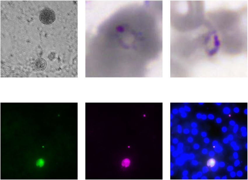
ARTICLE                                                               COMMUNICATIONS BIOLOGY | https://doi.org/10.1038/s42003-019-0737-3

T
        he majority of Plasmodium vivax infections is likely due to         (Fig. 1c). Taken together, these data indicate that the transgene
        relapses1. Not to be mixed up with recurrences, which may           expression of fluorescent proteins did not affect parasite develop-
        have other origins2, relapse infections have been posited to        ment in any stage of the life cycle, not only providing the oppor-
derive from hypnozoites3. After the identification of hypnozoites            tunity to visualize essential parasitic processes throughout the
in livers infected with Plasmodium cynomolgi4,5 and P. vivax6,              parasite life cycle, but also to study hypnozoite activation using this
the hypnozoite theory of relapse has been questioned7–9. One of             parasite line.
the arguments was that the actual transformation of the hypno-                 To determine the percentage of liver stage parasites that had
zoite into a schizont had never been observed7,10. The currently            lost the centromere plasmid as a result of the many nuclear
available in vitro models using nongenetically modified parasites            divisions that take place at the various stages of the life cycle in
all have in common that hypnozoites and developing forms are                the absence of pyrimethamine selection18, live images of
identified on the basis of morphological features in fixed tissue             hepatocyte cultures were acquired followed by the fixation and
cells. As a result, it is impossible to monitor the growth of indi-         IFA of the same wells. Indeed, GFP signals obtained were specific
vidual parasites over time, precluding direct observations of the           for liver stage parasites and both hypnozoites and schizonts
actual transition of a hypnozoite into a developing parasite.               expressed GFP (Fig. 1d). GFP signals were observed in 73% of the
   To overcome this hurdle, we here describe the construction of a          parasites as determined by IFA (counting > 200 parasites),
P. cynomolgi M strain dual fluorescent reporter line that can                indicating that 27% of the liver stage parasites had lost the
distinguish hypnozoites (green-fluorescent protein, GFP positive             centromere plasmid. This is similar to what has previously been
and mCherry negative) from developing forms (both GFP and                   reported for centromere plasmids11,12.
mCherry positive). Using this transgenic parasite line, we show
direct proof for reactivation of dormant hypnozoites in real time.
                                                                            mCherry is specifically expressed in liver schizonts. To observe
This demonstrates, almost 40 years after their discovery, that
                                                                            the timing of GFP and mCherry expression, live fluorescence
hypnozoites have the capacity to awaken to resume liver stage
                                                                            microscopy at a high magnification (63×; 1.40 NA) was per-
development, which in vivo ultimately provokes malaria relapses.
                                                                            formed at different time points of liver stage development. GFP
                                                                            was expressed throughout the liver stage development in both
                                                                            small and large forms (Fig. 2). At day 1 post sporozoite infection,
Results
                                                                            round liver stage forms were observed. In addition, some spor-
Transgenic P. cynomolgi recapitulates the full life cycle. To
                                                                            ozoites had not yet fully transitioned into rounded forms and the
express the fluorescent reporters GFP and mCherry in liver stage
                                                                            two distal ends of the sporozoites could still be observed. Similar
parasites, a P. cynomolgi reporter line was constructed using a
                                                                            to what has been described before for LISP2 expression15, lisp2-
centromere plasmid that is maintained throughout the life cycle11,12
                                                                            driven mCherry expression was absent in the first two days post
and a human dihydrofolate reductase (Hdhfr) selectable marker13
                                                                            sporozoite infection. In a minor parasite fraction at day 3 post
controlled by the constitutive P. cynomolgi hsp70 promoter.
                                                                            sporozoite invasion mCherry expression first emerged, often
Through the inclusion of a T2A self-cleaving peptide14 not only
                                                                            coinciding with a slightly more pronounced growth in size
hdhfr but also gfp was driven by the hsp70 promoter. In addition, to
                                                                            as compared to small forms (Fig. 2). From then onward, an
highlight stages that have initiated schizogony, the construct con-
                                                                            mCherry-positive population that grew in size and became
tained mCherry controlled by promoter and 3′UTR of the recently
                                                                            multinucleated could be differentiated from an mCherry-negative
described schizont-specific marker for early liver stage growth,
                                                                            population that remained single nucleated and small in size.
P. cynomolgi liver-specific protein 2 (lisp2)15 (Fig. 1a; see Methods
                                                                            During schizont maturation, mCherry expression levels rapidly
section for details). Following transfection16, pyrimethamine-
                                                                            increased with peak expression around days 8–10 (Fig. 2).
resistant parasites emerged at a growth rate comparable to wild-
                                                                            mCherry fluorescence remained visible in merosomes. To deter-
type parasites (Supplementary Fig. 1a). Ex vivo blood stage parasites
                                                                            mine whether mCherry expression coincided with nuclear divi-
showed strong GFP expression, while mCherry expression, was not
                                                                            sion, at different time points post sporozoite infection, liver stage
observed (Supplementary Fig. 1b), recapitulating the expression of
                                                                            cultures were fixed and 4′,6-diamidino-2-phenylindole, dilactate
the LISP2 protein15. When ex vivo blood stage schizonts and
                                                                            (DAPI, dilactate) was added for nuclear staining. GFP and
merozoites were imaged for prolonged times, merozoite invasion of
                                                                            mCherry signals could still be observed after fixation allowing
rhesus red blood cell (RBC) was observed (Supplementary movie 1).
                                                                            counting of the number of GFP- and mCherry-positive parasites
After feeding mosquitoes using a glass-feeder system, the trans-
                                                                            at each time point in relation to the number of parasite nuclei.
mission characteristics were determined. A side by side comparison
                                                                            The onset of mCherry expression corresponded with the initia-
of the transgenic line to wild-type parasites showed similar numbers
                                                                            tion of nuclear division, as all GFP-positive parasites with >1
of oocysts and sporozoites (Supplementary Fig. 1c). Gametocytes
                                                                            nucleus were mCherry positive (Table 1 and Supplementary
expressing GFP were readily observed in the blood used for mos-
                                                                            Fig. 2). These data indicate that the regulatory sequences used in
quito feeding and upon incubation at room temperature (RT) for
                                                                            the centromeric plasmid contained sufficient information for the
10–15 min the process of exflagellation could be viewed in real time
                                                                            correct timing of lisp2-driven mCherry expression19.
(Supplementary movie 2). Following mosquito transmission, pri-
                                                                               We also noted that cultures at days 2 and 3 contained
mary rhesus hepatocytes were infected with sporozoites. A side by
                                                                            substantially more parasites than cultures at later time points
side comparison of wild type and transgenic liver stage parasites at
                                                                            (Table 1). The loss of parasites during the initial stages of parasite
day 6 of development by immunofluorescence assay (IFA)17 showed
                                                                            development has been described before15,20 and may be a result
that numbers of exoerythrocytic forms (EEFs) and hypnozoite ratio
                                                                            of cytosolic immune responses, such as selective autophagy by the
of the transgenic line were similar to wild-type parasites (Fig. 1b).
                                                                            host cell20 or could reflect a parasite developmental failure.
Beyond day 6, parasite development appeared to progress normally
and schizonts had fully matured around day 10 post sporozoite
invasion, at which time merosomes could be observed (Fig. 1c and            Long-term live imaging of P. cynomolgi liver stage parasites.
Supplementary movie 3). To determine whether the full life cycle            Following the characterization of the parasites at high magnifi-
could be recapitulated, rhesus RBCs were added to wells that con-           cation, our next aim was to visualize the development of indivi-
tained merosomes and incubated overnight. The next day, blood               dual parasites over time. We initiated long-term liver stage
stage parasites were observed in these wells by Giemsa staining             cultures with the transgenic P. cynomolgi line in 96-well plates

2                          COMMUNICATIONS BIOLOGY | (2020)3:7 | https://doi.org/10.1038/s42003-019-0737-3 | www.nature.com/commsbio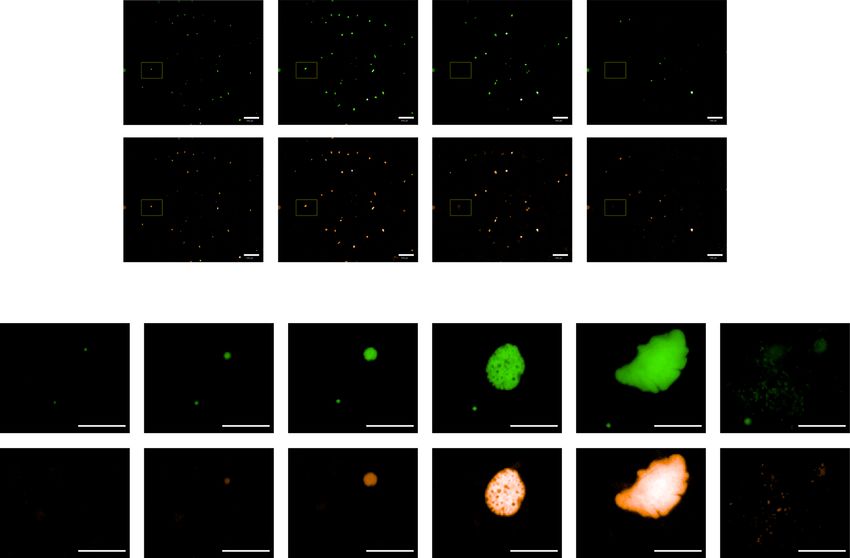
COMMUNICATIONS BIOLOGY | https://doi.org/10.1038/s42003-019-0737-3                                                                         ARTICLE

Fig. 1 The P. cynomolgi dual reporter line fully recapitulates the complete life cycle. a Schematic of the most important building blocks of the construct
used for transfection; for details of the complete construct, see methods and the Supplementary Data file 1. The plasmid contains two fluorescent reporters,
GFP driven by the hsp70 promoter and mCherry controlled by the lisp2 promoter. A centromere is included to maintain the construct throughout the life
cycle. b Dot plot of the number of small and large exoerythrocytic forms (EEFs) per well in a 96-well plate with s.e.m. from n = 6 wells. Results derive from
a side by side comparison of transgenic and wild-type parasites. c Left, P. cynomolgi merosomes (transgenic line) in culture at day 10 post sporozoite
invasion. Scale bar, 50 µm. Right, Giemsa staining of red blood cells collected one day post overlay on a liver stage culture containing merosomes shows
two ring stage blood forms. d GFP expression (live) in two small forms and one large form at day 6 post inoculation of a primary hepatocyte culture with
transgenic P. cynomolgi sporozoites; the same image after fixation and IFA with anti-HSP70 antibodies, and an overlay with DAPI. Scale bars, 25 µm.

using a Matrigel cover21 and performed daily or every other day                 mCherry levels increase over time, coinciding with parasite
imaging at a lower magnification using an Operetta high content                  growth (Fig. 3c). A clear distinction is visible between a
imager (PerkinElmer). When the development of parasites within                  population with background levels for mCherry (hypnozoites)
the same well was monitored between days 8–15 post sporozoite                   and an mCherry-positive population (schizonts). GFP expression
infection, well overviews made from stitched images revealed that               also increases over time, but this does not allow a clear
the majority of the schizonts burst in vitro between days 10 and                delineation of the transition between hypnozoites and schizonts,
13 (Fig. 3a and Supplementary movie 4). This closely mimics the                 likely reflecting the constitutive nature of the hsp70 promoter
maturation of P. cynomolgi EEFs in vivo, which are mature                       (Fig. 3c). The size and mCherry measurements indicate that
between days 9 and 1222. The development of individual parasites                at day 15 post sporozoite inoculation a number of schizonts
could be monitored in more detail in the nonstitched pictures,                  are present (Fig. 3c). To further quantitate this, for four wells
revealing GFP-positive parasites that remain small, and parasites               derived from three independent infections, the total number of
(schizonts) that in addition become mCherry positive and pro-                   hypnozoites and schizonts present at days 10 and 15 was
gressively grow in size and eventually rupture after day 10                     determined (Supplementary Table 1). This revealed important
(Fig. 3b). Taken together, this shows that the dual fluorescent                  issues regarding EEF kinetics. The total number of EEFs reduces
parasite line enables tracking of individual parasites in real time.            over time with 50% on day 15 compared to day 10. Both the
   To obtain more quantitative data, the GFP and mCherry                        number of schizonts and the number of hypnozoites are reduced
intensities, and the fluorescent surface area as a proxy for the                 (29% and 66% present at day 15, respectively, as compared to day
parasite size of individual hypnozoites and schizonts present in                10). This reduction is caused by culture technical reasons
the same ten fields of a well at different time points were                      (parasite death and hepatocyte detachment), by mature schizont
measured using the Operetta. This showed that in schizonts                      rupture and, possibly, by hypnozoite reactivation. The relatively

COMMUNICATIONS BIOLOGY | (2020)3:7 | https://doi.org/10.1038/s42003-019-0737-3 | www.nature.com/commsbio                                                   3
ARTICLE COMMUNICATIONS BIOLOGY | https://doi.org/10.1038/s42003-019-0737-3
Table 1 Quantitation of schizont specific expression of
mCherry.
# Parasites (1n) # Parasites (>1n)
Day p.i. GFP GFP and GFP GFP and
+ve only mCherry +ve +ve only mCherry +ve
2 170 0 0 0
3 143 1 0 0
4 71 9 0 24
6 58 1 0 37
10 36 3 0 19
Numbers of parasites in one well (96-well plate) expressing GFP only, or expressing both GFP
and mCherry. Parasites were scored after fixation at different time points p.i. (post infection).
Parasites were categorized as 1n (containing 1 nucleus) or >1n (multiple nuclei) as determined by
the DAPI signal
high levels of schizonts we observed on day 15 have also been
described by others using wild type P. cynomolgi EEF cultures15,
and ‘late’ schizonts have also been seen in liver biopsies from
P. cynomolgi-infected monkeys23. The complexity of the culture
dynamics precludes pinpointing where these late schizonts derive
from in standard cultures. Our transgenic parasite, however,
allowed us to trace back in time each late schizont, to determine
whether they had originated from hypnozoite reactivation.
Live visualization of P. cynomolgi hypnozoite activation. In
vivo, P. cynomolgi M is a fast relapsing parasite. While low-dose
inoculations (2000 sporozoites) followed by subcurative treat-
ment of blood stages gave rise to somewhat delayed relapses24,25,
high-dose sporozoite inoculations (105–106) resulted in early
relapses around days 18–33 post infection as assessed by thin film
analysis26,27. It is thus expected that activation of hypnozoites in
this parasite may start relatively soon after the completion of liver
stage development (merosome release) of the primary schizonts.
To identify parasites that had activated, images were acquired
daily or every other day. We then looked for the presence of
schizonts in images taken at days 15–17 and compared this to
images taken from the same field at day 10 post sporozoite
infection. Considering that the bulk of the primary schizonts have
reached full maturity at day 10, we chose this time point as
baseline. Only parasites that had remained small and GFP posi-
tive, but mCherry negative at day 10 were posited to be hypno-
zoites. Parasites that were hypnozoites at day 10 and schizonts at
days 15–17 were considered to originate from activated hypno-
zoites. This was further substantiated by monitoring parasite
development between these time points. Following this metho-
dology, we found that the overwhelming majority of schizonts
found at day 15 had already been present at day 10 as well,
indicating that in culture, schizonts can remain lingering on for
prolonged periods of time. However, in a few instances we could
indeed identify hypnozoites that initiate mCherry expression after
day 10 and develop into schizonts. Moreover, we could visualize
the full maturation of such a secondary schizont as highlighted by
the budding of merosomes (Fig. 4a). When we overlaid the well
overviews from different time points, generating a movie of
parasite development, it was clear that the hypnozoite activation
event took place after primary schizont development in the same
Fig. 2 Lisp2-driven mCherry differentiates P. cynomolgi hypnozoites from well (Supplementary movie 4).
developing forms. GFP and mCherry expression at different time
points of transgenic P. cynomolgi liver stage development. The panels
Early in vitro hypnozoite activation occurs infrequent. One of
at the righthand side show an overlay with brightfield images. Scale
the hallmarks of hypnozoites is that they are resistant to days 5–8
bars, 10 µm.
treatment with the PI4K inhibitor KDU69115,27. To further prove
that the events we observed are true hypnozoite activation events,
and not retarded schizonts, we pretreated cultures on days 5–8
4 COMMUNICATIONS BIOLOGY | (2020)3:7 | https://doi.org/10.1038/s42003-019-0737-3 | www.nature.com/commsbioCOMMUNICATIONS BIOLOGY | https://doi.org/10.1038/s42003-019-0737-3 ARTICLE Fig. 3 The dual fluorescent P. cynomolgi line reveals the real-time development of individual parasites. a Images of parasites visualized live by fluorescence microscopy using an Operetta High Content imager. Overviews are shown from stitched images of the same well (96-well plate) at different time points, showing GFP-expressing parasites (upper panel) and mCherry-expressing parasites (lower panel). Parasites from the yellow inset are depicted in b. Scale bar, 500 µm. b Images of a small and a developing form expressing GFP (upper panel) and mCherry (lower panel) at different time points after sporozoite invasion. Yellow arrows mark expression, and grey arrows mark absence of fluorescence. At day 11, individual merozoites can be observed. Scale bar, 50 µm. c Operetta measurements of fluorescent EEF surface areas (left panel), mCherry intensity (middle panel), and GFP intensity (right panel) in individual parasites (blue dots) followed at different days p.i. (post infection) in ten fields of a single well. The bar represents the median values. with KDU691 to eliminate schizonts from the cultures prior to ago demonstrates the transition of hypnozoites into a schizont, expected hypnozoite reactivation. The treatment resulted in an providing strong evidence for the hypnozoite theory of relapse. almost complete removal of schizonts (2 schizonts left at day 9, Next, in five independent infections we determined the number compared to 21 schizonts in a nontreated well; Supplementary of spontaneously activated hypnozoites up to day 22, depending Fig. 3). When comparing day 17 images with day 11, the well on length of cultivation. After scanning 21 untreated wells for overviews at day 17 revealed a schizont that was not present as reactivation events, corresponding to ±1175 day-15 hypnozoites, schizont at day 11. Tracing this parasite back in time revealed the we observed only three events of activation (deriving from two presence of a small form that started mCherry expression at day independent infections). This indicates that spontaneous hypno- 13 post infection (Fig. 4b, white arrow). From that time onward, zoite activation in vitro is an infrequent event during the first the parasite continues to grow, indicative of hypnozoite activa- 3 weeks of culture. tion. We conclude from this that the fluorescent parasite line To visualize the development of the observed events, mCherry for the first time since hypnozoite discovery over three decades intensities and EEF surfaces of the activated hypnozoites were COMMUNICATIONS BIOLOGY | (2020)3:7 | https://doi.org/10.1038/s42003-019-0737-3 | www.nature.com/commsbio 5
ARTICLE COMMUNICATIONS BIOLOGY | https://doi.org/10.1038/s42003-019-0737-3
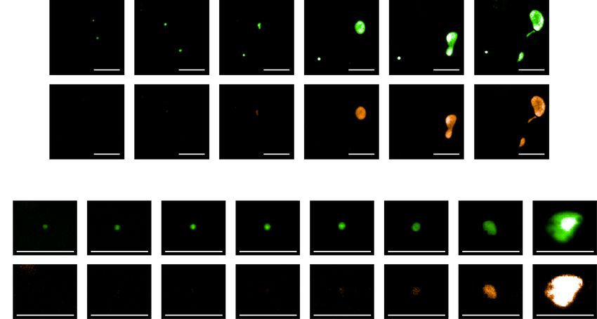
Fig. 4 Monitoring individual parasites over time by fluorescence microscopy reveals hypnozoite reactivation. a Hypnozoite reactivation resulting in a
fully mature liver stage schizont releasing merosomes. Operetta images show GFP expression (upper panel) and mCherry expression (lower panel) of a
hypnozoite (mCherry negative), and an activating hypnozoite at different time points after sporozoite invasion. Yellow arrows mark expression, and grey
arrows mark absence of fluorescence. Scale bar, 50 µm. b Images showing GFP expression (upper panel) and mCherry expression (lower panel) in an
activating hypnozoite at different time points after sporozoite invasion. A white arrow marks the onset of mCherry expression. Scale bar, 50 µm. c Operetta
measurements of activated hypnozoites with respect to EEF fluorescent surfaces (left panel) and mCherry intensity (right panel) at different days post
sporozoite (spz) inoculation (green lines). For reference, the measurements of five individual primary liver stage schizonts as already are also depicted (red
lines).
measured over time. Comparing this to measurements performed spontaneous event per 400 hypnozoites) suggest that only a min-
on several primary schizonts shows that the activated hypnozoites in ority of hypnozoites is prone to become activated at a certain
untreated wells (Fig. 4c, green lines) resume development after the time within the first 3 weeks of culture. In vivo, a similar phe-
primary schizonts have burst (Fig. 4c, red lines and Supplementary nomenon was observed when monkeys were inoculated with low
movie 4). Furthermore, this reveals that early hypnozoite activation, numbers of P. cynomolgi sporozoites8,28. Infection with 500 spor-
as defined by parasites initiating mCherry expression and growth, is ozoites induced multiple relapses in all monkeys infected, whereas
already evident between days 12–16 after sporozoite invasion. The monkeys infected with only five sporozoites induced a primary
speed of schizont maturation of activated hypnozoites appears to be infection in 3/5 monkeys, but relapses were not observed8. Monkeys
similar to that of primary schizonts (Fig. 4c). receiving 10–20 sporozoites all developed a patent parasitemia, and
3/8 monkeys relapsed over an observation period of 1 year28.
Interestingly, only one monkey showed an early relapse, at day 30
Discussion post infection. The second monkey relapsed at day 168 and the
In our hands, P. cynomolgi liver stage cultures could be routinely third monkey relapsed at days 58 and 75 post inoculation28. This
maintained at good quality for 3 weeks. Thereafter, cultures suggests that some sporozoites may be prone to activate early,
often started to deteriorate, and this is why we focused on reacti- whereas others may be prone to activate late. This may have a
vation events during the first 3 weeks of culture. The few hypno- biological reason, as the parasite’s main goal is to be transmitted to
zoite reactivation events measured in vitro (approximately one the next host. Activation of most or all hypnozoites at once may not
6 COMMUNICATIONS BIOLOGY | (2020)3:7 | https://doi.org/10.1038/s42003-019-0737-3 | www.nature.com/commsbioCOMMUNICATIONS BIOLOGY | https://doi.org/10.1038/s42003-019-0737-3 ARTICLE
be the best strategy to be transmitted in areas of low, unstable (continued)
transmission, because there would be only a single opportunity to Reagent or resource Source Identifier
be transmitted. For human malaria in tropical regions, the ability to
repeatedly induce new blood stage infections may be necessary Deposited data
Experimental models: cell lines
to compete with more virulent Plasmodium falciparum29. Instead of Parasite strain: Plasmodium Ref. 30 N/A
a predetermined duration of dormancy, reactivation of hypnozoites cynomolgi M
may occur stochastically. To study possible patterns in reactivation, Experimental models: organisms/
strains
culture conditions will have to be further optimized to obtain Monkey: Macaca mulatta In house N/A
healthy hepatocyte cultures lasting for at least 2 months. breeding colony
Despite the global importance of relapse infections, there is Mosquito: Anopheles stephensi RUNMC N/A
Sind–Kasur strain Nijmegen Nijmegen, NL
little knowledge on basic hypnozoite biology. We anticipate that Oligonucleotides
the dual fluorescent parasite line we describe here will be a Recombinant DNA
valuable tool to increase our knowledge on hypnozoite dormancy pCR-BluntII-PcyCEN Ref. 16 N/A
Building block from plasmid # 1 This paper N/A
and activation mechanisms. This parasite not only allows real- Building block from plasmid # 2 This paper N/A
time monitoring of parasite development, but also enables Building block from plasmid # 3 This paper N/A
FACSsort purification of reactivating hypnozoites for molecular pCyCEN_Lisp2mCherry_ This paper Addgene #137169
hsp70_GFP
characterization, as an addition to previously described hypno- Software and algorithms
zoite purification with another transgenic P. cynomolgi line16. Las X Leica https://www.leica-
Furthermore, the ability to now quantitate hypnozoite activation microsystems.com/
provides the opportunity to evaluate compounds that trigger products/microscope-
software/p/leica-las-x-ls/
hypnozoite activation. If we can identify compounds that provoke GraphPad Prism 7 GraphPad www.graphpad.com
hypnozoites to resume development, we can then eliminate the software
resulting schizonts with already available liver stage schizonticides Columbus 2.8.2 PerkinElmer www.perkinelmer.com
Other
in so-called ‘wake-and-kill’ strategies. This opens up new avenues CellCarrier-96 Ultra Microplates, PerkinElmer Cat. #6055700
to eliminate the hypnozoite reservoir from the human population, collagen coated
a key road block for malaria elimination.
Methods
Key resources table
Experimental model and subject details. Nonhuman primates: Nonhuman pri-
mates were used because no other models (in vitro or in vivo) were suitable for the
Reagent or resource Source Identifier aims of this project. The research protocol was approved by the central committee for
animal experiments (CCD license number AVD5020020172664) and the subprotocol
Antibodies was approved by the local independent ethical committee constituted conform Dutch
Rabbit anti-P. cynomolgi HSP70.1 Ref. 17 N/A law (BPRC Dier Experimenten Commissie, DEC; agreement number #708 and #007
polyclonal antibody C) prior to the start of the experiments. All experiments were performed according to
Alexa Fluor 647-conjugated Goat Thermo Fisher Cat. #A21245 Dutch and European laws. The Council of the Association for Assessment and
anti-Rabbit IgG (H + L) Scientific Accreditation of Laboratory Animal Care (AAALAC International) has awarded
Bacterial and virus strains BPRC full accreditation. Thus, BPRC is fully compliant with the international
Subcloning efficiency™ DH5α Thermo Fisher Cat. #18265-017 demands on animal studies and welfare as set forth by the European Council Directive
competent cells Scientific 2010/63/EU, and Convention ETS 123, including the revised Appendix A as well as
Biological samples the ‘Standard for humane care and use of Laboratory Animals by Foreign institutions’
Human serum A+ (pooled, heat Sanquin N/A
identification number A5539-01, provided by the Department of Health and Human
inactivated) blood bank
FBS Greiner Bio- Cat. #758093 Services of the United States of America’s National Institutes of Health (NIH) and
One Dutch implementing legislation. At least once a year, the health of the animals is
Primary rhesus hepatocytes BPRC N/A checked and prior to experimentation an additional health check is carried out,
Chemicals, peptides, and recombinant proteins including a physical examination and performing hematological, clinical-chemistry,
William’s E Thermo Fisher Cat. #32551-087 serology, and bacteriological and parasitological analyses. Only healthy animals were
Scientific included in the experiments. The rhesus monkeys used in this study (Macaca mulatta,
100 × Pen/strep Thermo Fisher Cat. #15140-122 either gender, age 4–16 years, Indian origin) were captive-bred and socially housed.
Scientific Animal housing was according to international guidelines for nonhuman primate care
100 mM sodium pyruvate Thermo Fisher Cat. #11360-036 and use. Besides their standard feeding regime and drinking water ad libitum via an
Scientific automatic watering system, the animals followed an environmental enrichment pro-
100 × Insulin/transferin/selenium Thermo Fisher Cat. #41400-045 gram in which, next to permanent and rotating nonfood enrichment, an item of food
supplement Scientific enrichment was offered to the macaques daily. All animals were monitored daily for
100 × MEM-NEAA Thermo Fisher Cat. #11140-035 health and discomfort. Monkeys were trained to voluntarily present for thigh pricks,
Scientific and were rewarded afterward. All intravenous injections and large blood collections
0.1 M Hydrocortisone Sigma Cat. #H0888 were performed under ketamine sedation, and all efforts were made to minimize
2-Mercaptoethanol Thermo Fisher Cat. #31350-010
suffering. Liver cells were derived from inhouse frozen batches of hepatocytes or from
Scientific
DMSO hybrimax Sigma Cat. #P1860 freshly collected liver lobes from monkeys that were euthanized in the course of
Pyrimethamine Sigma Cat. #P7771 unrelated studies (ethically approved by the BPRC DEC) or euthanized for medical
Matrigel Corning Cat. #354234 reasons, as assessed by a veterinarian. Therefore, none of the animals from which liver
Nycodenz Axis Shield Cat. #1002424 lobes were derived were specifically used for this work, according to the 3R rule
Leibovitz L15 medium Thermo Fisher Cat. #11415-056 thereby reducing the numbers of animals used. Euthanasia was performed under
Scientific ketamine sedation (10 mg/kg) and was induced by intracardiac injection of euthasol
Atovaquone Sigma Cat. #A7986 20%, containing pentobarbital.
KDU691 Novartis N/A Mosquitoes: Mosquito infections were performed as described previously17.
4% Paraformaldehyde in PBS Affymetrix Cat. #J19943 Anopheles stephensi mosquitoes Sind–Kasur strain Nijmegen31 were obtained from
4′,6-diamidino-2-phenylindole, Thermo Fisher Cat. #D3571 Nijmegen UMC, The Netherlands. At peak parasitemia, around 12 days after
dilactate (DAPI, dilactate) Scientific infection with blood stage P. cynomolgi M strain parasites, monkeys were bled for
Hoechst33342 Thermo Fisher Cat. #H3570 ex vivo mosquito feeding. Two to five-day-old female mosquitoes were fed on the
Scientific infected blood, using a water-jacketed glass feeder system kept at 37 °C,
Critical commercial assays female mosquitoes were fed on blood obtained from a monkey that had been
QIAfilter Plasmid Maxi Kit Qiagen Cat. #12263 infected with P. cynomolgi M strain blood stage parasites. Mosquitoes were housed
Human T Cell Nucleofector Kit Lonza Cat. #VPA-1002 for ~3 weeks in climate chambers at 25 °C and 80% humidity, and fed daily via
COMMUNICATIONS BIOLOGY | (2020)3:7 | https://doi.org/10.1038/s42003-019-0737-3 | www.nature.com/commsbio 7ARTICLE COMMUNICATIONS BIOLOGY | https://doi.org/10.1038/s42003-019-0737-3
cotton soaked in 5% D-glucose solution. One week after infection, oocysts were were performed entirely independently from each other: parasites were isolated
counted and mosquitoes were given an additional uninfected blood meal to from different donor monkeys; transmission experiments and hepatocyte cultures
promote sporozoite invasion of the salivary glands. were performed at different time points.
Malaria parasites: P. cynomolgi M strain stocks were originally provided by Dr. Live imaging of P. cynomolgi infected blood stage parasites: At the time of ex vivo
Bill Collins from the Center for Disease Control, Atlanta, USA32. Stocks were mosquito feeding using blood from a monkey infected with the transgenic
maintained as frozen stabilates in cryoprotectant (28% glycerol, 3% sorbitol, and P. cynomolgi line, a drop of blood was mixed 1:1 with Hoechst 33342 (resulting in a
0.65% NaCl in water) in liquid nitrogen. Parasites were thawed in a waterbath at final concentration of 10 µg/ml) and viewed under a Leica DMI6000B inverted
37 °C followed by sequential wash steps with equal volumes of 3.5% NaCl33. fluorescence microscope HC PL APO 63x/1.40–0.60 oil objective and pictures were
Parasites resuspended in phosphate-buffered saline (PBS) for i.v. injection (106 acquired with a DFC365FX camera.
parasites in 1 ml PBS) into a monkey. P. cynomolgi sporozoite isolation and liver stage culture: Two weeks post
Primary rhesus hepatocyte cultures: Primary hepatocyte cultures were initiated mosquito feeding on transgenic P. cynomolgi M strain-infected blood, salivary
either from freshly isolated M. mulatta hepatocytes through collagenase gland sporozoites were isolated and used for hepatocyte inoculation. Salivary
perfusion34, or from in-house cryopreserved stocks. Hepatocytes were seeded into glands were collected on ice in Leibovitz’s medium containing 3% fetal calf serum
collagen coated 96-well CellCarrier Ultra plates (PerkinElmer) at a density of and 2% Pen/Strep. Salivary glands were disrupted using a Potter-Elvehjem
~65 × 103 cells/well in William’s B medium: William’s E with glutamax containing homogenizer and debris was removed by a slow spin in a microfuge (3 min, 60×g,
10% human serum (A+), 1% MEM nonessential amino acids, 2% penicillin/ RT), before counting the sporozoites in a Bürker-Türk counting chamber. Primary
streptomycin, 1% insulin/transferrin/selenium, 1% sodium pyruvate, 50 µM rhesus hepatocytes seeded in 96-well CellCarrier Ultra plates (PerkinElmer) two
β-mercapto-ethanol, and 0.05 µM hydrocortisone. Following cell attachment, days earlier were inoculated with 5 x 10⁴ sporozoites per well in William’s B
medium was replaced by William’s B containing 2% dimethylsulfoxide (DMSO) to medium. Plates were spun for 5 min at 233×g (RT, low brake) before transfer back
prevent hepatocyte dedifferentiation. Prior to adding sporozoites, cells were washed into the humidified incubator (37 °C and at 5% CO2) for 2–3 h to allow for
twice in William’s B medium. Liver stage cultures were maintained in William’s B sporozoite invasion. Plates were washed with William’s B medium and for
medium in a humidified incubator at 37 °C and at 5% CO2. prolonged cultivation, 80 µl of Matrigel (Corning) was added on top of the infected
hepatocyte monolayer. After 30 min solidification at 37 °C, William’s B medium
was added and incubation was continued with regular medium refreshments until
Method details. Plasmid DNA cloning: Building blocks for the final construct were fixation with 4% paraformaldehyde (PFA) for 30 min at RT. For drug treatment,
synthesized and cloned into PUC18, PUC19, or PUC57 mini plasmids by Genscript P. cynomolgi liver stage cultures were daily treated with 0.5 µM KDU691 (Novartis)
(USA). See the Supplementary Data file 1 for an outline of the cloning strategy and in William’s B medium from days 5 to 8.
for sequences of the building blocks. Briefly, fragment Nluc-2A-mCherry was Live imaging of P. cynomolgi infected hepatocytes: Images of transgenic P. cynomolgi
excised (EcoRV/BglII) from plasmid #1 and ligated to the EcoRV/BglII-digested parasites were acquired using a Leica DMI6000B inverted fluorescence microscope
plasmid #2 containing flanking regions of P. cynomolgi lisp2 (PcyM_0307500) to equipped with a DFC365FX camera and a HC PL APO 63x/1.40–0.60 oil objective or
generate plasmid (a). The lisp2-mCherry_2A_Nluc expression cassette was cloned a HC PL APO 40x/1.30 oil objective. For monitoring liver stage development over
into the BamHI/ KpnI sites of building block plasmid #3 to generate plasmid (b). time, daily or every other day imaging was performed using an Operetta High Content
This plasmid was linearized with NotI and ligated to NotI-linearized plasmid pCR- Imaging system (PerkinElmer) and a 20x long WD objective. Acquisition times were
BluntII-TOPO containing a P. cynomolgi centromere16 (Genbank accession number kept constant. To determine whether the frequent imaging had any effect on parasite
JQ809338) to create construct pCyCEN_Lisp2mCherry_hsp70_GFP (Addgene, ID growth, images of parasites that had been imaged five times up to day 10 post infection
137169). All transformations were performed using subcloning efficiency DH5alpha were compared to parasites that were only imaged once at day 10. No differences in
cells (Invitrogen) according to the manufacturer’s instructions. For transfection, parasite sizes or numbers were observed indicating that repeated imaging apparently
maxiprep DNA was isolated (Qiagen). was not detrimental to the parasites.
Parasite transfection: Parasite transfection was essentially as described before16. To determine the number of reactivating hypnozoites versus nonreactivated
A donor monkey was infected (i.v.) with 106 P. cynomolgi M wild-type blood stage hypnozoites, the number of day-15 hypnozoites was determined in five
parasites from a cryopreserved stock. Parasitemia was monitored by reading independent infections by counting five wells, one well of each infection. This
Giemsa stained thin blood films prepared from thigh pricks. At a peak parasitemia resulted in an average of 56 day-15 hypnozoites per well, equaling 1176
of ±1% trophozoites, heparin blood was obtained and the monkey was cured from hypnozoites in 21 wells.
malaria by chloroquine treatment (i.m. 7.5 mg/kg) on three consecutive days. For Analysis of parasite size and fluorescence intensity: Parasite size and fluorescence
animal welfare reasons, we have previously developed a Nycodenz protocol to intensities were determined using a customized Columbus script. For detection of
enrich for P. cynomolgi blood stage parasites needed for transfection16. Using this live P. cynomolgi liver stage forms the following criteria were used: threshold for
method, parasite preparations of sufficient purity for transfection can be prepared, GFP expression was 0.50 and GFP-positive populations were counted when the
avoiding the need for splenectomy of the donor monkey in order to obtain high mean GFP intensity was >600. When confirmed manually that parasites were
yields of parasites. To this end, the infected blood was layered on top of a 55% identified by the script, the fluorescence and size characteristics of the parasites
Nycodenz/PBS cushion and centrifuged (25 min, 300×g, RT, low brake). The were recorded. The characteristics of the same parasites were determined at various
interface containing ±30% trophozoites was isolated and washed in RPMI. time points during liver stage culture.
Parasites were cultured at 2% hematocrit overnight in complete medium (RPMI- Immunofluorescence analysis: Liver stage cultures were fixed in 4% PFA for
1640 containing 20% heat inactivated human A+ serum and 15 µg/ml gentamicin) 30 min at RT and overnight incubated at 4 °C with rabbit anti-P. cynomolgi
at 37 °C and gas conditions of 5% CO2, 5% O2, and 90% N2. The next day, medium HSP70 primary antibody diluted 1:10,000 in antibody dilution buffer (0.3%
was refreshed and the culture was maintained until fully matured schizonts were Triton-X100, 1% bovine serum albumin in PBS). Samples were washed with PBS
observed by Giemsa stained thin films of the culture. To increase levels of and incubated for 2 h at RT with Alexa 647-conjugated goat-anti-rabbit IgG
successful transfection, two methods were used. Following a wash step in RPMI- (Thermo Fisher Scientific, 1:1000) and 2 µM DAPI (Thermo Fisher Scientific) in
1640, ±3 x 10⁸ parasites were gently resuspended in Cytomix (120 mM KCl, antibody dilution buffer. Samples were washed again in PBS and images were
0.15 mM CaCl2, 2 mM EGTA, 5 mM MgCl2, 10 mM K2HPO4, 10 mM KH2PO4, captured on a Leica DMI6000B inverted fluorescence microscope equipped with a
25 mM Hepes, pH 7.6) containing 45 µg plasmid DNA. The mixture was DFC365FX camera.
transferred to a 4 mm electroporation cuvette (Bio-Rad), electroporated at 25 µF,
2500 V and 200 Ω, and put on ice until injection. In addition, 2 x 10⁷ parasites were Statistics and reproducibility. Sample sizes and statistical analysis: Data were
gently resuspended in 100 µl Human T-Cell Nucleofector solution (Lonza), mixed analyzed using Prism 7.0 (GraphPad Software) or Columbus 2.8.2 (PerkinElmer).
with 10 µg plasmid DNA and electroporated using a Nucleofector device (Lonza, Results are presented as n values (the number of wells analyzed) ± s.e.m. as
program U-033). The mixtures from the two transfection methods were combined described in the figure legends.
in 0.5 ml PBS and i.v. injected into a recipient monkey. At day 4 post transfection,
blood stage parasites were observed in the recipient monkey. From the next day
onward (four times), the recipient monkey received every other day pyrimethamine Reporting summary. Further information on research design is available in
(1 mg/kg) hidden in a piece of fruit to select for transfectants. First parasites were the Nature Research Reporting Summary linked to this article.
observed at day 11 post transfection. At peak parasitemia, blood was taken for
mosquito feeding, stocks, and analyses. Subsequently, the monkey was cured from Data availability
malaria by chloroquine treatment (i.m. 7.5 mg/kg) on three consecutive days. The data, resources, and reagents that support the findings of this study are
For mosquito feeding, recipient monkeys were infected with 1 x 10⁶ transgenic available from the corresponding author upon reasonable request. The transfection
P. cynomolgi M strain parasites from a cryopreserved stock. To eliminate possible construct pCyCEN_Lisp2mCherry_hsp70_GFP can be requested through Addgene,
wild-type contaminant parasites, the monkeys received three dosages of ID 137169.
pyrimethamine (1 mg/kg, orally in a piece of fruit every other day), starting one day
post infection. Around peak parasitemia, on two consecutive days, generally
around days 11–13 post infection, 5–9 ml of heparin blood was taken to feed Received: 20 September 2019; Accepted: 12 December 2019;
mosquitoes and monkeys were cured from Plasmodium infection by intramuscular
treatment with chloroquine (7.5 mg/kg) on three consecutive days. Experiments
8 COMMUNICATIONS BIOLOGY | (2020)3:7 | https://doi.org/10.1038/s42003-019-0737-3 | www.nature.com/commsbioCOMMUNICATIONS BIOLOGY | https://doi.org/10.1038/s42003-019-0737-3 ARTICLE
References 27. Zeeman, A. M. et al. PI4 kinase is a prophylactic but not radical curative target
1. Robinson, L. J. et al. Strategies for understanding and reducing the in Plasmodium vivax-type malaria parasites. Antimicrob. Agents Chemother.
Plasmodium vivax and Plasmodium ovale hypnozoite reservoir in Papua New 60, 2858–2863 (2016).
Guinean children: a randomised placebo-controlled trial and mathematical 28. Contacos, P. G. & Collins, W. E. Letter: malarial relapse mechanism. Trans. R.
model. PLoS Med. 12, e1001891 (2015). Soc. Trop. Med. Hyg. 67, 617–618 (1973).
2. Markus, M. B. New evidence for hypnozoite-ndependent Plasmodium vivax 29. White, N. J. Why do some primate malarias relapse? Trends Parasitol. 32,
malarial recurrences. Trends Parasitol. 34, 1015–1016 (2018). 918–920 (2016).
3. Krotoski, W. A. Discovery of the hypnozoite and a new theory of malarial 30. Coatney, G. R. et al. Transmission of the M strain of Plasmodium cynomolgi
relapse. Trans. R. Soc. Trop. Med. Hyg. 79, 1–11 (1985). to man. Am. J. Trop. Med. Hyg. 10, 673–678 (1961).
4. Krotoski, W. A. et al. Relapses in primate malaria: discovery of two 31. Feldmann, A. M. & Ponnudurai, T. Selection of Anopheles stephensi for
populations of exoerythrocytic stages. Br. Med. J. 280, 153–154 (1980). refractoriness and susceptibility to Plasmodium falciparum. Med. Vet.
5. Krotoski, W. A. et al. Observations on early and late post-sporozoite tissue Entomol. 3, 41–52 (1989).
stages in primate malaria. I. Discovery of a new latent form of Plasmodium 32. Pasini, E. M. et al. An improved Plasmodium cynomolgi genome assembly
cynomolgi (the hypnozoite), and failure to detect hepatic forms within the first reveals an unexpected methyltransferase gene expansion. Wellcome Open Res.
24 h after infection. Am. J. Trop. Med. Hyg. 31, 24–35 (1982). 2, 42 (2017).
6. Krotoski, W. A. et al. Demonstration of hypnozoites in sporozoite-transmitted 33. Margos, G., Maier, W. A. & Seitz, H. M. Experiments on cryopreservation of
Plasmodium vivax infection. Am. J. Trop. Med. Hyg. 31, 1291–1293 (1982). Plasmodium falciparum. Trop. Med. Parasitol. 43, 13–16 (1992).
7. Shortt, H. E. Relapse in primate malaria: its implications for the disease in 34. Guguen-Guillouzo, C. et al. High yield preparation of isolated human adult
man. Trans. R. Soc. Trop. Med. Hyg. 77, 734–738 (1983). hepatocytes by enzymatic perfusion of the liver. Cell Biol. Int. Rep. 6, 625–628
8. Schmidt, L. H. Compatibility of relapse patterns of Plasmodium cynomolgi (1982).
infections in rhesus monkeys with continuous cyclical development and
hypnozoite concepts of relapse. Am. J. Trop. Med. Hyg. 35, 1077–1099 (1986).
9. Markus, M. B. Do hypnozoites cause relapse in malaria? Trends Parasitol. 31, Acknowledgements
239–245 (2015). We are grateful to the members of the Animal Science Department for excellent animal
10. Garnham, P. C. The liver in malaria with special reference to the care and veterinary assistance. We thank Anke Harupa for expert help in monitoring the
exoerythrocytic phase. Ann. Trop. Med. Parasitol. 81, 531–537 (1987). exflagellation process, Francisca van Hassel for preparing graphical representations and
11. Iwanaga, S. et al. Functional identification of the Plasmodium centromere and the mosquito breeding facilities in Nijmegen for provision of A. stephensi mosquitoes.
generation of a Plasmodium artificial chromosome. Cell Host Microbe 7, This work was funded by the Bill & Melinda Gates Foundation (OPP1141292).
245–255 (2010).
12. Voorberg-van der Wel, A. et al. A comparative transcriptomic analysis of Author contributions
replicating and dormant liver stages of the relapsing malaria parasite Conceptualization, A.V. and C.K.; methodology, A.V., C.K. and A.M.Z.; investigation,
Plasmodium cynomolgi. Elife 6, e29605 (2017). A.V., A.M.Z., I.N., N.W., E.K., O.K. and L.V.; resources, D.K.G., L.D. and T.T.D.;
13. de Koning-Ward, T. F. et al. The selectable marker human dihydrofolate writing—original draft, A.V. and C.K.; writing—review and editing, A.V., A.M.Z., D.K.
reductase enables sequential genetic manipulation of the Plasmodium berghei G., L.D., T.T.D. and C.K.
genome. Mol. Biochem. Parasitol. 106, 199–212 (2000).
14. Straimer, J. et al. Site-specific genome editing in Plasmodium falciparum using
engineered zinc-finger nucleases. Nat. Methods 9, 993–998 (2012). Competing interests
15. Gupta, D. K., et al. The Plasmodium liver-specific protein 2 (LISP2) is an early Devendra Kumar Gupta and Thierry Tidiane Diagana are employed by and/or are
marker of liver stage development. Elife 8, e43362 (2019). shareholders of Novartis Pharma AG. The other authors declare that no competing
16. Voorberg-van der Wel, A. et al. Transgenic fluorescent Plasmodium interests exist.
cynomolgi liver stages enable live imaging and purification of Malaria
hypnozoite-forms. PLoS One 8, e54888 (2013).
17. Zeeman, A. M. et al. KAI407, a potent non-8-aminoquinoline compound that
Additional information
Supplementary information is available for this paper at https://doi.org/10.1038/s42003-
kills Plasmodium cynomolgi early dormant liver stage parasites in vitro.
019-0737-3.
Antimicrob. Agents Chemother. 58, 1586–1595 (2014).
18. Barale, J. C. & Menard, R. Centromeric plasmids and artificial chromosomes:
Correspondence and requests for materials should be addressed to C.H.M.K.
new kids on the Plasmodium transfection block. Cell Host Microbe 7, 181–183
(2010).
19. Kocken, C. H. et al. Precise timing of expression of a Plasmodium falciparum-
Reprints and permission information is available at http://www.nature.com/reprints
derived transgene in Plasmodium berghei is a critical determinant of
subsequent subcellular localization. J. Biol. Chem. 273, 15119–15124 (1998).
Publisher’s note Springer Nature remains neutral with regard to jurisdictional claims in
20. Prado, M. et al. Long-term live imaging reveals cytosolic immune responses of
published maps and institutional affiliations.
host hepatocytes against Plasmodium infection and parasite escape
mechanisms. Autophagy 11, 1561–1579 (2015).
21. Dembele, L. et al. Persistence and activation of malaria hypnozoites in long-
term primary hepatocyte cultures. Nat. Med. 20, 307–312 (2014).
22. Garnham, P. C. C. Malaria parasites and other haemosporidia. (Blackwell,
Oxford, 1966). Open Access This article is licensed under a Creative Commons Attri-
23. Shortt, H. E., Bray, R. S. & Cooper, W. Further notes on the tissue stages bution 4.0 International License, which permits use, sharing, adaptation,
of Plasmodium cynomolgi. Trans. R. Soc. Trop. Med. Hyg. 48, 122–131 distribution and reproduction in any medium or format, as long as you give appropriate
(1954). credit to the original author(s) and the source, provide a link to the Creative Commons
24. Joyner, C. et al. Plasmodium cynomolgi infections in rhesus macaques display license, and indicate if changes were made. The images or other third party material in this
clinical and parasitological features pertinent to modelling vivax malaria article are included in the article’s Creative Commons license, unless indicated otherwise in
pathology and relapse infections. Malar. J. 15, 451 (2016). a credit line to the material. If material is not included in the article’s Creative Commons
25. Joyner, C. J. et al. Humoral immunity prevents clinical malaria during license and your intended use is not permitted by statutory regulation or exceeds the
Plasmodium relapses without eliminating gametocytes. PLoS Pathog. 15, permitted use, you will need to obtain permission directly from the copyright holder. To
e1007974 (2019). view a copy of this license, visit http://creativecommons.org/licenses/by/4.0/.
26. Deye, G. A. et al. Use of a rhesus Plasmodium cynomolgi model to screen for
anti-hypnozoite activity of pharmaceutical substances. Am. J. Trop. Med. Hyg.
86, 931–935 (2012). © The Author(s) 2020
COMMUNICATIONS BIOLOGY | (2020)3:7 | https://doi.org/10.1038/s42003-019-0737-3 | www.nature.com/commsbio 9You can also read