Optical coherence tomography in the 2020s-outside the eye clinic
←
→
Page content transcription
If your browser does not render page correctly, please read the page content below
Eye
                                    https://doi.org/10.1038/s41433-020-01263-6

                                        REVIEW ARTICLE

                                    Optical coherence tomography in the 2020s—outside the eye clinic
                                    Reena Chopra1 Siegfried K. Wagner1 Pearse A. Keane
                                                       ●                            ●
                                                                                                             1

                                    Received: 21 September 2020 / Revised: 20 October 2020 / Accepted: 27 October 2020
                                    © The Author(s), under exclusive licence to The Royal College of Ophthalmologists 2020

                                    Abstract
                                    Optical coherence tomography (OCT) is a paragon of success in the translation of biophotonics science to clinical practice.
                                    OCT systems have become ubiquitous in eye clinics but access beyond this is limited by their cost, size and the skill required
                                    to operate the devices. Remarkable progress has been made in the development of OCT technology to improve the speed of
                                    acquisition, the quality of images and into functional extensions of OCT such as OCT angiography. However, more needs to
                                    be done to radically improve the access to OCT by addressing its limitations and enable penetration outside of typical
                                    clinical settings and into underserved populations. Beyond high-income countries, there are 6.5 billion people with similar
                                    eye-care needs, which cannot be met by the current generation of bulky, expensive and complex OCT systems. In addition,
1234567890();,:
                  1234567890();,:

                                    advancing the portability of this technology to address opportunities in point-of-care diagnostics, telemedicine and remote
                                    monitoring may aid development of personalised medicine. In this review, we discuss the major milestones in OCT
                                    hardware development to reach those beyond the eye clinic.

                                    Introduction                                                              of this valuable diagnostic tool and enable point-of-care
                                                                                                              diagnostics, telemedicine and remote monitoring.
                                    Optical coherence tomography (OCT) imaging is rapid,
                                    safe, non-invasive and is increasingly being recognised as
                                    the gold standard for diagnosis of retinal disease [1].                   Miniature and low-cost OCT
                                    However, OCT machines have several limitations. First,
                                    they are costly; the price of a retinal OCT device ranges                 The construction of a portable, handheld, OCT device could
                                    from ~£30,000–£100,000 [2]. Second, the use of these                      increase the ease of access and expand OCT into settings
                                    devices are limited to typical clinical settings such as hos-             where use is currently prohibited by its cost and size. There
                                    pital clinics to optometry practices. They are housed in large            are currently three commercial handheld SD-OCT systems
                                    tabletop configurations and require alternating current                    available that address this issue. The Envisu C2300 OCT
                                    power, constraining their portability. As patients are                    (Leica Microsystems, Germany) became the first handheld
                                    required to sit upright and stabilise their head on a chinrest            OCT scanner to receive clearance from the Food and Drug
                                    whilst maintaining steady fixation, they are only suitable for             Administration [3]. The scanner consists of a 1.5 kg ima-
                                    ambulatory and cooperative patients. Third, a certain level               ging probe connected to a console by a 1.3-m flexible cable
                                    of skill is required to capture optimal images. A technician              with the most recent iteration, the C2300 additionally pro-
                                    is often required to align the device, capture the image and              viding a variety of lenses to enable both anterior- and
                                    perform a quality check to ensure that the image is accep-                posterior-segment imaging. The iVue system by Optovue
                                    table. In this review, we discuss several efforts that aim to             Inc. consists of a stand-mounted unit with a 2.2-kg remo-
                                    address these limitations and thus improve the accessibility              vable scanner [4]. Both systems benefit from rapid acqui-
                                                                                                              sition speed facilitating capture in paediatric populations. A
                                                                                                              third device, the Heidelberg Spectralis Flex Module (Hei-
                                                                                                              delberg Engineering, Heidelberg, Germany) incorporates a
                                    * Pearse A. Keane                                                         flexible ‘boom’ arm, which houses the acquisition lens and
                                      p.keane@ucl.ac.uk                                                       can be adjusted up to 100 cm from the main device body
                                    1                                                                         (Fig. 1) [5]. The portability of all these devices has allowed
                                         NIHR Biomedical Research Centre for Ophthalmology,
                                         Moorfields Eye Hospital NHS Foundation Trust and UCL Institute        for imaging of patients in different positions and beyond the
                                         of Ophthalmology, London, UK                                         outpatient clinic, from intensive care to the surgical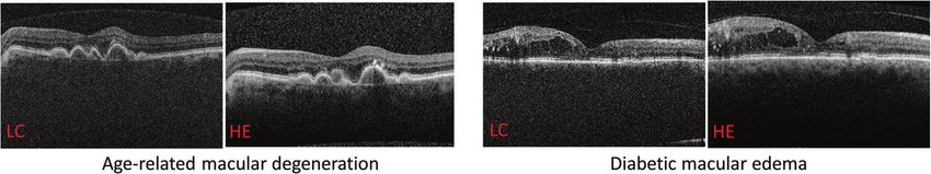
R. Chopra et al.

                                                                       et al., which had an acquisition speed of 350,000 A-scans
                                                                       per second—ten times greater than Envisu system [9].
                                                                       Speed of image acquisition is an important consideration,
                                                                       particularly in handheld systems, which are prone to arti-
                                                                       facts from operator motion, and compounded by subject
                                                                       motion. To alleviate impact of these artifacts on the output
                                                                       image, the system acquires two volumes orthogonal to each
                                                                       other, which can be processed to generate a motion-
                                                                       corrected merged volume. The high speed of the system
                                                                       also enables dense sampling of the retina, producing high
                                                                       definition in vivo retinal images with a field of view up to
                                                                       10 × 10 mm3 captured in this study. The shape and weight
                                                                       of the device is also a critical design consideration to ensure
                                                                       rapid and steady alignment of the device by the operator.
                                                                       With innovations in 3D printing, optical components can be
                                                                       housed in a lightweight ergonomic casing. The authors
                                                                       evaluated two different designs with identical optical com-
                                                                       ponents—their power grip design (0.50 kg) supported
Fig. 1 Heidelberg Engineering Spectralis Flex Module in use. The
system is affixed to a moveable stand with a flexible arm. Images
                                                                       ambidextrous operation but was overly sensitive to the
courtesy of Dr Xiaoxuan Liu and Dr Aditya Kale, University Hospitals   operator’s hand movements, whereas the camcorder design
Birmingham NHS Foundation Trust.                                       (0.42 kg) provided improved balancing but could only be
                                                                       operated using the right hand. The optical design also
                                                                       incorporated an iris camera and fixation target to further aid
environment [6]. However, these handheld systems are                   subject alignment. Although the images produced are
associated with a steep learning curve for operation, are              comparable to commercially available devices, the cost of
subject to motion artifacts and are still considerably heavy           the prototype was too high to be viable for use outside of
to hold or are attached to much larger systems that restrict           the eye clinic. The majority of the cost was due to the
use outside of the clinic.                                             VCSEL and data acquisition electronics. Similarly, Nanki-
   Several research groups have designed portable OCT                  vil et al. demonstrated a SS-OCT handheld device that
prototypes that incorporate miniaturised components known              incorporated a MEMS scanner with an iris camera to aid
as microelectromechanical systems (MEMS) in the spec-                  alignment. Uniquely, the system rapidly switches from
trometer. MEMS enable technology to be constructed at                  anterior to posterior-segment imaging, miniaturising whole-
micrometre scale, and is widely used in numerous consumer              eye OCT to a portable system [13].
products such as mobile phones, video game controllers and                The cost of OCT can be significantly reduced by
accelerometers and gyroscopes for drones. In OCT systems,              designing systems that utilise components that are manu-
MEMS scanning mirrors can be used instead of bulky                     factured at scale for other industries and are therefore
galvanometers to enable a lightweight compact design that              readily available and inexpensive. One biomedical engi-
can be integrated into probes or handheld devices, whilst              neering group from Duke University has built an OCT
simultaneously reducing the cost [7–10]. However, reduc-               system that uses commercial off-the shelf components to
tions in size and cost come at a significant sacrifice of                design the spectrometer, scanner, optics and electronics,
signal-to-noise ratio and reduced imaging depth. In addi-              costing a total of $7164 [14]. The system uses a variable
tion, OCT systems comprise several components including                focus liquid lens that can be precisely adjusted to the
the light source and the computer processing unit, all of              desired focal length, and enabling dynamic focus over
which can still be costly. Units integrating MEMS and                  multiple depths. An inexpensive microcontroller was used
vertical cavity surface emitting lasers (VCSELs) are also              to synchronise the MEMS mirror with the spectrometer.
available, combining the advantage of portability with                 The sensor of the spectrometer was inexpensive and is
extended depth range [11]. VCSELs are light sources with               designed to be more robust to system misalignment due to
large coherence lengths of up to 100 mm, greater than the              temperature fluctuations and mechanical stress. A mini PC
axial length of the eye [12]. Therefore, with VCSEL it is              accelerated using a graphics processing unit (GPU) was
possible to image both anterior and posterior segments                 used to increase the portability, eliminating the need for an
concurrently and without the need for additional attach-               external PC or laptop as is used in most commercial OCT
ments. A prototype swept-source OCT (SS-OCT) handheld                  systems. The increased processing power from a GPU
system incorporating VCSEL was demonstrated by Lu                      enables highly efficient image processing. To synchronise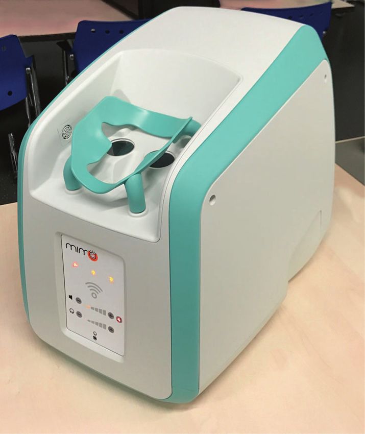
Optical coherence tomography in the 2020s—outside the eye clinic
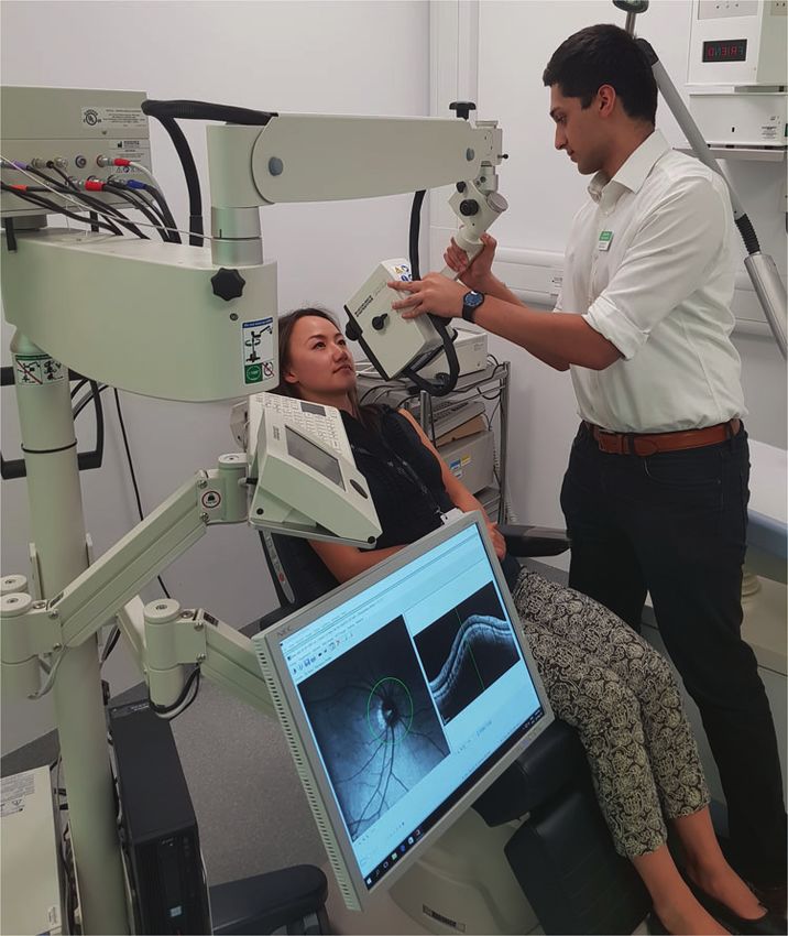
Fig. 2 Representative images from patients with pathology that were acquired by the low-cost OCT (LC) and the Heidelberg Engineering
Spectralis (HE) systems. Scale bars: 500 μm. Images courtesy of Prof. Adam Wax, Duke University.
Fig. 3 Low-cost, handhled
OCT system developed by
Duke University. A Complete
low-cost OCT system showing
PC, touchscreen and scanner. B
Detailed composition inside the
low-cost OCT handheld scanner.
Images courtesy of Prof. Adam
Wax, Duke University.
the movement of the MEMS mirror with the detector, an images with an operator hand holding the scanner without a
Arduino microcontroller was used to detect incoming sig- chinrest. The superluminescent diode in this system suf-
nals and subsequently senda a trigger to scan the MEMS fered from power fluctuations affecting the image contrast.
mirror without delay. The optical components were housed This engineering step was made to keep cost down, but
inside a lightweight 3D-printed casing with a total weight of given the impact on the imaging signal from the retina, the
2.7 kg (Fig. 1A, B). The system had an axial resolution of group aims to resolve this in future iterations. To enable
7 μm, an imaging depth of 2.8 mm with a 7.0 × 7.0-mm field complete stand-alone operation of the system, a 7-inch
of view, and a B-scan acquisition rate (512 × 512 pixels) of touchscreen was incorporated into the system body to allow
12 frames per second. To demonstrate the imaging cap- controls for data acquisition and display of retinal images
ability of the system in relevant biological tissue, ex vivo (Fig. 3). The total weight of the device was 2.3 kg, with a
porcine eyes and live mice retinas were imaged. The low- total cost of materials was $5037. While a productionised
cost OCT system can clearly resolve the tear film, indivi- version of this system is likely to be more expensive to
dual corneal layers, the lens and the iris of the porcine eye. offset the research and regulatory expenses, it has the
Retinal B-scan imaging of live mice could clearly resolve potential to offer adequate performance as a retinal
the individual retinal layers (Fig. 1C). Some limitations of screening tool at a fraction of the cost of current commercial
this prototype include the slow acquisition speed and lack of systems. The device will be commercialised through the
alignment tools such as eye tracking or iris cameras, making group’s start-up company called Lumedica (Durham, NC),
it prone to motion artifacts and thus reducing the ability to and are currently marketing an OCT microscope con-
take advantage of averaging to improve the signal-to- structed using similar technology for research purposes
noise ratio. [16, 17]. The OQ-LabScope boasts a low price of 10,000
The same group has further refined their low-cost OCT USD, a compact size (13 × 7.5 × 6 inches) and is lightweight
system and successfully applied the device to image the (2.7 kg). The device has an axial resolution of 5 μm in tissue
retina of 30 healthy volunteers and 30 patients with retinal and can acquire B-scan images at a rate of 22 per second. It
pathology [15]. The system offers an axial resolution of 8.0 can be connected to a PC and integrates a graphical user
μm, an imaging depth of 2.7 mm and a 6.6 × 6.6-mm field of interface for intuitive manipulation of imaging parameters.
view. The prototype OCT images could clearly resolve While this system is affordable and easy to use, it is not yet
relevant layers of the retina, comparable to the images from approved for use in retinal imaging.
the Heidelberg Spectralis system (Fig. 2). Although the One of the most exciting applications is the potential for
system was mounted on a chinrest for the study to aid ‘smart OCT’. The significant advancements in display,
stability for comparison with the Heidelberg device, it was sensor and battery technologies together have paved the
also operable as a handheld device. Evaluated in five way for multi-functional devices such as smartphones,
healthy volunteers, the investigators obtained high-quality tablets and smartwatches. Modern day smart devices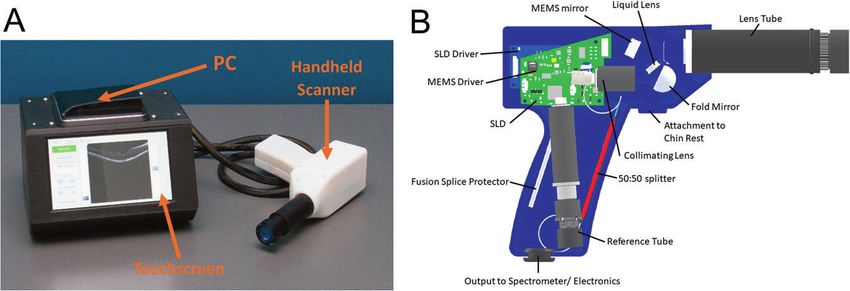
R. Chopra et al.

incorporate a number of embedded sensors that can be used         mechanically adjust the optical path length with the addition
to measure several health parameters such as cardiovascular       of a partial mirror close to the reference mirror, and thus
health [18], physical activity [19, 20] and sleep quality [21].   enabling a more compact design. The partial mirror propels
Smart devices have the ability to connect to a data network,      a portion of the backscattered light to travel to the detector,
enabling assessments to be remotely administered and              and the remaining portion is reflected back and forth
results to be wirelessly transmitted to health providers [22].    between the partial and reference mirror multiple times.
Nearly 60% of the world’s population is connected to the          Each successive reflection corresponds to regions deeper
internet [23], and nearly half own a smartphone [24],             within the target tissue. Every interference signal generated
making remote health monitoring a feasible option in large        by the reflections can be separated out using digital signal
parts of the world. Remote monitoring technologies would          processing techniques, and stitched together for each region
not only be valuable in low- and middle-income countries          of the A-scan to form a continuous image. Subhash et al.
where access to ophthalmologist offices is limited [25], but       have demonstrated the implementation of this technology
also for patients with chronic conditions such as age-related     using a smartphone interface [31]. In this work, an analogue
macular degeneration, which necessitate ongoing monitor-          front-end receives the optical signals. An analogue-to-
ing. Smartphone fundus imaging is already available that          digital converter connects the analogue front-end to a
utilises the smartphone camera with the addition of a clip-on     smartphone to digitise the received data. The smartphone
adaptor that provides the additional optics required [26].        acts as a signal processor and a graphical user interface for
However, integrating OCT into a smart device is more              the user to start and stop scan measurements and see a live
complex due to the need for emission of specialised               graph of incoming OCT measurements. These measure-
wavelengths. Advances in silicon photonics have demon-            ments can be transformed into an image and subsequently
strated the feasibility of integrating optical and electrical     analysed, saved or transmitted via email. The next steps for
circuits onto a chip that can be incorporated into portable       this technology will require improving the imaging perfor-
devices [27, 28]. OCTCHIP, a European project led by              mance to achieve real-time B-scan images [32]. Although
Wolfgang Drexler from the Medical University of Vienna,           the acquisition speed of MRO cannot compete with the
aims to use this technology to engineer a cost-effective,         latest Fourier domain systems, the moderate speed of the
handheld, wireless, OCT system that is the size of a 1 cent       system does not demand high specification computational
coin [27]. The project endeavours to radically transform          platforms therefore may be deployable on mobile systems.
OCT towards widespread adoption in point-of-care diag-            This technology will be commercialised by a start-up
nostics for the early diagnosis of retinal pathologies. In        company called Compact Imaging (Mountain View, CA) in
another European-funded project called Handheld OCT, the          partnership with Novartis (Basel, Switzerland) [33, 34].
same group aims to create a handheld system for point-of-
care diagnostics with partners from Carl Zeiss, University
College Cork, and several other partners in the electronics       Automated OCT
space [29]. This 5-year project commenced in
January 2020.                                                     Much of the burden of chronic eye conditions—to both
    Multiple-reference OCT (MRO) is a new type of OCT             patients and to healthcare systems—may be attributed to the
system that has a low-cost and compact configuration, with         limitations of the current eye examination. Patients with
a comparable footprint to the pick-up head contained within       such conditions commonly require long-term monitoring
a DVD unit [30]. The foundations of the technology are            with frequent, time-consuming visits to the eye clinic. The
similar to first-generation time-domain OCT. In time-              workflow in many eye clinics is inefficient, with patients
domain systems, a Michelson-type interferometer is used           being asked to wait multiple times, interact with several
to divide a broadband light, emitted from a super-                different healthcare staff and see their doctor on multiple
luminescent diode, into a reference arm of known path             occasions. The need for extensive testing also results in
length and time-delay, and a sample arm that is focused at        large staffing and equipment costs and extensive floor space
the tissue of interest, such as the retina. The two light beams   requirements. Moreover, the fundamental unit of the eye
are reflected back to a photodetector using a mirror to            examination—slit-lamp ophthalmoscopy—has little chan-
generate an interference pattern. By mechanically moving          ged since its original description in 1911 by Gullstrand [35],
the mirror over the sample, light is reflected from each           being time-consuming, subjective, non-quantitative and
depth in the tissue producing multiple A-scans that subse-        requiring an experienced clinician. A new form of eye
quently comprise a B-scan. This involves several moving           examination has been developed by Envision Diagnostics,
parts that travel considerable distances, consequently lim-       Inc. (El Segundo, CA, USA) termed binocular OCT [36],
iting the scan speed and making it prone to motion artifacts.     that aims to address the shortcomings of the current eye
The proposed MRO system reduces the need to                       exam, and adds many unique capabilities.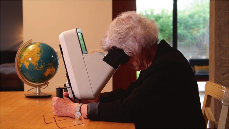
Optical coherence tomography in the 2020s—outside the eye clinic

   A prototype of this system has undergone usability
testing among patients with chronic eye conditions and
healthy volunteers [37]. Unlike other OCT devices, the
binocular OCT prototype consists of two oculars that align
to the patient’s eyes in an automated manner, enabling a
pair of eyes to be imaged simultaneously. This reduces the
labour required to align the device, and also reduces the
overall scan time. The binocular OCT also utilises a tune-
able swept-source laser system with adjustable optics that
can switch from anterior eye imaging to lens imaging, to
vitreous imaging and to posterior pole imaging, permitting
whole-eye OCT without the need for additional attach-              Fig. 4 Pupillometry in a patient with a right relative afferent
ments. Furthermore, the device incorporates ‘smart tech-           pupillary defect (RAPD), using the binocular optical coherence
nology’, offering more advanced display, input and                 tomography system. A Resting diameters pre-stimulus; B flash pre-
                                                                   sented to the left eye, constriction of both pupils observed; C both
computing capabilities than conventional OCT. A speaker
                                                                   pupils dilated to their resting diameter; D flash presented to the right
system is used to deliver audio instructions to guide the          eye. Constriction amplitude of both eyes is less than that observed
automated examination. In addition, the binocular aspect of        when the flash was presented to the left eye.
the device can be exploited so that OCT imaging can be
used for novel applications such as diagnostic functional
tests including pupillometry [38] (Fig. 4), strabismus mea-
surement [39] and ocular motility [37]. Traditionally these
tests have been subjective and required significant clinical
expertise to interpret.
   With these features, the binocular OCT aims to incor-
porate many aspects of the eye examination into a single
automated, patient-facing instrument, and has a number of
potential benefits if this was adopted in tertiary eye-care
clinics, including:
                                                                   Fig. 5 Proposed Home OCT, Notal Vision. Images courtesy of Prof.
                                                                   Anat Loewenstein,Tel Aviv University.
●   Increased efficiency of eye clinics, allowing patients
    with chronic eye disease to spend less time waiting
    during routine hospital eye examinations.                      Home OCT
●   Reduced costs, through a reduction in the total number
    of diagnostic instruments required and their associated        At-home disease monitoring devices are also in develop-
    labour costs.                                                  ment and may be more cost-effective for monitoring
●   Improved quality of eye care, through the introduction         patients at high-risk of developing neovascular AMD [40].
    of more, quantitative, standardised ocular measurements        Notal Vision (Tel Aviv, Israel) received FDA approval for
    and high-resolution imaging.                                   the ForeSeeHome in 2010, an at-home digital macular
                                                                   visual field and hyperacuity testing device that transmits
   The results of a usability study, and related focus group       data directly to the ophthalmologist. The device has since
testing, make it clear that patients are receptive to the          undergone a randomised clinical trial, concluding that
concept of an automated eye examination. To be attractive          individuals at high risk of conversion to neovascular AMD
to users, easy to use and effective at performing automated        would benefit from a home monitoring strategy for earlier
eye examinations, the system will need to be quick,                detection of choroidal neovascularisation development [41].
responsive and comfortable. Once established, binocular            Notal also plans to launch a ‘Home OCT’ device, a self-
OCT will offer objective, quantifiable information about            operated SD-OCT device that could be provided to patients
almost every aspect of the eye examination and has the             at risk of vision loss from AMD, or between visits to the
potential to supersede many traditional but flawed testing          ophthalmologist to customise appointments to the indivi-
methods. It is unlikely that the automated eye examination         dual patient’s needs (Fig. 5) [42]. The device acquires
will be suitable for use in all patients. However, if such a       images of the central 10 degrees of the macula and subse-
system can replace some aspects of the eye examination,            quently reads the image using the company’s artificial
workflows and waiting times are likely to improve and costs         intelligence platform, Notal OCT Analyzer (NOA). In a
are likely to reduce.                                              prospective clinical trial, 90% of 196 patients were able to
R. Chopra et al.
obtain a gradable image in at least one eye after a 2-min
video tutorial [43]. When evaluated on images of AMD
taken using the device, NOA demonstrated >97% sensi-
tivity and specificity for detection of intraretinal and sub-
retinal fluid [44]. The device received Breakthrough Device
Designation by the FDA in 2018, expediting assessment
and review for 510(k) clearance and market authorisation.
Though, the usability and validation of the OCT device and
software is yet to be determined through peer-reviewed
publications.
Self-examination low-cost full-field OCT (SELF-OCT)
is another proposed low-cost system that allows the patient
to independently examine the disease progression of AMD
at home without the presence of a physician [45, 46]. In
contrast to commercially available systems, the SELF-OCT
system sequentially acquires transversal en face images at
different depths instead of cross-sectional images in the
axial direction. The SELF-OCT prototype device captures
Fig. 6 Home-based MIMO-OCT system. Images courtesy of Dr
images at an axial resolution of 9.1 µm and a transversal
Peter Maloca, University of Basel.
resolution of 6.4 and 12.8 µm, respectively; although,
in vivo retinal scans from the system exhibit more artifacts
and a greater signal-to-noise ratio than clinical OCT sys- most ophthalmic units and increasingly in community
tems. Furthermore, the field of view of the SELF-OCT is optometry practices, the next decade will usher a further
1.4 × 4.8 × 1.5 mm, which is considerably smaller than democratisation of OCT through advances in hardware,
clinical OCT systems, but may be sufficient for monitoring cloud-based computing and artificial intelligence. Recent
of wet AMD [47]. This technology has been patented and is regulatory approvals and feasibility studies highlight the
being commercialised by Visotec [48]. emerging permeation of miniaturised, portable and handheld
A proposed novel home-based SD-OCT system termed OCT systems, affording their use as a point-of-care diag-
‘sparse OCT’ has recently been developed by a Swiss group nostic tool in non-traditional settings such as intensive care,
(Fig. 6) [49]. The portable device, named MIMO-OCT, as well as in the home environment for remote monitoring of
generates single 5-mm line scans at a resolution of 400 chronic conditions such as AMD. The latter in particular
pixels, or small sample 3.8 × 3.8-mm volume scans at a could be analogous to continuous monitoring and thus
resolution of up to 150 × 150 pixels. The downsampled provide opportunities for personalised treatment plans for
scanning pattern is less dense than scan protocols available conditions such as wet AMD that benefit from close mon-
on commercial SD-OCT systems, but reduces the size of the itoring and often require indefinite follow-up. Enabled by
instrument and enables fast scanning. The prototype the ability for secure wireless information transfer,
required the subject’s head to be inclined downwards onto improvements in the speed of data transmission through 5th
the headrest, enabling stable positioning to reduce moving generation telecommunication networks [50], and the cur-
artifacts. Manually graded central retinal thicknesses rent dynamism of teleophthalmology, scope also exists for
from sparse OCT data in 30 AMD patients were compared far-reaching impact of a low-cost OCT in low- and middle-
to automated measurements obtained from the Heidelberg income countries. Further efficiencies will come from
Spectralis device, showing no statistical difference. Sparse automated systems, such as the binocular OCT, which have
OCT may be one method of delivering a portable at-home the potential of reducing cost, increasing quality of data and
OCT system with adequate resolution for monitoring. introducing novel applications of OCT that can revolutionise
many aspects of the eye exam. Undoubtedly, OCT will
continue to transform ophthalmology, and portable, low-cost
Conclusion systems are likely to have their place in the OCT revolution.
In only two decades, OCT has evolved from an experimental Acknowledgements The authors would like to thank Dr Adam Dubis
for reviewing the manuscript.
investigation to redefining the landscape of ophthalmic
assessment and management, particularly within the sphere
Funding PAK is supported by a Moorfields Eye Charity Career
of retinal diseases. While such devices are now prevalent in Development Award (R190028A) and a UK Research & InnovationOptical coherence tomography in the 2020s—outside the eye clinic
Future Leaders Fellowship (MR/T019050/1). RC receives studentship ultrahigh speed swept source OCT with vertical-cavity surface
support from the College of Optometrists, United Kingdom. emitting lasers. Biomed Opt Express. 2012;3:2733–51.
13. Nankivil D, Waterman G, LaRocca F, Keller B, Kuo AN, Izatt JA.
Handheld, rapidly switchable, anterior/posterior segment swept
Compliance with ethical standards source optical coherence tomography probe. Biomed Opt Express.
2015;6:4516–28.
Conflict of interest PAK has acted as a consultant for DeepMind, 14. Kim S, Crose M, Eldridge WJ, Cox B, Brown WJ, Wax A. Design
Roche, Novartis and Apellis and is an equity owner in Big Picture and implementation of a low-cost, portable OCT system. Biomed
Medical. He has received speaker fees from Heidelberg Engineering, Opt Express. 2018;9:1232–43.
Topcon, Allergan and Bayer. RC is an employee of Google LLC and 15. Song G, Chu KK, Kim S, Crose M, Cox B, Jelly ET, et al. First
owns Alphabet stock. clinical application of low-cost OCT. Transl Vis Sci Technol.
2019;8:61.
Publisher’s note Springer Nature remains neutral with regard to 16. Lumedica. Lumedica. https://www.lumedicasystems.com/. Accessed
jurisdictional claims in published maps and institutional affiliations. 29 Aug 2020.
17. The Engineer. Low-cost OCT scanner promises to save eye-
sight. 2019. https://www.theengineer.co.uk/oct-scanner-sight-
duke-university/. Accessed 29 Aug 2020.
References 18. Strik M, Caillol T, Ramirez FD, Abu-Alrub S, Marchand H, Welte
N, et al. Validating QT-interval measurement using the Apple
1. Rosenfeld PJ. Optical Coherence Tomography and the Develop- watch ECG to enable remote monitoring during the COVID-19
ment of Antiangiogenic Therapies in Neovascular Age-Related pandemic. Circulation. 2020;142:416–8.
Macular Degeneration. Invest. Ophthalmol. Vis. Sci. 2016;57: 19. Case MA, Burwick HA, Volpp KG, Patel MS. Accuracy of
OCT14–26. smartphone applications and wearable devices for tracking phy-
2. Olson J, Sharp P, Goatman K, Prescott G, Scotland G, Fleming A, sical activity data. JAMA. 2015;313:625–6.
et al. Improving the economic value of photographic screening for 20. Degroote L, De Bourdeaudhuij I, Verloigne M, Poppe L, Crombez
optical coherence tomography-detectable macular oedema: a G. The accuracy of smart devices for measuring physical activity
prospective, multicentre, UK study. Health Technol Assess. in daily life: validation study. JMIR Mhealth Uhealth. 2018;6:
2013;17:1–142. e10972.
3. Food and Drug Administration. 510(k) Premarket notification, 21. Guillodo E, Lemey C, Simonnet M, Walter M, Baca-García E,
Envisu Spectral Domain Ophthalmic Imaging System (SDOIS). Masetti V, et al. Clinical applications of mobile health
2012. https://www.accessdata.fda.gov/scripts/cdrh/cfdocs/cfPMN/ wearable–based sleep monitoring: systematic review. JMIR
pmn.cfm?ID=K120057. Accessed 23 Aug 2020. Mhealth Uhealth. 2020;8:e10733.
4. Maloney R. The Optovue iVue OCT System from Grafton Opti- 22. Perez MV, Mahaffey KW, Hedlin H, Rumsfeld JS, Garcia A,
cal: the possibilities of hand-held OCT devices in ophthalmic Ferris T, et al. Large-scale assessment of a smartwatch to identify
practice. J Vis Commun Med. 2012;35:76–81. atrial fibrillation. N Engl J Med. 2019;381:1909–17.
5. Liu X, Kale AU, Capewell N, Talbot N, Ahmed S, Keane PA, 23. Statista. Global digital population as of July 2020. 2020.
et al. Optical coherence tomography (OCT) in unconscious and https://www.statista.com/statistics/617136/digital-population-w
systemically unwell patients using a mobile OCT device: a pilot orldwide/. Accessed 22 Aug 2020.
study. BMJ Open. 2019;9:e030882. 24. Statista. Smartphone users worldwide 2020. 2019. https://www.sta
6. Dayani PN, Maldonado R, Farsiu S, Toth CA. Intraoperative use tista.com/statistics/330695/number-of-smartphone-users-worldw
of handheld spectral domain optical coherence tomography ima- ide/. Accessed 22 Aug 2020.
ging in macular surgery. Retina. 2009;29:1457–68. 25. Sommer A, Taylor HR, Ravilla TD, West S, Lietman TM, Keenan
7. Larocca F, Nankivil D, Farsiu S, Izatt JA. Handheld simultaneous JD, et al. Challenges of ophthalmic care in the developing world.
scanning laser ophthalmoscopy and optical coherence tomography JAMA Ophthalmol. 2014;132:640–4.
system. Biomed Opt Express. 2013;4:2307–21. 26. Bastawrous A, Giardini ME, Bolster NM, Peto T, Shah N,
8. Demian D, Duma V-F, Sinescu C, Negrutiu ML, Cernat R, Topala Livingstone IAT, et al. Clinical validation of a smartphone-based
FI, et al. Design and testing of prototype handheld scanning adapter for optic disc imaging in Kenya. JAMA Ophthalmol.
probes for optical coherence tomography. Proc Inst Mech Eng H. 2016;134:151–8.
2014;228:743–53. 27. Drexler W. OCTChip. http://www.octchip.researchproject.at/.
9. Lu CD, Kraus MF, Potsaid B, Liu JJ, Choi W, Jayaraman V, et al. Accessed 1 Aug 2018.
Handheld ultrahigh speed swept source optical coherence tomo- 28. Cordis. Ophthalmic OCT on a Chip. European Commission;
graphy instrument using a MEMS scanning mirror. Biomed Opt 2016. https://cordis.europa.eu/project/id/688173. Accessed 31
Express. 2013;5:293–311. Aug 2020.
10. Sayegh SI, Nolan RM, Jung W, Kim J, McCormick DT, Chaney 29. Cordis. Handheld optical coherence tomography. European
EJ, et al. Comparison of a MEMS-based handheld OCT scanner Commission; 2020. https://cordis.europa.eu/project/id/871312.
with a commercial desktop OCT system for retinal evaluation. Accessed 31 Aug 2020.
Transl Vis Sci Technol. 2014;3:10. 30. Leahy MJ, Wilson C, Hogan J, O’Brien P, Dsouza R, Neuhaus K,
11. Potsaid B, Jayaraman V, Fujimoto JG, Jiang J, Heim PJS, Cable et al. The how and why of a $10 optical coherence tomography
AE. MEMS tunable VCSEL light source for ultrahigh speed 60 system. In: Optical coherence tomography and coherence domain
kHz–1 MHz axial scan rate and long range centimeter class OCT optical methods in biomedicine XX. San Francisco, California,
imaging. In: Optical coherence tomography and coherence domain United States: SPIE BiOS 2016; 2016. https://doi.org/10.1117/12.
optical methods in biomedicine XVI, vol 8213. San Francisco, 2213465.
California, United States: SPIE BiOS; 2012. p. 82130M. 31. Subhash HM, Neuhaus K, Dsouza R, Hogan J, Wilson C, Leahy
12. Grulkowski I, Liu JJ, Potsaid B, Jayaraman V, Lu CD, Jiang J, MJ. Smartphone-based Multiple Reference Optical coherence
et al. Retinal, anterior segment and full eye imaging using tomography (MROTM) system. In: Miami, Florida United States:R. Chopra et al.
Biomedical optics 2014; 2014. https://doi.org/10.1364/biomed. neovascularization home monitoring of the Eye (HOME) study.
2014.bt3a.72. Ophthalmology. 2014;121:535–44.
32. McNamara PM, Dsouza R, O’Riordan C, Collins S, O’Brien P, 42. Notal Vision. Home OCT longitudinal home-based study with
Wilson C, et al. Development of a first-generation miniature patient self-operated device has begun. GlobalNewsWire. 2020.
multiple reference optical coherence tomography imaging device. https://www.globenewswire.com/news-release/2020/06/17/
J Biomed Opt. 2016;21:126020. 2049409/0/en/Home-OCT-longitudinal-home-based-study-with-
33. Compact Imaging. Compact imaging. https://compactimaging. patient-self-operated-device-has-begun.html. Accessed 20 Aug
com/. Accessed 29 Aug 2020. 2020.
34. Novartis. Novartis signs collaboration agreement to fund Medtech 43. Healio. Patients with AMD can successfully self-operate OCT
Development of low cost eye monitor. 2019. https://www.nova device at home. https://www.healio.com/news/ophthalmology/
rtis.ie/stories/hope/novartis-signs-collaboration-agreement-fund- 20191111/patients-with-amd-can-successfully-selfoperate-oct-
medtech-development-low-cost-eye-monitor. Accessed 3 Sep device-at-home. Accessed 15 Sep 2020.
2020. 44. Lally D, Kim JE, Elman MJ, Tomkins-Netzer O, Alon Y, Berg-
35. Timoney PJ, Breathnach CS. Allvar Gullstrand and the slit lamp man E, et al. Performance of a novel deep learning algorithm for
1911. Ir J Med Sci. 2013;182:301–5. Automatic Retinal Fluid Quantification in Home OCT Images.
36. Walsh AC. Binocular optical coherence tomography. Ophthalmic Invest Ophthalmol Vis Sci. 2020;61:2571.
Surg Lasers Imaging. 2011;42(Suppl):S95–105. 45. Sudkamp H, Koch P, Spahr H, Hillmann D, Franke G, Münst M,
37. Chopra R, Mulholland PJ, Dubis AM, Anderson RS, Keane PA. et al. In-vivo retinal imaging with off-axis full-field time-domain
Human factor and usability testing of a binocular optical optical coherence tomography. Opt Lett. 2016;41:4987–90.
coherence tomography system. Transl Vis Sci Technol. 46. Sudkamp H, Hillmann D, Koch P, Endt MV, Spahr H, Münst M,
2017;6:16. et al. Simple approach for aberration-corrected OCT imaging of
38. Chopra R, Mulholland PJ, Petzold A, Ogunbowale L, Gazzard G, the human retina. Opt Lett. 2018;43:4224–7.
Bremner F, et al. Automated pupillometry using a prototype 47. von der Burchard CC, Tode J, Ehlken C, Roider J. 2 mm Central
binocular optical coherence tomography system. Am J Ophthal- macular volume scan is sufficient to detect exudative age-related
mol. 2020. https://doi.org/10.1016/j.ajo.2020.02.013. macular degeneration activity in optical coherence tomography.
39. Chopra R, Mulholland PJ, Tailor VK, Anderson RS, Keane PA. Invest Ophthalmol Vis Sci. 2017;58:374.
Use of a binocular optical coherence tomography system to 48. Visotec. Visotec. https://visotec.health/. Accessed 20 August
evaluate strabismus in primary position. JAMA Ophthalmol. 2020.
2018;136:811–7. 49. Maloca P, Hasler PW, Barthelmes D, Arnold P, Matthias M,
40. Wittenborn JS, Clemons T, Regillo C, Rayess N, Liffmann Kruger Scholl HPN, et al. Safety and feasibility of a novel sparse optical
D, Rein D. Economic evaluation of a home-based age-related coherence tomography device for patient-delivered retina home
macular degeneration monitoring system. JAMA Ophthalmol. monitoring. Transl Vis Sci Technol. 2018;7:8.
2017;135:452–9. 50. Li J-PO, Liu H, Ting DSJ, Jeon S, Chan RVP, Kim JE, et al.
41. AREDS2-HOME Study Research Group, Chew EY, Clemons TE, Digital technology, tele-medicine and artificial intelligence in
Bressler SB, Elman MJ, Danis RP, et al. Randomized trial of a ophthalmology: A global perspective. Prog. Retin. Eye Res. 2020:
home monitoring system for early detection of choroidal 100900.You can also read