Middle East respiratory syndrome: what we learned from the 2015 outbreak in the Republic of Korea - The Korean Journal of Internal ...
←
→
Page content transcription
If your browser does not render page correctly, please read the page content below
REVIEW

Korean J Intern Med 2018;33:233-246
https://doi.org/10.3904/kjim.2018.031

Middle East respiratory syndrome: what we learned
from the 2015 outbreak in the Republic of Korea
Myoung-don Oh, Wan Beom Park, Sang-Won Park, Pyoeng Gyun Choe, Ji Hwan Bang, Kyoung-Ho Song,
Eu Suk Kim, Hong Bin Kim, and Nam Joong Kim

Department of Internal Medicine,                      Middle East Respiratory Syndrome coronavirus (MERS-CoV) was first isolated
Seoul National University College of
Medicine, Seoul, Korea                                from a patient with severe pneumonia in 2012. The 2015 Korea outbreak of MERS-
                                                      CoV involved 186 cases, including 38 fatalities. A total of 83% of transmission
Received : February 5, 2018                           events were due to five superspreaders, and 44% of the 186 MERS cases were the
Accepted : February 13, 2018
                                                      patients who had been exposed in nosocomial transmission at 16 hospitals. The
Correspondence to                                     epidemic lasted for 2 months and the government quarantined 16,993 individuals
Myoung-don Oh, M.D.                                   for 14 days to control the outbreak. This outbreak provides a unique opportunity
Department of Internal Medi-
                                                      to fill the gap in our knowledge of MERS-CoV infection. Therefore, in this paper,
cine, Seoul National University
College of Medicine, 101 Dae-                         we review the literature on epidemiology, virology, clinical features, and preven-
hak-ro, Jongno-gu, Seoul 03080,                       tion of MERS-CoV, which were acquired from the 2015 Korea outbreak of MERS-
Korea
                                                      CoV.
Tel: +82-2-2072-2945
Fax: +82-2-762-9662
E-mail: mdohmd@snu.ac.kr                              Keywords: Coronavirus infections; Middle East respiratory syndrome coronavi-
                                                      rus; Coronavirus; Korea; Disease outbreaks
*This paper was contributed by
the Korean Society of Infectious
Diseases.

INTRODUCTION                                                                                      [5]. Public Health England also published an extensive
                                                                                                  review of literature for clinical decision-making support
Middle East respiratory syndrome coronavirus (MERS-                                               for treatment of MERS-CoV patients [6].
CoV) was first isolated from a patient with severe pneu-                                             The 2015 Korea outbreak of MERS-CoV is the largest
monia in September 2012 [1]. Since then, as of 31 January                                         outside the Arabian Peninsula and provides a unique
2018, the World Health Organization (WHO) has been                                                opportunity to fill the gap in our knowledge about
notified of 2,143 laboratory-confirmed cases of MERS-                                             MERS-CoV infection. Since it has now been almost 3
CoV infection, including at least 750 deaths, from 27                                             years since the 2015 MERS outbreak in the Republic of
countries (Fig. 1) [2]. Most of MERS-CoV human infec-                                             Korea, we reviewed the literature to summarize the les-
tions have occurred in the Arabian Peninsula. Saudi                                               sons we have learned from the Korea outbreak.
Arabia reported 1,783 cases, including 726 deaths (fatal-
ity rate, 40.7%) [3]. A recent article summarized the cur-
rent knowledge on the epidemiology, virology, human                                               SEARCH STRATEGY
pathogenesis, clinical management, and prevention of
MERS-CoV infection [4]. Another comprehensive review                                              PubMed databases were searched for studies on MERS
article on risk factors and determinants of transmission                                          cases during the 2015 outbreak in the Republic of Korea.
of MERS-CoV from human to human in the commu-                                                     The search terms used were combinations of ‘Middle
nity and hospital settings has been published recently                                            East respiratory syndrome’ in all fields and ‘Korea’ in all

Copyright © 2018 The Korean Association of Internal Medicine                                                                                                     pISSN 1226-3303
This is an Open Access article distributed under the terms of the Creative Commons Attribution Non-Commercial License (http://creativecommons.org/licenses/      eISSN 2005-6648
by-nc/4.0/) which permits unrestricted noncommercial use, distribution, and reproduction in any medium, provided the original work is properly cited.
                                                                                                                                                              http://www.kjim.org
The Korean Journal of Internal Medicine Vol. 33, No. 2, March 2018

                       2,103 Reported to WHO as of November 17, 2017
               110
               105
               100
                95                            Republic of Korea
                90                            Other countries
                85                            Saudi arabia
                80
                75
                70
                65
No. of cases

                60
                55
                50
                45
                40
                35
                30
                25
                20
                15
                10
                 5
                 0
                     12 18 24 30 36 42 48 01 07 13 19 25 31 37 43 49 03 09 15 21 27 33 39 45 51 05 11 17 23 29 35 41 47 01 07 13 19 25 31 37 43 49 03 09 15 21 27 33 39
                             2012                     2013                      2014                    2015                       2016                    2017
                                                                                         Week of onset
                                                                                             Year
Figure 1. Confirmed global cases of Middle East respiratory syndrome coronavirus reported to World Health Organization
(WHO) as of November 17, 2017 (n = 2,103). Other countries: Algeria, Austria, Bahrain, China, Egypt, France, Germany, Greece,
Iran, Italy, Jordan, Kuwait, Lebanon, Malaysia, the Netherlands, Oman, Philippines, Qatar, Thailand, Tunisia, Turkey, United
Arab Emirates, United Kingdom, United States of America, Yemen. Adapted from World Health Organization [2].

fields. As of 31 January 2018, a total of 221 articles were                                    The first case of MERS
retrieved, and 216 of them were written in English. The                                        The first patient (index case) with MERS-CoV infection
full texts of the English articles were reviewed and crit-                                     was a 68-year-old Korean man returning from the Mid-
ically assessed.                                                                               dle East. He ran a commercial greenhouse business in
                                                                                               Bahrain. He flew from Seoul to Bahrain on April 24,
                                                                                               2015, and traveled through the United Arab Emirate and
EPIDEMIOLOGY                                                                                   the Kingdom of Saudi Arabia, and came back to Seoul
                                                                                               on May 4, 2015. During his 10-day business trip, he did
Summary of the Korea outbreak                                                                  not visit a camel farm or a hospital. He did not eat camel
The 2015 Korea outbreak of MERS-CoV resulted in 186                                            meat or any other camel products. He did not remember
cases including 38 fatalities [7]. The index case was a re-                                    meeting any persons with respiratory symptoms [9-12].
turning traveler from the Middle East. The infection                                             Seven days after returning home, on May 11, he devel-
had spread within the hospital, and subsequently to                                            oped fever and myalgia. He visited a local clinic on May
other hospitals because of patient movement, resulting                                         12, 14, and 15. His symptoms, however, did not improve
in nosocomial transmission at 16 clinics and hospitals.                                        and a new non-productive cough developed on 15th
The epidemic lasted for 2 months, with the government                                          May. He visited Pyeongtaek St. Mary’s (PTSM) Hospital
declaring a “virtual” end to the epidemic on July 6, 2015.                                     (65 km south from Seoul), a secondary hospital in Gyeo-
In order to control the outbreak, the government quar-                                         nggi Province. His chest X-ray showed a consolidation
antined 16,993 individuals for 14 days, and the economic                                       in the upper zone of right lung, and he was admitted to
loss was estimated at 9.311 trillion Korean won (8.5 bil-                                      the hospital for pneumonia on May 15. Ceftriaxone and
lion US dollars) [8].                                                                          amikacin were started, but his pneumonia progressed
                                                                                               despite the antibiotic treatment. He was transferred to

234                    www.kjim.org                                                                                              https://doi.org/10.3904/kjim.2018.031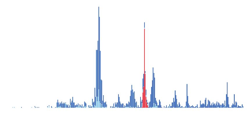
Oh MD, et al. MERS outbreak in South Korea

the Samsung Medical Center (SMC), a university-affil-         Table 1. Demographic and epidemiological characteristics of
iated tertiary hospital in Seoul on May 17 [9]. An infec-     186 patients with MERS-CoV infection [17]
tious disease physician suspected MERS pneumonia              Characteristic                                  No. (%)
and requested a laboratory test from the Korea Centers
                                                              Age, yr
for Disease Control (CDC). MERS-CoV was confirmed
                                                                ≤9                                            0
on May 20.
                                                                10–19                                         1 (0.5)
                                                                20–29                                        13 (7.0)
The chain of transmission/hospital to hospital trans-
                                                                30–39                                        26 (14.0)
mission
                                                                40–49                                        29 (15.6)
The chain of transmission was identified in all 186
                                                                50–59                                        42 (22.6)
MERS patients [13,14]. Of the 86 healthcare institutions
                                                                60–69                                        36 (19.4)
that had taken in MERS-CoV-infected patients, noso-
                                                                70–79                                        30 (16.1)
comial transmission occurred in 12 hospitals and four
                                                                ≥ 80                                          9 (4.8)
clinics (plus two ambulances) [13,15,16]. The epidemio-
                                                              Sex
logical characteristics are summarized in Table 1 [17].
                                                                Male                                        111 (59.7)
The epidemic curve is shown in Fig. 2.
                                                                Female                                       75 (40.3)
                                                              Exposure category
The first wave of the epidemic: outbreak at PTSM
                                                                Patients                                     82
Hospital
                                                                Family member/visitors                       63
During his admission from May 15 to 17 at PTSM Hospi-
                                                                Doctors                                        8
tal, the first patient was unknowingly spreading MERS-
                                                                Nurses                                        15
CoV. Patients, visitors, and healthcare workers (HCWs)
                                                                Paid care givers                               8
were exposed to MERS-CoV. Therefore, by May 20,
                                                                Technologist at Radiology Department           2
when the Korea CDC noticed the importation of MERS-
                                                                Ambulance attendants                           2
CoV into the Republic of Korea, individuals staying on
                                                                Security guards                                2
the same floor as the first case had already been exposed
to MERS-CoV. As a result, a total of 36 MERS cases oc-          Patient transporter                            1

curred at PTSM Hospital. Of them, 26 patients devel-            Employee at IT Department                      1

oped symptoms within 14 days after exposure, suggest-           Exposure category uncertain                    2

ing they were first-generation cases; the remaining 10        Place of exposure
patients may be second-generation cases [7,10].                 Hospitals/clinics                           172 (92.5)
   The mode of transmission of MERS-CoV at PTSM                 Home                                          2 (1.1)
Hospital remains uncertain. In addition to family mem-          Either hospital or home                       6 (3.2)
bers of the first patient, only one patient shared the same     Ambulance                                     3 (1.6)
room (#8104) with him; all the other cases stayed in oth-       Uncertain                                     3 (1.6)
er rooms, which were located > 2 m apart from room            Generation of transmission
#8104. All patient rooms were equipped with an air ven-         1st (index case)                              1 (0.5)
tilating system except for room #8104. A recent study           2nd                                          28 (15.1)
using a multi-agent modeling framework suggested the            3rd                                         120 (64.4)
transmission occurred via a long-range airborne route           4th                                          26 (14.0)
or via a combination of a long-range airborne route and         Either 2nd, 3rd, or 4th                       8 (4.4)
direct contact transmission [18]. A chest computed to-          Uncertain                                     3 (1.6)
mography (CT) scan taken in the early stages (day 1 or        MERS-CoV, Middle East respiratory syndrome coronavirus;
2) of illness showed very small, ground glass nodules in      IT, information technology.
the periphery of the lungs, suggesting inhalation of air-
borne particles [19].

https://doi.org/10.3904/kjim.2018.031                                                          www.kjim.org             235
The Korean Journal of Internal Medicine Vol. 33, No. 2, March 2018
20
Index patient Hospital C Hospital H Hospital M, N Others
15
No. of cases
10
5
0
11 12 13 14 15 16 17 18 19 20 21 22 23 24 25 26 27 28 29 30 31 1 2 3 4 5 6 7 8 9 10 11 12 13 14 15 16 17 18 19 20 21 22 23 24 25 26 27 28 29 30 1
A
15 Hospital C Secondary Tertiary Quaternary
10
5
0
15 Hospital H
No. of cases
10
5
0
15 Hospital M, N
10
5
0
11 12 13 14 15 16 17 18 19 20 21 22 23 24 25 26 27 28 29 30 31 1 2 3 4 5 6 7 8 9 10 11 12 13 14 15 16 17 18 19 20 21 22 23 24 25 26 27 28 29 30 1
B May June July
Date of symptom onset
Figure 2. Epidemiological curve for the 2015 Middle East respiratory syndrome coronavirus (MERS-CoV) outbreak in the Re-
public of Korea. (A) Overall epidemiologic curve by date of symptom onset. Red color denotes the index patient. (B) Epidemic
curves for each of the three main clusters. Outbreaks at Hospital C, Hospital H, and Hospital M and N are depicted in yellow,
green, and purple color, respectively. Adapted from Korea Centers for Disease Control and Prevention [7].
The index case for the outbreak at the SMC an estimated 683 visitors, and 218 HCWs as contacts of
The index case (superspreader-1) of the outbreak at the superspreader-1. Of them, 278 were quarantined and 617
SMC was a 35-year-old man who had been exposed to were under active monitoring. As a result of exposure
MERS-CoV at PTSM Hospital during May 15 to 17 [19,20]. to superspreader-1, 82 had laboratory-confirmed MERS-
He developed a fever on May 21. Despite antibiotic treat- CoV infection, and an additional 10 were secondarily in-
ment, his pneumonia became aggravated and tachypnea fected from them [20,21].
developed, so he eventually visited the emergency room
(ER) at SMC on May 27. During his 58-hour stay at the The outbreaks in Daejeon: Daechung & Geonyang
ER, he experienced vigorous coughing and watery diar- Hospitals
rhea. On May 29, the Korea CDC found he was staying The index case (superspreader-2) for the Daejeon (100
there. A laboratory test confirmed MERS-CoV infection km south from Pyeontaek city) outbreak was infected
and he was transferred to the Seoul National University at PTSM Hospital. He developed symptoms on May 20
Hospital, a government-designated isolation facility on and was admitted to Daechung Hospital on May 22. As
May 30. his pneumonia progressed despite the treatments, he
was transferred to Geonyang Hospital on May 28. A total
The second wave of the epidemic: the outbreak at SMC of 25 secondary cases (14 in Daechung Hospital and 11
Many individuals had been exposed to the superspread- in Geonyang Hospital) were detected. Although super-
er-1 at the ER, and contact tracing identified 675 patients, spreader-2 caused the two hospital outbreaks, the me-
236 www.kjim.org https://doi.org/10.3904/kjim.2018.031Oh MD, et al. MERS outbreak in South Korea
dian time of incubation (8.8 days vs. 4.6 days), secondary ary infections, and the transmission potential decreased
attack rates (4.7% vs. 3.0%), and case fatality rate (28.6% sharply in subsequent generations [30,33]. A total of 83%
vs. 63.6%) were different between the two outbreaks. The of transmission events were due to five superspreaders,
high fatality rate was associated with pre-existing pneu- and 44% of the 186 MERS cases were in-patients who
monia or poor underlying pulmonary function [22-24]. had been exposed within the hospitals [7]. The spreaders
transmitted MERS-CoV from days 1 to 11 of their illness
One patient traveled to China (median, 7 days), and the number of patients infected by
A 44-year-old male, who had been exposed to the first each spreader ranged from 1 to 84 (interquartile range,
index case on May 16, developed back pain on May 21. 1 to 12) [27].
He is a son of the third MERS case. His sister had also The median incubation period and serial interval was
been exposed to MERS-CoV and was confirmed to have 7 days and 12.5 days, respectively [7,34]. R0 ranged from
an MERS-CoV infection on May 25. On May 26, against 2.5 to 7.2, higher than the previous estimate of < 1 [35]. It
medical advice, he flew to Hong Kong and subsequently was estimated at 4.04 for the outbreak cluster in PTSM
travelled to Huizhou through an entry point in Shen- Hospital, and 5.0 for the outbreak in SMC [36]. A mathe-
zhen City, Guangdong Province. The Korea CDC noti- matical modeling study demonstrated that although R0
fied the WHO of his travel, and the Chinese health au- < 1 overall, cluster sizes of over 150 cases are not unex-
thority found him on May 27 and immediately isolated pected for MERS-CoV infection [37].
him. He was confirmed to have MERS-CoV infection on Compared with the non-spreaders, the spreaders had
May 29. The Chinese authority identified 78 close con- a higher frequency of fever (≥ 38.5°C) and chest infiltrates
tacts. After 14 days of quarantine, none of the contacts in more than three lung zones, and longer non-isolat-
presented symptoms and all tested negative for MERS- ed in-hospital days [27]. The spreaders had higher viral
CoV [25,26]. load in sputum samples with the cycle thresholds for
the upE and ORF1a genes of 22.7 vs. 27.2 and 23.7 vs. 27.9,
Epidemiological characteristics respectively. The spreaders with more than three trans-
Epidemiological characteristics have been reported by missions had higher numbers of contacts and ER visits
several groups (Table 2) [7,16,20,21,23,27-30]. The defin- [38]. A brief exposure of a 10-minute stay and 2 minutes
ing epidemiological characteristics were superspreading of talking was enough for the transmission of MERS-
events at hospitals [30-32]. Early superspreading events CoV [39].
generated a disproportionately large number of second-
Table 2. Epidemiological findings and clinical outcomes of 2015 MERS-CoV outbreak in the Republic of Korea
Incubation time: 2–14 days
Infectious period: 1–11 day of illness onset
Duration of fever: 8 days (median)
Symptom onset to rRT-PCR (–) conversion: 17 days (median)
Five superspreaders infected 83% of cases
Pneumonia (CXR infiltrates): 80.8% of the laboratory confirmed MERS-CoV (+) patients
Pneumonia progressed suddenly at around day 7 of illness onset
Symptom onset to mechanical ventilation: 9 days (median)
Mechanical ventilation required in 24.5% of the laboratory confirmed MERS-CoV (+) patients
Symptom onset to death: 14 days (median)
Case fatality ratio: 20.4% (38/186)
MERS-CoV, Middle East respiratory syndrome coronavirus; rRT-PCR, real-time reverse transcription polymerase chain reac-
tion; CXR, chest X-ray.
https://doi.org/10.3904/kjim.2018.031 www.kjim.org 237The Korean Journal of Internal Medicine Vol. 33, No. 2, March 2018
VIROLOGY ease severity. In a serologic study of 42 MERS patients,
the seroconversion rate was 0% in asymptomatic infec-
Characterization of the MERS-CoV isolates tion, 60.0% in symptomatic infection without pneumo-
The whole genome sequences of MERS-CoV isolated nia, 93.8% in pneumonia without respiratory failure, and
from the Korea outbreak were reported [26,40-43]. The 100% in pneumonia with respiratory failure [53]. The se-
virus most similar to the Korean isolates was a camel vi- rological response remained detectable for > 12 months
rus (Camel/Riyadh/Ry159/2015) that belonged to lineage in all survivors (11/11) who had severe disease. Antibody
5, a recombinant of lineage 3 and 4 [44,45]. Viruses from titers were not detectable in four of six patients who had
lineage 5 had been predominant in Saudi Arabian cam- mild pneumonia, suggesting that MERS-CoV seroepi-
els since November 2014, but human viruses of this lin- demiological studies may underestimate the extent of
eage were reported from February 2015 [44]. A phyloge- mild and asymptomatic infection [54].
netic study of spike glycoprotein genes also showed the
Korean strains were closely related to the viral strains
from Riyadh, Saudi Arabia [46]. In a molecular study, 11 CLINICAL FEATURES
of 13 MERS-CoV genome had an I529T mutation and
one D510G mutation in the receptor binding domain Clinical manifestations
of viral spike protein, resulting in reduced affinity to The Korean Society of Infectious Diseases compiled the
the human cognate receptor, CD26 [47]. Heterogeneity clinical data of the 186 MERS patients [55]. The median
analysis revealed the combined frequency of I529T and age was 55 years (range, 16 to 86). The male to female
D510G was high (87.7%), although the frequency of both ratio was 3:2. The most common coexisting medical
mutations varied greatly among specimens [48]. conditions were hypertension (31.7%), diabetes (18.8%),
Another genome sequence study also reported dele- solid organ malignancy (13.4%), and chronic lung dis-
tions of 414 and 419 nucleotides between ORF5 and the E ease (10.2%). Patients with MERS-CoV infection devel-
protein, suggesting that the virus might be defective in oped a spectrum of illnesses, ranging from asymptom-
its ability to package MERS CoV [49]. A microevolution atic to fulminant pneumonia with fatal outcome. The
study found no evidence of changes in the evolutionary respiratory symptoms were similar to other acute viral
rate [43]. However, whether the transmissibility and vir- respiratory infections. At the time of presentation, fever
ulence of the Korean isolates are different from those of had developed in 81.7%, cough in 56.7%, and sputum in
the previous isolates from the Kingdom of Saudi Arabia 39.8% of patients. Symptoms of upper respiratory-tract
remains to be determined by phenotypic assays. infections were infrequent: sore throat was present in
9.1% and rhinorrhea in 1.6% of patients. Gastrointesti-
Kinetics of virus shedding nal symptoms were also observed: diarrhea was present
A viral shedding study showed that the copies of MERS- in 19.4%, nausea or vomiting in 14.0%, and abdominal
CoV RNA detected by real-time reverse transcription pain in 8.1% of patients. Diarrhea may be due to the side
polymerase chain reaction (rRT-PCR) in respiratory effect of lopinavir/ritonavir, as 87% of the patients re-
samples peaked during week 2, and the median value ceived antiviral drugs for MERS-CoV treatment [55]. In
was 7.21 log10 in the severe group and 5.54 log10 in mild the early stages of illness, cough and sputum were not
cases [50]. The study also showed that viral titers were prominent even in patients who later developed overt
higher in throat samples than in nasopharyngeal sam- pneumonia [56]. At admission, 68% (123/180) of patients
ples. Another study also demonstrated higher viral loads had abnormalities on chest radiographs, while 80.8%
were associated with severity and mortality [51]. (147/182) of them developed the abnormalities during
the course of the disease [55]. Sudden progression of
Antibody response kinetics pneumonia occurred around day 7 of illness [50,57]. Pre-
After MERS-CoV infection, antibody responses devel- dictive factors for development of pneumonia included
oped by the third week of illness in most patients [52]. older age, high fever, thrombocytopenia, lymphopenia,
Seroconversion rates increased with the increase of dis- C-reactive protein (CRP) ≥ 2 mg/dL, and a high viral load
238 www.kjim.org https://doi.org/10.3904/kjim.2018.031Oh MD, et al. MERS outbreak in South Korea
in sputum (threshold cycle value of rRT-PCR < 28.5) [58]. Laboratory diagnosis of MERS-CoV
Forty-five patients (24.5%) were treated with mechanical The guidelines for the molecular diagnosis of MERS-
ventilation and 15 patients (8.2%) needed extracorporeal CoV infection have been published by the Korean So-
membrane oxygenation [55]. The typical course of pneu- ciety for Laboratory Medicine [63,64]. Specimen type
monia is shown in Fig. 3. and quality is important in the laboratory diagnosis of
Acute kidney injury (AKI) developed in 14.0% of the pa- MERS-CoV infection. Lower respiratory-tract samples,
tients, 42.7% (44/103) had proteinuria, and 35.0% (36/103) such as sputum and tracheal aspirates, have higher viral
had hematuria. Fifteen patients were treated with renal loads than upper respiratory-tract samples [50]. How-
replacement therapy [55]. Old age is an independent risk ever, MERS patients may not shed the virus during the
factor for the occurrence of AKI [59]. Neuromuscular early stage of their illness. Therefore, initial negative re-
manifestations were not uncommon; hypersomnolence, sults should not rule out the possibility of MERS [65],
weakness and tingling in the extremities were reported and patients suspected of having MERS-CoV infection
during the treatment of MERS, suggesting Guillain-Bar- should be retested using a lower respiratory-tract sam-
ré syndrome or virus-related sensory neuropathy [60]. ple [63,66]. When sputum cannot be obtained, throat
swabs may be an alternative source of diagnostic sam-
Radiological findings ples [50]. Special attention should be given to diagnos-
Serial changes in chest radiographs was reported in five ing MERS-CoV infection in immunocompromised pa-
patients [56]. Chest CT scans revealed rapidly developed tients, as they may present with atypical features, such as
multifocal nodular consolidations with ground-glass a longer incubation period, a longer period from initial
opacity halo and mixed consolidation, mainly in the de- PCR positivity to symptom onset, and persistent viral
pendent and peripheral areas [61]. shedding [67].
Laboratory findings Antiviral treatment
There are few laboratory findings specific to MERS-CoV An antiviral treatment guideline has been published by
infection, although monocytosis with normal white the writing committee with support from the Korean
blood cell count and low CRP level were more common Society of Infectious Diseases and the Korean Society for
in MERS patients at initial presentation [57,62]. Chemotherapy [68]. The guideline recommended a tri-
25 25
20 20
Chest radiograph score
Chest radiograph score
15 15
10 10
5 5
0 0
1 3 5 7 9 11 13 15 17 19 21 23 25 27 1 3 5 7 9 11 13 15 17 19 21 23 25 27
Days after symptom onset Days after symptom onset
A B
Figure 3. Progression of Middle East respiratory syndrome (MERS) pneumonia. (A) Severe cases. (B) Mild cases. In severe cases,
pneumonia progressed suddenly around 7 days after symptom onset. Adapted from Oh et al., with permission from Massa-
chusetts Medical Society [50].
https://doi.org/10.3904/kjim.2018.031 www.kjim.org 239The Korean Journal of Internal Medicine Vol. 33, No. 2, March 2018
Live Live
30 Death 30 Death
25 25
2
20 20
No. of cases
No. of cases
4
15 4 15 8 7
25
10 10
4 1
12 21 20 15 11 12
5 13 7 5 6
8
1 1 1 1
2
0 0
10–19 20–9 30–39 40–49 50–59 60–69 70–79 ≥ 80 10–19 20–29 30–39 40–49 50–59 60–69 70–79 ≥ 80
A Age (yr)
B Age (yr)
Figure 4. Case fatality ratio of Middle East respiratory syndrome during the 2015 Korea outbreak. Case fatality ratio was 10.1%
(11/109) in patients without underlying diseases (A), and 35.1% (27/77) with underlying diseases (B). Overall case fatality ratio was
20.4% (38/186).
ple combination regimen of type 1 interferon, ribavirin, 35 weeks of gestational age developed dyspnea and
and lopinavir/ritonavir for 10 to 14 days. However, the her chest radiograph showed diffuse infiltrates in the
guideline was mostly based on expert opinions and fur- left lower lung zone. She recovered from the infection
ther study for the optimal antiviral treatment in MERS and delivered a healthy full-term baby without vertical
is warranted. MERS-CoV transmission [74].
A patient developed organizing pneumonia 7 days af-
Risk factors for mortality and prognosis ter the resolution of MERS-CoV infection. He was suc-
The median duration of fever was 8 days. The median cessfully treated with corticosteroids [75].
time to negative conversion of MERS-CoV in sputum In a mental health study, 7.6% of 1,656 patients who
samples by rRT-PCR was 17 days (range, 4 to 45) [55]. were quarantined showed symptoms of anxiety, and
The median time from symptom onset to death was 6.4% reported feelings of anger during the 2 weeks of
14 days. The case fatality rate was 20.4% (38/186) (Fig. 4). quarantine [76]. Mental health support, accurate in-
The in-hospital mortality, 7-day mortality, and 28-day formation, and appropriate supplies, including food,
(from symptom onset) mortality were 19.4% (36/186), clothes, and accommodation, should be provided to iso-
3.8% (7/186), and 17.7% (33/186), respectively [55]. lated persons [76]. A case of possible transfusion-related
Host factors associated with mortality were old age (> acute lung injury following transfusion of convalescent
60 years), smoking history, pre-existing pneumonia, ab- plasma was also reported [77].
normal renal function, and comorbid conditions [24,69-
71]. Low albumin, altered mentality, and high pneumo-
nia severity index score at admission were risk factors INFECTION CONTROL AND PREVENTION
for mortality [69]. MERS-CoV RNA in blood samples
was detected in 33% (7/21) of patients at presentation, Guidelines for infection control and prevention
and it was associated with a worse clinical outcome [72]. A comprehensive “MERS infection prevention and
A shorter incubation period was also associated with an control guideline for healthcare facilities” was drafted
increased risk of death [24,73]. by the guideline development committee with gener-
A pregnant woman infected with MERS-CoV in her ous support from Korean Academic Societies [78]. The
240 www.kjim.org https://doi.org/10.3904/kjim.2018.031Oh MD, et al. MERS outbreak in South Korea
guideline included practical aspects of infection control Healthcare workers and MERS-CoV infection
and prevention based on the experience from the Korea There was a case of a 39-year-old female nurse who was
outbreak, such as the composition of members for the infected with MERS-CoV during a cardiopulmonary re-
MERS emergency committee, a space plan for an ante- suscitation lasting 1 hour for a MERS patient who had
room for donning and doffing, isolation of in-patients pneumonia and hemoptysis [88]. Serological surveillance
and HCWs in a hospital affected by an outbreak, and conducted after the MERS outbreak for asymptomatic
management of the deceased and autopsy [78]. infection among HCWs involved in the direct care of
The hemodialysis unit may become an epicenter for MERS patients showed that 0.3% (2/737) of them were
MERS-CoV outbreak because hemodialysis patients MERS-CoV IgG positive by an indirect immunofluores-
must receive renal replacement therapy even during cent assay. Among the HCWs who did not use appropri-
the outbreak, and the risk of exposure to MERS-CoV ate personal protective equipment (PPE), seropositivity
continues. During the Korea outbreak, a hemodialysis was 0.7% (2/294) compared with 0% (0/443) in HCWs
unit was found to have a MERS patient, and a total of with appropriate PPE use [89]. Another serological sur-
104 patients and 18 HCWs were exposed to MERS-CoV. vey also showed that none of the 285 HCWs were posi-
With support from the Korean Society of Nephrology, tive for MERS-CoV immunoglobulin G, although 38.2%
the dialysis unit could continue to operate while the (109/285) of the HCWs reported experiencing MERS-like
exposed patients were isolated in the unit. Fortunately, symptoms while caring for the MERS patients [90]. In a
no further transmission occurred at the unit [79]. After third study, 0 of 189 HCWs showed seroconversion by
this successful experience, the Society published a clini- a plaque reduction neutralization test, although 20% to
cal practice guideline for hemodialysis facilities dealing 25% of HCWs reported MERS-like symptoms [91].
with MERS patients [80]. During the outbreak in SMC, all HCWs assigned to
The Korea outbreak was driven by the superspreaders MERS patients were screened for MERS-CoV, regard-
who visited multiple healthcare facilities; thus, generat- less of the presence or absence of symptoms. Of the 591
ing a large number of secondary cases. Limiting unnec- HCWs, three (0.5%) asymptomatic HCWs (two nurses
essary contacts with patients with respiratory symptoms and one physician) were found to be MERS-CoV (+), but
in healthcare settings, especially in emergency depart- they did not transmit the virus to others [21]. In anoth-
ments, is of critical importance [33]. er study, an asymptomatic nurse without PPE contacted
82 HCWs, including 33 close contacts who were exposed
Environmental contamination within 2 m from the index nurse and were not using
MERS-CoV could survive for longer than 48 hours at PPE. None of the exposed HCWs were infected [92].
20°C and 40% of relative humidity, suggesting contact However, the potential for transmission from asymp-
or fomite transmission might occur in healthcare set- tomatic rRT-PCR positive individuals is still unknown.
tings [81]. MERS-CoV was detected by rRT-PCR in speci- Therefore, asymptomatic HCWs who are rRT-PCR-pos-
mens taken from the medical equipment [82-84]. MERS- itive for MERS-CoV should be isolated and should not
CoV was also isolated by cell culture of the air and swab return to work until two consecutive respiratory-tract
samples taken from a MERS isolation unit, suggesting samples test negative on rRT-PCR [93].
extensive contamination of the isolation unit [85,86]. Of
the 186 cases, 23% were infected by undocumented con- Control of the outbreak by private-public collaboration
tact between cases (i.e., indirect transmission of MERS- The government requested the Korean Society of Infec-
CoV via environmental contact) [87]. Therefore, fomites tious Diseases to participate in the control of the MERS
with possible MERS-CoV contamination should be san- outbreak. On June 8, 2015, the Society organized the
itized, and a minimum room ventilation rate of six air Rapid Response Team, consisting of 15 infectious-dis-
changes per hour should be implemented to minimize ease doctors and two infection-control professionals
recirculation of pathogen-bearing droplets. Meticulous [94]. Critical suggestions to prevent future epidemics
environmental cleaning may be important for prevent- were made regarding a rapid alerting system for index
ing transmission in healthcare settings. cases, provision of a sufficient airborne infection isola-
https://doi.org/10.3904/kjim.2018.031 www.kjim.org 241The Korean Journal of Internal Medicine Vol. 33, No. 2, March 2018
Table 3. Lessons learned from the 2015 outbreak of MERS-CoV in the Republic of Korea
A single, missed case may trigger a huge, nationwide outbreak
The first line of defense is not the thermal scanner at the airport. It is doctors in the community clinics/hospitals.
Due to sudden deterioration of pneumonia around day 7 of illness, patients may visit emergency department, or be admitted
to intensive care unit.
Superspreading events may occur in healthcare settings, especially at the emergency department.
Patients may transmit MERS-CoV as early as 2 days after symptom onset. Early detection and isolation is of critical impor-
tance.
Aggressive strategy for quarantine maybe necessary, especially when large number of individuals are exposed in the health-
care settings.
Huge socioeconomic impact with an estimated 8.5 billion US dollars
MERS-CoV, Middle East respiratory syndrome coronavirus.
tion facility, education and training of HCWs for im- REFERENCES
portant infectious diseases, and overcrowding and visi-
tor control in the hospital [95]. 1. Zaki AM, van Boheemen S, Bestebroer TM, Osterhaus
AD, Fouchier RA. Isolation of a novel coronavirus from
a man with pneumonia in Saudi Arabia. N Engl J Med
CONCLUSIONS 2012;367:1814-1820.
2. World Health Organization. Middle East respiratory
The 2015 Korea outbreak was the largest outbreak out- syndrome coronavirus (MERS-CoV) [Internet]. Geneva:
side of the Middle East. Researchers had a unique op- World Health Organization, c2018 [cited 2018 Feb 8].
portunity to compare the nature of MERS-CoV infection Available from: http://who.int/emergencies/mers-cov/en/.
with the Middle East experience. The outbreak unveiled 3. Ministry of Health Kingdom of Saudi Arabia. MERS-
the weak points of infrastructure in our medical system, CoV daily update [Internet]. Riyadh (SA): Ministry of
especially of preparedness for emerging global infec- Health Kingdom of Saudi Arabia, c2014 [cited 2018 Feb
tious diseases. Nosocomial transmission was one of the 8]. Available from: https://www.moh.gov.sa/en/CCC/
core features of MERS-CoV infection. The introspec- PressReleases/Pages/statistics-2018-01-27-001.aspx.
tion for loose medical referral systems, overcrowding 4. Arabi YM, Balkhy HH, Hayden FG, et al. Middle East
at the ER, a lack of expert resources and infection con- respiratory syndrome. N Engl J Med 2017;376:584-594.
trol infrastructure, and lack of organized preparedness 5. Hui DS, Azhar EI, Kim YJ, Memish ZA, Oh MD, Zumla
for medical crises prompted the government to reform A. Middle East respiratory syndrome coronavirus: risk
the healthcare system, and healthcare sectors to invest factors and determinants of primary, household, and
further in infectious diseases and infection control. Al- nosocomial transmission. Lancet Infect Dis. 2018 In Press.
though the improvement is still ongoing, the speed and 6. Public Health England. Treatment of MERS-CoV:
content are still currently insufficient. International co- information for clinicians. Clinical decision-making
operation to prepare for and defeat emerging infectious support for treatment of MERS-CoV patients [Internet].
diseases should also be emphasized. Lessons we learned London (UK): Public Health England, 2017 [cited 2018
from the outbreak are summarized in Table 3. Feb 8]. Available from: https://www.gov.uk/government/
uploads/system/uploads/attachment_data/file/638628/
Conflict of interest MERS_CoV_guidance_for_clinicians.pdf.
No potential conflict of interest relevant to this article 7. Korea Centers for Disease Control and Prevention.
was reported. Middle East respiratory syndrome coronavirus outbreak
in the Republic of Korea, 2015. Osong Public Health Res
Perspect 2015;6:269-278.
242 www.kjim.org https://doi.org/10.3904/kjim.2018.031Oh MD, et al. MERS outbreak in South Korea
8. Oh MD. The Korean Middle East respiratory syndrome Air 2018;28:51-63.
coronavirus outbreak and our responsibility to the global 19. Oh MD, Choe PG, Oh HS, et al. Middle East respiratory
scientific community. Infect Chemother 2016;48:145-146. syndrome coronavirus superspreading event involving 81
9. Lee JY, Kim YJ, Chung EH, et al. The clinical and persons, Korea 2015. J Korean Med Sci 2015;30:1701-1705.
virological features of the first imported case causing 20. Cho SY, Kang JM, Ha YE, et al. MERS-CoV outbreak
MERS-CoV outbreak in South Korea, 2015. BMC Infect following a single patient exposure in an emergency
Dis 2017;17:498. room in South Korea: an epidemiological outbreak study.
10. Kim KM, Ki M, Cho SI, et al. Epidemiologic features of Lancet 2016;388:994-1001.
the first MERS outbreak in Korea: focus on Pyeongtaek 21. Park GE, Ko JH, Peck KR, et al. Control of an outbreak of
St. Mary's Hospital. Epidemiol Health 2015;37:e2015041. Middle East respiratory syndrome in a tertiary hospital in
11. Yang JS, Park S, Kim YJ, et al. Middle East respiratory Korea. Ann Intern Med 2016;165:87-93.
syndrome in 3 persons, South Korea, 2015. Emerg Infect 22. Park SH, Kim YS, Jung Y, et al. Outbreaks of Middle East
Dis 2015;21:2084-2087. respiratory syndrome in two hospitals initiated by a
12. Park YS, Lee C, Kim KM, et al. The first case of the 2015 single patient in Daejeon, South Korea. Infect Chemother
Korean Middle East respiratory syndrome outbreak. 2016;48:99-107.
Epidemiol Health 2015;37:e2015049. 23. Park JW, Lee KJ, Lee KH, et al. Hospital outbreaks of
13. FluTrackers. South Korea coronavirus MERS case list: Middle East respiratory syndrome, Daejeon, South Korea,
including imported and exported cases. Ministry of Health 2015. Emerg Infect Dis 2017;23:898-905.
& WHO confirmed data only: 2015 outbreak [Internet]. 24. Nam HS, Park JW, Ki M, Yeon MY, Kim J, Kim SW. High
Winter Park (FL): FluTrackers, c2018 [cited 2018 Feb 8]. fatality rates and associated factors in two hospital
Available from: https://flutrackers.com/forum/forum/novel- outbreaks of MERS in Daejeon, the Republic of Korea.
coronavirus-ncov-mers-2012-2014/novel-coronavirus-who- Int J Infect Dis 2017;58:37-42.
chp-wpro-ecdc-oie-fao-moa-reports-and-updates/south- 25. Wu J, Yi L, Zou L, Zhong H, et al. Imported case of MERS-
korea-coronavirus/732065-south-korea-coronavirus-mers- CoV infection identified in China, May 2015: detection
case-list-including-imported-and-exported-cases-ministry- and lesson learned. Euro Surveill 2015;20:21158.
of-health-who-confirmed-data-only-2015-outbreak. 26. Wang Y, Liu D, Shi W, et al. Origin and possible genetic
14. Korea Broadcasting System Digital News. MERS-CoV recombination of the Middle East respiratory syndrome
infection: current status-interactive and infographics coronavirus from the first imported case in china: phylo-
[Internet]. KBS Digital News, c2013 [cited 2018 Feb 8]. genetics and coalescence analysis. MBio 2015;6:e01280-15.
Available from: http://dj.kbs.co.kr/resources/2015-06-08/. 27. Kang CK, Song KH, Choe PG, et al. Clinical and epidemi-
15. Lee SS, Wong NS. Probable transmission chains of ologic characteristics of spreaders of Middle East respi-
Middle East respiratory syndrome coronavirus and the ratory syndrome coronavirus during the 2015 outbreak in
multiple generations of secondary infection in South Korea. J Korean Med Sci 2017;32:744-749.
Korea. Int J Infect Dis 2015;38:65-67. 28. Park SH, Kim WJ, Yoo JH, Choi JH. Epidemiologic pa-
16. Kim KH, Tandi TE, Choi JW, Moon JM, Kim MS. rameters of the Middle East respiratory syndrome out-
Middle East respiratory syndrome coronavirus (MERS- break in Korea, 2015. Infect Chemother 2016;48:108-117.
CoV) outbreak in South Korea, 2015: epidemiology, 29. Park HY, Lee EJ, Ryu YW, et al. Epidemiological investi-
characteristics and public health implications. J Hosp gation of MERS-CoV spread in a single hospital in South
Infect 2017;95:207-213. Korea, May to June 2015. Euro Surveill 2015;20:1-6.
17. Ministry of Health and Welfare. The 2015 MERS outbreak 30. Chowell G, Abdirizak F, Lee S, et al. Transmission charac-
in the Republic of Korea: learning from MERS (The teristics of MERS and SARS in the healthcare setting: a
White Paper). Seoul (KR): Ministry of Health and Welfare, comparative study. BMC Med 2015;13:210.
2016:473. 31. Chun BC. Understanding and modeling the super-spread-
18. Xiao S, Li Y, Sung M, Wei J, Yang Z. A study of the ing events of the Middle East respiratory syndrome out-
probable transmission routes of MERS-CoV during the break in Korea. Infect Chemother 2016;48:147-149.
first hospital outbreak in the Republic of Korea. Indoor 32. Ki M. 2015 MERS outbreak in Korea: hospital-to-hospital
https://doi.org/10.3904/kjim.2018.031 www.kjim.org 243The Korean Journal of Internal Medicine Vol. 33, No. 2, March 2018
transmission. Epidemiol Health 2015;37:e2015033. 2016;22:327-330.
33. Nishiura H, Endo A, Saitoh M, et al. Identifying 44. Sabir JS, Lam TT, Ahmed MM, et al. Co-circulation of
determinants of heterogeneous transmission dynamics three camel coronavirus species and recombination of
of the Middle East respiratory syndrome (MERS) MERS-CoVs in Saudi Arabia. Science 2016;351:81-84.
outbreak in the Republic of Korea, 2015: a retrospective 45. Kim JI, Kim YJ, Lemey P, et al. The recent ancestry of
epidemiological analysis. BMJ Open 2016;6:e009936. Middle East respiratory syndrome coronavirus in Korea
34. Virlogeux V, Fang VJ, Park M, Wu JT, Cowling BJ. has been shaped by recombination. Sci Rep 2016;6:18825.
Comparison of incubation period distribution of human 46. Kim DW, Kim YJ, Park SH, et al. Variations in spike
infections with MERS-CoV in South Korea and Saudi glycoprotein gene of MERS-CoV, South Korea, 2015.
Arabia. Sci Rep 2016;6:35839. Emerg Infect Dis 2016;22:100-104.
35. Zhang XS, Pebody R, Charlett A, et al. Estimating and 47. Kim Y, Cheon S, Min CK, et al. Spread of mutant Middle
modelling the transmissibility of Middle East respiratory East respiratory syndrome coronavirus with reduced
syndrome coronavirus during the 2015 outbreak in affinity to human CD26 during the South Korean
the Republic of Korea. Influenza Other Respir Viruses outbreak. MBio 2016;7:e00019.
2017;11:434-444. 48. Park D, Huh HJ, Kim YJ, et al. Analysis of intrapatient
36. Choi S, Jung E, Choi BY, Hur YJ, Ki M. High reproduction heterogeneity uncovers the microevolution of Middle
number of Middle East respiratory syndrome coronavirus East respiratory syndrome coronavirus. Cold Spring Harb
in nosocomial outbreaks: mathematical modelling in Mol Case Stud 2016;2:a001214.
Saudi Arabia and South Korea. J Hosp Infect 2017 Sep 25 49. Xie Q, Cao Y, Su J, et al. Two deletion variants of Middle
[Epub]. https://doi.org/10.1016/j.jhin.2017.09.017. East respiratory syndrome coronavirus found in a patient
37. Kucharski AJ, Althaus CL. The role of superspreading in with characteristic symptoms. Arch Virol 2017;162:2445-
Middle East respiratory syndrome coronavirus (MERS- 2449.
CoV) transmission. Euro Surveill 2015;20:14-18. 50. Oh MD, Park WB, Choe PG, et al. Viral load kinetics of
38. Kim SW, Park JW, Jung HD, et al. Risk factors for MERS coronavirus infection. N Engl J Med 2016;375:1303-
transmission of Middle East respiratory syndrome 1305.
coronavirus infection during the 2015 outbreak in South 51. Min CK, Cheon S, Ha NY, et al. Comparative and kinetic
Korea. Clin Infect Dis 2017;64:551-557. analysis of viral shedding and immunological responses
39. Kim T, Jung J, Kim SM, et al. Transmission among in MERS patients representing a broad spectrum of
healthcare worker contacts with a Middle East respiratory disease severity. Sci Rep 2016;6:25359.
syndrome patient in a single Korean centre. Clin Microbiol 52. Park WB, Perera RA, Choe PG, et al. Kinetics of serologic
Infect 2016;22:e11-e13. responses to MERS coronavirus infection in humans,
40. Kim YJ, Cho YJ, Kim DW, et al. Complete genome se- South Korea. Emerg Infect Dis 2015;21:2186-2189.
quence of Middle East respiratory syndrome coronavirus 53. Ko JH, Muller MA, Seok H, et al. Serologic responses of
KOR/KNIH/002_05_2015, isolated in South Korea. Ge- 42 MERS-coronavirus-infected patients according to the
nome Announc 2015;3:e00787-15. disease severity. Diagn Microbiol Infect Dis 2017;89:106-
41. Lu R, Wang Y, Wang W, et al. Complete genome sequence 111.
of Middle East Respiratory syndrome coronavirus 54. Choe PG, Perera RAPM, Park WB, et al. MERS-CoV
(MERS-CoV) from the first imported MERS-CoV case in antibody responses 1 year after symptom onset, South
China. Genome Announc 2015;3:e00818-15. Korea, 2015. Emerg Infect Dis 2017;23:1079-1084.
42. Park WB, Kwon NJ, Choe PG, et al. Isolation of Middle 55. Choi WS, Kang CI, Kim Y, et al. Clinical presentation and
East respiratory syndrome coronavirus from a patient of outcomes of Middle East respiratory syndrome in the
the 2015 Korean outbreak. J Korean Med Sci 2016;31:315- Republic of Korea. Infect Chemother 2016;48:118-126.
320. 56. Rhee JY, Hong G, Ryu KM. Clinical implications of 5
43. Seong MW, Kim SY, Corman VM, et al. Microevolution of cases of Middle East respiratory syndrome coronavirus
outbreak-associated Middle East respiratory syndrome infection in a South Korean outbreak. Jpn J Infect Dis
coronavirus, South Korea, 2015. Emerg Infect Dis 2016;69:361-366.
244 www.kjim.org https://doi.org/10.3904/kjim.2018.031Oh MD, et al. MERS outbreak in South Korea
57. Kim ES, Choe PG, Park WB, et al. Clinical progression 69. Hong KH, Choi JP, Hong SH, et al. Predictors of
and cytokine profiles of Middle East respiratory mortality in Middle East respiratory syndrome (MERS).
syndrome coronavirus infection. J Korean Med Sci Thorax 2018;73:286-289.
2016;31:1717-1725. 70. Majumder MS, Kluberg SA, Mekaru SR, Brownstein
58. Ko JH, Park GE, Lee JY, et al. Predictive factors for JS. Mortality risk factors for Middle East respiratory
pneumonia development and progression to respiratory syndrome outbreak, South Korea, 2015. Emerg Infect Dis
failure in MERS-CoV infected patients. J Infect 2015;21:2088-2090.
2016;73:468-475. 71. Mizumoto K, Endo A, Chowell G, Miyamatsu Y, Saitoh M,
59. Cha RH, Joh JS, Jeong I, et al. Renal complications and Nishiura H. Real-time characterization of risks of death
their prognosis in Korean patients with Middle East associated with the Middle East respiratory syndrome
respiratory syndrome-coronavirus from the central (MERS) in the Republic of Korea, 2015. BMC Med
MERS-CoV designated hospital. J Korean Med Sci 2015;13:228.
2015;30:1807-1814. 72. Kim SY, Park SJ, Cho SY, et al. Viral RNA in blood as
60. K i m J E , He o J H , K i m H O, e t a l . Ne u r o l o g i c a l indicator of severe outcome in Middle East respirato-
complications during treatment of Middle East ry syndrome coronavirus infection. Emerg Infect Dis
respiratory syndrome. J Clin Neurol 2017;13:227-233. 2016;22:1813-1816.
61. Choi WJ, Lee KN, Kang EJ, Lee H. Middle East respiratory 73. Virlogeux V, Park M, Wu JT, Cowling BJ. Association
syndrome-coronavirus infection: a case report of serial between severity of MERS-CoV infection and incubation
computed tomographic findings in a young male patient. period. Emerg Infect Dis 2016;22:526-528.
Korean J Radiol 2016;17:166-170. 74. Jeong SY, Sung SI, Sung JH, et al. MERS-CoV infection
62. Park GE, Kang CI, Ko JH, et al. Differential cell count in a pregnant woman in Korea. J Korean Med Sci
and CRP level in blood as predictors for Middle East 2017;32:1717-1720.
respiratory syndrome coronavirus infection in acute 75. Kim I, Lee JE, Kim KH, Lee S, Lee K, Mok JH. Successful
febrile patients during nosocomial outbreak. J Korean treatment of suspected organizing pneumonia in
Med Sci 2017;32:151-154. a patient with Middle East respiratory syndrome
63. Lee H, Ki CS, Sung H, et al. Guidelines for the laboratory coronavirus infection: a case report. J Thorac Dis
diagnosis of Middle East respiratory syndrome 2016;8:E1190-E1194.
coronavirus in Korea. Infect Chemother 2016;48:61-69. 76. Jeong H, Yim HW, Song YJ, et al. Mental health status of
64. Ki CS, Lee H, Sung H, et al. Korean Society for Laboratory people isolated due to Middle East respiratory syndrome.
Medicine practice guidelines for the molecular diagnosis Epidemiol Health 2016;38:e2016048.
of Middle East respiratory syndrome during an outbreak 77. Chun S, Chung CR, Ha YE, et al. Possible transfusion-
in Korea in 2015. Ann Lab Med 2016;36:203-208. related acute lung injury following convalescent plasma
65. Huh HJ, Ko JH, Kim YE, et al. Importance of specimen transfusion in a patient with Middle East respiratory
type and quality in diagnosing Middle East respiratory syndrome. Ann Lab Med 2016;36:393-395.
syndrome. Ann Lab Med 2017;37:81-83. 78. Kim JY, Song JY, Yoon YK, et al. Middle East respiratory
66. Lee JH, Lee CS, Lee HB. An appropriate lower respiratory syndrome infection control and prevention guideline for
tract specimen is essential for diagnosis of Middle healthcare facilities. Infect Chemother 2015;47:278-302.
East respiratory syndrome (MERS). J Korean Med Sci 79. Moon SY, Son JS, Lee YH, et al. Middle East respiratory
2015;30:1207-1208. syndrome coronavirus transmission in dialysis unit and
67. Kim SH, Ko JH, Park GE, et al. Atypical presentations of infection control interventions in Korea. Infect Control
MERS-CoV infection in immunocompromised hosts. J Hosp Epidemiol 2016;37:1514-1516.
Infect Chemother 2017;23:769-773. 80. Park HC, Lee YK, Lee SH, et al. Middle East respiratory
68. Chong YP, Song JY, Seo YB, Choi JP, Shin HS; Rapid syndrome clinical practice guideline for hemodialysis
Response Team. Antiviral treatment guidelines for facilities. Kidney Res Clin Pract 2017;36:111-116.
Middle East respiratory syndrome. Infect Chemother 81. van Doremalen N, Bushmaker T, Munster VJ. Stability of
2015;47:212-222. Middle East respiratory syndrome coronavirus (MERS-
https://doi.org/10.3904/kjim.2018.031 www.kjim.org 245The Korean Journal of Internal Medicine Vol. 33, No. 2, March 2018
CoV) under different environmental conditions. Euro confirmed MERS patients: incidence and risk factors
Surveill 2013;18:20590. of MERS-CoV seropositivity. Clin Microbiol Infect
82. Bin SY, Heo JY, Song MS, et al. Environmental contam- 2016;22:880-886.
ination and viral shedding in MERS patients during 90. Lee JY, Kim G, Lim DG, et al. Seroprevalence of Middle
MERS-CoV outbreak in South Korea. Clin Infect Dis East respiratory syndrome coronavirus among healthcare
2016;62:755-760. personnel caring for patients with middle east respirato-
83. Oh MD. Environmental contamination and viral ry syndrome in South Korea. Infect Control Hosp Epide-
shedding in MERS patients. Clin Infect Dis 2016;62:1615. miol 2016;37:1513-1514.
84. Song JY, Cheong HJ, Choi MJ, et al. Viral shedding and 91. Ko JH, Lee JY, Baek JY, et al. Serologic evaluation of
environmental cleaning in Middle East respiratory MERS screening strategy for healthcare personnel during
syndrome coronavirus infection. Infect Chemother a hospital-associated outbreak. Infect Control Hosp
2015;47:252-255. Epidemiol 2017;38:234-238.
85. Kim SH, Chang SY, Sung M, et al. Extensive viable 92. Moon SY, Son JS, Lee YH, et al. Middle East respiratory
Middle East respiratory syndrome (MERS) coronavirus syndrome coronavirus transmission in dialysis unit and
contamination in air and surrounding environment in infection control interventions in Korea. Infect Control
MERS isolation wards. Clin Infect Dis 2016;63:363-369. Hosp Epidemiol 2016;37:1514-1516.
86. Oh MD. Transmissibility of Middle East respiratory 93. World Health Organization. Management of asymptomatic
syndrome by the airborne route. Clin Infect Dis persons who are RT-PCR positive for Middle East
2016;63:1143. respiratory syndrome coronavirus (MERS-CoV): Interim
87. Majumder MS, Brownstein JS, Finkelstein SN, Larson guidance [Internet]. Geneva: World Health Organization,
RC, Bourouiba L. Nosocomial amplification of MERS- c2018 [cited 2018 Feb 8]. Available from: http://apps.who.int/
coronavirus in South Korea, 2015. Trans R Soc Trop Med iris/bitstream/10665/180973/1/WHO_MERS_IPC_15.2_eng.
Hyg 2017;111:261-269. pdf?ua=1&ua=1.
88. Nam HS, Yeon MY, Park JW, Hong JY, Son JW. Healthcare 94. Lee J; Rapid Response Team, Kim WJ. Collaborative
worker infected with Middle East Respiratory Syndrome intervention of Middle East respiratory syndrome: rapid
during cardiopulmonary resuscitation in Korea, 2015. response team. Infect Chemother 2016;48:71-74.
Epidemiol Health 2017;39:e2017052. 95. Jeon MH, Kim TH. Institutional preparedness to prevent
89. Kim CJ, Choi WS, Jung Y, et al. Surveillance of the future Middle East respiratory syndrome coronavirus-
Middle East respiratory syndrome (MERS) coronavirus like outbreaks in Republic of Korea. Infect Chemother
(CoV) infection in healthcare workers after contact with 2016;48:75-80.
246 www.kjim.org https://doi.org/10.3904/kjim.2018.031You can also read