2021 ABSTRACT BOOK - Virtual congress & live talk show - OCT Forum 2021
←
→
Page content transcription
If your browser does not render page correctly, please read the page content below
Virtual congress
    & live talk show

APRIL 9 / 23 / 30

      2021

   ABSTRACT BOOK
APRIL 9 / 23 / 30

                                                                            2021
          Virtual congress
          & live talk show
INDEX

AGE-RELATED MACULAR DEGENERATION                                                          4
BROCCOLI-DERIVED COMPOUND INDUCES PROTECTIVE EFFECTS IN AN IN VITRO MODEL OF AGE-RELATED
MACULAR DEGENERATION: AN ‘OMICS’ APPROACH                                                4
SHORT-TERM OUTCOMES OF PATIENTS WITH NEOVASCULAR EXUDATIVE AMD:
THE EFFECT OF COVID-19 PANDEMIC                                                           5
RATE OF MISDIAGNOSIS AND CLINICAL USEFULNESS OF THE CORRECT DIAGNOSIS IN EXUDATIVE
NEOVASCULAR MACULOPATHY SECONDARY TO AMD VS. PACHYCHOROID DISEASE                         6

ANGIOID STREAKS                                                                           7
PROGNOSTIC FACTORS BY MULTIMODAL IMAGING ANGOID STREAKS ASSOCIATED WITH PATTERN
DYSTROPHY IN PSEUDOXANTHOMA ELASTICUM AND NON SYSTEMIC DISEASE PATIENTS                   7

CHALLENGING CASES                                                                         8
C OPTIC NEURITIS AS A POSSIBLE LATE MANIFESTATION OF PREVIOUS COVID-19 INFECTION          8

C MULTIPLE EXUDATIVE CHOROIDAL NODULES RESISTANT TO INMUNOSUPPRESSIVE THERAPY.
   A CHALLENGING DIAGNOSIS                                                                9
CHOROIDAL NEOVASCULAR MEMBRANE SECONDARY TO OPTIC NERVE DRUSEN: A CASE REPORT            10
POSTERIOR UVEITIS, AN UNCOMMON COMPLICATION OF MULTIPLE SCLEROSIS. CASE REPORT           11

DIABETIC RETINOPATHY                                                                     12
CATARACT SURGERY WITH COMBINED VERSUS DEFERRED INTRAVITREAL DEXAMETHASONE
IMPLANT FOR DIABETIC MACULAR EDEMA: LONG-TERM OUTCOMES FROM A REAL-WORLD SETTING         12
THREE-YEAR OCT PREDICTIVE FACTORS OF DISEASE RECURRENCE IN EYES WITH SUCCESSFULLY
TREATED MYOPIC CHOROIDAL NEOVASCULARIZATION                                              13

FUNCTIONAL IMAGING                                                                       14
VOLUME RENDERED OCTA ASSESSMENT OF MACULAR ISCHEMIA IN PATIENTS WITH TYPE
1 DIABETES AND WITHOUT DIABETIC RETINOPATHY                                              14

MACULAR TELANGIECTASIA                                                                   15
ANATOMICAL AND FUNCTIONAL CORRELATION BASED ON MULTIMODAL IMAGING IN PATIENTS
WITH TYPE 2 IDIOPATHIC MACULAR TELANGIECTASIA                                            15
CHOROIDAL VASCULATURE IN PATIENTS WITH MACULAR TELANGIECTASIA                            16

C CASES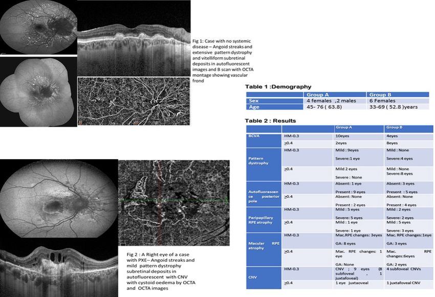
APRIL 9 / 23 / 30

                                                                            2021
       Virtual congress
OPTICAL&COHERENCE   TOMOGRAPHY ANGIOGRAPHY
         live talk show                                                                   17
OCT ANGIOGRAPHY IN A CASE OF TUBEROUS SCLEROSIS COMPLICATED WITH MACULAR
CHOROIDAL NEOVASCOLARIZATION                                                              17
THREE-DIMENSIONAL MODEL OF THE MICROVASCULAR NETWORK OBTAINED BY USING OPTICAL
COHERENCE TOMOGRAPHY ANGIOGRAPHY IN FOVEA PLANA AND NORMAL FOVEAL DEPRESSION.             18
VASCULAR AND STRUCTURAL FINDINGS IN UNAFFECTED FELLOW EYES OF PATIENTS WITH UNILATERAL
PRIMARY OPEN ANGLE GLAUCOMA USING OPTICAL COHERENCE TOMOGRAPHY ANGIOGRAPHY                19
COMPARISON OF AUTOMATED AND MANUAL SEGMENTATION OF SPECTRALIS OCT-ANGIOGRAPHY
EN FACE SLABS FOR THE DETECTION OF CHOROIDAL NEOVASCULARIZATION                           20
OPTICAL COHERENCE TOMOGRAPHY ANGIOGRAPHY OF THE OPTIC DISC AND SURROUNDING
TISSUES FOLLOWING SLOTTED PLAQUE BRACHYTHERAPY FOR CHOROIDAL MELANOMA                     21
RETINAL CHANGES AND ANGIOOCT AS A POSSIBLE BIOMARKER IN THE PRECLINICAL PHASE
OF ALZHEIMER DISEASE                                                                      22
OCT RISK FACTORS FOR 3-YEAR DEVELOPMENT OF MACULAR COMPLICATIONS IN EYES
WITH “RESOLVED” CHRONIC CENTRAL SEROUS CHORIORETINOPATHY                                  23
QUANTITATIVE VASCULAR CHANGES IN THE AGEING RETINA AS MEASURED BY SWEPT SOURCE
OPTICAL COHERENCE TOMOGRAPHY ANGIOGRAPHY                                                  24

RETINAL & MACULAR DYSTROPHIES                                                             25
CASES OF AUTOSOMAL RECESSIVE BESTROFINOPATHY MASKING AS OTHER DISEASES                    25

RETINAL ARTERY OCCLUSION & PARACENTRAL ACUTE MIDDLE MACULOPATHY                           26
C AN ATYPICAL OPTIC NEURITIS CASE                                                         26
PARACENTRAL ACUTE MIDDLE MACULOPATHY CHARACTERISTICS IN MULTIMODAL IMAGE                  27

STRUCTURAL OPTICAL COHERENCE TOMOGRAPHY                                                   28
RETINAL MICROVASCULAR AND CHOROIDAL VESSEL ABNORMALITIES AND THEIR CORRELATION
WITH CHOROIDAL NODULES IN NEUROFIBROMATOSIS TYPE 1                                        28

ULTRA-WIDE FIELD RETINAL IMAGING                                                          30
C PERIPHERAL RETINAL BREAKS AND RETINAL HOLES CAPTURED BY SPECTRAL-DOMAIN OPTICAL COHERENCE
   TOMOGRAPHY 2 PERFORMED BEFORE AND AFTER BARRAGE ARGON LASER PHOTO-COAGULATION          30
IN VIVO MAPPING OF THE CHORIOCAPILLARIS IN HIGH MYOPIA:
A WIDEFIELD SWEPT SOURCE OPTICAL COHERENCE TOMOGRAPHY ANGIOGRAPHY                         31

C CASES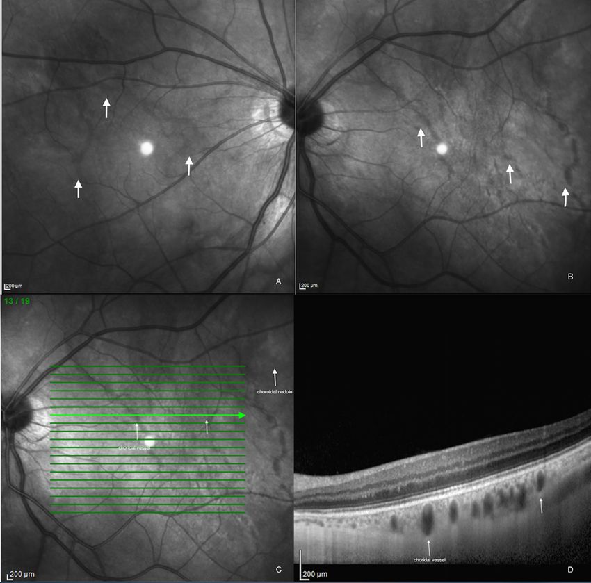
APRIL 9 / 23 / 30

                                                                                                          2021
            Virtual congress
            & live talk show
                                    AGE-RELATED MACULAR DEGENERATION
BROCCOLI-DERIVED COMPOUND INDUCES PROTECTIVE EFFECTS IN AN IN VITRO MODEL OF AGE-RELATED
MACULAR DEGENERATION: AN ‘OMICS’ APPROACH
 F. KWA 1, N. DULULL 1, U. ROESSNER 2, D. DIAS 1, T. RUPASINGHE 3
1
  School of Health and Biomedical Sciences, Royal Melbourne Institute of Technology University, Melbourne, Australia, 2
School of Biosciences, Faculty of Science, The University of Melbourne, Melbourne, Australia, 3 Metabolomics Australia, Bio21
Institute, The University of Melbourne, Melbourne, Australia 4 School of Health Sciences, Swinburne University of Technology,
Melbourne, Australia
Purpose: Age-related macular degeneration (AMD) is a leading cause of blindness in the ageing population. Impaired
innate immune responses to oxidative stress observed in AMD can result from the formation of oxidised lipids. Low levels
of polyunsaturated fatty acids have shown to increase AMD risk. The broccoli-derived isothiocyanate, L-sulforaphane (LSF), is
reported to be a powerful antioxidant in various cell types including neuroblastoma cells. Here, we examine the effects of LSF
on human retinal cells and reveal key elements that regulate oxidative stress and fatty acid/lipid metabolism to facilitate a
cytoprotective state.
Method: he adult retinal pigment epithelium-19 cell line was treated with 3-30µM LSF. Cell proliferation was assessed by tetrazolium
salt-based assays, under normal or hydrogen peroxide-induced oxidative stress conditions. Changes in gene expression of redox
enzymes, glutathione S transferase M1 (GSTM1) and heme oxygenase-1 (HO-1) were established using quantitative real-time PCR.
Fatty acid and lipid profiling were performed by gas and liquid chromatography-mass spectrometry, respectively.
Results: Under normal conditions, LSF was not toxic to the retinal cells. However, LSF rescued these cells from oxidative damage
in a dose-dependent manner by upregulating GSTM1 and HO-1 gene expression. Treatment with LSF increased levels of
unsaturated oleic and eicosatrienoic acids. Lipid levels of phosphatidylcholine, cholesteryl ester and oxo-phytodienoic acid
were increased upon LSF treatment but levels of phosphatidylethanolamines were decreased.
Conclusions: Retinal cells at risk of oxidative damage can be pre-conditioned with LSF to regulate levels of redox enzymes and
fatty acids/lipids known to be implicated in the pathogenesis of AMD.

                                                                                      BACK TO THE INDEX         u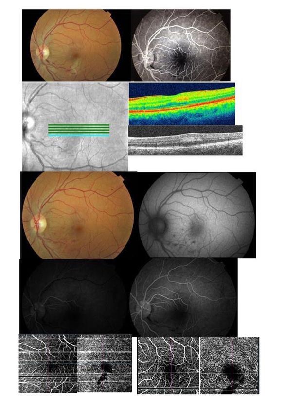
APRIL 9 / 23 / 30

                                                                                                          2021
         Virtual congress
         & live talk show
SHORT-TERM OUTCOMES OF PATIENTS WITH NEOVASCULAR EXUDATIVE AMD: THE EFFECT OF COVID-19
PANDEMIC
C. SENNI 1, E. BORRELLI 1, D. GROSSO 1, G. VELLA 1,2, R. SACCONI 1, M. BATTISTA 1, L. QUERQUES 1, I. ZUCCHIATTI 1,
F. PRASCINA 1, G. QUERQUES 1 , F. BANDELLO 1
1
  Department of Ophthalmology, University Vita-Salute, IRCCS Ospedale San Raffaele, Milan, Italy
2
  Ophthalmology, Department of Surgical, Medical, Molecular Pathology and of Critical Area, University of Pisa, Pisa, Italy

Purpose: To estimate the impact of delayed care during the coronavirus disease 2019 (COVID-19) pandemic on the outcomes
of patients with neovascular age-related macular degeneration (AMD).
Methods: Consecutive patients with diagnosis of neovascular AMD were consecutively enrolled between 9th March 2020 and
12th June 2020 (during and immediately after the Italian COVID-19 quarantine). During the inclusion (or pandemic) visit (V0),
patients received a complete ophthalmologic evaluation, including optical coherence tomography (OCT). Best-corrected visual
acuity (BCVA) and OCT findings from the two preceding visits (V-1 and V-2) were compared with data at V0.
Results: One-hundred patients (112 eyes) were enrolled in this study. The time interval between following visits was 110.7±37.5
days within V0 and V-1 and 80.8±39.7 days within V-1 and V-2, respectively (P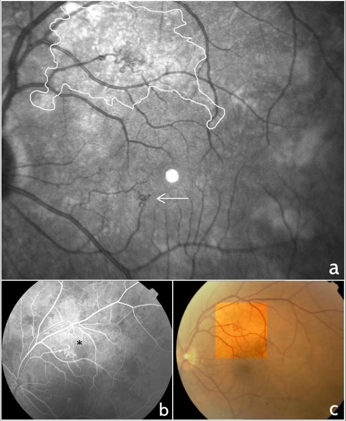
APRIL 9 / 23 / 30

                                                                                                          2021
         Virtual congress
         & live talk show
RATE OF MISDIAGNOSIS AND CLINICAL USEFULNESS OF THE CORRECT DIAGNOSIS IN EXUDATIVE NEOVASCULAR
MACULOPATHY SECONDARY TO AMD VS. PACHYCHOROID DISEASE
M. BATTISTA 1,2 , E. BORRELLI 1,2, F. GELORMINI 1,2, R. SACCONI 1,2, L. QUERQUES 1,2, G. VELLA 2,3, C. VIGANÒ 1,2, G. QUERQUES 1,2,
F. BANDELLO 1,2
1
  Vita-Salute San Raffaele University Milan, Italy 2 IRCCS San Raffaele Scientific Institute, Milan, Italy 3 Ophthalmology,
Department of Surgical, Medical, Molecular Pathology and of Critical Area, University of Pisa, Pisa, Italy

The aim of this study was to explore the relative prevalence and clinical differences between age-related macular degeneration
(AMD) and pachychoroid disease in patients older than 50 years with newly diagnosed exudative neovascular maculopathy,
and also assess the rate of misdiagnosis between these two disorders.
In this retrospective observational study, we reviewed data from patients 50 years of age and older with newly diagnosed
treatment-naïve exudative macular neovascularization (MNV) secondary to AMD or pachychoroid disease. Of the 139 patients
(139 eyes) who fulfilled the inclusion criteria, 35 patients were graded as being affected by pachychoroid disease complicated
by exudative MNV and 104 subjects had neovascular AMD.
Therefore, prevalence of pachychoroid disease complicated by exudative MNV was 25.2% (confidence interval - CI, 18.2-33.2%).
Mean±SD age was 67.0±8.8 years in the pachychoroid disease group and 80.6±6.6 years in the neovascular AMD group
(P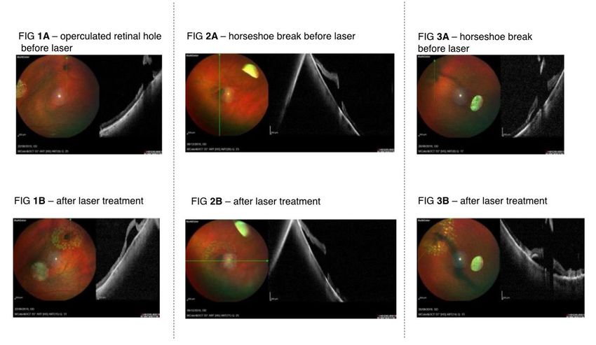
APRIL 9 / 23 / 30
2021
Virtual congress
& live talk show
ANGIOID STREAKS
PROGNOSTIC FACTORS BY MULTIMODAL IMAGING ANGOID STREAKS ASSOCIATED WITH PATTERN DYSTROPHY
IN PSEUDOXANTHOMA ELASTICUM AND NON SYSTEMIC DISEASE PATIENTS
T.K. GERGES ¹
1
Al Watany Eye Hospital, Cairo, Egypt
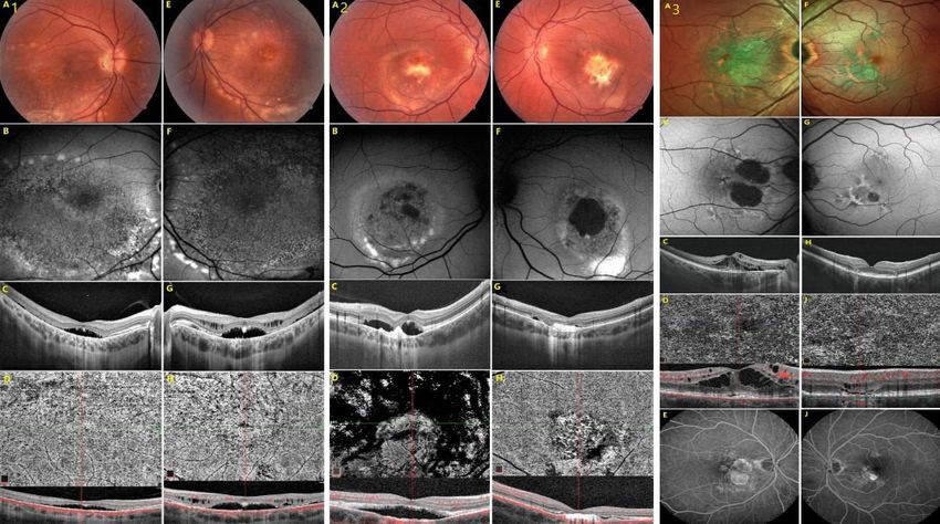
Introduction: Angoid streaks(AS) are crack-like dehiscences in Bruch’s membrane. Angoid streaks may be associated with
pseudoxanthoma elasticum(PXE),other systemic disease or without any systemic disease. Pattern dystrophy(PDS) is an
autosomal dominant condition that may occur in isolation or with PXE. PD and AS have previously been reported in PXE, but
has not been previously reported in non PXE.
Purpose : Using multimodal imaging to detect AS associated with PD , in groupA :PXE and groupB non-systemic disease
patients comparing retinal manifestations that may affect visual prognosis between 2 groups.
Method: GroupA12 eyes GroupB 12eye subjected to ocular examination , autofluorescence(and OCT to determine optimal
imaging method to detect fundus AS, PD, geographical atrophy in macula and peripapillary region, autofluorecent zone within
the posterior pole and CNV.
Results : Best corrected visual acuityBCVA : HM-0.3 ( GrpA= 10eyes, GrpB= 4eyes) and BVCA>0.4: (GrpA= 2eyes, GrpB=
8eyes),Pattern Dystrophy: Mild( GrpA= 5eyes, GrpB= none) , Severe ( GrpA= 4eyes, GrpB= 12 eyes); AF zone: (GrpA= 9eyes,
Grp 9eyes= none) ; Peripapillary atrophy: Mild (GrpA= 6eyes, GrpB= 7eyes) , Severe (GrpA= 7eyes, GrpB= 5eyes) ; Macular
RPE changes : Mild RPE changes ( Grp A= 4eyes, Grp B= 7eyes) , Macular geographical atrophy(GA) ( GrpA= 8eyes, GrpB=
5eyes), CNV (GrpA= 10eyes, GrpB= 5eyes).
Conclusion: Angoid streaks in PXE and non-systemic disease are associated with several retinal manifestations that are best
evaluated by multimodal imaging. GA and CNV are more common in PXE with wore visual prognosis than non-systemic disease
patients with AS and PDS. optic neuritis not only as a symptom of active Covid-19 infection, as already reported in literature,
but also as its consequence.
BACK TO THE INDEX u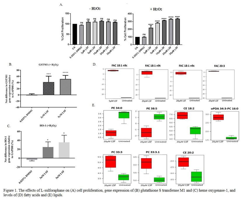
APRIL 9 / 23 / 30

                                                                                                          2021
            Virtual congress
            & live talk show
                                                  CHALLENGING CASES

 C OPTIC NEURITIS AS A POSSIBLE LATE MANIFESTATION OF PREVIOUS COVID-19 INFECTION
     A.V. BUX 1, A. NACUCCHI 1, A. STELLA ¹, C. IACULLI ¹
     1
       Department of Ophtalmology, Ospedali Riuniti University of Foggia, Foggia, Italy

Purpose: To describe a case of optic neuritis in a patient with an history of unknown COVID-19 infection.
Case Report: A 21 years-old patient with no medical history, referred to our Clinic for a sudden loss of vision in her left eye with
pain on eye movement.
At baseline visit, the patient underwent complete evaluation, including fundus autofluorescence, fluorescein angiography,
optical coherence tomography, computerized perimetry, B-scan ultrasonography and orbit computer assisted tomography.
Visual acuity was 20/20 in right eye and 20/200 in left eye, with relative afferent pupillary defect. A diagnosis of monolateral
papilledema was made. Afterwards, also brain and spinal cord magnetic resonance imaging(MRI), sierologic tests for vasculitis
and anti-Sars-Cov-2 antibodies were performed. MRI showed enhancement in the left more than the right optic nerve suggestive
of optic neuritis with no other abnormalities.
Both IgM and IgG anti-Sars-Cov-2 antibodies resulted positive (18,5 AU/ml and 48,3 AU/ml respectively). Then, the patient,
tested negative for Covid-19 by molecular nasopharyngeal swab.
The patient was started on intravenous methylprednisolone 1 g every 24 hours for five days, and her vision and eye pain
completely resolved.
Conclusion: To our Knowledge, this is the first report on late optic neuritis, in an otherwise asymptomatic previous Covid-19
infection. We hypothesize a plausible autoimmune pathogenesis of the optic neuritis. To our opinion is important to consider
optic neuritis not only as a symptom of active Covid-19 infection, as already reported in literature, but also as its consequence.

 C CASES

                                                                                      BACK TO THE INDEX         u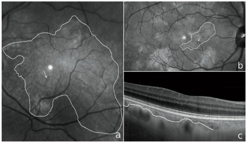
APRIL 9 / 23 / 30

                                                                                                    2021
        Virtual congress
        & live talk show
 C MULTIPLE EXUDATIVE CHOROIDAL NODULES RESISTANT TO INMUNOSUPPRESSIVE THERAPY.
   A CHALLENGING DIAGNOSIS
    G LIAÑO 1, C ARRUABARRENA 1, J CAÑAS MARTÍN 1
    1
      University Hospital Príncipe de Asturias, Department of Ophthalmology, Alcalá de Henares, Spain

Purpose: To introduce one singular case of a patient with choroidal nodules resistant to inmunossuppressive treatments and
uncertain diagnosis.
Methods: A 52 year-old lady came to our emergency room complaining about decreased vision in the right eye. She reported a
medical history of polyarthritis and purpura episodes with unknown etiology on maintenance treatment with oral prednisone.
No ophthalmic history of interest. BCVA (best corrected visual accuracy) was 20/30, and unspecific alterations in epitelium
pigmentarium were found in fundoscopy. Swept source OCT scan revealed a neurosensorial detachment(NSD) affecting the
macula. Thus, central serous choroidopathy (CSC) diagnosis was proposed and previous corticoid treatment was removed. At
fifth month-follow up BCVA improved but NSD persisted so oral eplerenone was prescribed.Two months later, we performed
fluorescein angiography (FAG) and indocyanine angiography (IAG) since BCVA dropped to 20/40.This exam spotted seven
hypocianescent choroidal nodules all over the posterior pole.Laboratory test (including the whole set of serologies, mantoux
and ACE sarcoidosis screening) were strictly normal. PET and visual field study 24.2 were also negative.
As BVCA was still getting worse (20/100) by the following months, IAG was repeated, probing an increase of exudation from
the central lesion as well as a newfound peripapillary nodule. Adalimumab treatment was tried, but any clinical efficacy was
observed after four months.
Subsequently, we added a first dose of intravitreal bevacizumab whose efficacy is yet to be assessed.
Conclusions: Pending the clinical response to anti-VEGF, either an atypical form of CSC or choroidal granuloma should be
considered as differential diagnosis.

 C CASES

                                                                                 BACK TO THE INDEX        u
APRIL 9 / 23 / 30

                                                                                                   2021
       Virtual congress
       & live talk show
CHOROIDAL NEOVASCULAR MEMBRANE SECONDARY TO OPTIC NERVE DRUSEN: A CASE REPORT
J. CAÑAS MARTÍN 1, C. ARRUABARRENA SÁNCHEZ 1, M. I. MARTÍNEZ SÁNCHEZ 1
1
 University Hospital Príncipe de Asturias, Department of Ophthalmology, Alcalá de Henares, Spain

Purpose: Describe optic nerve drusen (OND) morphology and one of its complications, the choroidal neovascular membrane
(CNVM).
Methods: The case of a 63-year-old woman that refers decreased
visual acuity (VA) and metamorphopsia in left eye is presented. As
ophthalmological background, a previous follow-up due to CNVM
secondary to OND in right eye stands out.
Results: A optic nerve head (ONH) of scalloped and diffuse edges and
a yellowish macular lesion are observed in the funduscopy. Fluorescein
angiography (FA) shows nodular staining of the ONH and early macular
hyperfluorescence. Optical coherence tomography (OCT) of the ONH shows
round hyperreflective deposits, and macular OCT shows a hyperreflective
subretinal lesion with associated subretinal fluid. Angio-OCT confirms the
CNVM image in choriocapillaris and external retinal plexus. The visual
field shows peripheral nasal defects and a increased blind spot.
The diagnosis of CNVM secondary to OND is made. Treatment with aflibercept
in pro re nata (PRN) strategy achieves anatomical and functional improvement.
Currently, the patient remains stable, with VA greater than 0’8.
Conclusions: Optic nerve drusen appear in up to 2’4% of the population,
being bilateral in most cases. The choroidal neovascular membrane
secondary to OND is a rare complication whose macular progression can
result in loss of VA. Both lesions can be diagnosed with OCT and angio-
OCT. Optic nerve drusen should be monitored on a regular basis to rule
out complications that could potentially threaten vision.

                                                                                 BACK TO THE INDEX   u
APRIL 9 / 23 / 30
2021
Virtual congress
& live talk show
POSTERIOR UVEITIS, AN UNCOMMON COMPLICATION OF MULTIPLE SCLEROSIS. CASE REPORT
C. METRANGOLO 1, S. DONATI 1,2, L. FONTANEL 2, W. MESSINA 2, G. D’ALTERIO 1, P. RADICE 1, C. AZZOLINI 2
1
Ophthalmology Unit, Ospedale di Circolo e Fondazione Macchi, ASST Sette Laghi, Varese, Italy
2
Department of Medicine and Surgery, University of Insubria, Varese, Italy
Introduction: Multiple sclerosis might lead to several ocular diseases, including the more common optic neuritis, intermediate
or posterior uveitis. Ocular complications due to medicines used for its treatment have also been reported, such as cystoid
macular edema.
Clinical case: A patient diagnosed with multiple sclerosis, recently receiving Fingolimod (Gilenya, Novartis) for exacerbation of
his disease, has a unilateral visual loss. OCT examination and fluorangiography revealed unilateral cystoid macular edema and
multifocal peripheral vascular leakage, with bilateral involvement.
Discussion: Retinal involvement can be due to different etiologies. Macular edema is known to be secondary to Fingolimod, but
in the presence of retinal vasculitis, we consider it as a complication of posterior uveitis, which originated from autoimmune
disorders due to multiple sclerosis
Conclusion: Systemic steroid therapy led to a complete resolution of uveitis, which remains a negative pathognomonic sign for
an increase in the inflammatory process of the underlying disease.
BACK TO THE INDEX uAPRIL 9 / 23 / 30
2021
Virtual congress
& live talk show
DIABETIC RETINOPATHY
CATARACT SURGERY WITH COMBINED VERSUS DEFERRED INTRAVITREAL DEXAMETHASONE IMPLANT FOR
DIABETIC MACULAR EDEMA: LONG-TERM OUTCOMES FROM A REAL-WORLD SETTING
E. CORBELLI ¹, F. FASCE ¹, L. IULIANO ¹, R. SACCONI ¹, R. LATTANZIO ¹, F. BANDELLO ¹, G. QUERQUES ¹
1
University Vita-Salute, IRCCS Ospedale San Raffaele, Milan, Italy
Purpose: To evaluate the long-term functional and anatomical outcomes of eyes undergoing cataract surgery combined with
dexamethasone (DEX) implant in eyes with pre-existing diabetic macular edema (DME), compared with a matched sample
undergoing the same procedure but with a 1-month deferred steroid injection.
Methods: Retrospective observation of all consecutive patients undergoing combined cataract surgery and intravitreal DEX
for pre-existing DME from 2010 to 2017 at our institution. In the investigational combined group, best-corrected visual acuity
(BCVA) and central retinal thickness (CRT) were extracted at baseline (combined surgery and DEX), 1, 4, 12 and 24 months after
procedure. The deferred group was analyzed the day of surgery, the day of DEX injection (baseline), then according to the same
time points of the investigational group.
Results: Forty eyes were analyzed, 20 in combined and 20 in deferred group. BCVA disclosed akin trends in both, increasing
from similar starting values (p=0.9913) to comparable scores 1 month after DEX (p=0.4229). After 4 months BCVA similarly
dropped down in both groups, with nonsignificant variations within each group. CRT was similar at the time of surgery
(p=0.6134). DEX injection positively reduced CRT in both sample, with a superior beneficial effect in the combined group after
1 month (p=0.0010). At 4 months, CRT further elevated and remained overall stable, without differences between groups.
Conclusion: DEX implant performed at the time of surgery achieved the same long-term functional and anatomical outcomes
of 1-month deferred DEX implant in treating eyes with pre-existing DME that should undergo cataract extraction.
BACK TO THE INDEX uAPRIL 9 / 23 / 30
2021
Virtual congress
& live talk show
THREE-YEAR OCT PREDICTIVE FACTORS OF DISEASE RECURRENCE IN EYES WITH SUCCESSFULLY TREATED
MYOPIC CHOROIDAL NEOVASCULARIZATION
F. BORGHESAN 1,2, E. BORRELLI 1,2, M. BATTISTA 1,2, G. VELLA 2,3, R. SACCONI 1,2, L. QUERQUES 1,2, D. GROSSO 1,2, G. QUERQUES 1,2,
F. BANDELLO 1,2
1
Vita-Salute San Raffaele University Milan, Italy 2 IRCCS San Raffaele Scientific Institute, Milan, Italy 3 Ophthalmology,
Department of Surgical, Medical, Molecular Pathology and of Critical Area, University of Pisa, Pisa, Italy
Running head: OCT risk factors for relapses in myopic MNV
Purpose: To assess the relationship of demographics, clinical characteristics and structural optical coherence tomography
(OCT) findings to disease recurrence in a cohort of patients with newly diagnosed myopic choroidal neovascularization (CNV).
Methods: In this retrospective, longitudinal study, a total of 64 participants (64 eyes) with successfully treated myopic CNV who
had obtained resolution of exudation after treatment (study baseline) and with 3 years of regular follow-ups. Several baseline
OCT qualitative features and quantitative measurements were assessed at baseline and included in the analysis. Main outcome
measures included incidence of disease recurrence and hazard ratio (HR) for demographics, clinical characteristics and OCT risk
factors.
Results: At month 36, 40 eyes (62.5%) developed disease recurrence (active CNV). Multivariate linear regression analysis
revealed that final visual acuity (dependent variable) was associated with visual acuity at the first visit after complete resolution of
exudation (pAPRIL 9 / 23 / 30
2021
Virtual congress
& live talk show
FUNCTIONAL IMAGING
VOLUME RENDERED OCTA ASSESSMENT OF MACULAR ISCHEMIA IN PATIENTS WITH TYPE 1 DIABETES AND
WITHOUT DIABETIC RETINOPATHY
D. GROSSO ¹, E. BORRELLI ¹, M. PARRAVANO 2, E. COSTANZO 2, M. BRAMBATI 1, C. VIGANÒ 1, R. SACCONI 1, L. QUERQUES 1,
A. PINA 1, D. DE GERONIMO 2, G. QUERQUES 1, F. BANDELLO 1
1
Department of Ophthalmology, University Vita-Salute, IRCCS Ospedale San Raffaele, Milan, Italy
2
IRCCS Fondazione Bietti, Rome, Italy
Purpose: To measure macular perfusion in patients with type 1 diabetes and no signs of diabetic retinopathy (DR) using volume
rendered three-dimensional (3D) optical coherence tomography angiography (OCTA).
Methods: We collected data from 35 patients with diabetes and no DR who had OCTA obtained. An additional control group of
35 eyes from 35 healthy subjects was included for comparison. OCTA volume data were processed with a previously presented
algorithm in order to obtain the 3D vascular volume and 3D perfusion density. In order to weigh the contribution of different
plexuses’ impairment to volume rendered vascular perfusion, OCTA en face images were binarized in order to obtain two-
dimensional (2D) perfusion density metrics.
Results: Mean±SD age was 27.2±10.2 years [range 19-64 years] in the diabetic group and 31.0±11.4 years [range 19-61
years] in the control group (p=.145). The BCVA was 0.0±0.1 LogMAR in the diabetic group and 0.0±0.0 LogMAR in healthy
eyes (p=.871). The 3D vascular volume was 0.27±0.05 mm3 in the diabetic group and 0.29±0.04 mm3 in the control group
(p=.020). The 3D perfusion density was 9.3±1.6 % and 10.3±1.6 % in diabetic patients and controls, respectively (p=.005).
Using a 2D visualization, the perfusion density was lower in diabetic patients, but only at the deep vascular complex (DVC) level
(38.9±3.7 % in diabetes and 41.0±3.1 % in controls, p=.001), while no differences were detected at the superficial capillary
plexus (SCP) level (34.4±3.1 % and 34.3±3.8 % in the diabetic and healthy subjects, respectively, p=.899).
Conclusions: Eyes without signs of DR of patients with diabetes have a reduced volume rendered macular perfusion compared
to control healthy eyes. The comparison between 3D and 2D visualizations highlights the significant contribution of DVC to
reduced volume rendered macular perfusion.
BACK TO THE INDEX uAPRIL 9 / 23 / 30
2021
Virtual congress
& live talk show
MACULAR TELANGIECTASIA
ANATOMICAL AND FUNCTIONAL CORRELATION BASED ON MULTIMODAL IMAGING IN PATIENTS WITH TYPE 2
IDIOPATHIC MACULAR TELANGIECTASIA
A.L. AMBRÓSIO MARTA 1, P.M. BAPTISTA 1, I. CARNEIRO 1, M.J. FURTADO 1, M. LUME 1
1
Centro Hospitalar Universitário do Porto, Ophthalmology Department, Porto, Portugal
Purpose: Our study evaluates the contribution of multimodal imaging in type 2 idiopathic macular telangiectasia (MT2) and
correlates functional and anatomical findings.
Methods: Retrospective consecutive case series of 26 eyes with MT2. All patients underwent Spectral-Domain Optical Coherence
Tomography (SD-OCT), Optical Coherence Tomography Angiography (OCTA), Fundus Autofluorescence (FAF), Fluorescein
Angiography (FA) and Microperimetry (MP). Quantitative parameters on OCTA were compared to a control group.
Results: Loss of hypoautofluorescent centre on blue-light FAF was found in all patients. On SD-OCT, intraretinal hyporreflective
spaces were identified in all eyes, ILM draping in 45% and in average a lower central foveal thickness (CFT) (p=0.001). On
OCTA, MT2 showed a higher macular vascular density of deep capillary plexus (DCP) (p=0.001) and fractal dimension value (D)
was higher (p=0.007). FA showed staining of the abnormal juxtafoveal temporal microvasculature.
On MP, 70% of eyes achieved a stable fixation and retinal sensitivity appears to be normal in the absence of outer retinal
atrophy. The fixations ratio within the central 2degrees correlates negatively with age (p=0,006;r=-0,594) and with area
(p=0.036,r=-0.471) and perimeter (p=0.029,r=-0.488) of the foveal avascular zone (FAZ) on the DCP; and positively with
CFT (p=0,012;r=0,549) and with D on the DCP (p=0.009,r=567). Patients with stable fixation were younger (p=0.026), had
higher SCT (p=0.010) and D on the DCP (p=0.002), and lower area and perimeter of the FAZ on superficial capillary plexus
(p=0.011 and 0.018) and DCP (p=0.006 and 0.012).
Conclusions: Multimodal imaging, including OCTA and MP, allows for a better anatomical and functional characterization of
MT2.
BACK TO THE INDEX uAPRIL 9 / 23 / 30
2021
Virtual congress
& live talk show
CHOROIDAL VASCULATURE IN PATIENTS WITH MACULAR TELANGIECTASIA
J. H. MARQUES ¹, A.L. AMBRÓSIO MARTA ¹, R. VIEIRA 1, M. J. FURTADO 1, M. LUME 1
1
Ophthalmology Department, Centro Hospitalar Universitário do Porto, Porto, Portugal
Purpose: To evaluate changes in choroidal structure and vascularization in eyes with type 2 macular telangiectasia (MacTel).
Methods: Observational cross-sectional study at Centro Hospitalar Universitário do Porto. Eyes of 14 patients with
MacTel (group 1, 28 eyes) were compared with 10 age-matched healthy controls (group 0, 20 eyes). All subjects
underwent a complete ophthalmologic examination, fluorescein angiography, blue peak autofluorescence, spectral
domain (SD) optical coherence tomography (OCT) and OCT-angiography (OCT-A) on Heidelberg® Spectralis platform.
Eyes with intraretinal/subretinal fluid, media opacities or erroneous OCT segmentation were excluded from analysis.
Choroidal vascularity index (CVI) was calculated in a single foveal-centered high-resolution 20º OCT B-scan. Choroidal limits
were manually defined by a single investigator, through Image J® software. The outer border of the hyperreflective line
representing the retinal pigment epithelium was considered the inner choroidal limit. The outer choroidal limit was defined
as the inner border of the hyperreflective sclera. As previously described by other groups, the image was binarized using the
Niblack method. Choroidal area (CA), hyperreflective stromal area and hyporeflective vascular luminal areas were automatically
calculated in the full 5mm-width image (G) and also in 1-mm width nasal (N) and temporal (T) sectors. CVI was calculated as
luminal area divided by CA. CA was converted to average choroidal thickness (CT) in the corresponding areas (G, N and T).
Choriocapillaris flow density (CCD) and choroidal flow density (CHD) were calculated from an 10x10º OCT-A C-scan,
segmented automatically in the choriocapillaris and choroid layers, respectively, using the OCT proprietary software. The
image was binarized in ImageJ® using the Phansalkar´s method and CCD and CHD were defined as the ratio between flow
signal area and total area (G). CCD and CHD were also calculated in 1mm nasal (N) and temporal (T) sectors of the C-scan.
The ratio between nasal and temporal measurements was also considered (rCT, rCVI, rCCD, rCHD)
Results: Mean age was 64.3±13.6 in group 1 and 65.9±10.3 in group 0 (p=0.669). In OCT, CT was increased in group 1
(293±60um vs 210±56um in G; p0.643 in all sectors).
The OCT-A analysis showed, in group 1, a reduced CCD (43.2.5±16.5% vs 51.5±8.6% for G; p0.246 in G and N). rCCD, rCDH and
rCT were higher in group 1 (pAPRIL 9 / 23 / 30
2021
Virtual congress
& live talk show
OPTICAL COHERENCE TOMOGRAPHY ANGIOGRAPHY
OCT ANGIOGRAPHY IN A CASE OF TUBEROUS SCLEROSIS COMPLICATED WITH MACULAR CHOROIDAL
NEOVASCOLARIZATION
A. TURTORO 1, M. FORTUNATO 2, G. CENNAMO 3, C. NARDELLA 4
1
Ospedale Pugliese, Catanzaro, Italy 2 Ospedale Bambino Gesù, Roma, Italy 3 Federico II Univertity, Napoli, Italy 4 Ospedale
Santa Maria delle Grazie, Velletri, Italy
Purpose: to describe the optical coherence tomography angiography (OCTA) characteristics of a case of a Tuberous sclerosis
patient complicated with unilateral macular choroidal neovascularization (CNV) during the ranibizumab therapy
Method: a 9 year-old male suffering of incomplete form of tuberous sclerosis complex (formes frustes) presented with
headache and decrease in vision in his right eye and since three months. He underwent a complete ophthalmic examination,
fluorescein angiography, OCT-SD and OCTA confirmed a diagnosis of CNV in his right eye. Four montly intravitreal injections
were made in his right eye.
Results: visual acuity improved from 20/60 to 20/30 in the right eye after Ranibizumab therapy.SD-OCT showed the resolution
of intraretinal fluid and OCTA revealed a decrease of size of CNV after treatment.
Conclusions: OCTA is a reproducible and non-invasive examination, which provides a reliable follow-up over time as for the
neovascular size area and activity in child with Tuberous sclerosis
BACK TO THE INDEX uAPRIL 9 / 23 / 30
2021
Virtual congress
& live talk show
THREE-DIMENSIONAL MODEL OF THE MICROVASCULAR NETWORK OBTAINED BY USING OPTICAL COHERENCE
TOMOGRAPHY ANGIOGRAPHY IN FOVEA PLANA AND NORMAL FOVEAL DEPRESSION.
S. FRAGIOTTA 1, C. CIANCIMINO 1, A. PERDICCHI 1, S. ABDOLRAHIMZADEH 1, G. SCUDERI 1
1
Ophthalmology Unit, Department NESMOS, St. Andrea Hospital, University of Rome La Sapienza, Rome, Italy
Purpose: To analyze microvascular architecture and possible alternative arrangements in eyes with idiopathic fovea plana and
controls through a three-dimensional reconstruction of optical coherence tomography angiography (OCTA).
Methods: A retrospective analysis was conducted on 2 patients with fovea plana and 2 controls who underwent a 3 x 3 mm
OCTA scan protocol with a scan quality of 9/10. A total of 12 enface OCTA slabs and 408 projection-resolved OCTA b-scans
were analyzed with Fiji software (Fiji, http://fiji.sc;) and then imported and elaborated using a volume rendering software
(ImageVis3D).
Results: In eyes with fovea plana, a superficial vascular plexus and a single deeper vascular monolayer with multiple short
interconnections characterized the foveal zone. In contrast, eyes with normal foveal pit and foveal avascular zone, characterized
by an identifiable three-laminar organization of the vascular plexus, exhibited most interconnections between the superficial
and intermediate plexus and rare direct connections between superficial and deep plexus.
Conclusions: An abnormal development of foveal depression pursued a microvascular rearrangement of the microvascular
interconnections and plexuses. The use of a volume rendering reconstruction aids the understanding of anatomical variations
in vascular plexuses.
BACK TO THE INDEX uAPRIL 9 / 23 / 30
2021
Virtual congress
& live talk show
VASCULAR AND STRUCTURAL FINDINGS IN UNAFFECTED FELLOW EYES OF PATIENTS WITH UNILATERAL PRIMARY
OPEN ANGLE GLAUCOMA USING OPTICAL COHERENCE TOMOGRAPHY ANGIOGRAPHY
A. RAGKOUSIS 1, E. GKOUMAS 1, S. KYRIAZI 1, A. PAPADOPOULOS 1, N. KOUTROPOULOU 1, E. BOUTOURI 1, G. MANGOURITSAS 1
1
Red Cross General Hospital - Ophthalmology Department, Athens, Greece
Purpose: To investigate peripapillary vessel density (ppVD), peripapillary retinal nerve fiber layer (RNFL) and macular ganglion
cell complex (GCC) thickness in clinically unaffected fellow eyes (F) of patients with unilateral primary open angle glaucoma
(POAG) using optical coherence tomography angiography (OCTA).
Methods: This cross-sectional observational study included 20 untreated patients with unilateral glaucomatous damage
manifested with abnormal RNFL and GCC. F eyes were classified as clinically unaffected if they had statistically within normal
limits or borderline RNFL and GCC thickness and normal visual fields. Twenty healthy eyes of age-matched individuals (H)
served as controls. RNFL, GCC and ppVD were measured by means of spectral-domain OCT/OCTA (RTVueXR Avanti, Optovue,
Fremont, CA) in both F and H groups. F group was compared with H group using Student’s t – test.
Results: Mean RNFL and GCC average thickness in microns differed significantly (p0.9)
diagnostic ability for structural parameters and a poor one (APRIL 9 / 23 / 30
2021
Virtual congress
& live talk show
COMPARISON OF AUTOMATED AND MANUAL SEGMENTATION OF SPECTRALIS OCT-ANGIOGRAPHY EN FACE
SLABS FOR THE DETECTION OF CHOROIDAL NEOVASCULARIZATION
N. SILVA 1, A. ABREU 1, M. FURTADO 1, M. LUME 1
1
Centro Hospitalar Universitário do Porto, Porto, Portugal
Purpose: To compare the sensitivity of choroidal neovascularization (CNV) detection between automatically generated
“avascular complex” or “choriocapillaris” en face slabs and manually adjusted segmentation slab using Spectralis optical
coherence tomography angiography (OCTA) in neovascular age-related macular degeneration (nAMD).
Methods: Cross-sectional prospective study of previously treated eyes with nAMD-associated type 1 or 2 choroidal
neovascularization (CNV). Patients with polypoidal choroidal vasculopathy were excluded. OCTA imaging was acquired in
a 3x3 millimeters area centered on the lesion using the Spectralis OCTA (Heidelberg Engineering, Germany). OCTA en face
images were automatically generated using the “superficial vascular complex”, “deep vascular complex”, “avascular complex”,
“choriocapillaris” and “choroid” slabs provided by the software. Manually adjusted segmentation was also performed.
Results: Sixty-three eyes were included. All patients were under anti-vascular endothelial growth factor treatment on a
treat-and-extend regimen. Sixty-two percent (n=39) had active CNV, while 38% (n=24) had inactive CNV. The neovascular
membrane was visualized in 76% (n=48) with both automated and manual segmentation. Manually modified images didn’t
identify any neovascular lesion not visualized with automated segmentation. Automatically generated “avascular complex”
and “choriocapillaris” slabs showed 75% (n=47) and 8% (n=5) of CNV, respectively. In only 1 case, the choroidal neovascular
membrane was visualized in “choriocapillaris” slab but not in “avascular complex” slab.
Conclusions: Automated and manual segmentation of Spectralis OCTA en face slabs were similar in detecting CNV, although
manual adjustment may allow a better delineation of the neovascular lesion. Concerning automatically generated slabs the
“avascular complex” had higher sensitivity to detect CNV than the “choriocapillaris” slab.
BACK TO THE INDEX uAPRIL 9 / 23 / 30
2021
Virtual congress
& live talk show
OPTICAL COHERENCE TOMOGRAPHY ANGIOGRAPHY OF THE OPTIC DISC AND SURROUNDING TISSUES
FOLLOWING SLOTTED PLAQUE BRACHYTHERAPY FOR CHOROIDAL MELANOMA
A. FAM 1, P. FINGER 1, A. TOMAR 1
1
The New York Eye Cancer Center, New York, USA
Purpose: To evaluate the radial peripapillary capillary vessel density
(CVD) in eyes irradiated with slotted palladium-103 (103Pd) plaques for
peripapillary, juxtapapillary, and circumpapillary choroidal melanoma.
Methods: This retrospective cross-sectional review evaluated consecutive
patients treated with slotted 103Pd plaque irradiation. The radial
peripapillary CVD was assessed using optical coherence tomography
angiography (OCTA). Contralateral eyes served as controls. Data collected
included demographics, eye treated, radiation doses and dose-rates to
the macula, optic disc and surrounding tissues, and quantified radial
peripapillary CVD in treated and control eyes. The paired t-test was used
to compare the change in radial peripapillary CVD after radiation and its
correlation to radiation dose and dose-rate.
Results: Fourteen patients, with a mean age was 66.4 years, were
recruited. Their mean follow-up duration from initial radiation treatment
was 4.29 years (2.97, 1-19). Mean radiation dose to the optic disc was
85.5 Gy (mean dose-rate: 57.2 Gy/hr). Dose to macula was 63.6 Gy
(mean dose-rate: 37.8 Gy/hr). There was a significantly reduced radial
peripapillary CVD in treated eyes compared to control (p=0.001).
The difference in CVD between the treated eyes and control showed
strong negative correlations with optic nerve radiation dose (r=-0.647,
p=0.012) and dose-rate (r=-0.579, p=0.030).
Conclusion: OCTA imaging shows that radial peripapillary CVD is
significantly reduced in eyes treated with slotted, circumpapillary
plaques used for treatment of choroidal melanoma. This reduction was
linearly correlated to radiation dose and dose-rate delivered to the optic
disc.
BACK TO THE INDEX uAPRIL 9 / 23 / 30
2021
Virtual congress
& live talk show
RETINAL CHANGES AND ANGIOOCT AS A POSSIBLE BIOMARKER IN THE PRECLINICAL PHASE OF ALZHEIMER
DISEASE
C. IACOBUCCI 1, A. CHIARAVALLOTI 2, B. BILLI 3, V. DI MARTINO 4, A.E. CASTELLANO 5
1
Diagnostica e Microchirurgia oculare, Centro Vitale Campobasso, Italy, 2 University of Tor Vergata, Roma, Italy,
3
Fondazione Bietti, Clinica San Domenico, Roma, Italy, 4 Statistical Private Practice, Roma, Italy 5 IRCCS Neuromed La Sapienza
University, Pozzilli, Italy
Purpose: Anatomical changes in Retina, since the pre-clinical onset of AD, represent the opportunity to identify vascular and
structural changes associated with amyloid ocular burden: not only the thinning of RFNL and the RCG retinal ganglion cell
layer, but especially the narrowing and rarefaction of the circumfoveal vascular plexus identified and measured with AngioOCT.
Methods: Patients with Initial Cognitive Impairment underwent to: neuropsychological test, RMN and brain PET with tracer
for amyloid. The characteristic trait of AD, beta-amyloid deposits in the brain is also present in the retina of these subjects; the
study, investigating retinal vascular networks with AngioOCT highlights such alterations as possible AD Biomarker. 42 patients
with mild Cognitive Impairment non Dementia (MC I) with C TPET positive for beta amyloid deposits in the brain had similar
retinal deposits by examination with AngioOCT, with decreasing vascular flow and rarefaction of vascular tree design, with
highly significant frequency (100%), when compared with healthy groups of people of similar age.
Results: 42 subjects between 47 years old and 66, suffering from Mild CognitiveImpairment (MC I) according to current
diagnostic criteria, have been included.
They underwent to: complete neuropsychological assessment for MCI, including MMSE Cerebral MRI and Amy-PET.
Complete ophthalmological examination in both eyes.
Macular OCT, RFNL, GCL+IPL by OCT were performed.
Finally Angio OCT examination was executed
Conclusions: The authors propose the possibility that the retinal and choroidal vascular alterations highlighted with AngioOC
T in subjects with prodromal AD, can represent an effective and economic Biomarker to diagnose this disease at the beginning
BACK TO THE INDEX uAPRIL 9 / 23 / 30
2021
Virtual congress
& live talk show
OCT RISK FACTORS FOR 3-YEAR DEVELOPMENT OF MACULAR COMPLICATIONS IN EYES WITH “RESOLVED”
CHRONIC CENTRAL SEROUS CHORIORETINOPATHY
F. NADIN 1, E. BORRELLI 1, M. BATTISTA 1, R. SACCONI 1, F. GELORMINI 1, L. QUERQUES 1, D. GROSSO 1, G. VELLA 1,2, G. QUERQUES 1,
F. BANDELLO 1
1
Ophthalmology Department, San Raffaele University Hospital, Milan, Italy 2 Ophthalmology, Department of Surgical, Medical,
Molecular Pathology and of Critical Area, University of Pisa, Pisa, Italy
Purpose: To assess the relationship of demographics, clinical characteristics and structural optical coherence tomography (OCT)
findings to development of sight-threatening macular complications (choroidal neovascularization [CNV], large areas of retinal
pigment epithelium [RPE] atrophy and cystoid macular degeneration [CMD]) in a cohort of eyes with “resolved” chronic central
serous chorioretinopathy (CSC) at inclusion (study baseline).
Design: Retrospective cohort study.
Methods: In this study, a total of 71 participants (71 eyes) with “resolved” (absence of subretinal fluid) chronic CSC at baseline
and 3 years (36 months) of regular follow-ups were retrospectively enrolled. Structural OCT scans were reviewed. Baseline OCT
qualitative features reflecting a distress of the neuroretina, RPE or choroid were assessed and included: ellipsoid zone (EZ)
discontinuity, outer nuclear layer (ONL) thinning, presence of hyperreflective intraretinal foci, dome-shaped pigment epithelium
detachment (PED), hyperreflective flat irregular PED, hyporeflective flat irregular PED, and inner choroidal attenuation. OCT
images obtained at follow-up visits were also reviewed for the development of macular complications (CNV, large areas of RPE
atrophy [at least 250 µm in diameter] and CMD). Main outcome measures included Incidence of macular complications and
hazard ratio (HR) for demographics, clinical characteristics and OCT risk factors.
Results: At month 36, 20 eyes (28.2%) developed macular complications: 9 eyes (12.7%) displayed CNV, whereas 9 eyes (12.7%)
had large areas of RPE atrophy, and 2 eyes (2.8%) developed cystoid macular degeneration. Several factors were associated
with an increased risk of CNV developing: intraretinal hyperreflective foci had an HR of 11.58 (95% CI, 1.10-37.24; P=0.040);
inner choroidal attenuation had an HR of 9.66 (95% CI, 1.07-22.34; P=0.043); presence of macular complications in the fellow
eye had an HR of 20.17 (95% CI, 1.34-39.41; P=0.030). We also identified factors associated with the development of RPE
atrophy: ONL thinning had an HR of 13.47 (95% CI, 1.10-39.86; P=0.042); dome-shaped PED had an HR of 21.40 (95% CI,
1.50-41.10; P=0.031); inner choroidal attenuation had an HR of 13.20 (95% CI, 1.07-39.32; P=0.044).
Conclusion: We identified OCT risk factors for the development of macular complications in eyes with chronic CSC. Our
findings may help in the identification of high-risk patients.
BACK TO THE INDEX uAPRIL 9 / 23 / 30
2021
Virtual congress
& live talk show
QUANTITATIVE VASCULAR CHANGES IN THE AGEING RETINA AS MEASURED BY SWEPT SOURCE OPTICAL
COHERENCE TOMOGRAPHY ANGIOGRAPHY
V. STARACE 1, R. SACCONI 1, M. TREVISI 1, E. BORRELLI 1, G. QUERQUES 1, F. BANDELLO 1
1
Department of Ophthalmology, IRCCS Ospedale San Raffaele, University Vita-Salute, Milan, Italy
Purpose: To quantitate regional differences in the retinal perfusion and its age-related changes in healthy subjects with optical
coherence tomography angiography (SS-OCTA) imaging.
Methods: Healty subjects were imaged with the SS-OCTA system (PLEX Elite 9000, Carl Zeiss Meditec Inc, Dublin, California,
USA). 3x3mm and 6x6-mm scans centered on the fovea were acquired and analyzed post-processed with thresholding and
binarization processes. The FAZ area was excluded from analysis to avoid its confounding factor. Eyes where grouped for decades
(at least 10 eyes for decades up to 80 years). We analyzed retinal vessel density and perfusion and its changes with age.
Results: One hundred eyes (100 patients) with no significant ocular pathologies were enrolled. The datas showed a reduction
in retinal perfusion related to age.
Conclusions: Aging induces a reduction in retinal vascular perfusion.
BACK TO THE INDEX uAPRIL 9 / 23 / 30
2021
Virtual congress
& live talk show
RETINAL & MACULAR DYSTROPHIES
CASES OF AUTOSOMAL RECESSIVE BESTROFINOPATHY MASKING AS OTHER DISEASES
N. MATCKO 1, M. GATSU 1
1
The S. Fyodorov Eye Microsurgery Federal State Institution, Saint-Petersburg, Russia
Purpose: Using multimodal imaging, describe the complex cases of autosomal recessive bestrophinopathy (ARB) encountered
in clinical practice.
Methods: In addition to the standard ophthalmological examination, patients underwent fundus autofluorescence imaging
(AF) (Heidelberg Engineering, Heidelberg, Germany), spectral-domain optical coherence tomography Optovue RTVue XR
Avanti (Optovue, Inc, Fremont, CA), and fundus photography. One patient underwent fluorescence angiography (FA).
Results: We present 3 clinical cases from our practice. The lack of a family history assessment, the failure to perform AF led to
the patient 1 with cystoid fluid initially was misdiagnosed with CNV, and was treated with anti-VEGF therapy, which did not lead
to a proper result. In the case of patient 2 it led to the fact that scar changes were regarded as a manifestation of chorioretinitis,
and therefore the patient 2 was treated with systemic prednisolone without effect. In the case of patient 3 cystoid changes
in neuroepithelium were mistakenly regarded as manifestations of Irvine-Gass syndrome, and therefore anti-inflammatory
therapy was carried out without the expected effect.
Conclusions: In clinical practice, ARB masking as a more common form of retinal pathology, and therefore differential
diagnosis is often difficult. This is facilitated by the identification in patients of cystoid changes in neuroepithelium, subretinal
scarring (without choroidal neovascularization). In this regard, we recommend a multimodal approach, paying attention to
the characteristic variants of hyperautofluorescence in the form of a “bead” and cruciform pattern, the absence of signs of
inflammatory edema according to FA, and clarification of the family history.
BACK TO THE INDEX uAPRIL 9 / 23 / 30
2021
Virtual congress
& live talk show
RETINAL ARTERY OCCLUSION & PARACENTRAL ACUTE MIDDLE MACULOPATHY
C AN ATYPICAL OPTIC NEURITIS CASE
C. ARRUABARRENA SÁNCHEZ 1, J. CAÑAS MARTÍN 1, G. LIAÑO DE ULZURUN 1
1
Department of Ophthalmology, University Hospital Principe de Asturias, Madrid, Spain
Introduction: Retinal artery occlusion is easy clinical diagnosis in acute phase. But can be difficult in chronic phases.
Case Report: 60-year-old male with a personal history of high blood pressure, diabetes mellitus and left occlusive carotid
disease, complains of acute, painless, lower hemisphere vision loss in his right eye of one month of evolution.
Best corrected visual acuity was 1 in both eyes. Fundus exploration was normal. Retinal nerve fiber layer on Optical Coherence
Tomography (OCT) in the right eye showed a decrease in the upper rim and the right eye visual field showed a lower altitudinal
defect with respect of the midline.
It was initially diagnosed as a right ischemic optic neuritis. But macular OCT showed a thinning of the upper half of the macular
with an obvious disorganization of the retinal inner layers and a careful evaluation of the fundus showed a crossing of the
superior temporal arcade vein over the artery near the optic nerve with a decrease in arterial caliber.
An angio-OCT was performed showing a showing a decrease in the superficial capillary plexus and an abolition of the deep
plexus in the upper half of the macula. And the final diagnosis was occlusion of the superior temporal arterial branch and a
carotid ultrasound was requested in addition to the erythrocyte sedimentation rate.
Discussion: Some cases of retinal arterial occlusion can be very difficult to diagnose and the characteristics of OCT and OCT
angiography carefully assessed can be very helpful.
C CASES
BACK TO THE INDEX uAPRIL 9 / 23 / 30
2021
Virtual congress
& live talk show
PARACENTRAL ACUTE MIDDLE MACULOPATHY CHARACTERISTICS IN MULTIMODAL IMAGE
M.I. MARTÍNEZ SÁNCHEZ 1, C. ARRUABARRENA SÁNCHEZ 1, J. CAÑAS MARTÍN 1, G. LIAÑO SANZ DIEZ ULZURRUN 1
1
University Hospital Principe de Asturias, Alcalá De Henares, Madrid, Spain
Purpose: Paracentral acute middle maculopathy (PAMM) is a vascular disease characterized by paracentral scotomas and band-
like located in the inner nuclear layer (INL) in spectral-domain optic coherence tomography (SD-OCT). It is the result of ischemia
of the intermediate and deep capillary plexuses and this can be demonstrated in OCT angiography (OCT-A).
The purpose of this case is to present a case of a macula branch retinal vein occlusion (BRVO) and a PAMM lesion associated.
Methods: A 43-year-old woman complaining of unilateral paracentral scotoma, came to the emergency unit.
Results: Visual acuity was 20/20 in both eyes. She had a normal anterior chamber exam and intraocular pressure. The fundus
examination disclosured tortuosity and dilation of the inferior temporal macular vein, intraretinal hemorrhages and focal
edema associated with a patchy area of deep retinal whitening inferior to the macula. SD-OCT revealed focal hyper-reflective
lesion at the level of the INL in the whitening area and OCT-A showed absence of the deep capillary plexus. The diagnosis of
BRVO associated with a PAMM lesion was made.
Conclusions: Most cases complaining of paracentral scotoma with normal fundus are a challenge for the clinician. The finding
of sudden loss of visual acuity with paracentral scotoma associated with the appearance of a hyperreflective band in the
parafoveal INL at the SD-OCT should make us suspect the existence of a PAAM. In these patients, it is essential to use SD-OCT
and OCT-A for diagnosis and monitoring; and a systemic study of associated risk factors must be done.
BACK TO THE INDEX uAPRIL 9 / 23 / 30
2021
Virtual congress
& live talk show
STRUCTURAL OPTICAL COHERENCE TOMOGRAPHY
RETINAL MICROVASCULAR AND CHOROIDAL VESSEL ABNORMALITIES AND THEIR CORRELATION WITH
CHOROIDAL NODULES IN NEUROFIBROMATOSIS TYPE 1
S. ABDOLRAHIMZADEH 1
1
University of Rome Sapienza, NESMOS department, Faculty of Medicine and Psychology, St. Andrea Hospital, Rome, Italy
Background: Neurofibromatosis type 1 (NF1) is part of a group of heredo-familial disorders characterized by the presence of
disseminated hamartoma. The gene for NF1 was cloned on chromosome 17q11.2. Loss of the protein neurofibomin in NF1
leads to alterations of cell growth and proliferation.
In ophthalmology hamartoma of the iris, optic pathway gliomas, and choroidal alterations are diagnostic markers of NF1. Recent
methods of imaging with near infrared reflectance (NIR), spectral domain optical coherence tomography (SD-OCT), enhanced
depth imaging (EDI), and the novel OCT angiography have provided further evidence of distinctive ophthalmic features in NF1.
Methods: Subjects: 17 consecutive patients with diagnosis of NF1 based on NIH criteria.
17 age and gender-matched healthy control patients.
Examination: Comprehensive ophthalmological examination, fundus photography, NIR Imaging, EDI - SDOCT
Results: Microvascular retinal abnormalities were found with NIR in 6 patients with NF1 (35%). They were localized in the
vascular arcade area; second or third order, small tributaries of the superior or inferior temporal vein consisting of small
tortuous vessels with a “spiral” or “corkscrew” appearance. NIR images showed that the retinal microvessels were all overlying
patchy choroidal alterations, which on cross-sectional EDI images were hyperreflective nodules in the choroidal thickness.
Fluorescein angiography showed that the microvascular alterations did not leak fluorescein. Anomalous choroidal vessels
using NIR imaging were observed bilaterally in 4 patients. EDI scans positioned on NIR evidenced vessels confirmed unusually
dilated choroidal vessels. These distinct vessels had a diameter that extended from the choroid-scleral junction to the outer
border of the hyperreflective band corresponding to the retinal pigment epithelium/Bruch’s layer. There was an absence of the
choriocapillaris or Sattler’s layer above the dilated vessels. BCVA was 20/20 in all patients.
Discussion: Retinal angiomatosis was reported by Frenkel in 1967 in a patient with NF1. Muci-Mendoza et al using direct
ophthalmoscopy in 2002, reported 12 of 32 patients (37.5%) with NF1 with retinal microvascular abnormalities. In 2014 we
used NIR imaging for the first time and confirmed these results. We observed retinal microvascular alterations in six of 17
patients with NF1 (35%). All abnormal retinal microvessels were overlying patchy choroidal alterations (placcoid type). However,
they were localized to the retina and did not involve the choroid, as shown by spectralis EDI-OCT cross-sections. Cassiman et
al. confirmed our results that corkscrew vessels were found in association with placcoid type choroidal nodules using OCT
angiography and found microvascular alterations were in the superficial vascular network. A successive large population
study in 2019, having adopted our method of NIR examination in NF1 patients, confirmed our pilot study results showing
microvascular retinal abnormalities in105 of 334 NF1 patients (31.4%), and reported that these correlated with cutaneous
neurofibromas. A further study using NIR imaging and OCT angiography showed retinal vascular abnormalities in 6.1% of
294 patients but did not find any correlation with other NF1 alterations. The authors found that the microvascular alterations
localized at the superficial vascular network were associated with anomalous and crowded vessels of the deep vascular network
in 75% of cases.The choroid is a multifunctional vascular tissue. Our report of unusually dilated choroidal vessels may constitute
another distinctive ophthalmological feature in NF1 that requires study in a larger population of patients. There is aberrant
proliferation of tissues of neural crest origin in NF1. It has been speculated that functional disorders of vasomotor nerve cells
that originate in the embryonal neural crest can lead to microvascular alterations in NF1. Furthermore, neurofibomin is present
in endothelium cells leading to the speculation that the loss of neurofibomin expression in endothelial cells may cause the
proliferation of vascular smooth muscle cells.
Conclusions: The microavascular retinal alterations and possibly the novel choroidal vessel alterations may be considered
biomarkers of Nf1. Together with the high frequency of systemic arterial disease in NF1, reaching up to 10%, and the
neurological disorders such as epilepsy and headaches, possibly correlated with microvascular cerebral alterations, vascular
alterations warrant further study.
BACK TO THE INDEX uAPRIL 9 / 23 / 30
2021
Virtual congress
& live talk show Fig. 1 Near infrared reflectance and optical
coherence tomography cross-section of retinal
and choroidal alterations in NF1. Near infrared
reflectance image of “corkscrew” or “spiral”
microvascular alteration (arrow) overlying
large patchy choroidal alteration delineated
with a white line (a), raster optical coherence
tomography image and corresponding
cross-section of the choroidal nodule
delineated with a yellow line (b and c). (From:
Abdolrahimzadeh S, et al. Retinal microvascular
abnormalities overlying choroidal nodules in
neurofibromatosis type 1. BMC Ophthalmol
2014, 14:146).
Fig. 3 Bilateral near infrared (NIR) images (A, B) and enhanced
Fig. 2 Retinal microvascular abnormality with depth imaging spectral domain optical coherence tomography
“hemangioma-like” or “ball of thread” appearance in (EDI-SDOCT) raster (C), and cross sectional images (D) of the left eye
NF1. Near infrared reflectance image where the white in a 58-year old female patient with NF1. Arrows on NIR indicate
line delineates the patchy choroidal alteration around unusual choroidal vessels which are shown to occupy the entire
the hemangioma-like microvascular abnormality and the choroidal thickness extending from the choroid-scleral junction to
arrow indicates a smaller microvascular abnormality (a), the RPE/Bruch’s complex layer on cross sectional EDI-SDOCT images.
fluorescein angiography where the asterisk indicates the Note the morphology of the choroid where the chorioocapillaris,
hemangioma-like microvascular abnormality (b) and fundus Sattler’s layer and Haller’s layer cannot be differentiated. Spherical
photograph with colour enhancement of the microvascular equivalent: right eye -0.50 D, left eye -1.50 D, central subfoveal
alteration (c). (From: Abdolrahimzadeh S, et al. Retinal choroidal thickness: 160μm and 252μm in the right and left eye,
microvascular abnormalities overlying choroidal nodules in respectively. (From: Abdolrahimzadeh S, et al. Unusual choroidal
neurofibromatosis type 1. BMC Ophthalmol 2014, 14:146). vessels in neurofibromatosis type 1 observed with near infrared
reflectance and spectral domain optical coherence tomography. Acta
Ophthalmologica 2016; 94: e815-e816.)
BACK TO THE INDEX uYou can also read