Microbiota of the Digestive Gland of Red Abalone (Haliotis rufescens) Is Affected by Withering Syndrome
←
→
Page content transcription
If your browser does not render page correctly, please read the page content below
microorganisms
Article
Microbiota of the Digestive Gland of Red Abalone
(Haliotis rufescens) Is Affected by
Withering Syndrome
Alejandro Villasante 1 , Natalia Catalán 1,2 , Rodrigo Rojas 3,4 , Karin B. Lohrmann 4,5
and Jaime Romero 1,4, *
 1   Laboratorio de Biotecnología de los Alimentos, Unidad de Alimentos, Instituto de Nutrición y Tecnología de
     los Alimentos (INTA), Universidad de Chile, El Líbano 5524, Macul, Santiago 783090, Chile;
     alejandro.villasante@gmail.com (A.V.); nataliabcatalant@gmail.com (N.C.)
 2   Doctorado en Acuicultura, Programa Cooperativo Universidad de Chile, Universidad Católica del Norte,
     Pontificia Universidad Católica de Valparaíso, Santiago 783090, Chile
 3   Laboratorio de Patobiología Acuática, Departamento de Acuicultura, Facultad de Ciencias del Mar,
     Universidad Católica del Norte, Larrondo 1281, Coquimbo 1780000, Chile; rrojas@ucn.cl
 4   Centro de Innovación Acuícola AquaPacífico, Larrondo 1281, Coquimbo 1780000, Chile; klohrman@ucn.cl
 5   Departamento de Biología Marina, Facultad de Ciencias del Mar, Universidad Católica del Norte,
     Larrondo 1281, Coquimbo 1780000, Chile
 *   Correspondence: jromero@inta.uchile.cl

 Received: 28 July 2020; Accepted: 3 September 2020; Published: 13 September 2020

 Abstract: Withering syndrome (WS), an infectious disease caused by intracellular bacteria Candidatus
 Xenohaliotis californiensis, has provoked significant economic losses in abalone aquaculture.
 The pathogen infects gastroenteric epithelia, including digestive gland, disrupting the digestive system
 and causing a progressive wilting in abalone. Nonetheless, our knowledge about WS implications in
 digestive gland microbiota, and its role in diseases progress remains largely unknown. This study
 aims to determine whether digestive gland-associated microbiota differs between healthy red abalone
 (Haliotis rufescens) and red abalone affected with WS. Using high-throughput sequencing of the V4
 region of the 16S rRNA gene, our results revealed differences in microbiota between groups. Bacterial
 genera, including Mycoplasma, Lactobacillus, Cocleimonas and Tateyamaria were significantly more
 abundant in healthy abalones, whilst Candidatus Xenohaliotis californiensis and Marinomonas were
 more abundant in WS-affected abalones. Whilst Mycoplasma was the dominant genus in the healthy
 group, Candidatus Xenohaliotis californiensis was dominant in the WS group. However, Candidatus
 Xenohaliotis californiensis was present in two healthy specimens, and thus the Mycoplasma/Candidatus
 Xenohaliotis californiensis ratio appears to be more determinant in specimens affected with WS.
 Further research to elucidate the role of digestive gland microbiota ecology in WS pathogenesis
 is required.

 Keywords: withering syndrome; next-generation sequencing (NGS); microbiota; microbiome; abalone;
 Haliotis; rickettsia-like organism (RLO); Candidatus Xenohaliotis californiensis

1. Introduction
      In Chile, aquaculture has become one of the larger food productive sectors, with a sustained
expansion rate that has positioned the country in the top ten major aquaculture producers worldwide [1].
However, the Chilean aquaculture industry is highly dependent on salmon culture, which constitutes
a risk factor when considering the impact of salmonids disease outbreaks as well as fluctuations in the
economy and consumers market. Promoting diversification of Chilean aquaculture is a feasible strategy

Microorganisms 2020, 8, 1411; doi:10.3390/microorganisms8091411           www.mdpi.com/journal/microorganisms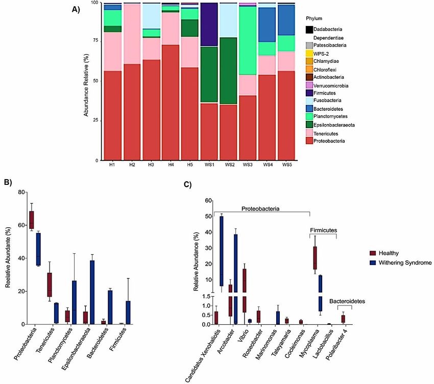
Microorganisms 2020, 8, 1411                                                                        2 of 13

to ensure the well-being of this industry. Aquaculture of abalone, a marine herbivorous gastropod,
including red abalone and Japanese abalone (Haliotis rufescens and Haliotis discus hannai, respectively),
is a promising avenue to achieve this goal, based on a confluence of factors, such as adequate
seawater temperature and availability of protected coastal areas for abalone culture, competitive
labor cost and an abundant supply of kelp as abalone food [2]. Most of Chilean abalone aquaculture
involves intensive farming of red abalone due to facile husbandry management during its life cycle [3].
However, infectious diseases might jeopardize the success of abalone farming in Chile. Withering
syndrome (WS) is an infectious disease that was introduced to Chile by importing infected red
abalone [4]. The etiology is a rickettsia-like organism identified as the gastrointestinal intracellular
prokaryote Candidatus Xenohaliotis californiensis [5]. WS is characterized by a chronic degenerative
condition that causes important economic loss, particularly a severely shrunken body, in abalone
aquaculture. Candidatus Xenohaliotis californiensis infects gastrointestinal epithelia provoking severe
morphological abnormalities within the digestive gland of abalone, which ends in physiological
starvation followed by anorexia, cachexia (i.e., absorption of pedal musculature), lethargy and
death [5]. However, whether digestive-tract-associated microbiota is modulated in abalone with WS
has not been reported to date. It is well known that the gastrointestinal tract harbors an enormous
number of microbes within the lumen, which constitute the gut microbiota, an ecosystem in dynamic
equilibrium [6]. Gut microbiota carry out essential functions, such as breakdown of foods, uptake of
nutrients, enhancement of intestinal development, development and stimulation of mucosal immune
system, and antagonistic effects against pathogens, all of which are of relevance to the well-being of the
host [6,7]. Host immune system can be modulated by gut microbiota via multiple factors, which include
microbial components and their metabolites [7]. Indeed, some infectious diseases have their origin in
the disruption of the established community structure and subsequent function changing the overall
balance between the microbiota and host, resulting in altered infection susceptibility [8]. Taking this
antecedent into account, it is of interest to determine whether WS affects the composition of digestive
tract microbiota in abalone, since this is a first step to elucidate how does microbial community respond
to WS. Thus, the aim of this study was to characterize the composition of microbiota associated with
digestive gland in red abalone with WS. Knowing this aspect of the disease can contribute to a better
understanding of the pathogenesis of WS as well as help to implement strategies, including the use of
probiotics and/or prebiotics to improve growth performance, inhibition of adherence and colonization
of pathogenic bacteria, immune response and WS resistance in red abalone farming.

2. Materials and Methods

2.1. Sample Collection
     Red abalone ranging in size (defined as the maximum length of the elliptical shell) from 35 to 90 mm,
were collected from an abalone farm center located at Región de Coquimbo, Chile. During farming,
specimens were fed a commercial diet ABLKELP® (specifications at https://www.kelproducts.com/
aquacultural). Distinction between healthy specimens (n = 5) and specimens with WS disease (n = 5)
was purely based on the presence of morphological and histological signs. This is because the presence
of the pathogens per se is not a feasible criterion for grouping red abalones in healthy or with WS
disease, as previously suggested [9]. Moreover Friedman et al. [10] found both low and non-significant
Spearman rank correlation coefficients between intensity of rickettsia-like organism in abalones with WS
and morphological and histological signs (Figure S1). After collection, specimens were placed in sterile
bags and preserved on ice until sent to laboratory, where they were euthanized by freezing. All samples
were processed within 24 h of collection at Laboratorio de Biotecnología de los Alimentos at Instituto
de Nutrición de los Alimentos (INTA) of Universidad de Chile for further analysis. Tissue samples
from digestive gland were dissected from specimens, flash frozen in liquid nitrogen and further stored
at −80 ◦ C until DNA extraction to perform microbiota analysis.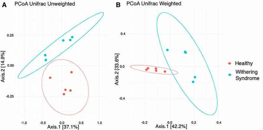
Microorganisms 2020, 8, 1411                                                                        3 of 13

2.2. DNA Extraction and Sequencing
     DNA was extracted from digestive gland lysed homogenates using QIAamp PowerFecal DNA
Kit (Qiagen, Germantown, MD, USA) according to manufacturer’s protocol. The V4 region of the
16S rRNA gene was amplified following the fusion primer method using primers 515F and 806R
as described [11]. The resulting amplicons were of suitable length to be used in the Illumina® Inc.
(Shirley, NY, USA) sequencing platform. All PCR reactions were performed in duplicates in a 30-µL
reaction mixture containing 1.5 U (5 U/µL) GoTaq® G2 Flexi DNA Polymerase (Promega, Madison,
WI, USA), 6 µL of Buffer (5×), 2.4 µL of Mg (25 mM), 1.2 µL of nucleotide mix (5 mM each), 0.3 µL
of primers (20 µM) and 18.5 µL of nuclease-free water. In addition, a negative PCR control without
DNA template was run. PCR conditions included an initial denaturation at 94 ◦ C for 5 min, followed
by 35 cycles of denaturation at 94 ◦ C for 30 s, annealing at 56 ◦ C for 30 s, and extension at 68 ◦ C for
45 s. After the procedure, amplicons from each sample were pooled and run on a 1% agarose gel.
Amplicon concentrations were determined using Qubit® dsDNA HS Assay kit (Life Technologies,
Grand Island, NY, USA). Subsequently, amplicons were purified with QIAquick® PCR Purification
kit (Qiagen, Germantown, MD, USA). Libraries were sequenced on the paired-end Illumina platform
Hiseq PE250 adapted for 300-bp paired-end reads at CD Genomics (http://www.cd-genomics.com).

2.3. Bioinformatic Analysis
     Quality-filtered reads were assembled into error-corrected amplicon sequence variants (ASVs)
using Devisive Amplicon Denoising Algorithm (DADA2) v1.6.0 microbiome pipeline (available at
https://github.com/benjjneb/dada2) to identify the presence and abundance of different microbial taxa
based on the assembly of the 16S rRNA sequence reads. Forward and reverse reads were truncated at
285 and 275 bp, respectively, by using read quality scores for dataset via filterAndTrim function set with
standard parameters (maxN = 0, truncQ = 2, and maxEE = 2). Singleton sequences were automatically
removed by DADA2’s error model, followed by a sample inference step using the inferred error model.
In addition, chimeric sequences were removed using removeBimeraDenovo function. Assembled
ASVs were assigned into the corresponding taxonomy (phylum to genus) level using Silva Database
(Silva) naïve Bayesian classifier (implemented in DADA2) and “Silva nr v132 train set” [12]. Using R
package Phyloseq [13], we eliminated any ASV without a bacterial phylum assignment, and also those
assigned to Cyanobacteria/Chloroplast. Using DADA2, no rarefying of sequence reads was necessary.
Finally, Illumina next-generation DNA sequences were deposited in the Sequencing Read Archive
(SRA) of the National Centre for Biotechnology Information under Bioproject accession PRJNA636409,
SRA run accessions SRX8460649-SRX8460658.

2.4. Ethical Notes
   The study was conducted in accordance with the guidelines of the Bioethics and Biosecurity
Committee of Instituto de Nutrición and Tecnología de los Alimentos (INTA) at Universidad de Chile.

2.5. Statistical Analysis
      Statistical analyses were performed using “R” v. 3.4.3 (http://www.R-project.org). R Packages
Phyloseq and Vegan [14] were used for microbiota data analyses. Alpha diversity measured by Shannon
and Simpson diversity index and species richness measured by Chao1 was calculated using Phyloseq.
Normality was tested with Shapiro–Wilk test and alpha diversity indexes were further analyzed for
differences using a Student’s t-test with a 5% of significance level. Unweighted and Weighted UniFrac
distances were calculated as β diversity measures using Phyloseq package in R. To statistically test the
homogeneity of microbial community composition, we performed permutational multivariate analysis
of variance (PERMANOVA) using package Vegan analyses on distance metrics. Differential taxa
abundance was performed using LefSe [15]. This method involves Kruskal–Wallis (KW) sum-rank
test between classes of data followed by (unpaired) Wilcoxon rank-sum test to conduct pairwise tests
Microorganisms 2020, 8, 1411 4 of 13
among subclasses. LDA is then used to estimate the effect size for each of the identified taxa. We used
LEfSe (Galaxy Version 1.0) with default parameters (KW = 0.05; Wilcoxon = 0.05; LDA score threshold
= 2.0) as well as using the all-against-all strategy for multi-class analysis. All other comparisons were
made using either Welch’s t-test or Kruskal–Wallis (KW). Mann–Whitney U test was conducted to
detect differences in Mycoplasma/Candidatus Xenohaliotis californiensis ratio between groups.
3. Results
3.1. High-Throughput Sequence Data
A total of 1,018,662 initial raw reads were obtained, from which 646,093 reads were retained after
removing low-quality reads and chimeras (Table S1). Further, 306,616 reads were measured in the
group of healthy red abalones and 339,477 reads were measured in the group of red abalones with WS.
3.2. Analysis of Microbiota Diversity
No significant differences in richness (Chao1: p < 0.739) and either alpha diversity index
(Shannon: p < 0.283; Simpson: p < 0.140) between healthy red abalones and red abalones with WS
were observed after t-test (Figure 1). Regarding beta diversity, the composition of digestive gland
microbiota was significantly (p = 0.009) different between healthy red abalones and red abalones
with WS when considering weighted distances (quantitative UniFrac phylogenetic distance matrices).
However, no differences (p = 0.177) between groups were detected when considering unweighted
distances (qualitative UniFrac phylogenetic distance matrices). In order to depict similarity in
digestive gland microbiota obtained from both healthy red abalones and red abalones with WS
(based on unweighted and weighted UniFrac analyses) the principal coordinates plot (PCoA) was
used (Figure 2A and B, respectively). In the PCoA of the unweighted UniFrac analyses, the two main
components explained 52% of the total variance (axis 1, 37.1%; axis 2, 14.9%) while in the PCoA of the
weighted UniFrac analyses, the two main components explained 75.8% of the total variance (axis 1,
42.2%; axis 2, 33.6%).Microorganisms 2020, 8,
Microorganisms 1411
2020, 8, x FOR PEER REVIEW 5 of 13
5 of 13
Microorganisms 2020, 8, x FOR PEER REVIEW 5 of 13
1. Comparison
FigureFigure of alpha
1. Comparison diversity
of alpha indexes;
diversity indexes;Chao
Chao 1, Shannonand
1, Shannon andSimpson,
Simpson, between
between healthy
healthy red red
Figure
abalones and 1.
redComparison affected
of alpha diversity indexes;syndrome
Chao 1, Shannon and Simpson, between scale
healthy red
abalones andabalones withwith
red abalones affected withering
withering syndrome disease.
disease.Note
Notethe
the different
different scale in Y-
in Y-axis
abalones and red abalones affected with withering syndrome disease. Note the different scale in Y-
due toaxis
different
due to indexes
different ranging values.values.
indexes ranging
axis due to different indexes ranging values.
FigureFigure 2. Principal
2. Principal coordinatesanalysis
coordinates analysis(PCoA)
(PCoA) of
of the
the bacterial
bacterialcommunities
communities derived fromfrom
derived the the
Figure 2. Principal coordinates analysis (PCoA) of the bacterial communities derived from the
unweighted
unweighted (A) (A) and
and weigthed
weigthed (B)(B) UniFrac
UniFrac distancematrix.
distance matrix. Circles
Circles represent
representindividual samples
individual fromfrom
samples
unweighted (A) and weigthed (B) UniFrac distance matrix. Circles represent individual samples from
red abalone digestive gland microbiota. Red circles correspond to samples from healthy abalones
(n = 5) and blue light circles correspond to samples from red abalones affected with withering syndrome
disease (n = 5).Microorganisms 2020, 8, 1411 6 of 13
3.3. Taxonomic Composition and Differential Abundance of Bacterial Communities of Digestive Gland of
Healthy Red Abalone and Red Abalone with WS Disease
Taxonomic composition reveled Proteobacteria, Tenericutes and Planctomycetes were the dominant
phyla in digestive gland microbiota from healthy red abalones (62.8%, 23.4% and 4.8%, respectively)
while Proteobacteria, Epsilonbacteraeota and Planctomycetes were the most abundant phyla in red abalones
with WS (44.6%, 15.5% and 12.3%, respectively; Figure 3A,B). At the genus level, the dominant bacteria
genus in digestive gland microbiota from healthy red abalones were Mycoplasma (23.1%), Vibrio (8.9%)
and Arcobacter (3.1%) (Figure 3C). Candidatus Xenohaliotis californiensis and Arcobacter were the
dominant bacteria genus detected in the digestive gland microbiota in red abalones with WS (25.1% and
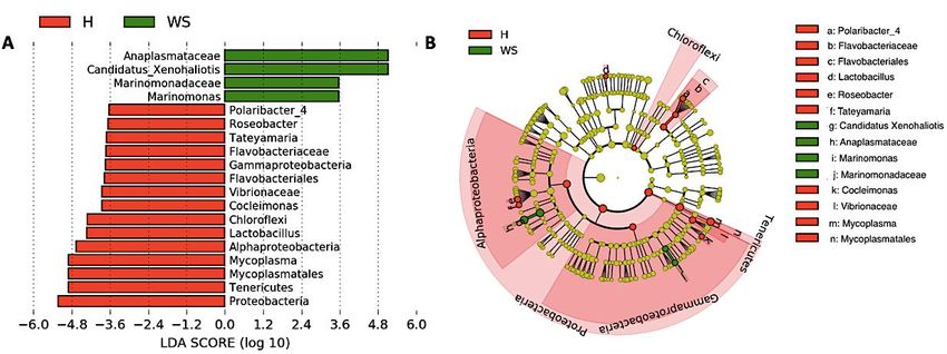
15.5%, respectively; Figure 3C). Further, significant differences in relative abundance (LDA effect side
score ≥ 3.5; Figure 4A) at different taxon levels (using phylum to genus-level data) between healthy red
abalones and red abalones with WS were detected by LEfSe analysis. These differences are illustrated in
a cladogram in Figure 4B. Phylum Proteobacteria, Tenericutes and Chloroflexi were differentially abundant
in healthy red abalones compared with red abalones with WS (Figure 4A). Class Alphaproteobacteria
and Gammaproteobacteria were differentially abundant in healthy red abalones compared with red
abalones with WS. Order Flavobacteriales and Mycoplasmatales were significantly more abundant in
healthy red abalones compared with red abalones with WS. At the family level, Flavobacteriaceae and
Vibrionaceae were significantly more abundant in healthy red abalones compared with red abalones
affected with WS, and at the bacteria genus level, Mycoplasma, Lactobacillus, Cocleimonas, Tateyamaria,
Roseobacter and Polaribacter_4 were significantly more abundant in healthy red abalones compared with
red abalones with WS. On the other hand, in red abalones with WS, bacterial family Anaplasmataceae and
Marinomonadaceae as well as bacteria genera Candidatus Xenohaliotis californiensis and Marinomonas
were significantly more abundant compared with healthy red abalones. Finally, we observed that
the Mycoplasma/Candidatus Xenohaliotis californiensis ratio was significantly (p = 0.011) lower in red
abalones affected with WS compared with healthy specimens (0.3 v/s 76.8, respectively).Microorganisms 2020, 8, 1411 7 of 13
Microorganisms 2020, 8, x FOR PEER REVIEW 7 of 13
(A)Relative
Figure3.3.(A)
Figure Relativeabundance
abundance(%) (%) atat phylum
phylum level for each
each sample
samplein indigestive
digestivegland
glandmicrobiota
microbiota
fromhealthy
from healthyredredabalone
abalone(H1,
(H1,H2,
H2,H3,
H3,H4H4 and
and H5)
H5) and
and red abalone affected
affectedwith
withwithering
witheringsyndrome
syndrome
disease(WS1,
disease (WS1,WS2,
WS2,WS3,
WS3,WS4
WS4and
andWS5).
WS5). Comparison
Comparison ofof digestive
digestive gland
gland microbiota
microbiotafrom
fromhealthy
healthyred
red
abalone (red boxes) and red abalone affected with Withering Syndrome (blue boxes) in
abalone (red boxes) and red abalone affected with Withering Syndrome (blue boxes) in terms of terms of relative
abundance
relative (%) at phylum
abundance level (B)level
(%) at phylum and (B)
genus
andlevel (C).level (C).
genus
Figure 4. Differences in digestive gland microbiota of healthy red abalones (H) compared with red
abalones with withering syndrome disease (WS). Analysis of 16S rRNA reveals the differential
composition of microbiota depending on the origin of the sample (H or WS). LEfSe was used toFigure 3. (A) Relative abundance (%) at phylum level for each sample in digestive gland microbiota
from healthy red abalone (H1, H2, H3, H4 and H5) and red abalone affected with withering syndrome
disease (WS1, WS2, WS3, WS4 and WS5). Comparison of digestive gland microbiota from healthy red
abalone (red boxes) and red abalone affected with Withering Syndrome (blue boxes) in terms of
Microorganisms 2020, 8, 1411 8 of 13
relative abundance (%) at phylum level (B) and genus level (C).
Differencesinindigestive
Figure4.4.Differences
Figure digestive gland
gland microbiota
microbiota of of healthy
healthy redred abalones
abalones (H)(H) compared
compared with
with red
red abalones with withering syndrome disease (WS). Analysis of 16S rRNA reveals
abalones with withering syndrome disease (WS). Analysis of 16S rRNA reveals the differential the differential
compositionof
composition of microbiota
microbiota depending
depending on
on the
the origin
origin of
of the
the sample
sample (H(H or
or WS).
WS). LEfSe
LEfSe was
wasused
usedtoto
determine the statistical significance and the effect size of the differential abundance of taxa between H
and WS. Section (A) shows LDA score of abundance of taxa; Section (B) shows cladogram showing
differentially abundant taxa (phylum to genus) of digestive gland microbiota in healthy red abalones
and red abalones with WS disease.
4. Discussion
Red abalone are the primary species of abalone aquaculture production in Chile. Withering syndrome
is a chronic, slow progressing disease causing important economic loss (i.e., low growth rate, reduced
weight condition index and high mortality) in red abalone farming. It has been said that the causative
agent is a rickettsia-like organism known as Candidatus Xenohaliotis californiensis, which initially infects
the epithelium of the gastroenteric system (i.e., posterior esophagus and digestive gland) followed by a
progressive deterioration in the mussel pedal muscle [5,10]. Although this is an infectious disease that
initially affects the digestive tract of abalone, no study on potential changes of microbiota associated
with digestive tract (i.e., digestive gland) of abalone with WS has been conducted. Thus, the present
study is the first report describing structural changes in the bacterial communities of digestive gland
microbiota in red abalones affected with WS.
Taxonomic composition analyses revealed differences in the ranking of major bacteria phyla of
digestive gland microbial communities between both conditions in red abalones (healthy specimens:
Proteobacteria > Tenericutes > Planctomycetes and specimens with WS disease: Proteobacteria > Epsilonbacteraeota
> Planctomycetes). However, in both conditions, Proteobacteria was the dominant bacteria phylum in
digestive gland microbiota. In agreement with our results, previous works have reported Proteobacteria
as dominant phylum in digestive tract tissues in several marine gastropods species. Neu et al. [16]
reported Proteobacteria to be the most abundant phylum in the digestive gland and whole-body
samples of intertidal gastropods species, such as owl limpet (Lottia gigantea), rough periwinkle
(Littorina keenae) and black turban snail (Chlorostoma funebralis). Ito et al. [17] found that Proteobacteria
as the most abundant phylum in the gut microbiota of marine gastropods species, including sea hare
(Dolabella auricularia) and sea snail (Batillus cornutus). Moreover, Proteobacteria have been reported
to be the dominant phylum in the gut microbiota of marine bivalve molluscs, such as blue mussel
(Mytilus edulis). However, the dominance of Proteobacteria phylum in digestive tract-associated
microbiota in molluscs is not consistent across the literature. For instance, Gobet et al. [18] detected
Fusobacteria as the dominant phylum of microbiota associated with the digestive gland in European
abalone (Haliotis tuberculate) fed different macroalgae-based diets (Palmaria palmata, Ulva lactuca,
Saccharina latissima, Laminaria digitate). Aceves et al. [19] observed Tenericutes was the dominantMicroorganisms 2020, 8, 1411 9 of 13
phylum, followed by Proteobacteria, Fusobacteria and Bacteroidetes in the digestive gland microbiota
of the freshwater mussel Alabama rainbows (Villosa nebulosa). Therefore factors, including host
species, digestive tract section, diet composition, seasonal patterns and environment (i.e., freshwater
or seawater, water temperature and biogeography) most likely influence digestive-system-associated
microbial communities of the host.
Our results indicate that heathy and diseased abalone had similar microbiota species
diversity, in terms of both species richness (Chao Index) and abundance levels (Shannon Index).
However, we observed significant differences between healthy and diseased groups in terms of the
phylogenetic distances of the microbiota composition when considering the weighted UniFrac analysis,
which accounts for the presence and absence of species. We can attribute this variation to Mycoplasma
species and Candidatus Xenohaliotis californiensis; for example, digestive gland microbiota were
strongly dominated (23.09%) by genus Mycoplasma in healthy red abalones whilst Mycoplasma’s
share of digestive gland microbiota decreased to 7.88% in red abalones with WS disease (Figure 3B).
On the contrary, Candidatus Xenohaliotis californiensis, causative agent of WS disease, was dominant
(25.12%) genus in digestive gland microbiota of red abalones with WS disease while Candidatus
Xenohaliotis californiensis’s share of digestive gland microbiota decreased to 0.7% in healthy red
abalones. Moreover, these observed differences in relative abundance were significant based on LEfSe
analysis, which revealed that order Mycoplasmatales and genus Mycoplasma were significantly more
abundant in healthy red abalones, whilst genus Candidatus Xenohaliotis californiensis was significantly
more abundant in red abalones with WS disease (Figure 4A). Interestingly, recent research has detected
Mycoplasma in high abundance in algae [20,21], which suggests that feeding an algae-based diet
to abalone might constitute a route of access into digestive system for Mycoplasma. Similar to our
study, previous works have reported that genus Mycoplasma either exhibits a preponderant share of
digestive system-associated microbiota or a ubiquitous presence (member of the core microbiota) in
digestive microbiota in several mollusc species. For example, Cicala et al. [22] found that the genus
Mycoplasma dominated gastrointestinal microbiome in northeast Pacific blue (Haliotis fulgens) and
yellow (Haliotis corrugate) abalone. Gobet et al. [18] found that the genus Mycoplasma, along with genera
Psychrilyobacter and Vibrio, were part of the core microbiota, constantly dominating the digestive gland
microbiota, in the European abalone. In other mollusc species, such as the freshwater mussel Alabama
rainbows, digestive gland microbiota was overwhelmingly dominated by genus Mycoplasma [19].
The authors suggested that Mycoplasma-like sequences could not be ascribed unequivocally to the
genus Mycoplasma and probably represent new lineages within the class Mollicutes. Aronson et al. [23]
observed that the genus Mycoplasma was predominant in the bacterial community of the digestive gland
and stomach of the gastropod deep-sea, bone-eating snail (Rubyspira osteovora), while Neu et al. [16]
highlighted that the high abundance of genus Mycoplasma was characteristic of microbial communities
of gastropod species, such as C. funebralis, C. eiseni and L. gigantea. Similarly, Pierce and Ward [24]
reported that the genus Mycoplasma was a member of the core bivalve gut microbiome in both eastern
oyster and blue mussel. Although Mycoplasma has been considered an obligate vertebrate pathogen
causing several diseases in humans [25], the consistently observed high abundance of this genus
in gastrointestinal-associated microbiota in healthy bivalves and gastropods species suggests that
Mycoplasma or Mycoplasma-like organisms play an important ecological role in the gut and health of
various marine molluscs. In line with this statement, Register et al. [26] proposed that Mycoplasma
might be highly host-specific and Wang et al. [27] suggested the existence of a mutualistic relationship
between Mycoplasma and their hosts. This idea was further supported by Cicala et al. [22] who
suggested that Mycoplasma contribute to their abalone host by playing pivotal roles in several metabolic
functions. In this regard, mutualism between Mycoplasma-like symbionts and their host is built upon
Mycoplasma’s inability to perform many basic metabolic functions due to their small genomes [28].
An example of this mutualism is that Mycoplasma require molecules such as sterols and fatty acids
provided by a host to survive [29] while Mycoplasma contribute to animals by digesting nutrient-poor
foods as well as supplying their hosts with amino sugars and simple carbohydrates. This is especiallyMicroorganisms 2020, 8, 1411 10 of 13
true, as the Mycoplasma genome presents a high number of genes involved in the degradation of
glycans, proteins and complex oligosaccharides [27,30,31]. Another benefit of this aforementioned
mutualism is that Mycoplasma might protect their hosts against microbial pathogen infections by
competitive inhibition of the binding site at the epithelium cell surface of the host. Mycoplasma strongly
adheres to host cell surfaces using outer membrane proteins, such as adhesin [32], thus blocking
microbial pathogens binding to the epithelium cell surface of host. In addition, Mycoplasma likely
protects their hosts against microbial pathogen infections by breaking down sialic acid residues of
outer membranes proteins used by microbial pathogens to avoid the host’s innate immune response,
based on the presence of sialic acid lyase genes in Mycoplasma genome [27,33].
In our study, Mycoplasma was the dominant genus in healthy specimens and was sharply reduced
in specimens with WS disease, while quite the opposite occurred with genus Candidatus Xenohaliotis
californiensis. Indeed, we observed the presence of this Candidatus Xenohaliotis californiensis in
digestive gland microbiota in two healthy specimens, suggesting that the mere presence of this
pathogen might not be enough to cause WS disease [9,22], and thus other cofactors might be required
to promote the proregression of the disease. Therefore, we hypothesize that WS disease requires the
presence of the Candidatus Xenohaliotis californiensis along with a significant drop in the relative
abundance of Mycoplasma, causing a concomitant decrease in Mycoplasma/Candidatus Xenohaliotis
californiensis ratio, as observed in our study. A potential mechanism of Mycoplasma-induced protection
might be based on the presence of sialic acid lyase activity degrading the α 2,3 sialic acid residues at the
receptor domains of that host cell, which are recognized by the outer membrane proteins A (OmpA) of
rickettsia-like organism [34]. The OmpA has previously been described as putative adhesin favoring
Rickettsia attachment to epithelial cells in vitro [35]. Further, this receptor–ligand interaction has been
referred to as essential for the efficient cellular entry of Anaplasma phagocytophilum, a member of the
order Rickettsiales, into human myeloid cells [36]. Remarkably, the authors described that competitive
inhibition of binding and entry using OmpA can be achieved by either a monoclonal antibody directed
against the sLex α 2,3 sialic acid residue of the P-selectin glycoprotein1 (PSGL-1) receptor or sialidase
treatment of host cell.
In addition, the genus Lactobacillus was significantly more abundant in healthy specimens in
our study. However, Lactobacillus’s share of digestive gland microbiota was quite low in healthy
red abalones (Figure 3B). Lactobacillus is a member of lactic acid bacteria (LAB), which have been
used as probiotics in animal production systems and fish aquaculture, due to their role in adjusting
intestinal environment, regulating intestinal mucosal immunity and maintaining intestinal barrier
function [37–39]. In shellfish aquaculture industry most of the reported beneficial effects of these
probiotics, including improved growth performance, enzymatic contribution to nutrition, inhibition of
adherence and colonization of pathogenic bacteria in digestive tract, modulation of gut microbiota,
and increase hematological parameters and immune response, have been observed in shrimp
farming [40]. On the other hand, less antecedents exist regarding LAB administration as probiotics in
marine mussel’s aquaculture [39,41–44]. Therefore, contrary to what has been observed in fish and
shrimp aquaculture species, our results suggest that Lactobacillus’s role adjusting intestinal environment
and promoting health in abalone host may be marginal.
In recent years, there has been growing consumer concern regarding the indiscriminate use of
antibiotics to treat infectious diseases in aquaculture farming due to the development and dissemination
of antibiotic resistance, food safety hazards and environmental issues, such as residue accumulation
and aquatic biodiversity toxicity [40,45]. As a reaction to this concern, in order to ensure safe
aquaculture food production, several government agencies have established maximum residual limits
for antibiotics in food [45]. In other more extreme cases, such as the European Union, the use of
antibiotics in food production has been banned since 2003 [40]. Therefore, efforts should be focused on
reducing antibiotics to safeguard the environment and ensure safety of consumers, feed industry and
aquaculture workers [45]. In this regard, the use of probiotics is a feasible supplementary strategy to
vaccine and chemicals in aquaculture [40]. Based on this fact, it is of interest to explore whether theMicroorganisms 2020, 8, 1411 11 of 13
administration of Mycoplasma to promote red abalone health could constitute a strategy to improve
productivity in red abalone farming. This is especially true since WS disease management should
be approached from a sustainable point of view considering aspects such as whether methods are
environmentally friendly, safe for human consumption and cost-effective.
5. Conclusions
This study provided evidence of differences in the structure of bacteria communities of red abalone
digestive microbiota between healthy specimens and specimens with WS disease. Mycoplasma and
Candidatus Xenohaliotis californiensis were the most abundant genera, and their relative abundance
most strongly contributed to the microbial profile variations between diseased and healthy abalone in
this study. Similar to previous studies, we observed that the mere presence of Candidatus Xenohaliotis
californiensis was not associated with WS in abalone; however, a decrease in the Mycoplasma/Candidatus
Xenohaliotis californiensis ratio appears to be more indicative of specimens affected with the disease.
Further research to explore the potential use of Mycoplasma as a probiotic to promote abalone health,
and thus improve productivity in farming, is required.
Supplementary Materials: The following are available online at http://www.mdpi.com/2076-2607/8/9/1411/s1,
Figure S1: Macroscopic and histologic images of healthy and WS-affected abalones. Table S1: Reads obtained
from healthy and disease specimens from Illumina sequencing.
Author Contributions: Conceptualization, J.R. and A.V.; methodology, R.R. and K.B.L.; formal analysis, N.C.;
investigation, R.R. and N.C.; writing—original draft preparation, A.V. and N.C.; writing—review and editing,
A.V., J.R., R.R. and K.B.L. All authors have read and agreed to the published version of the manuscript.
Funding: This research was funded by FONDECYT, grant number 1200523.
Acknowledgments: N.C. acknowledges support from Lab Biotecnología.
Conflicts of Interest: The authors declare no conflict of interest.
References
1. FAO. The State of World Fisheries and Aquaculture 2018. Available online: http://www.fao.org/state-of-
fisheries-aquaculture (accessed on 18 October 2018).
2. Flores-Aguilar, R.A.; Gutiérrez, A.; Ellwanger, A.; Searcy-Bernal, R. Development and current status of
abalone aquaculture in Chile. J. Shellfish Res. 2007, 26, 705–711. [CrossRef]
3. Enríquez, R.; Villagrán, R. La experiencia del desarrollo del cultivo de abalón (Haliotis spp.) en Chile:
Oportunidades y desafíos. OIE Rev. Sci. Tech. 2008, 27, 103–112. [CrossRef] [PubMed]
4. Brokordt, K.; González, R.; Farías, W.; Winkler, F.E.; Lohrmann, K.B. First insight into the heritable variation
of the resistance to infection with the bacteria causing the withering syndrome disease in Haliotis rufescens
abalone. J. Invertebr. Pathol. 2017, 150, 15–20. [CrossRef] [PubMed]
5. Crosson, L.M.; Wight, N.; VanBlaricom, G.R.; Kiryu, I.; Moore, J.D.; Friedman, C.S. Abalone withering
syndrome: Distribution, impacts, current diagnostic methods and new findings. Dis. Aquat. Org. 2014, 108,
261–270. [CrossRef]
6. Honda, K.; Littman, D.R. The microbiome in infectious disease and inflammation. Annu. Rev. Immunol. 2012,
30, 759–795. [CrossRef]
7. Corthier, G.; Doré, J. Une ère nouvelle dans le domaine des interactions entre le microbiote et la santé
humaine. Gastroenterol. Clin. Biol. 2010, 34, S1–S6. [CrossRef]
8. Libertucci, J.; Young, V.B. The role of the microbiota in infectious diseases. Nat. Microbiol. 2019, 4, 35–45.
[CrossRef]
9. Cáceres-Martínez, J.; Vásquez-Yeomans, R.; Flores-Saaib, R.D. Intracellular prokaryote Xenohaliotis
californiensis in abalone Haliotis spp. from Baja California, México. Cienc. Pesq. 2011, 19, 5–11.
10. Friedman, C.S.; Biggs, W.; Shields, J.D.; Hedrick, R.P. Transmission of withering syndrome in black abalone,
Haliotis cracherodii Leach. J. Shellfish Res. 2002, 21, 817–824.Microorganisms 2020, 8, 1411 12 of 13
11. Caporaso, J.G.; Kuczynski, J.; Stombaugh, J.; Bittinger, K.; Bushman, F.D.; Costello, E.K.; Fierer, N.; Peña, A.G.;
Goodrich, J.K.; Gordon, J.I.; et al. QIIME allows analysis of high-throughput community sequencing data.
Nat. Methods 2010, 7, 335–336. [CrossRef]
12. Wang, Q.; Garrity, G.M.; Tiedje, J.M.; Cole, J.R. Naive Bayesian classifier for rapid assignment of rRNA
sequences into the new bacterial taxonomy. Appl. Environ. Microbiol. 2007, 73, 5261–5267. [CrossRef]
[PubMed]
13. McMurdie, P.J.; Holmes, S. Phyloseq: An R package for reproducible interactive analysis and graphics of
microbiome census data. PLoS ONE 2013, 8, e61217. [CrossRef] [PubMed]
14. Oksanen, J.; Blanchet, F.G.; Kindt, R.; Legendre, P.; O’Hara, R.B.; Simpson, G.L.; Solymos, P.; Stevens, M.H.H.;
Wagner, H. Vegan: Community Ecology Package 2011. R Package Version 1.17–10. Available online:
https://cran.r-project.org/web/packages/vegan/index.html (accessed on 10 March 2019).
15. Segata, N.; Bornigen, D.; Morgan, X.C.; Huttenhower, C. PhyloPhlAn is a new method for improved
phylogenetic and taxonomic placement of microbes. Nat. Commun. 2013, 4. [CrossRef] [PubMed]
16. Neu, A.T.; Allen, E.E.; Roy, K. Diversity and composition of intertidal gastropod microbiomes across a major
marine biogeographic boundary. Environ. Microbiol. Rep. 2019, 11, 434–447. [CrossRef]
17. Ito, M.; Watanabe, K.; Maruyama, T.; Mori, T.; Niwa, K.; Chow, S.; Takeyama, H. Enrichment of bacteria
and alginate lyase genes potentially involved in brown alga degradation in the gut of marine gastropods.
Sci. Rep. 2019, 9, 2129. [CrossRef]
18. Gobet, A.; Mest, L.; Perennou, M.; Dittami, S.M.; Caralp, C.; Coulombet, C.; Huchette, S.; Roussel, S.;
Michel, G.; Leblanc, C. Seasonal and algal diet-driven patterns of the digestive microbiota of the European
abalone Haliotis tuberculata, a generalist marine herbivore. Microbiome 2018, 6, 60. [CrossRef]
19. Aceves, A.K.; Johnson, P.; Bullard, S.A.; Lafrentz, S.; Arias, C.R. Description and characterization of the
digestive gland microbiome in the freshwater mussel Villosa nebulosa (Bivalvia: Unionidae). J. Molluscan Stud.
2018, 84, 240–246. [CrossRef]
20. Hollants, J.; Leroux, O.; Leliaert, F.; Decleyre, H.; De Clerck, O.; Willems, A. Who is in there? Exploration of
endophytic bacteria within the siphonous green seaweed Bryopsis (Bryopsidales, Chlorophyta). PLoS ONE
2011, 6, e26458. [CrossRef]
21. Hollants, J.; Leliaert, F.; Verbruggen, H.; Willems, A.; De Clerck, O. Permanent residents or temporary lodgers:
Characterizing intracellular bacterial communities in the siphonous green alga Bryopsis. Proc. R. Soc. B.
2013, 280, 20122659. [CrossRef]
22. Cicala, F.; Cisterna-Céliz, J.A.; Moore, J.D.; Rocha-Olivares, A. Structure, dynamics and predicted functional
role of the gut microbiota of the blue (Haliotis fulgens) and yellow (H. corrugata) abalone from Baja California
Sur, Mexico. PeerJ 2018, 6, e5830. [CrossRef]
23. Aronson, H.S.; Zellmer, A.J.; Goffredi, S.K. The specific and exclusive microbiome of the deep-sea bone-eating
snail, Rubyspira osteovora. FEMS Microbiol. Ecol. 2017, 93. [CrossRef] [PubMed]
24. Pierce, M.L.; Ward, J.E. Gut microbiomes of the eastern oyster (Crassostrea virginica) and the blue mussel
(mytilus edulis): Temporal variation and the influence of marine aggregate-associated microbial communities.
MSphere 2019, 4, e00730-19. [CrossRef] [PubMed]
25. Razin, S.; Yogev, D.; Naot, Y. Molecular biology and pathogenicity of mycoplasmas. Microbiol. Mol. Biol. Rev.
1998, 62, 1094–1156. [CrossRef]
26. Register, K.B.; Thole, L.; Rosenbush, R.F.; Minion, F.C. Multilocus sequence typing of Mycoplasma bovis
reveals host-specific genotypes in cattle versus bison. Vet. Microbiol. 2015, 175, 92–98. [CrossRef]
27. Wang, Y.; Huang, J.M.; Wang, S.L.; Gao, Z.M.; Zhang, A.Q.; Danchin, A.; He, L.S. Genomic characterization
of symbiotic mycoplasmas from the stomach of deep-sea isopod bathynomus sp. Environ. Microbiol. 2016,
18, 2646–2659. [CrossRef] [PubMed]
28. Brown, D.R.; Zacher, L.A.; Wendland, L.D.; Brown, M.R. Emerging mycoplasmoses in wildlife. In Mycoplasmas
Molecular Biology, Pathogenicity and Strategies for Control, 1st ed.; Blanchard, A., Browing, G., Eds.; Horizon
Bioscience: Norwich, UK, 2005; pp. 383–414.
29. Ludwig, W.; Euzéby, J.; Whitman, W.B. Road map of the phyla Bacteroidetes, Spirochaetes, Tenericutes
(Mollicutes), Acidobacteria, Fibrobacteres, Fusobacteria, Dictyoglomi, Gemmatimonadetes, Lentisphaerae,
Verrucomicrobia, Chlamydiae, and Planctomycetes. In Bergey’s Manual of Systematic Bacteriology; Krieg, N.R.,
Ludwig, W., Whitman, W., Hedlund, B.P., Paster, B.J., Staley, J.T., Ward, N., Brown, D., Parte, A., Eds.;
Springer: New York, NY, USA, 2010; Volume 4, pp. 1–19.Microorganisms 2020, 8, 1411 13 of 13
30. Fraune, S.; Zimmer, M. Host-specificity of environmentally transmitted mycoplasma-like isopod symbionts.
Environ. Microbiol. 2008, 10, 2497–2504. [CrossRef] [PubMed]
31. Duperron, S.; Pottier, M.A.; Leger, N.; Gaudron, S.M.; Puillandre, N.; Le Prieur, S.; Sigwart, J.D.; Ravaux, J.;
Zbinden, M. A tale of two chitons: Is habitat specialisation linked to distinct associated bacterial communities?
FEMS Microbiol. Ecol. 2013, 83, 552–567. [CrossRef]
32. Henrich, B.; Feldmann, R.C.; Hadding, U. Cytoadhesins of Mycoplasma hominis. Infect. Immun. 1993, 61,
2945–2951. [CrossRef]
33. Severi, E.; Hood, D.W.; Thomas, G.H. Sialic acid utilization by bacterial pathogens. Microbiology 2007, 153,
2817–2822. [CrossRef]
34. Pérez-Acosta, J.; Martínez-Porchas, M.; Gollas-Galván, T.; Martínez-Córdova, L.R.; Gutiérrez-Millán, L.E.;
López-Torre, M. Proteínas transmembranales de organismos tipo rickettsia (OTR) en animales acuáticos:
Factores de adherencia, invasión e infección. Rev. Biol. Mar. Oceanogr. 2017, 52, 19–32. [CrossRef]
35. Li, H.; Walker, D.H. rOmpA is a critical protein for the adhesion of Rickettsia rickettsii to host cells.
Microb. Pathog. 1998, 24, 289–298. [CrossRef] [PubMed]
36. Palmer, G.H.; Noh, S.M. Rickettsial entry into host cells: Finding the keys to unlock the doors. Infect. Immun.
2012, 80, 3746–3747. [CrossRef] [PubMed]
37. Nayak, S.K. Probiotics and immunity: A fish perspective. Fish Shellfish Immunol. 2010, 29, 2–14. [CrossRef]
38. Catalán, N.; Villasante, A.; Wacyk, J.; Ramírez, C.; Romero, J. Fermented Soybean Meal Increases Lactic Acid
Bacteria in Gut Microbiota of Atlantic Salmon (Salmon salar). Probiotics Antimicrob. Proteins 2018, 10, 566–576.
[CrossRef]
39. Hoseinifar, S.H.; Sun, Y.Z.; Wang, A.; Zhou, Z. Probiotics as Means of Diseases Control in Aquaculture, a
Review of Current Knowledge and Future Perspectives. Front. Microbiol. 2018, 9, 2429. [CrossRef]
40. Ringø, E. Probiotics in shellfish aquaculture. Aquac. Fish. 2020, 5, 1–27. [CrossRef]
41. Martínez, P.; Ibáñez, A.L.; Monroy, O.A.; Ramírez, H.C. Use of probiotics in aquaculture. ISRN Microbiol.
2012, 2012, 916845. [CrossRef]
42. Mohapatra, S.; Chakraborty, T.; Kumar, V.; DeBoeck, G.; Mohanta, K.N. Aquaculture and stress management:
A review of probiotic intervention. J. Anim. Physiol. Anim. Nutr. 2013, 97, 405–430. [CrossRef]
43. Wang, Y.B.; Li, J.R.; Lin, J. Probiotics in aquaculture: Challenges and outlook. Aquaculture 2008, 281, 1–4.
[CrossRef]
44. Pandiyan, P.; Balaraman, D.; Thirunavukkarasu, R.; Jothi, E.D.; Subaramaniyan, K.; Manikkam, S.;
Sadayappan, B. Probiotics in aquaculture. Drug Invent. Today 2013, 5, 55–59. [CrossRef]
45. Lulijwa, R.; Rupia, E.J.; Alfaro, A.C. Antibiotic use in aquaculture, policies and regulation, health and
environmental risk: A review of the top 15 major producers. Rev. Aquacult. 2019, 12, 640–663. [CrossRef]
© 2020 by the authors. Licensee MDPI, Basel, Switzerland. This article is an open access
article distributed under the terms and conditions of the Creative Commons Attribution
(CC BY) license (http://creativecommons.org/licenses/by/4.0/).You can also read