A Rabbit Model of Aqueous-Deficient Dry Eye Disease Induced by Concanavalin A Injection into the Lacrimal Glands: Application to Drug Efficacy ...
←
→
Page content transcription
If your browser does not render page correctly, please read the page content below
Journal of Visualized Experiments                                                                                             www.jove.com

Video Article
A Rabbit Model of Aqueous-Deficient Dry Eye Disease Induced by
Concanavalin A Injection into the Lacrimal Glands: Application to Drug
Efficacy Studies
                      1               2,3             2
Robert A. Honkanen , Liqun Huang , Basil Rigas
1
Department of Ophthalmology, Stony Brook University
2
Department of Preventive Medicine, Stony Brook University
3
Medicon Pharmaceuticals, Inc.

Correspondence to: Robert A. Honkanen at Robert.Honkanen@stonybrookmedicine.edu

URL: https://www.jove.com/video/59631
DOI: doi:10.3791/59631
Keywords: Medicine, Issue 155, dry eye, dry eye disease, dry eye syndrome, rabbit model, Concanavalin A, tear break-up time, Schirmer’s tear
test, tear osmolarity, rose bengal staining, ocular drug development
Date Published: 1/24/2020
Citation: Honkanen, R.A., Huang, L., Rigas, B. A Rabbit Model of Aqueous-Deficient Dry Eye Disease Induced by Concanavalin A Injection into the
Lacrimal Glands: Application to Drug Efficacy Studies. J. Vis. Exp. (155), e59631, doi:10.3791/59631 (2020).

Abstract
    Dry eye disease (DED), a multifactorial inflammatory disease of the ocular surface, affects 1 in 6 humans worldwide with staggering implications
    for quality of life and health care costs. The lack of informative animal models that recapitulate its key features impedes the search for new
    therapeutic agents for DED. Available DED animal models have limited reproducibility and efficacy. A model is presented here in which DED
    is induced by injecting the mitogen concanavalin A (Con A) into the orbital lacrimal glands of rabbits. Innovative aspects of this model are the
    use of ultrasound (US) guidance to ensure optimal and reproducible injection of Con A into the inferior lacrimal gland; injection of Con A into
    all orbital lacrimal glands that limits compensatory production of tears; and use of periodic repeat injections of Con A that prolong the state of
    DED at will. DED and its response to test agents are monitored with a panel of parameters that assess tear production, the stability of the tear
    film, and the status of the corneal and conjunctival mucosa. They include tear osmolarity, tear break-up time, Schirmer's tear test, rose bengal
    staining, and tear lactoferrin levels. The induction of DED and the monitoring of its parameters are described in detail. This model is simple,
    robust, reproducible, and informative. This animal model is suitable for the study of tear physiology and of the pathophysiology of DED as well as
    for the assessment of the efficacy and safety of candidate agents for the treatment of DED.

Video Link
    The video component of this article can be found at https://www.jove.com/video/59631/

Introduction
                                                                                        1,2,3,4                                                   5,6
    Dry eye disease (DED) is a chronic condition with high prevalence and morbidity        . Inflammation plays a key role in its pathogenesis . The
    pathophysiology of DED is conceptualized as deriving from either under-production or over-evaporation of tears; the former is also known as
                              7
    aqueous-deficient DED . Sjögren's syndrome, an extensively studied prototypical cause of DED, affects primarily the lacrimal glands (LGs) and
    is a striking example of their importance in the pathogenesis of DED. DED is often treated with artificial tears which provide temporary relief, or
    with cyclosporine or lifitegrast, both of which suppress ocular inflammation. None of the available treatments for DED are optimal, necessitating
                                       8,9
    the development of new agents .

    The search for new therapeutic agents for DED is hampered by three major challenges: the lack of a recognized druggable molecular target,
    which may be elusive given the pathophysiological complexity of DED; the sparsity of promising agents; and the lack of animal models that
    recapitulate key features of DED.

    As with most drug development efforts, informative animal models of DED are a crucial investigative tool, notwithstanding the axiomatic
    statement that no animal model completely recapitulates a human disease. Mouse, rat, and rabbit models of DED are the most commonly
                                                          10,11
    used while dogs and primates are used infrequently         . Most of the more than 12 rabbit DED models reported to date attempt to reduce tear
                                                                       12,13,14,15,16
    production by either removing LGs or by impeding their function                  . Such approaches include the surgical resection of the ILG; closure of
    its excretory duct; and impairing LG function by irradiation or injection of one of the following: activated lymphocytes, mitogens, botulinum toxin,
    atropine, or benzalklonium. Major limitations of these methods are their inconsistency and the frequent partial suppression of tear production.
                                                                                                                                                        17
    Concanavalin A (Con A), a lectin of plant origin, is a potent stimulator T-cell subsets and has been used in experimental models of hepatitis
             18
    and DED . The original Con A-based model was reported to offer significant advantages, including its relative simplicity; inflammatory cell influx
    in the LGs, mimicking diseases such as Sjogren's; stimulation of the proinflammatory cytokines IL-1β, IL-8, and TGF-β1; reduced tear function

Copyright © 2020 Creative Commons Attribution-NonCommercial-NoDerivs 3.0 Unported                            January 2020 | 155 | e59631 | Page 1 of 20
License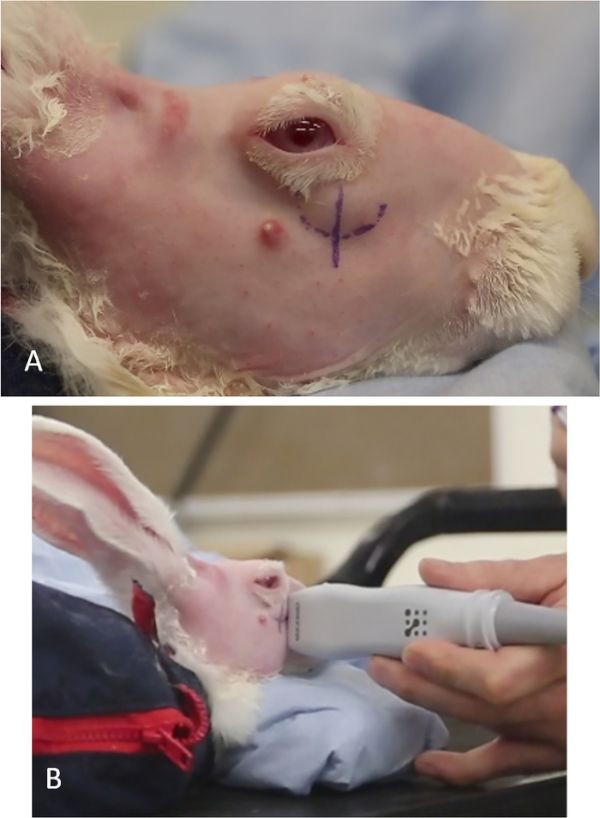
Journal of Visualized Experiments                                                                                        www.jove.com

  monitored by measuring tear fluorescein clearance and tear break-up time (TBUT); and drug responsiveness shown for an anti-inflammatory
  corticosteroid.

  When this promising method was applied, in addition to its advantages, limitations were identified that necessitated its overall revision and
  drastic improvements. Three critical shortcomings of the method are documented. First, the model was an acute one; the induced DED subsided
  after about 1 week. Second, the response of the animals was inconsistent. As demonstrated, in "blind" transcutaneous injections to the Inferior
  LG (ILG), Con A was delivered only randomly to the targeted gland. Detailed study of the anatomy of the ILG revealed that its size could vary
                   19
  as much as 4-fold making such injections "hit-or-miss" efforts. Finally, even when the ILG was injected, the superior LG (SLG) frequently
  compensated for the reduced tear flow, making the model problematic.

  These key limitations were overcome by introducing three modifications to the method, generating a superior animal model of DED. First, the
  injection of Con A into the ILG was performed under ultrasound (US) guidance, ensuring that Con A entered the gland. The success of the
  injection was confirmed by obtaining a post-injection US image, as shown in Figure 1. Second, to remove the compensatory tear contribution
  of the SLG, both the palpebral and orbital portions of this gland were injected with Con A. Finally, this acute model of DED was converted to a
  chronic one by repeated injections of Con A every 7-10 days. DED of 2 months' duration is readily achieved in these rabbits. The success of this
                                          19
  approach has been amply documented .

  As already mentioned, an important application of animal models of DED is to determine the efficacy and safety of candidate therapeutic
                                                                                                                                          20,21
  agents. The utility of this model was demonstrated by the study of phosphosulindac (OXT-328), a novel anti-inflammatory small molecule
                                                                                                   19
  administered as eye drops. Its efficacy was demonstrated based on a panel of parameters of DED . The relative simplicity and informative
  nature of this model also allowed side-by-side comparison of phosphosulindac to the two FDA approved drugs for DED, cyclosporine and
  lifitegrast, demonstrating its strong preclinical superiority.

Protocol
  All animal studies were approved by the Institutional Review Board of Stony Brook University and performed in accordance with the ARVO
  Statement for the Use of Animals in Ophthalmic and Vision Research.

1. Animals and housing
  1.   Acquire New Zealand White (NZW) rabbits weighing 2-3 kg.
  2.   House the rabbits singly in cages with strict temperature (65 ± 5 °F) and humidity (45 ± 5%) control. Lighting should have a 12 h on/off cycle.
  3.   Provide unlimited access to water and standard rabbit chow. Eliminate dietary enrichments as they may contain vitamin A that affects the eye.
  4.   Acclimate the animals for at least 2 weeks prior to baseline measures or induction of dry eye.

2. Methods of anesthesia and euthanasia
  NOTE: All procedures require mild sedation except for Con A injection that requires moderate sedation.

  1. For mild sedation, inject acepromazine (1 mg/kg) subcutaneously over the shoulders using a 26-gauge needle. Endpoint for mild sedation:
     animals maintain a relaxed head position with ear lobes no longer fully upright.
     NOTE: If the appropriate endpoint is not reached, an additional injection of acepromazine may be given. Animals should always remain
     awake, responsive to touching of their whiskers, and never show slowed breathing.
  2. For moderate sedation, first give the animals acepromazine as above. After the endpoint is reached (see the note above), give isoflurane
     using a gas mask with O2 flow set at 1 L/min and isoflurane delivery set to 5% (Figure 2).
  3. Administer isoflurane until the rabbit's body tone is completely relaxed and ears are completely floppy.
     NOTE: No compensatory muscle movements should occur when the animal is turned on its side; breathing always remains spontaneous.
  4. Spontaneous recovery occurs within 2-5 min: signs include spontaneous head movements and increased or normal muscular tone. After the
     experimental procedure is completed with moderate sedation, observe the rabbits for about 30 min or until their behavior returns to normal.
     NOTE: Ophthalmic ointment is not required during either form of sedation. 1) In mild sedation, animals are still alert and maintain a blink
     reflex. In moderate sedation, the inhibition of the blink reflex is so short that the ocular surface is not at risk. 2) Placement of ophthalmic
     ointment on the ocular surface precludes visualization of structures assessed during testing.
  5. Euthanasia: Use an overdose of intravenous pentobarbital (100 mg/kg).

3. Removal of nictitating membrane
  1. Perform the removal during the acclimation period (usually the first week) to allow complete and accurate evaluation of the cornea.
  2. Injection to the right nictitating membrane
     NOTE: If the nictitating membrane of both eyes is to be removed, it is simplest to do this in one session. Start with one eye and proceed as
     described. For clarity of description, this method starts with the right eye.
       1. Place the rabbit in an appropriately sized restraining bag.
       2. Induce mild sedation as described in step 2.1.
       3. Apply 25 μL of preservative-free lidocaine to the right eye using a micropipette.
       4. Place a flexible wire lid speculum between the eyelids.
       5. Using 0.3 forceps (or equivalent), grasp the nictitating membrane at its apex and extend it over the cornea.
       6. Inject lidocaine 1% with 1:100,000 epinephrine subconjunctivally into the base of the nictitating membrane using a 26-gauge sharp
            needle (Figure 3A). A moderate bleb should form over the nictitating membrane.

Copyright © 2020 Creative Commons Attribution-NonCommercial-NoDerivs 3.0 Unported                        January 2020 | 155 | e59631 | Page 2 of 20
License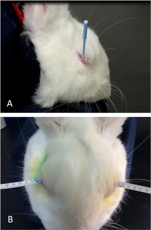
Journal of Visualized Experiments                                                                                           www.jove.com

        7. Remove the speculum.

  3. Perform an identical injection to the left nictitating membrane.
  4. Cutting the nictitating membrane
       1. After approximately 5 min, place the lid speculum back in the right eye. Grasp and retract the nictitating membrane at its apex using 0.3
           forceps (or similar).
       2. Cut the nictitating membrane off at its base using Westcott scissors or equivalent (Figure 3B).
           NOTE: Bleeding is minimal and does not typically require cautery. Nevertheless, a high-temperature battery cautery is always kept
           nearby in case additional hemostasis is needed.
       3. Remove the speculum.
       4. Place topical antibiotic ointment on the eye (e.g., neomycin, polymyxin, bacitracin, and hydrocortisone).

  5. Leave the Harderian gland intact. The Harderian gland is sometimes seen when the nictitating membrane is retracted.
     NOTE: If a large white mass or tissue elevation is seen in the nasal or superior subconjunctival region after the nictitating membrane is
     removed, the membrane was resected too close to its base allowing the Harderian gland to spontaneously prolapse. To prevent this in
     subsequent procedures, leave more of the nictitating membrane at the base.
  6. Allow the ocular surface to heal for at least 1 week before further manipulations are made or ocular surface assays are performed.

4. Measurement of dry eye parameters and collection of tear samples
  NOTE: Measure DED parameters based on study protocol needs (e.g., at baseline and specified time points thereafter). Measurements for DED
  should be done in the following order, with rigorous effort to faithfully replicate them each time. Test all animals at approximately the same time of
  day (± 1 h) to minimize circadian variations. These measurements usually require a team of two investigators.

  1. Place the rabbit in a restraining bag. Induce mild sedation.
                       22
  2. Tear Osmolarity
       1. Manually blink the eyelids 5-10 times to evenly distribute the tear layer on the ocular surface.
       2. Gently retract the lower lid.
       3. Sample tears with the TearLab Osmometer at the junction of the palpebral and bulbar conjunctiva along the lower fornix, just posterior
           to the base of the truncated nictitating membrane.
       4. Measure osmolarity using the TearLab Osmolarity Test following the manufacturer's instructions.

  3. Tear break-up time (TBUT)
       1. Darken the room for this assay.
       2. Place a wire lid speculum between the eyelids.
       3. Apply a 50 µL drop of 0.2% fluorescein over the corneal surface using a micropipette. If even distribution of the fluorescein over the
           cornea is not obtained with the first drop, place a second drop.
       4. Immediately start a timer.
       5. Observe the pre-corneal tear film under a blue light. The TBUT is defined as the time taken to develop black dots, lines or obvious
           disruption of the fluorescein film (Figure 4). If needed, use surgical loupes that provide +1.50 magnification to better visualize early
           signs of break-up. Monitor for up to 1 min; if break-up as defined here occurs after 1 min, record TBUT for only 60 s.

  4. Schirmer's Tear Test (STT)
       1. Apply a 25 μL drop of preservative-free lidocaine to the ocular surface.
       2. Place a Weck cell surgical spear into the inferior fornix to absorb residual lidocaine and any tear fluid. If needed, use the lower eyelid to
          cover the proximal end of the sponge to help keep it in place (Figure 5A).
       3. After approximately 30 s, remove the Weck sponge.
       4. Immediately insert a Schirmer's tear test strip into the space between the cornea and the palpebral conjunctiva at the mid-point of the
          lower lid.
       5. Immediately start a timer (Figure 5B).
       6. After 5 min, measure the length of the moistened portion of the strip; this is the STT value.
       7. Perform measures in triplicate and report the average of the 3 readings as the STT value.

  5. Collection of tear samples
      1. To collect tear samples for assaying levels of various analytes in them such as lactoferrin, after the STT value is recorded at 5 min,
           leave the strip in place until wetting of at least 20 mm is obtained.
           NOTE: If adequate wetting does not occur after DED has been induced, advance the strip deeper into the lower fornix to help reach
           this endpoint in reasonable time.
      2. Cut the moistened strip and place immediately into 490 µL of chilled Tear Collection Buffer (4% BSA, 1M NaCl, 0.1% Tween-20 in PBS
           with proteinase inhibitor cocktail).
      3. Keep the samples on ice until they can be stored at -80 °C, where they should remain until assayed.

  6. Rose Bengal Staining (RBS)
      1. Apply 50 µL of 1% preservative-free lidocaine to the cornea using a micro-pipette.
      2. After 30 s, place 25 µL of 1% rose bengal on the ocular surface and manually blink the eyelid to distribute it evenly.
      3. Immediately start a timer.
      4. At 3.5 min, place a wire lid speculum between the lids.
      5. At 4.0 min, photograph the superior conjunctival and corneal surface (Figure 6).
          NOTE: Adjust to the type of camera being used. Typical settings: digital single-lens reflex camera, aperture priority mode (aperture
          13 or greater), ISO 6000, 100 mm macro lens attached with two 12.5 mm extension tubes, manual focus mode, lens at maximum

Copyright © 2020 Creative Commons Attribution-NonCommercial-NoDerivs 3.0 Unported                         January 2020 | 155 | e59631 | Page 3 of 20
License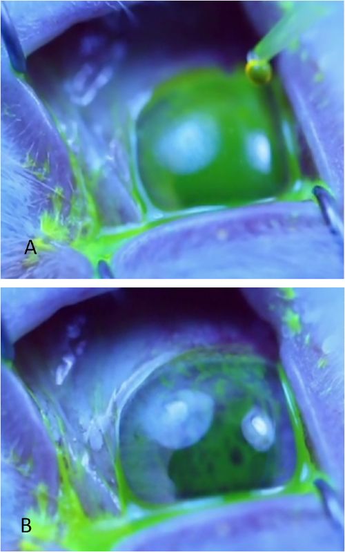
Journal of Visualized Experiments www.jove.com
magnification, and illumination from macro/ring flash set to automatic with through-the-lens mode. The ring flash's focus lamp is turned
on to aid focus on the cornea.
6. Complete all photos for both eyes within 1 min.
23
7. Score ocular surface staining using the NEI method modified as follows. Don not grade 6 separate conjunctival zones. Score the superior
conjunctiva of each eye. This is the portion of the conjunctival surface easily photographed without manipulating the globe. Manipulation
could artefactually change the staining of the ocular surface.
8. Tear lactoferrin levels are a surrogate measure of tear production from the lacrimal glands. Assay tear lactoferrin in tears collected as above
24
by using an enzyme-linked immunosorbent assay kit following the instructions of the manufacturer.
5. Induction and treatment of dry eye
NOTE: Three portions of the orbital lacrimal gland system are injected.
1. Sedate the rabbits with acepromazine 0.2 mg/kg subcutaneously.
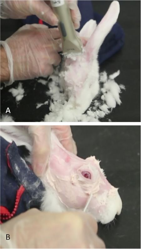
2. Shear off the fur in the periorbital and scalp area and completely remove any residual fur using Nair. Leave the skin entirely smooth for better
visualization of the anatomical hallmarks and US-guided injection of concanavalin A (Figure 7).
3. Induce moderate sedation as described above.
4. Injection of the palpebral portion of the superior lacrimal gland (PSLG)
NOTE: Perform injection of PSLG first.
1. Apply to the appropriate eye 25 µL of preservative-free lidocaine 1% with a micropipette.
2. Evert the upper eyelid and apply gentle medial pressure to the posterior orbital rim until the protuberance marking the palpebral portion
of the gland is seen. The PSLG appears as a small bulbous elevation in the posterior (temporal) portion of upper lid.
NOTE: To view the gland tissue during the learning process, apply 5% fluorescein to the area (Figure 8A). Tears can be seen
streaming from the bulbous PSLG. Application of fluorescein is not needed for the administration of Con A; it is done only for illustration
purposes to show the gland tissue.
3. Using fine-toothed forceps and a 27-gauge needle on a tuberculin syringe, directly penetrate the gland using a transconjunctival
approach. Advance the needle 2 mm into the tissue and inject 500 µg of Con A in a volume of 0.1 mL (Figure 8B).
NOTE: This injection can sometimes be painful. If necessary, keep the animals under isoflurane until this injection is completed.
5. Injection of the orbital superior lacrimal gland (OSLG)
NOTE: OSLG follows in rapid succession.
25
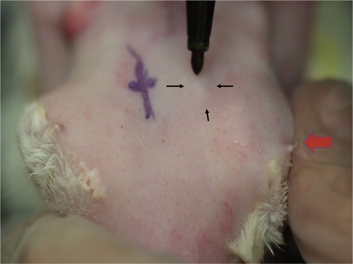
1. Apply medial pressure to the globe causing the OSLG to protrude from the posterior incisure (see ref for anatomy, if needed). Apply
medial pressure to the globe (Figure 9, red arrow) with the protuberance of the OSLG from the posterior incisure. The protuberance
serves as gross localization to find the posterior incisure.
2. Use curved forceps with tips closed to indent the area until the bony opening in the skull is felt. This will be slit-like with an anterior/
posterior direction under the protuberance.
3. Apply modest pressure with forceps to leave an indentation in the skin, which will serve as the landmark for needle placement (Figure
10A).
4. Insert a needle (tuberculin syringe with a 27-gauge, 5/8-inch needle) perpendicular to the skin over the indentation mark (Figure
10B) ~ ¼ inch into the incisure, then redirect the needle posteriorly and externally towards the lateral canthus aiming for the midpoint
between the injection site and bony orbital rim.
NOTE: If the incisure is not precisely targeted with the needle, the skull blocks its advancement.
5. Once the hub of the needle is reached, slowly inject 1000 µg of Con A in a volume of 0.2 mL (Figure 10C).
6. Complete the injection of both the PSLG and OSLG within 2-3 min.
7. Remove the animal from isoflurane sedation (if not yet done). Injection of the inferior lacrimal gland (ILG) can usually be completed without
further sedation.
8. Injection of the inferior lacrimal gland
1. View the animal from the side. The prominence of the ILG can be seen along the lower anterior portion of the orbit (Figure 11A).
2. Draw a vertical line using a surgical marking pen or suitable permanent marker on the skin where the superficial part of the ILG gland
transitions from its superficial (more external) resting place on the zygomatic bone to its more medial location in the orbit. This is
typically inferior to the anterior limbus (Figure 11A).
3. Identify the end of the zygomatic bone by sweeping the vertically-held US probe across this line on the skin. The ILG transition occurs
where the image of the gland changes from clearly circumscribed (hyperechoic line of the zygomatic bone is seen along the lower edge
of the gland in the image) to one without a recognizable medial border (the zygomatic bone echo is no longer present, Figure 1).
4. Observe the relative position of the hand-piece to the line drawn on the skin when the US screen shows this change. This is the
"injection site" where Con A should be given.
5. Control the depth of injection so as to place Con A into the gland at a point just medial to the zygomatic arch bone.
6. Determine the depth of injection as follows: Set the desired depth of injection as the depth of the zygomatic bone (hyperechoic signal)
plus 1 mm. Subtract this value from the known length of the needle (15 mm in this example).
7. Insert the needle into the gland at the "injection site" ~12 mm, then slowly withdraw it until the length of the exposed needle outside the
body (measured with surgical calipers) is equal to the difference calculated in 5.8.6 (Figure 12). Inject 1000 µg of Con A in 0.2 mL.
NOTE: To ensure that the capsule of the gland is pierced and not simply pushed by the needle, the needle should be inserted ~12 mm
or almost to the hub before its withdrawal begins.
8. Repeat the US to confirm the success of the injection. The ILG should show a characteristic hypoechoic space (Figure 1).
26
NOTE: The ILG injection is the one best tolerated by the animals and is, therefore, done last.
9. Complete the entire procedure to inject all glands of both eyes within 10 min. This will require achievement of competency in the procedure.
NOTE: A single set of injections into the 2 orbital lacrimal glands will induce acute DED lasting 1-2 weeks.
Copyright © 2020 Creative Commons Attribution-NonCommercial-NoDerivs 3.0 Unported January 2020 | 155 | e59631 | Page 4 of 20
License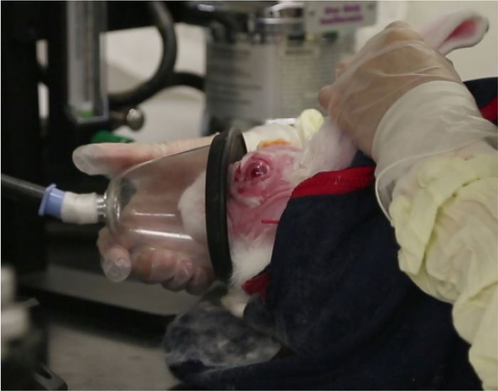
Journal of Visualized Experiments                                                                                         www.jove.com

  10. For DED of longer duration, inject Con A exactly as above every 7 days. Up to 6 such injections have been successfully performed.

6. Post-procedure care
  1. Following injection of Con A, monitor the animals in their restraining bags for at least 10-20 min, or until the anesthetic effect has worn off.
  2. Do not leave the animals unattended until they have regained sufficient consciousness to maintain sternal recumbency. Do not return them to
     their individual cages until they are fully recovered.
  3. Post-procedure pain is usually mild and lasts less than 48 h. Assess pain with the rabbit grimace scale. If needed, give a single dose of
     subcutaneous ketorolac (5 mg/kg). For more severe pain, give subcutaneous buprenorphine 0.1 mg/kg every 8 h.

Representative Results
  Con A injections induced a strong inflammatory response in the lacrimal glands characterized by a dense lymphocytic infiltrate (Figure 13),
  accompanied by decreased tear production. All tear parameters were markedly altered (Table 1 and Table 2). In addition, tear lactoferrin levels
  were suppressed (control = 3.1±0.45 vs. Con A injected = 2.7±0.02 ng/mg protein (mean ± SEM); p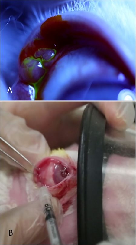
Journal of Visualized Experiments                                                                                          www.jove.com

  Figure 1: Ultrasound image of the inferior lacrimal gland. Upper Panel: The ILG as it moves deeper in orbit to lie beneath the zygomatic
  arch. The dashed line represents the line on skin across which the US probe is swept. Middle Panels: As the hand-piece is swept across this
  line, the examiner looks for loss of the zygomatic bone echo that is present in the left image (arrow) and disappears in the right. Lower Panels:
  Images of the ILG taken before (left) and after (right) injection of Con A. Development of a large cystic space within the gland confirms proper
                                       19
  delivery. Reprinted with permission . Please click here to view a larger version of this figure.

Copyright © 2020 Creative Commons Attribution-NonCommercial-NoDerivs 3.0 Unported                       January 2020 | 155 | e59631 | Page 6 of 20
License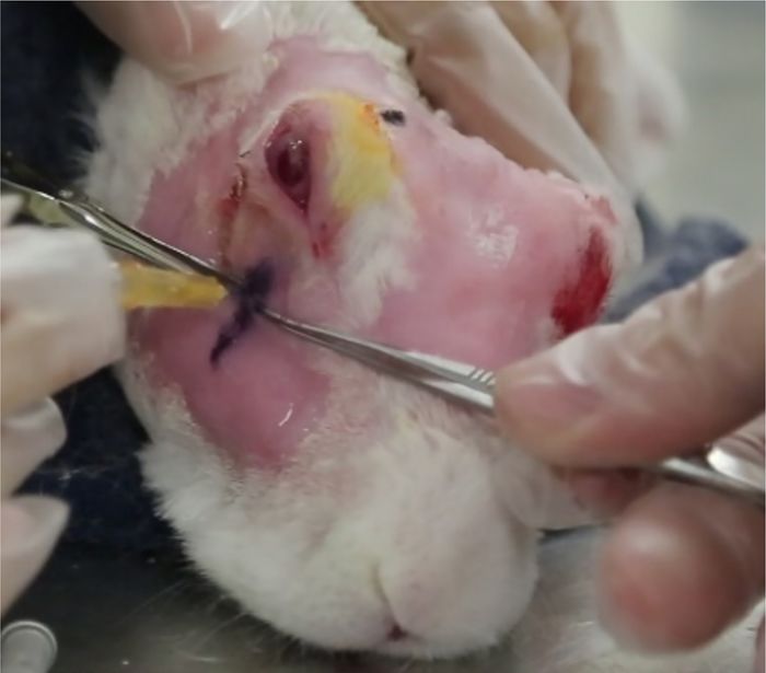
Journal of Visualized Experiments                                                                                   www.jove.com

  Figure 2: Gas mask sedation. This photograph shows the gas mask providing brief moderate sedation with isoflurane. Please click here to view
  a larger version of this figure.

Copyright © 2020 Creative Commons Attribution-NonCommercial-NoDerivs 3.0 Unported                  January 2020 | 155 | e59631 | Page 7 of 20
License
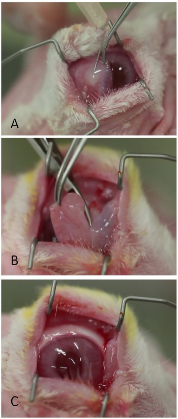
Journal of Visualized Experiments                                                                                      www.jove.com

  Figure 3: Removal of the nictitating membrane. (A) Injection of lidocaine/epinephrine. (B) Truncation of the nictitating membrane at its base
  with Westcott scissors. (C) The ocular surface visualized more readily after removal of the nictitating membrane. Please click here to view a
  larger version of this figure.

Copyright © 2020 Creative Commons Attribution-NonCommercial-NoDerivs 3.0 Unported                    January 2020 | 155 | e59631 | Page 8 of 20
License
Journal of Visualized Experiments                                                                                         www.jove.com

  Figure 4: Tear break-up time measurement. (A) Uniform green tear film appearance of the corneal surface under blue light immediately
  following application of fluorescein drops. (B) Corneal surface that has already undergone marked break-up evidenced by multiple dark circles
  and linear streaks in the fluorescein. Break-up time is recorded as soon as the first dark spot or line develops. The two light blue circles are
  reflections of the light source off of the cornea. Please click here to view a larger version of this figure.

Copyright © 2020 Creative Commons Attribution-NonCommercial-NoDerivs 3.0 Unported                      January 2020 | 155 | e59631 | Page 9 of 20
License
Journal of Visualized Experiments                                                                                             www.jove.com

  Figure 5: Schirmer's tear test. (A) The proper placement of the Weck-Cel sponge in the lower fornix to remove any residual topical lidocaine
  solution and baseline tears. By placing the posterior edge of the triangular sponge under the lower lid margin, one can maintain a very uniform
  technique to dry the ocular surface prior to placement of the tear strips. (B) A tear strip appropriately placed at the mid-position of the lower lid
  between the globe and lower lid (palpebral conjunctiva). Please click here to view a larger version of this figure.

Copyright © 2020 Creative Commons Attribution-NonCommercial-NoDerivs 3.0 Unported                         January 2020 | 155 | e59631 | Page 10 of 20
License
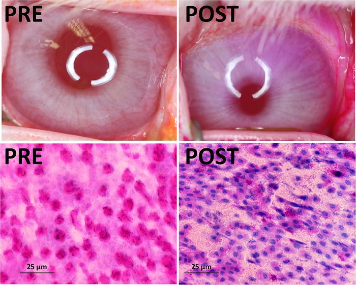
Journal of Visualized Experiments                                                                                         www.jove.com

  Figure 6: Rose bengal staining. Upper: Photographs of the corneal surface. Left: No rose bengal staining is present before treatment with
  Con A. Right: Diffuse corneal and conjunctival staining is seen in the upper nasal quadrant post-injection (upper right). Lower: Conjunctival
  impression cytology from the superior bulbar conjunctiva. Left: Numerous goblet cells are present before treatment. Right: Epithelial cells are
  present but goblet cells are absent post-treatment. Please click here to view a larger version of this figure.

Copyright © 2020 Creative Commons Attribution-NonCommercial-NoDerivs 3.0 Unported                     January 2020 | 155 | e59631 | Page 11 of 20
License
Journal of Visualized Experiments                                                                                            www.jove.com

  Figure 7: Preparation of rabbit for concanavalin A injections. (A) Small shears are used to remove fur, allowing easier visualization of
  landmarks to identify the orbital superior lacrimal gland. (B) Nair is used to remove hair that remains after shearing. Please click here to view a
  larger version of this figure.

Copyright © 2020 Creative Commons Attribution-NonCommercial-NoDerivs 3.0 Unported                       January 2020 | 155 | e59631 | Page 12 of 20
License
Journal of Visualized Experiments                                                                                          www.jove.com

  Figure 8: Injection of the palpebral lacrimal gland. (A) The palpebral lacrimal gland, appearing as a bulbous elevation in the posterior
  temporal portion of the upper lid. Tears are seen streaming from the surface of this gland after applying a drop of 2% fluorescein. (B) The
  palpebral lacrimal gland is being injected while the rabbit is receiving moderate sedation. One investigator retracts the eyelid, optimizes exposure
  of the gland, and secures the mask while the second investigator injects the gland. Please click here to view a larger version of this figure.

Copyright © 2020 Creative Commons Attribution-NonCommercial-NoDerivs 3.0 Unported                      January 2020 | 155 | e59631 | Page 13 of 20
License
Journal of Visualized Experiments                                                                                            www.jove.com

  Figure 9: Localization of the orbital superior lacrimal gland. Changes in skin contours indicate the location of the OSLG as it protrudes
  through the posterior incisure. Alternating medial pressure on the globe (large arrow) causes the superior orbital gland to prolapse, which is seen
  as a small elevation in the skin. This elevation will increase in size each time the pressure is applied (small arrows). The location of this gland is
  usually in line with the posterior orbital rim. Please click here to view a larger version of this figure.

Copyright © 2020 Creative Commons Attribution-NonCommercial-NoDerivs 3.0 Unported                       January 2020 | 155 | e59631 | Page 14 of 20
License
Journal of Visualized Experiments                                                                                         www.jove.com

  Figure 10: Injection of the orbital superior lacrimal gland. (A) Application of gentle pressure to the skull with fine-toothed forceps in the area
  which prolapsed as in Figure 9. A thin slit-like opening in the skull can be palpated. Leaving a small indentation mark with the forceps greatly
  aids placement of the needle during injection. (B) The needle is being inserted perpendicularly through the incisure. If placed incorrectly, its
  passage is stopped by the bony skull. (C) The needle is in final position angled towards the lateral canthus. Please click here to view a larger
  version of this figure.

Copyright © 2020 Creative Commons Attribution-NonCommercial-NoDerivs 3.0 Unported                     January 2020 | 155 | e59631 | Page 15 of 20
License
Journal of Visualized Experiments                                                                                        www.jove.com

  Figure 11: Localization of the inferior lacrimal gland. (A) The prominence of the superficial portion of the ILG seen through the lower lid. The
  curvilinear pen mark denotes the lower position of the gland. The vertical line, under the nasal limbus, denotes the approximate position where
  the ILG transitions to a deeper position within the orbit and serves as a visual reference for the US. (B) US hand-piece sweeping across the area
  of the vertical line; the US monitor will show where the zygomatic bone ends, where the ILG transitions and where the Con A injection should be
  given ("injection site"). Please click here to view a larger version of this figure.

Copyright © 2020 Creative Commons Attribution-NonCommercial-NoDerivs 3.0 Unported                    January 2020 | 155 | e59631 | Page 16 of 20
License
Journal of Visualized Experiments www.jove.com Figure 12: Injection of the inferior lacrimal gland. Injection of the ILG is done at the location identified by US. The depth of injection is calculated as described in the text (step 5.8.6). Calipers (seen behind the needle) ensure that the needle is placed at the proper depth before injection. Please click here to view a larger version of this figure. Figure 13: Histology of the lacrimal glands. Tissue sections of a normal inferior lacrimal gland with typical tubulo-alveolar structure (A) and following injection of Con A (B), showing marked lymphocytic infiltrate with effacement of structure. Similar inflammatory infiltrates are seen in the superior lacrimal glands. Please click here to view a larger version of this figure. Copyright © 2020 Creative Commons Attribution-NonCommercial-NoDerivs 3.0 Unported January 2020 | 155 | e59631 | Page 17 of 20 License
Journal of Visualized Experiments                                                                                                www.jove.com

Injection Method     TBUT, sec                                     Tear Osmolality, Osm/L                      STT, mm
                     Baseline                Post-injection        Baseline            Post-injection          Baseline               Post-injection
                     mean ± SEM, % change
ILG without US       58.5 ± 1.5              44.5 ± 7.7            297 ± 4.9           300 ± 3.8               15.2 ± 0.9             12.9 ± 2.2
guidance
%change                                      -23%                                      1%                                             -15%
                                             p=0.05                                    p=0.36                                         p=0.21
US-guided ILG +      59.4 ± 0.6              11.4 ± 4.2            296 ± 4.7           326 ± 3.7               15.1 ± 1.3             10.7 ± 1.8
PSLG
% change                                     -81%                                      10%                                            -29%
                                             p
Journal of Visualized Experiments www.jove.com
Also keep in mind the variation in the size of the ILG. The corollary to this is that the success of Con A must always be confirmed with follow
up imaging. Variations in the response to Con A in a group of rabbits is most often due to the injection technique (unsuccessful or partially
successful injection) or to ignoring the capacity of residual lacrimal gland tissues to compensate with overproduction of tears. For those who wish
to master the injection technique, injecting methylene blue followed by prompt anatomic dissection can be helpful; visualization is achieved if it
reaches the lacrimal gland or spills onto neighboring tissues. To date, this injection method has been performed over 270 times by the authors
without a single complication.
Assaying the five parameters of DED presented above can be as tricky as is their determination in clinical practice. Although circadian variations
28
have not yet been formally documented in any of them, there is enough background evidence of such phenomena in the eye that they should
be assayed at the same time of day (± 1 h), especially when repeat assays are to be performed and compared to each other. Consistency in
performing these assays is essential. A team of two is required. Four or more investigators in the same room participating in the assays can be
disruptive, given that some steps require strict timing. Appropriate and high-quality photographic documentation, where indicated, is important.
19
This model is ideally suited for drug development studies. Mastery of the animal model and assay techniques ensured excellent reproducibility
of efficacy and safety studies.
This is a powerful experimental approach because it eliminates the confounding variability of prior models, has streamlined the animal model
and essentially standardized assaying the five parameters of DED. The successful application of this model to the study of a candidate
therapeutic agent has affirmed its practical utility as an informative animal model for a disease in desperate need of novel agents and of a deeper
understanding of its pathogenesis.
Disclosures
The authors declare no competing interests except for BR who has an equity position in Medicon Pharmaceuticals, Inc. and Apis Therapeutics,
LLC; and LH, an employee of Medicon Pharmaceuticals, Inc. with an equity position in Apis Therapeutics, LLC.
Acknowledgments
All animal studies were completed in accordance and compliance with all relevant regulatory and institutional guidelines. All studies were
approved by the Institutional Review Board of Stony Brook University and performed in accordance with the ARVO Statement for the Use of
Animals in Ophthalmic and Vision Research.
These studies were supported in part by a Targeted Research Opportunities grant from the Stony Brook University School of Medicine (Grant
Number 1149271-1-82502) and a research grant from Medicon Pharmaceuticals, Inc., Setauket, NY. The authors thank Michele McTernan for
editorial support.
References
1. Paulsen, A. J. et al. Dry eye in the Beaver Dam Offspring Study: prevalence, risk factors, and health-related quality of life. American Journal
of Ophthalmology. 157 (4), 799-806 (2014).
2. Vehof, J., Kozareva, D., Hysi, P. G., & Hammond, C. J. Prevalence and risk factors of dry eye disease in a British female cohort. British
Journal of Ophthalmology. 98 (12), 1712-1717 (2014).
3. Tan, L. L., Morgan, P., Cai, Z. Q., & Straughan, R. A. Prevalence of and risk factors for symptomatic dry eye disease in Singapore. Clinical
and Experimental Optometry. 98 (1), 45-53 (2015).
4. Craig, J. P. et al. TFOS DEWS II Report Executive Summary. The Ocular Surface. 15 (4), 802-812 (2017).
5. Baudouin, C. et al. Clinical impact of inflammation in dry eye disease: proceedings of the ODISSEY group meeting. Acta Ophthalmologica
(Copenhagen). 96 (2), 111-119 (2018).
6. Calonge, M. et al. Dry eye disease as an inflammatory disorder. Ocular Immunology and Inflammation. 18 (4), 244-253 (2010).
7. Pflugfelder, S. C., & de Paiva, C. S. The Pathophysiology of Dry Eye Disease: What We Know and Future Directions for Research.
Ophthalmology. 124 (11S), S4-S13 (2017).
8. Buckley, R. J. Assessment and management of dry eye disease. Eye (London, England). 32 (2), 200-203 (2018).
9. Clayton, J. A. Dry Eye. New England Journal of Medicine. 378 (23), 2212-2223 (2018).
10. Barabino, S. Animal models of dry eye. Archivos de la Sociedad Española de Oftalmología. 80 (12), 693-694; 695-696 (2005).
11. Stern, M. E., & Pflugfelder, S. C. What We Have Learned From Animal Models of Dry Eye. International Ophthalmology Clinics. 57 (2),
109-118 (2017).
12. Chen, Z. Y., Liang, Q. F., & Yu, G. Y. Establishment of a rabbit model for keratoconjunctivitis sicca. Cornea. 30 (9), 1024-1029 (2011).
13. Gilbard, J. P., Rossi, S. R., & Gray, K. L. A new rabbit model for keratoconjunctivitis sicca. Investigative Ophthalmology & Visual Science. 28
(2), 225-228 (1987).
14. Guo, Z. et al. Autologous lacrimal-lymphoid mixed-cell reactions induce dacryoadenitis in rabbits. Experimenal Eye Research. 71 (1), 23-31
(2000).
15. Burgalassi, S., Panichi, L., Chetoni, P., Saettone, M. F., & Boldrini, E. Development of a simple dry eye model in the albino rabbit and
evaluation of some tear substitutes. Ophthalmic Research. 31 (3), 229-235 (1999).
16. Xiong, C. et al. A rabbit dry eye model induced by topical medication of a preservative benzalkonium chloride. Investigative Ophthalmology &
Visual Science. 49 (5), 1850-1856 (2008).
17. Heymann, F., Hamesch, K., Weiskirchen, R., & Tacke, F. The concanavalin A model of acute hepatitis in mice. Lab Animal. 49 (1 Suppl),
12-20 (2015).
Copyright © 2020 Creative Commons Attribution-NonCommercial-NoDerivs 3.0 Unported January 2020 | 155 | e59631 | Page 19 of 20
LicenseJournal of Visualized Experiments www.jove.com
18. Nagelhout, T. J., Gamache, D. A., Roberts, L., Brady, M. T., & Yanni, J. M. Preservation of tear film integrity and inhibition of corneal injury by
dexamethasone in a rabbit model of lacrimal gland inflammation-induced dry eye. Journal of Ocular Pharmacology and Therapeutics. 21 (2),
139-148 (2005).
19. Honkanen, R. A., Huang, L., Xie, G., & Rigas, B. Phosphosulindac is efficacious in an improved concanavalin A-based rabbit model of
chronic dry eye disease. Translational Research. 198 58-72 (2018).
20. Huang, L. et al. The novel phospho-non-steroidal anti-inflammatory drugs, OXT-328, MDC-22 and MDC-917, inhibit adjuvant-induced arthritis
in rats. British Journal of Pharmacology. 162 (7), 1521-1533 (2011).
21. Mattheolabakis, G. et al. Topically applied phospho-sulindac hydrogel is efficacious and safe in the treatment of experimental arthritis in rats.
Pharmaceutical Research. 30 (6), 1471-1482 (2013).
22. Osmalek, T., Froelich, A., & Tasarek, S. Application of gellan gum in pharmacy and medicine. International Journal of Pharmaceutics. 466
(1-2), 328-340 (2014).
23. Lemp, M. A. Report of the National Eye Institute/Industry workshop on Clinical Trials in Dry Eyes. Contact Lens Association of
Ophthalmologists Journal. 21 (4), 221-232 (1995).
24. Dal Piaz, F., Braca, A., Belisario, M. A., & De Tommasi, N. Thioredoxin system modulation by plant and fungal secondary metabolites.
Current Medicinal Chemistry. 17 (5), 479-494 (2010).
25. Davis, F. A. The Anatomy and Histology of the Eye and Orbit of the Rabbit. Transactions of the American Ophthalmological Society. 27 400
402-441 (1929).
26. Lima, L., Lange, R. R., Turner-Giannico, A., & Montiani-Ferreira, F. Evaluation of standardized endodontic paper point tear test in New
Zealand white rabbits and comparison between corneal sensitivity followed tear tests. Veterinary Ophthalmology. 18 (Suppl 1), 119-124
(2015).
27. Zheng, W. et al. Therapeutic efficacy of fibroblast growth factor 10 in a rabbit model of dry eye. Mol Med Report. 12 (5), 7344-7350 (2015).
28. Wiechmann, A. F., & Summers, J. A. Circadian rhythms in the eye: the physiological significance of melatonin receptors in ocular tissues.
Progress in Retinal and Eye Research. 27 (2), 137-160 (2008).
Copyright © 2020 Creative Commons Attribution-NonCommercial-NoDerivs 3.0 Unported January 2020 | 155 | e59631 | Page 20 of 20
LicenseYou can also read