Involvement of different α2 adrenoceptor subtypes in effects of dexmedetomidine on porcine coronary artery
←
→
Page content transcription
If your browser does not render page correctly, please read the page content below
ORIGINAL ARTICLES

Applied Science and Environmental Studies1, Department of Science, School of Science and Technology, The Open
University of Hong Kong; Department of Pharmacology & Pharmacy2, LKS Faculty of Medicine, the University of Hongkong,
Hong Kong, China

Involvement of different α2 adrenoceptor subtypes in effects of
dexmedetomidine on porcine coronary artery
E. SZE-WAN WONG1,*, JINGJING LI2, R. RENKAI LI2, G. PAK-HENG LEUNG2,*, J. KWOK-FU NG2

        Received October 28, 2020, accepted November 23, 2020
        *Corresponding authors: Dr Emily Sze-Wan Wong, Applied Science and Environmental Studies,
        Department of Science, School of Science and Technology, The Open University of Hong Kong; Hong Kong
        eswwong@ouhk.edu.hk
        Dr George Pak-Heng, Department of Pharmacology & Pharmacy, The University of Hong Kong
        gphleung@hku.hk
        Pharmazie 76: 12-17 (2021)                                                                  doi: 10.1691/ph.2021.0878

        Objective: Although the α2-adrenoceptor receptor (α2-AR) agonist dexmedetomidine causes coronary vaso-
        constriction, activation of endothelial α2-ARs is known to induce vascular relaxation. Here, we used functional
        studies and molecular techniques to explore the involvement of different α2-AR subtypes (α2A-AR, α2B-AR α2C-AR)
        in the effects of dexmedetomidine on the coronary artery. Methods: After precontraction with prostaglandin F2a,
        changes in tension caused by dexmedetomidine, in the presence or absence of different antagonists, were
        studied in isolated right porcine coronary rings in organ chambers. The presence or absence of different subtypes
        of α2-ARs in right coronary arteries and in blocks of subendocardial myocardium containing coronary resistance
        arterioles was investigated using immunofluorescence, western blotting and reverse transcription-polymerase
        chain reaction (RT-PCR). Results: Dexmedetomidine caused relaxation of isolated large porcine coronary
        arteries, which was attenuated by rauwolscine (a non-selective α2-AR antagonist), BRL44408 (an α2A-AR antag-
        onist) and ARC239 (an α2B-AR antagonist), and abolished by L-NAME (a nitric oxide synthase inhibitor) and
        pertussis toxin (a Gi protein inhibitor), and by removal of the endothelium. Although the relaxation could also be
        inhibited by MK912 (an α2C-AR antagonist), the pattern of inhibition suggested that it was due to a non-selective
        effect. The presence of α2A-AR in the endothelium of the right coronary artery, of α2B-AR in both the endothe-
        lium and vascular smooth muscle of the same artery, and of α2C-AR in the coronary resistance arterioles was
        confirmed by western blotting, RT-PCR and immunofluorescence. Conclusions: Expression of α2-AR subtypes is
        different in porcine coronary arteries of different sizes. Dexmedetomidine relaxes the large right coronary artery
        in an endothelium-dependent, α2A-AR, α2B-AR and nitric oxide-mediated manner. In contrast, α2C-AR is the only
        type of α2-AR in the resistance coronary arterioles and may be linked to vasoconstriction.

1. Introduction                                                        tomidine in large mammals, both dexmedetomidine and another
The α2-adrenoceptor (α2-AR) agonist dexmedetomidine is widely          α2-AR agonist, BHT933, reduce coronary flow in humans (Snapire
used in operating rooms and intensive care units because of its        et al. 2006).
sedative and analgesic effects. Emerging studies have shown that       The effects of dexmedetomidine on vascular tone involve inter-
perioperative use of dexmedetomidine during both open heart and        play between the nervous system and the blood vessels them-
non-cardiac surgery may reduce perioperative cardiac morbidity         selves. Dexmedetomidine mediates sympathetic vasoconstriction
(Biccard et al. 2008; Chi et al. 2016; Elgebaly et al. 2020; Gong      (sympatholysis) by activating α2-ARs in the central and peripheral
et al. 2017; Wijeysundera et al. 2008). The cardioprotective effects   nervous systems. In blood vessels, agonism of postsynaptic α2-ARs
of dexmedetomidine may involve mitochondrial K+ channels               on vascular smooth muscle cells may cause vasoconstriction
(Raupach et al. 2020), activation of PI3K/Akt signaling (Chang         (Talke et al. 2003), whereas agonism of postsynaptic α2-ARs on
et al. 2020), miR-208b-3p/Med13/Wnt/β-catenin signaling (Wang          endothelial cells may cause vasorelaxation (Figueroa et al. 2001).
et al. 2020) and HIF-a signaling (Peng et al. 2020) pathways, as       Dexmedetomidine is known to act on three subtypes of α2-AR,
well as reduction of oxidative stress (Han et al. 2019).               α2A-AR, α2B-AR and α2C-AR (Hunter et al. 1997; Moura et al.
Myocardial oxygen supply, which depends on normal coronary             2006). Using receptor binding and pharmacological studies, it has
relaxation and contraction, is the determining factor for main-        been shown that stimulation of α2D-AR and α2A-AR subtypes can
taining normal cardiac function. Although dexmedetomidine is           induce endothelium-dependent vasorelaxation in rats (Bockman et
cardioprotective, it is generally considered to be a coronary vaso-    al. 1996) and pigs (Bockman et al. 1993). In contrast, the α2B-AR
constrictor, which reduces coronary blood flow and myocardial          subtype seems to mediate vasoconstrictor responses since α2-AR
perfusion, and increases coronary vascular resistance, in dogs and     agonist-induced pressor responses are absent in α2B-AR knockout
goats (Coughlan et al. 1992; Flacke et al. 1993; Lawrence et al.       mice (Link et al. 1996). In line with these studies, we previously
1997; Pagel et al. 1998; Rockacrts et al. 1996). Dexmedetomidine       reported a biphasic effect of dexmedetomidine on isolated rat
also increases coronary vascular resistance in pigs (Jalonen et al.    mesenteric artery and aorta (Wong et al. 2010). At low concen-
1995; Kundra and Kaur 2016). Similar to the effects of dexmede-        trations (< 30 nM), dexmedetomidine evoked relaxation of the

12                                                                                                                Pharmazie 76 (2021)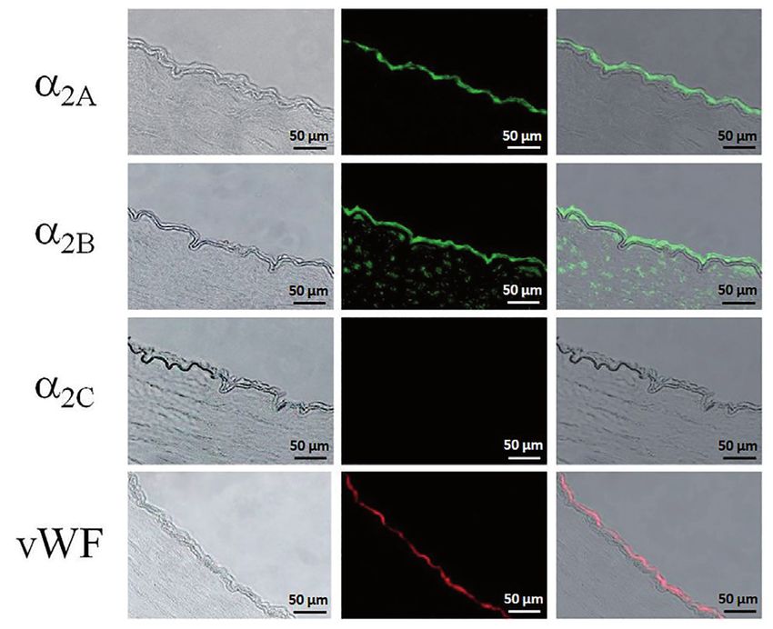
ORIGINAL ARTICLES

mesenteric artery, followed by contraction at higher concentrations                     2. Investigations and results
(> 30 nM). Dexmedetomidine-induced relaxation depends on nitric
oxide (NO), endothelium and Gi protein, and is mediated mainly                          2.1. Relaxation studies
by α2A/D-ARs, and possibly α2B-ARs. Contraction, on the other                           Dexmedetomidine induced a concentration-dependent relaxation
hand, is mediated mainly by α2B-ARs and α1-ARs and involves the                         of porcine coronary artery at concentrations between 100 pM
actions of prostanoids. The effect of dexmedetomidine on the aorta                      and 100 nM, followed by a rebound in tension at concentrations
is significantly smaller than on the mesenteric arteries, suggesting                    between 100 nM and 10 μM. Treatment with l-NAME (endo-
different vascular effects on different vascular beds.                                  thelial NO synthase inhibitor) or removal of the endothelium

Fig. 1: Dexmedetomidine concentration-response curves in porcine right coronary rings treated with indomethacin (10 μM) and precontracted with prostaglandin F2α. (A) With
        intact endothelium (EC), with intact EC and treatment with l-NAME (300 μM), and with removal of endothelium (n = 10); (B) Treatment with rauwolscine (100 nM) or
        pertussis toxin (PTx, 400 ng/ mL) (n = 10). Data are the mean ± S.E.M. *P < 0.05 compared with control.

There have been very few reports describing the direct effect of                        abolished dexmedetomidine-induced relaxation (Fig. 1A). Treat-
dexmedetomidine on the coronary artery and the AR subtypes                              ment with pertussis toxin (Gi protein inhibitor, 400 ng/mL) or
involved. It is important to understand the direct effect of                            rauwolscine (non-selective α2-AR antagonist, 100 nM) also led
dexmedetomidine on blood vessel cells, especially the effect on                         to a significant reduction in endothelium-dependent relaxation
endothelial cells, since this is an important mechanism in coun-                        (Fig. 1B).
teracting vasoconstriction caused by the sympatholytic effect of                        BRL44408 (α2A/D-AR antagonist) and ARC239 (α2B-AR antag-
dexmedetomidine. The objectives of this study were to evaluate                          onist) caused a rightward shift of the concentration-response
the distribution of α2-AR subtypes in large coronary arteries and                       curves compared with the control. The corresponding Schild
small coronary arterioles, and to correlate this distribution with                      regression line was not different from unity for BRL44408. The
functional pharmacological effects.                                                     apparent binding affinity (pKB) was 8.28 (95% CI, 7.86–9.19),

Fig. 2: Dexmedetomidine concentration-response curves in porcine right coronary rings with intact endothelium treated with indomethacin (10 μM) and precontracted with
        prostaglandin F2α. (A) Treatment with BRL44408 (BRL, 3–300 nM) (n = 10); (B) Treatment with ARC239 (10 nM to 1 μM) (n = 10); (C) Treatment with MK912 (0.03–3
        nM) (n = 10). Data are the mean ± S.E.M. Insets show Schild regression analyses of concentration-response curves for BRL44408 and ARC239. *P < 0.05 compared with
        control.

Pharmazie 76 (2021)                                                                                                                                                   13
ORIGINAL ARTICLES
which is very similar to the known binding affinity (pKB = 8.25) 2.4. Immunofluorescence assay
of BRL44408 for cloned human α2A-AR (Uhlen et al. 1994). The immunofluorescence assay showed that endothelial cells of
Since the slope of the Schild regression line was different from coronary arteries were stained by the α2A-AR antibody. Immunore-
unity for ARC239, no attempt was made to determine pKB active α2B-AR was localized to both endothelial cells and vascular
for ARC239 (Fig. 2A and 2B). MK912 (α2C-AR antagonist) smooth muscle cells of coronary arteries, whereas no α2C-AR could
caused a rightward shift, as well as a reduction in the maximal be detected in coronary arteries (Fig. 5). In contrast, subendocar-
relaxation (Fig. 2C), suggesting a mixture of competitive and dial myocardium containing coronary arterioles was negatively
non-competitive antagonism. The slope of the Schild regression stained by α2A-AR and α2B-AR antibodies, but immunoreactive
line was also different from unity for MK912 so pKB value was α2C-AR was detected in the endothelial cells and vascular smooth
not determined. muscle cells of the arterioles (Fig. 6).
2.2. Reverse transcription-polymerase chain reaction 3. Discussion
RT-PCR showed that α2A-AR and α2B-AR mRNAs, but not α2C-AR In this study, we found a differential distribution of α2-AR subtypes
mRNA, were expressed in the right coronary artery. In contrast, in porcine large coronary arteries and intramyocardial coronary
expression of α2B-AR and α2C-AR mRNAs, but not α2A-AR mRNA, resistance arterioles. The α2A-AR is expressed exclusively in the
was detected in subendocardial myocardium (Fig. 3).
Fig. 3: RT-PCR analysis of α2-AR mRNA in porcine right coronary arteries and
subendocardial myocardium. In the coronary artery, PCR products are seen
in reactions using oligonucleotide primer pairs for α2A-AR and α2B-AR, but
not for α2C-AR. In subendocardial myocardium, PCR products are seen in
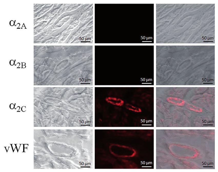
Fig. 5: Immunofluorescence staining of α2-AR proteins in porcine right coronary
reactions using oligonucleotide primer pairs for α2B-AR and α2C-AR, but not
arteries. Middle column: Positive immunoreactivity for α2A-AR (green) was
for α2A-AR. Expected sizes of amplified fragments: 363-bp (α2A-AR), 459-bp
detected in endothelial cells. α2B-AR (green) was localized to endothelial
(α2B-AR) and 403-bp (α2C-AR). DNA size markers are shown at the left.
cells and vascular smooth muscle cells but no α2C-AR was detected. Von-Wil-
lebrand factor (VWF, red) was used as a marker of endothelial cells. Left col-
umn: Phase contrast images of corresponding stained sections. Right column:
Merged light micrographs and fluorescence micrographs.
Fig. 4: Western blot analysis of α2-AR proteins in porcine right coronary arteries and
subendocardial myocardium. In the coronary arteries, α2A-AR and α2B-AR
proteins were detected, but α2C-AR was absent. In contrast, α2C-AR protein
was expressed in subendocardial myocardium, whereas α2A-AR and α2B-AR
proteins were not detected. b-actin, which served as a positive control, was
detected in both coronary arteries and subendocardial myocardium.
2.3. Western blotting
Protein bands of approximately 82 kDa were expressed in coronary
arteries when using the α2A-AR and α2B-AR antibodies. The sizes
of these protein bands were close to the reported molecular sizes
of α2A-AR (~72 kDa) and α2B-AR (~70 kDa) in rat heart (Diebold Fig. 6: Immunofluorescence staining of α2-AR proteins in porcine subendocardial
et al. 2005). No protein expression of α2C-AR could be detected in myocardium. Middle column: Positive immunoreactivity for α2C-AR (red)
coronary arteries and no protein expression of α2A-AR or α2B-AR was detected in endothelial cells and vascular smooth muscle cells, but no
was found in the subendocardial myocardium. However, a protein α2A-AR or α2B-AR was detected. Von-Willebrand factor (VWF, red) was used
as a marker of endothelial cells. Left column: Phase contrast images of cor-
band that corresponds to α2C-AR protein was detected in the suben- responding stained sections. Right column: Merged light micrographs and
docardial myocardium (Fig. 4). fluorescence micrographs.
14 Pharmazie 76 (2021)ORIGINAL ARTICLES
endothelium of large coronary arteries. The α2B-AR is expressed α2-AR agonists, it also relaxes large coronary arteries via activation
in endothelial cells and vascular smooth muscle of large coronary of α2A-AR. In patients with coronary artery disease and stenotic
arteries, but the α2C-AR is absent. In contrast, subendocardial segments in the larger coronary arteries, constriction of small
myocardium does not express α2A-AR. Although α2B-AR mRNA coronary resistance arterioles may not necessarily be harmful. An
was detected in the subendocardial myocardium by RT-PCR, the acute drop in coronary vascular resistance may, in fact, give rise to
protein is not detectable in arterioles and cardiac muscle. This the phenomenon of “coronary steal” and further reduce perfusion
discrepancy may be due to low translation rate and/or high protein in the ischemic regions of the myocardium (Seiler et al. 1997). On
turnover in the cells, which leads to very low levels of α2B-AR the other hand, dilatation of larger coronary arteries may improve
protein expression that are below the detection thresholds of flow characteristics in those arteries. Sudden vessel contraction,
western blot analysis and immunofluorescence staining. Expres- with an associated increase in flow velocity, or conversion to turbu-
sion of α2C-AR mRNA and protein was observed in the suben- lent flow with a concomitant increase in local shear stress, may
docardial myocardium. Immunofluorescence staining showed play an important role in plaque rupture (Fukumoto et al. 2008).
that α2C-AR is localized to endothelial cells and vascular smooth These differential effects of dexmedetomidine on large conduit and
muscle cells of the coronary arterioles. small resistance coronary arteries may explain in part the benefi-
We also showed that dexmedetomidine induces endothelium-de- cial effects of the perioperative use of different α2-AR agonists in
pendent relaxation of isolated porcine large coronary artery, which patients with cardiovascular risk factors, as reported in a previous
is likely mediated by release of NO. Our results are consistent with meta-analysis (Wijeysundera et al. 2003).
those of another recent study (Zhou et al. 2017), which showed that Dexmedetomidine probably modulates the vascular tone of large
a low concentration of dexmedetomidine relaxed porcine coronary coronary arteries by releasing NO from the endothelium. Although
artery through an endothelium- and NO-dependent mechanism. In we did not directly measure NO release in this study, the aboli-
that study, however, the α2-AR antagonist yohimbine failed to block tion of relaxation after treatment with l-NAME strongly suggests
the vasorelaxing effect on the coronary arteries. There are two possible that this is the case. Endothelial NO release is known to confer
explanations for this discrepancy. Firstly, in our study, we used right, advantages other than modulation of vascular tone (Kawai 1994;
large coronary arteries but the other study used left, distal coronary Vanhoutte 2003). For instance, endothelial NO plays an important
arteries. It has previously been reported that the vascular response to role in reducing excessive platelet activation, particularly at
α2-AR agonists varies in different blood vessels (Wong et al. 2010). atheromatous plaques (Freedman et al. 1998; Radomski et al.
Secondly, different α2-AR antagonists were used in the two studies. In 1987; Roberts et al. 2008; Schafer and Bauersachs 2008; Schafer
an earlier study, we found that the vascular effect of dexmedetomidine et al. 2004). NO release is also known to favorably modulate the
is different from that of the alternative α2-AR agonists, clonidine and inflammatory response, which may also reduce the expression of
UK14304 (Wong et al. 2010), probably because of differences in thrombogenic mediators around a plaque (Tziros and Freeman
receptor selectivity. A similar explanation can be used to explain the 2006; Vanhoutte 2003). With probable NO release in coronary
difference between yohimbine, which is a non-selective α2-AR antag- arteries following dexmedetomidine administration, these effects
onist with moderate affinity for adrenergic α1, 5-HT1 and dopamine may further augment the beneficial effects of dexmedetomidine in
D2 receptors, and dexmedetomidine. In our study, using more selective patients with coronary artery disease.
antagonists, we showed that α2A-AR is the major receptor subtype Dexmedetomidine exerts its anesthetic effects via α2A-ARs in the
responsible for this relaxation. The dexmedetomidine-induced relax- central nervous system (Hunter et al. 1997; Nelson et al. 2003).
ation can be inhibited by pertussis toxin, suggesting the involvement Since, as we have shown here, release of NO and coronary dilata-
of Gi-protein, which is known to be the transducing mechanism for tion are also mediated by α2A-ARs, more subtype-selective α2-AR
α2A-ARs. Our conclusion is also supported by estimation of antagonist agonist anesthetics, particularly those with predominant α2A-AR
affinity using the Schild regression approach. The results of immuno- selectivity, may be superior to agonists that are not subtype-selec-
fluorescence staining, RT-PCR and western blotting all showed that tive when used in patients with high cardiovascular risk.
α2A-AR is expressed in endothelial cells of coronary arteries. Although In conclusion, we demonstrated a differential distribution of α2-AR
the α2B-AR antagonist ARC239 also produced a shift in the relaxation subtypes in the coronary circulation, and showed that dexmedeto-
curves for dexmedetomidine, the results of the Schild regression anal- midine dilates large coronary arteries via activation of α2A-ARs and
ysis did not suggest simple single receptor competitive antagonism. subsequent release of NO. Dexmedetomidine probably constricts
Since the α2B-AR is present on both the endothelium and vascular small resistance coronary arterioles via activation of α2C-ARs.
smooth muscle in this artery, it is unclear whether this receptor
subtype contributes to contraction or relaxation of coronary arteries. 4. Experimental
Development of more specific α2B-AR antagonists would provide
useful pharmacological tools to address this question. On the other 4.1. Drugs and chemicals
hand, the α2C-AR antagonist MK912 produced a mixed shift. Taken Bradykinin, prostaglandin F2α, rauwolscine, BRL44408 (2-[2H-(1-methyl-1,3-di-
hydroisoindole)methyl]-4,5-dihydroimidazole maleate), indomethacin, MK912
together with the immunofluorescence, western blotting and RT-PCR (L-657,743, 2S-trans-1,3,4,5′,6,6′,7,12b-octahydro-1′,3′-dimethyl-spiro[2H-ben-
results, this may indicate that MK912 has non-selective effects on zofuro[2,3-a]quinolizine-2,4′(1′H)-pyrimidin]-2′(3′H)-one hydrochloride hydrate),
ω
other receptor subtypes. N -nitro-L-arginine methyl ester hydrochloride (l-NAME) and protease inhibitor
Although we did not perform direct functional studies on isolated cocktail were purchased from Sigma-Aldrich (St. Louis, MO, USA). ARC239
(2-[2-(4-(2-Methoxyphenyl)piperazin-1-yl)ethyl]-4,4-dimethyl-1,3-(2H,4H)-isoquin-
coronary arterioles in this study, the absence of α2A-AR and α2B-AR olindione dihydrochloride) was purchased from Tocris Bioscience (Ellisville, MO,
in these arterioles suggests that dexmedetomidine may not relax USA). Dexmedetomidine was purchased from Abbott Laboratories (Chicago, IL,
these arterioles. Consistent “coronary vasoconstriction” with USA). Pertussis toxin was purchased from List Biological Laboratories, Inc. (Camp-
α2-AR agonists, measured as reduced coronary blood flow or bell, CA, USA). All primary antibodies were bought from Abcam (Cambridge, MA,
USA) and secondary antibodies were bought from Millipore (Billerica, MA, USA).
raised coronary resistance, has been shown by others in different Stock solutions of dexmedetomidine was prepared in DMSO. A stock solution of
species, including human, in numerous studies (Baumgart et al. indomethacin was prepared in 5 mM sodium bicarbonate solution. A stock solution of
1999; Coughlan et al. 1992; Flacke et al. 1993; Indolfi et al. 1992; pertussis toxin was prepared in 0.1 M sodium sulfate buffer. All other solutions were
Lawrence et al. 1997; Pagel et al. 1998; Rockacrts et al. 1996). prepared in deionized water. Concentrations are expressed as final molar concentra-
tion in the bath solution.
Dexmedetomidine thus most likely causes constriction of these
arterioles via activation of α2C-ARs.
Taken together, our results show that dexmedetomidine should not 4.2. Porcine hearts
be considered to be simply a coronary vasodilator or a coronary Pig hearts were collected from the local abattoir, where the animals were killed
vasoconstrictor. While it probably causes constriction of small according to the regulations of the Food and Environmental Hygiene Department of
the Hong Kong Special Administrative Region. The hearts were immediately rinsed
coronary resistance arterioles, in agreement with the observations and transported back to the laboratory in ice cold, oxygenated Krebs-Ringer solution
by other investigators of reduced coronary blood flow and increased (composition: NaCl, 118.3 mM; KCl, 4.7 mM; MgSO4, 1.2 mM; KH2PO4, 1.2 mM;
coronary vascular resistance after administration of different CaCl2, 2.5 mM; NaHCO3, 25.0 mM; glucose, 11.1 mM).
Pharmazie 76 (2021) 15ORIGINAL ARTICLES
4.3. Large coronary arteries were incubated in blocking serum for 30 min and then with rabbit polyclonal anti-
The right coronary arteries were dissected free, separated from the surrounding fat α2A-AR, anti-α2B-AR or anti-α2C-AR antibody diluted 1:100 (v/v) with diluting buffer
and connective tissue, and cut into rings 4–5 mm in length. In some experiments, the (PBS with 0.01% (v/v) Triton X-100, 0.01% (v/v) Tween 20, and 0.1% (w/v) bovine
endothelium was removed by perfusion with saponin in Krebs–Ringer solution (1 serum albumin) at 4 °C overnight. Anti-von-Willebrand-factor-antibody was used
mg/mL, 1 mL) for 20 s before cutting the rings. The rings were suspended between as a marker for endothelial cells. Sections were washed three times with PBS and
stainless steel stirrups in jacketed organ chambers filled with Krebs-Ringer solution incubated with FITC- (for -α2A-AR and anti-α2B-AR) or rhodamine- (for α2C-AR and
(5 mL), and the isometric contractile force was measured using a PowerLab 4SP von Willebrand factor) conjugated goat anti-rabbit secondary antibody [1: 100 (v/v)
data acquisition system (ADInstruments, Oxford, UK). The rings were subjected to a with diluting buffer]. The slides were then washed and mounted for observation with
resting tension of 5 g, which was found to be the optimal tension in pilot experiments. a confocal microscope.
The solution was maintained at 37 °C and continuously aerated with 95% O2/5% CO2.
4.9. Statistical analysis
4.4. Protocol for functional studies on porcine coronary arteries Data are presented as the mean±S.E.M., with n representing the number of animals
After equilibration, indomethacin (10 mM) was added to the rings, with or without (pig hearts). For dose response curves, the concentration response curves were fitted
endothelium, the rings were contracted with prostaglandin F2a (1–3 mM), and the to the Hill equation using Prism 4.0 (GraphPad Software, Inc., San Diego, CA, USA).
relaxation to cumulative doses of dexmedetomidine (100 pM to 10 mM) was In curves showing parallel rightward shifts without change in maximal response,
measured. Several antagonists were used to define the receptors and mechanisms antagonist affinities were estimated using the method of Arunlakshana and Schild
involved. Rauwolscine (non-selective α2-AR antagonist, 100 nM), BRL44408 (1959). If the Schild regression slope was not significantly different from unity, the
(α2A-AR antagonist, 3 nM – 300 nM), ARC239 (α2B-AR antagonist, 10 nM – 1 mM) antagonist affinity was estimated from the intercept of the regression line as described
and MK912 (α2C-AR antagonist, 0.03–3 nM), alone or in combination, were used by Jenkinson and co-workers (1995).
to evaluate the role of different α2-AR subtypes. Nw-nitro-l-arginine methyl ester
(l-NAME, inhibitor of NO synthase, 100 mM) and pertussis toxin (inactivates Gi Analysis of variance (ANOVA) was used for comparison of curves among multiple
protein, 400 ng/mL) were used alone or in combination to evaluate the roles of NO treatment groups. Post hoc comparison was performed by the Newman-Keuls multiple
and Gi-protein, respectively. comparison test, where appropriate. Student’s t-test was used in the case of two group
comparisons. P < 0.05 was considered to indicate statistically significant differences.
4.5. Characterization of a2-adrenoreceptor subtype distribution Funding: This study was supported by the R&D Funding of the Open University of
Hong Kong (2018/1.3).
Studies on the anatomy of porcine coronary arteries have confirmed that coronary
resistance arterioles run diffusely across the ventricular myocardium (Rodrigues Conflicts of interest: The authors declare that there is no conflict of interest.
et al. 2005). Tiny blocks of subendocardial myocardium, which contain numerous
intramyocardial resistance coronary arterioles, were harvested from identical sites on
the left ventricular wall. These blocks were then used to map out the distribution References
of different α2-AR subtypes, using reverse transcription-polymerase chain reaction
(RT-PCR), western blotting and immunofluorescence. The results were compared Arunlakshana O, Schild HO (1959) Some quantitative uses of drug antagonists. Br J
with those obtained using segments of porcine right coronary artery. Pharmacol Chemother 14: 48–58.
Baumgart D, Naber C, Haude M, Oldenburg O, Erbel R, Heusch G, Siffert W (1999)
G protein beta3 subun96it 825T allele and enhanced coronary vasoconstriction on
4.6. Reverse transcription-polymerase chain reaction alpha(2)-adrenoceptor activation. Circ Res 85: 965–969.
Biccard BM, Goga S, de Beurs J (2008) Dexmedetomidine and cardiac protection for
Total RNA was isolated from coronary arteries and subendocardial myocardium using non-cardiac surgery: a meta-analysis of randomised controlled trials. Anaesthesia
TRIzol reagent (Invitrogen, Grand Island, NY, USA). Total RNA (2 mg) was used 63: 4–14.
for first-strand cDNA synthesis, using random hexamer primers and Superscript II Bockman CS, Gonzalez-Cabrera I, Abel PW (1996) Alpha-2 adrenoceptor subtype
RNase H reverse transcriptase (SuperScript Preamplification System, Invitrogen). causing nitric oxide-mediated vascular relaxation in rats. J Pharmacol Exp Ther
The resulting first-strand cDNA was directly used for PCR amplification.
278: 1235–1243.
Sets of primers were designed and synthesized for PCR analysis. The two primers
Bockman CS, Jeffries WB, Abel PW (1993) Binding and functional characterization
used for amplifying α2A-AR (accession no. NM_214400) were 5’-GAGCGCAG-
of alpha-2 adrenergic receptor subtypes on pig vascular endothelium. J Pharmacol
GCCCAATGGCCTA-3’ (sense) and 5’-GTTCTGCCTTCCGCGCCAGCG-3’
Exp Ther 267: 1126–1133.
(antisense), which generated a 363-bp PCR product. The two primers for α2B-AR
Chang JH, Jin MM, Liu JT (2020) Dexmedetomidine pretreatment protects the heart
(accession no. DQ182110) were 5’-AGGGCTAAGGGGGGCCCTGGG-3’ (sense)
against apoptosis in ischemia/reperfusion injury in diabetic rats by activating
and 5’-GGTCAGCTGCGCCCGCCGACG-3’ (antisense), which generated a 459-bp
PI3K/Akt signaling in vivo and in vitro. Biomed Pharmacother 127: 110188.
product. Reactions were carried out for 60 cycles with the following parameters:
Chi X, Liao M, Chen X, Zhao Y, Yang L, Luo A, Yang H (2016) Dexmedetomidine
denaturation at 94 °C for 30 s, annealing at 50 °C for 1 min, and extension at 72
attenuates myocardial injury in off-pump coronary artery bypass graft surgery. J
°C for 1.5 min. Porcine α2C-AR has not been completed cloned so human primers
Cardiothorac Vasc Anesth 30: 44–50.
were used instead. The two primers for α2C-AR (accession no. NM_000683) were
Coughlan MG, Lee JG, Bosnjak ZJ, Schmeling WT, Kampine JP, Warltier DC (1992)
5’-TGCGCGCGCCACAGAACCTCTTCCT-3’ (sense) and 5’- ATGCAGGAGGA-
Direct coronary and cerebral vascular responses to dexmedetomidine. Significance
CAGGATGTACCA-3’ (antisense), which yielded a 403-bp PCR product (Mchrotra
et al. 2006). Reactions were carried out for 60 cycles with the following parameters: of endogenous nitric oxide synthesis. Anesthesiology 77: 998–1006.
denaturation at 94 °C for 45 s, annealing at 58 °C for 45 s, and extension at 72 °C for Diebold Y, Enriquez de SA, Calonge M, Saez V, Callejo S, Stern ME (2005)
1.5 min. PCR products were analyzed by agarose gel electrophoresis and visualized Alpha2-adrenergic receptors are present in normal human conjunctiva. Curr Eye
by staining with ethidium bromide. Res 30: 1121–1129.
Elgebaly AS, Fathy SM, Sallam AA, Elbarbary Y (2020) Cardioprotective effects
of propofol-dexmedetomidine in open-heart surgery: A prospective double-blind
study. Ann Card Anaesth 23: 134–141.
4.7. Western blotting
Figueroa XF, Poblete MI, Boric MP, Mendizábal VE, Adler-Graschinsky E, Huidobro-
Protein expression of α2-ARs was determined by western blotting, as previously Toro JP (2001). Clonidine-induced nitric oxide-dependent vasorelaxation mediated
described (Li et al. 2019). Artery segments and blocks of myocardium were homoge- by endothelial alpha(2)-adrenoceptor activation. Br J Pharmacol 134: 957–968.
nized in ice-cold sodium phosphate buffer containing 1:1000 (v/v) protease inhibitor Flacke WE, Flacke JW, Bloor BC, McIntee DF, Sagan M (1993). Effects of dexme-
cocktail (5 mM, pH 8). The homogenate was centrifuged at 3000 × g for 10 min to detomidine on systemic and coronary hemodynamics in the anesthetized dog. J
remove nuclei and unbroken cells. The amount of protein in the supernatant was then Cardiothorac Vasc Anesth 7: 41-49.
determined using the Bradford protein assay. Proteins (20 mg) were resolved on 10% Freedman JE, Ting B, Hankin B, Loscalzo J, Keaney JF, Jr., Vita JA (1998) Impaired
sodium dodecyl sulfate-polyacrylamide gels and electrotransferred to nitrocellulose platelet production of nitric oxide predicts presence of acute coronary syndromes.
membranes. The membranes were subsequently blocked with 5% (w/v) nonfat dry Circulation 98: 1481–1486.
milk in PBS overnight at 4 °C, followed by incubation with polyclonal rabbit anti- Fukumoto Y, Hiro T, Fujii T, Hashimoto G, Fujimura T, Yamada J, Okamura T,
α2A-AR, anti-α2B-AR or anti-α2C-AR antibody [1:100 (v/v) dilution in blocking solu- Matsuzaki M (2008) Localized elevation of shear stress is related to coronary
tion] at room temperature for 2 h. The nitrocellulose membranes were then washed plaque rupture: a 3-dimensional intravascular ultrasound study with in-vivo color
extensively with 0.02% (v/v) Triton X-100 in PBS and incubated with horseradish mapping of shear stress distribution. J Am Coll Cardiol 51: 645–650.
peroxidase-conjugated goat anti-rabbit secondary antibody [1:5000 (v/v) dilution Gong Z, Ma L, Zhong YL, Li J, Lv J, Xie YB (2017) Myocardial protective effects
in blocking solution] at room temperature for 2 h. Excess secondary antibody was of dexmedetomidine in patients undergoing cardiac surgery: A meta-analysis and
again removed by washing. The bound secondary antibody was detected by enhanced systematic review. Exp Ther Med 13: 2355–2361.
chemiluminescence. Protein expression of b-actin, which served as a positive control, Han H, Dai D, Hu J, Zhu J, Lu L, Tao G, Zhang R (2019) Dexmedetomidine improves
was similarly detected using mouse monoclonal anti-b-actin antibody. cardiac function and protects against maladaptive remodeling following myocar-
dial infarction. Mol Med Rep 20: 5183–5189.
Hunter JC, Fontana DJ, Hedley LR, Jasper JR, Lewis R, Link RE, Secchi R, Sutton
4.8. Immunofluorescence assay J, Eglen RM (1997) Assessment of the role of alpha2-adrenoceptor subtypes in
Localization of different α2-AR protein was investigated by immunofluorescence the antinociceptive, sedative and hypothermic action of dexmedetomidine in trans-
staining (Leung et al. 2001). Paraffin-embedded sections of paraformaldehyde-fixed genic mice. Br J Pharmacol 122: 1339–1344.
coronary arteries and subendocardial myocardium (3 mm) were dewaxed and Indolfi C, Piscione F, Villari B, Russolillo E, Rendina V, Golino P, Condorelli M,
hydrated. Antigens were retrieved by treatment with citrate buffer (0.01 M, pH 6.0) Chiariello M (1992) Role of alpha 2-adrenoceptors in normal and atherosclerotic
for 5 min in a microwave oven. After rinsing with pure water and PBS, the sections human coronary circulation. Circulation 86: 1116–1124.
16 Pharmazie 76 (2021)ORIGINAL ARTICLES
Jalonen J, Halkola L, Kuttila K, Perttila J, Rajalin A, Savunen T, Scheinin M, Valtonen Roberts W, Riba R, Homer-Vanniasinkam S, Farndale RW, Naseem KM (2008) Nitric
M (1995) Effects of dexmedetomidine on coronary hemodynamics and myocardial oxide specifically inhibits integrin-mediated platelet adhesion and spreading on
oxygen balance. J Cardiothorac Vasc Anesth 9: 519–524. collagen. J Thromb Haemost 6: 2175–2185.
Jenkinson DH, Barnard EA, Hoyer D, Humphrey PP, Leff P, Shankley NP (1995) Rodrigues M, Silva AC, Aguas AP, Grande NR (2005) The coronary circulation of the
International Union of Pharmacology Committee on Receptor Nomenclature and pig heart: comparison with the human heart. Eur J Anat 9: 67–87.
Drug Classification. IX. Recommendations on terms and symbols in quantitative Roekaerts PM, Prinzen FW, de Lange S (1996) Coronary vascular effects of dexme-
pharmacology. Pharmacol Rev 47: 255–266. detomidine during reactive hyperemia in the anesthetized dog. J Cardiothorac Vasc
Kawai C (1994) Pathogenesis of acute myocardial infarction. Novel regulatory Anesth 10: 619–626.
systems of bioactive substances in the vessel wall. Circulation 90: 1033–1043. Schafer A, Bauersachs J (2008) Endothelial dysfunction, impaired endogenous
Kundra TS, Kaur P (2016) Effect of dexmedetomidine on normal coronary vessel platelet inhibition and platelet activation in diabetes and atherosclerosis. Curr Vasc
diameter. Pharmacology 98: 217–219. Pharmacol 6: 52–60.
Lawrence CJ, Prinzen FW, de Lange S (1997) Hemodynamic and coronary vascular Schafer A, Wiesmann F, Neubauer S, Eigenthaler M, Bauersachs J, Channon KM
effects of dexmedetomidine in the anesthetized goat. Acta Anaesthesiol Scand 41: (2004) Rapid regulation of platelet activation in vivo by nitric oxide. Circulation
830–836. 109: 1819–1822.
Leung GP, Tse CM, Chew SB, Wong PY (2001) Expression of multiple Na+/H+ Seiler C, Fleisch M, Meier B (1997) Direct intracoronary evidence of collateral steal
exchanger isoforms in cultured epithelial cells from rat efferent duct and cauda in humans. Circulation 96: 4261–4267.
epididymidis. Biol Reprod 64: 482–490. Snapir A, Posti J, Kentala E, Koskenvuo J, Sundell J, Tuunanen H, Hakala K, Scheinin
Li J, Wu Y, Wang D, Zou L, Fu C, Zhang J, Leung GP (2019) Oridonin synergistically H, Knuuti J, Scheinin M (2006) Effects of low and high plasma concentrations of
enhances the anti-tumor efficacy of doxorubicin against aggressive breast cancer dexmedetomidine on myocardial perfusion and cardiac function in healthy male
via pro-apoptotic and anti-angiogenic effects. Pharmacol Res 146: 104313. subjects. Anesthesiology 105: 902–910.
Link RE, Desai K, Hein L, Stevens ME, Chruscinski A, Bernstein D, Barsh GS, Talke P, Lobo E, Brown R (2003) Systemically administered alpha2-agonist-induced
Kobilka BK (1996) Cardiovascular regulation in mice lacking alpha2-adrenergic peripheral vasoconstriction in humans. Anesthesiology 99: 65–70.
receptor subtypes b and c. Science 273: 803–805. Tziros C, Freedman JE (2006) The many antithrombotic actions of nitric oxide. Curr
Mehrotra S, Gupta S, Garrelds IM, Villalon CM, Saxena PR, Bogers AJ, Maassen- Drug Targets 7: 1243–1251.
vandenbrink A (2006) Effects of current and prospective antimigraine drugs on the Uhlen S, Porter AC, Neubig RR (1994) The novel alpha-2 adrenergic radioligand
porcine isolated meningeal artery. Naunyn Schmiedebergs Arch Pharmacol 374: [3H]-MK912 is alpha-2C selective among human alpha-2A, alpha-2B and
163–175. alpha-2C adrenoceptors. J Pharmacol Exp Ther 271: 1558–1565.
Moura E, Afonso J, Hein L, Vieira Coelho MA (2006) Alpha2-adrenoceptor subtypes Vanhoutte PM (2003) Endothelial control of vasomotor function: from health to coro-
involved in the regulation of catecholamine release from the adrenal medulla of nary disease. Circ J 67: 572–575.
mice. Br J Pharmacol 149: 1049–1058. Wang Z, Yang Y, Xiong W, Zhou R, Song N, Liu L, Qian J (2020) Dexmedetomi-
Nelson LE, Lu J, Guo T, Saper CB, Franks NP, Maze M (2003) The alpha2-adre- dine protects H9C2 against hypoxia/reoxygenation injury through miR-208b-3p/
noceptor agonist dexmedetomidine converges on an endogenous sleep-promoting Med13/Wnt signaling pathway axis. Biomed Pharmacother 125: 110001.
pathway to exert its sedative effects. Anesthesiology 98: 428–436. Wijeysundera DN, Naik JS, Beattie WS (2003) Alpha-2 adrenergic agonists to
Pagel PS, Proctor LT, Devcic A, Hettrick DA, Kersten JR, Tessmer JP, Farber NE, prevent perioperative cardiovascular complications: a meta-analysis. Am J Med
Schmeling WT, Warltier DC (1998) A novel alpha 2-adrenoceptor antagonist atten- 114: 742–752.
uates the early, but preserves the late cardiovascular effects of intravenous dexme- Wong ES, Man RY, Vanhoutte PM, Ng KF (2010) Dexmedetomidine induces both
detomidine in conscious dogs. J Cardiothorac Vasc Anesth 12: 429–434. relaxations and contractions, via different {alpha}2-adrenoceptor subtypes, in the
Peng K, Chen WR, Xia F, Liu H, Meng XW, Zhang J, Liu HY, Xia ZY, Ji H (2020) isolated mesenteric artery and aorta of the rat. J Pharmacol Exp Ther 335: 659–664.
Dexmedetomidine post-treatment attenuates cardiac ischaemia/reperfusion injury Zhou SZ, Li ZM, Liu XR, Zhou J, Tan XQ, Yang Y, Wei JC (2017) Bidirectional
by inhibiting apoptosis through HIF-1α signalling. J Cell Mol Med 24: 850–861. regulatory effects of dexmedetomidine on porcine coronary tone in vitro. Med Sci
Radomski MW, Palmer RM, Moncada S (1987) Endogenous nitric oxide inhibits Monit 23: 1621–1626.
human platelet adhesion to vascular endothelium. Lancet 2: 1057–1058.
Raupach A, Karakurt E, Torregroza C, Bunte S, Feige K, Stroethoff M, Brandenburger
T, Heinen A, Hollmann MW, Huhn R (2020) Dexmedetomidine provides cardi-
oprotection during early or late reperfusion mediated by different mitochondrial
K+-channels. Anesth Analg Sep 1; doi: 10.1213/ANE.0000000000005148.
Pharmazie 76 (2021) 17You can also read