Divergent Pathways Mediate Spine Alterations and Cell Death Induced by Amyloid- , Wild-Type Tau, and R406W Tau
←
→
Page content transcription
If your browser does not render page correctly, please read the page content below
The Journal of Neuroscience, November 18, 2009 • 29(46):14439 –14450 • 14439
Neurobiology of Disease
Divergent Pathways Mediate Spine Alterations and
Cell Death Induced by Amyloid-, Wild-Type Tau,
and R406W Tau
Christian Tackenberg and Roland Brandt
Department of Neurobiology, University of Osnabrück, D-49076 Osnabrück, Germany
Alzheimer’s disease is characterized by synaptic alterations and neurodegeneration. Histopathological hallmarks represent amyloid
plaques composed of amyloid- (A) and neurofibrillary tangles containing hyperphosphorylated tau. To determine whether synaptic
changes and neurodegeneration share common pathways, we established an ex vivo model using organotypic hippocampal slice cultures
from amyloid precursor protein transgenic mice combined with virus-mediated expression of EGFP-tagged tau constructs. Confocal
high-resolution imaging, algorithm-based evaluation of spines, and live imaging were used to determine spine changes and neurodegen-
eration. We report that A but not tau induces spine loss and shifts spine shape from mushroom to stubby through a mechanism
involving NMDA receptor (NMDAR), calcineurin, and GSK-3 activation. In contrast, A alone does not cause neurodegeneration but
induces toxicity through phosphorylation of wild-type (wt) tau in an NMDAR-dependent pathway. We show that GSK-3 levels are
elevated in APP transgenic cultures and that inhibiting GSK-3 activity or use of phosphorylation-blocking tau mutations prevented
A-induced toxicity of tau. FTDP-17 tau mutants are differentially affected by A. While R406W tau shows increased toxicity in the
presence of A, no change is observed with P301L tau. While blocking NMDAR activity abolishes toxicity of both wt and R406W tau, the
inhibition of GSK-3 only protects against toxicity of wt tau but not of R406W tau induced by A. Tau aggregation does not correlate with
toxicity. We propose that A-induced spine pathology and tau-dependent neurodegeneration are mediated by divergent pathways
downstream of NMDAR activation and suggest that A affects wt and R406W tau toxicity by different pathways downstream of NMDAR
activity.
Introduction been developed (Hardy and Selkoe, 2002), in which changes in tau
Alzheimer’s disease (AD) is characterized by massive neurode- are considered downstream of the A pathology.
generation and altered neuronal connectivity. Histopathological Several studies showed that synaptic changes and neurode-
hallmarks are the presence of extracellular amyloid plaques con- generation are separated temporally. However, whether both
sisting of aggregated A and intracellular neurofibrillary tangles share common pathways remains to be determined. Synaptic al-
(NFTs) containing hyperphosphorylated tau. Most of AD cases terations as evidenced by a reduction in dendritic spine numbers
are spontaneous; however, familial AD (FAD) with an early onset and changes in spine shape are an early event in AD and are
also occurs. FAD cases are due to mutations in the presenilin (PS) thought to be caused by soluble low-molecular-weight A oli-
genes 1 and 2 and in the APP gene, which increase generation of gomers (Haass and Selkoe, 2007; Tackenberg et al., 2009). Tau
the most amyloidogenic form of A, A42 (Steiner et al., 1999). hyperphosphorylation and the formation of NFTs occur later.
While mutations in the tau gene have been reported in fronto- While the amount of NFTs correlates with the degree of de-
temporal dementia and parkinsonism linked to chromosome 17 mentia (Braak and Braak, 1991), little is known whether and
(FTDP-17), in which they cause tau aggregation and neurodegen- how tau affects spines and how tau and A interfere. Rapoport
eration, no tau mutations have been identified in AD (Shahani and et al. (2002) provided evidence for an essential role of tau in
Brandt, 2002). Based on this, the amyloid cascade hypothesis has A-induced cell death. Interestingly, a hyperphosphorylation-
mimicking tau mutant induced massive degeneration by itself
when overexpressed in neurons (Fath et al., 2002; Shahani et al.,
2006), suggesting that the extent of phosphorylation determines
Received July 24, 2009; revised Sept. 1, 2009; accepted Sept. 7, 2009.
tau toxicity. In agreement with this, A can induce tau phosphor-
Funds have been provided by the Deutsche Forschungsgemeinschaft (DFG BR1192/11-1). We thank Dr. Sondra
Schlesinger (Washington University School of Medicine) for the generous gift of pSinRep5 vector and helper DH (26S) ylation at disease-relevant sites (Ferreira et al., 1997; Zheng et al.,
DNA and Dr. Peter Davies for PHF1 antibody. We appreciate the help of Adnan Ghori and Christoph Kessler with 2002; Leschik et al., 2007). It has been shown that FTDP-17 tau
morphological analyses and the help of Angelika Hilderink with Western blotting. We also thank Lidia Bakota for mutants can induce neurodegeneration in vivo (Lewis et al., 2000;
helpful suggestions on this manuscript. Miyasaka et al., 2005; Santacruz et al., 2005). However, the mech-
Correspondence should be addressed to Prof. Dr. Roland Brandt, Department of Neurobiology, University of
Osnabrück, Barbarastraße 11, D-49076 Osnabrück, Germany. E-mail: brandt@biologie.uni-osnabrueck.de.
anism by which the toxicity of wild-type (wt) tau and FTDP-17
DOI:10.1523/JNEUROSCI.3590-09.2009 tau mutants is induced and whether they share a common path-
Copyright © 2009 Society for Neuroscience 0270-6474/09/2914439-12$15.00/0 way remain to be shown.
14440 • J. Neurosci., November 18, 2009 • 29(46):14439 –14450 Tackenberg and Brandt • Spine Changes and Cell Death Induced by A and Tau
To dissect the role of A and tau on spine changes and cell (Micro-Tech-Lab) and coverslipped. To compare the expression levels of
death and to determine the mechanisms and signaling pathways EGFP-coupled tau constructs, the fluorescence intensity of infected neu-
involved in both processes, we established an ex vivo model based rons was determined. Hippocampal slice cultures were infected with the
on organotypic hippocampal slice cultures. Cultures were pre- respective construct and fixed on day 3 postinfection. Confocal z-stack
images were acquired using identical microscope settings for the respec-
pared from transgenic mice expressing mutated APP (APPSDL) in
tive constructs. Using the ImageJ program, all optical sections were
combination with virus-mediated expression of EGFP-coupled
scaled to 32-bit format and summed up. It was verified by software tools
wt tau and FTDP-17 mutated tau constructs. Confocal high- that no pixels were saturated. Integrated fluorescence intensities were
resolution imaging and algorithm-based evaluation were used to determined relative to background fluorescence for 10 neurons per
determine effects on spines. Cell death was visualized by live construct.
imaging. Treatment of hippocampal slice cultures. The following inhibitors were
Our data indicate that spine changes and wt tau-mediated cell used to treat hippocampal slice cultures at the indicated concentrations
death are both caused by A through NMDA glutamate receptors (in M): 0.5 and 1 ␥-secretase inhibitor DAPT, 20 NMDAR antagonist
(NMDARs) and GSK-3 activation but involve different path- 3-(2-carboxypiperazin-4-yl)propyl-1-phosphonic acid (CPP), 1 cal-
ways. Furthermore, we show that the FTDP-17 mutant R406W cineurin inhibitor tacrolimus (FK-506), and 10 GSK-3 inhibitor
tau is also affected by A but in a GSK-3-independent pathway. 4-benzyl-2-methyl-1,2,4-thiadiazolidine-3,5-dione (TDZD). For analy-
ses of effects on spines, all media were continuously supplemented with
the respective inhibitor. For assessment of cell death, inhibitors were
Materials and Methods added only after replacing the culture medium with Nb-N1.
Animals. Heterozygous APPSDL transgenic C57BL/6 mice (Aventis Antibodies. The following primary antibodies were used: phosphorylation-
Pharma) and nontransgenic littermates (C57BL/6 mice; Charles River independent tau antibody Tau-5 (mouse; PharMingen), phosphorylation-
Laboratories and Harlan Winkelmann) were used. APPSDL transgenic dependent tau antibody PHF-1 (mouse; a generous gift from Peter
mice express human APP695 with three familial Alzheimer’s disease mu- Davies, Albert Einstein College of Medicine, Bronx, NY), anti-synapto-
tations in one construct, the Swedish (KM595/596NL), Dutch (E618Q), physin (mouse; Millipore), tubulin antibody DM1A (mouse; Sigma), and
and London (V642I) mutations, under the control of the platelet-derived anti-GSK-3 and phospho-GSK-3␣/ (mouse; Cell Signaling Technol-
growth factor  promoter (Blanchard et al., 2003). All animals were ogy). As secondary antibodies, cyanine 3 (Cy3)-coupled anti-mouse an-
maintained and killed according to National Institutes of Health guide- tibody (Dianova) and peroxidase-conjugated anti-mouse antibodies
lines and German animal care regulations. Genotyping was performed by (Jackson ImmunoResearch) were used.
PCR from DNA extracted from mouse tail using the following primers: Immunohistochemistry. Infected hippocampal slices were left attached
APP-forward, 5⬘-GTAGCAGAGGAGGAAGAAGTG-3⬘; and APP- on the insert membranes throughout the immunostaining protocol to
reverse, 5⬘-CATGACCTGGGACATTCTC-3⬘. Primers were purchased preserve the hippocampal structure. Immunostaining was performed
from Biomers. free floating to ensure penetration of antibodies inside the tissue. Slices
Materials. Chemicals were purchased from Sigma. Culture medium were first washed with PBS and fixed with 4% paraformaldehyde in PBS
and supplements were obtained from Sigma and Invitrogen, culture dishes containing 4% sucrose for 2 h at 4°C. After washing with PBS, slices were
and plates from Nunc, and membrane culture inserts from Millipore. treated with 1% Triton X-100 in PBS for 90 min and 50 mM ammonium
Gamma-secretase inhibitor N-[N-(3,5-difluorophenacetyl)-1-alanyl]-S- chloride for 45 min at room temperature. Slices were blocked with PBS
phenylglycine t-butyl ester (DAPT) was purchased from Merck. containing 5% fetal calf serum, 1% BSA, and 0.1% Triton X-100 at 4°C
Sindbis virus constructs. Construction of virus was performed as de- overnight followed by incubation for 5 d at 4°C with primary antibodies
scribed previously (Shahani et al., 2006). The following virus constructs diluted in blocking solution. After being washed with PBS, slices were
were used for experiments: pSinRep5-EGFP, pSinRep5-EGFP-352wt tau, incubated with Cy3-coupled anti-mouse antibody for 3 d at 4°C. The
pSinRep5-EGFP-352 pseudohyperphosphorylated (PHP) tau, pSinRep5- slices were then washed in PBS, mounted in Confocal-Matrix (Micro-
EGFP-352 Ala tau, pSinRep5-EGFP-441 R406W tau, and pSinRep5-EGFP- Tech-Lab), and coverslipped.
441 P301L tau. For PHP tau and Ala tau, 10 sites (Ser198, Ser199, Ser202, Immunoblot analysis. Cultured hippocampal slices were harvested on
Thr231, Ser235, Ser396, Ser404, Ser409, Ser413, and Ser422) were mutated day 15, sonicated in RIPA buffer (50 mM Tris-HCl, 150 mM NaCl, 2 mM
to glutamate and alanine, respectively (Eidenmüller et al., 2001). EDTA, 1% NP-40, 0.5% deoxycholate, and 0.1% SDS, pH 8.0) contain-
Organotypic hippocampal slice cultures and Sindbis virus infection. Or- ing protease inhibitors (1 mM PMSF, 10 g/ml each of leupeptin and
ganotypic hippocampal slice cultures were prepared and cultured ac- pepstatin, 1 mM EGTA) and phosphatase inhibitors (1 mM sodium or-
cording to the study by Stoppini et al. (1991). In short, 6- to 7-d-old APP thovanadate, 20 mM sodium fluoride, and 1 mM sodium pyrophosphate),
transgenic and nontransgenic C57BL/6 mice were decapitated, brains and centrifuged for 15 min at 13,000 ⫻ g at 4°C. The supernatant (lysate)
were removed, and both hippocampi were isolated and cut into 400-m- was collected, frozen, and stored at ⫺80°C. Twenty percent of lysates
thick slices by use of a McIllwain tissue chopper (Gabler). Slices were were subjected to SDS-PAGE and transferred to Immobilon-P (Milli-
cultured on Millicell culture plate inserts (0.4 m, Millipore) in six-well pore) followed by immunoblotting with various antibodies. As second-
plates containing 1 ml of culture medium (46% minimum essential me- ary antibody, peroxidase-coupled anti-mouse antiserum was used.
dium Eagle with HEPES modification, 25% basal medium with Earle’s Detection used enhanced chemiluminescence using SuperSignal West
modification, 25% heat-inactivated horse serum, 2 mM glutamine, 0.6% Dura extended-duration substrate (Pierce) and was performed accord-
glucose, pH 7.2). Culture plates were kept at 37°C in a humidified atmo- ing to the manufacturer’s protocol. Blots were quantified with Gel-Pro
sphere containing 5% CO2. Slices were kept in culture for 12 d before the Analyzer 4.0 (Media Cybernetics).
experiments. Culture medium was exchanged every second or third day. Sequential tau extraction. Tau solubility profiles were generated as pre-
On day 11 culture medium was replaced by low-serum Nb-N1 medium viously described (Shahani et al., 2006) but without sonication, using the
(94.5% neurobasal medium, 0.5% heat-inactivated horse serum, 2 mM following buffers: (1) high-salt buffer (750 mM NaCl, 50 mM Tris buffer,
glutamine, 0.6% glucose, 1⫻ N1 supplement, pH 7.2). On day 12 in vitro pH 7.4), (2) 1% Triton in high-salt buffer, (3) RIPA buffer, (4) 2% SDS,
slice cultures were infected with Sindbis virus using a droplet method and (5) 70% formic acid (FA). All buffers were supplemented with phos-
(Shahani et al., 2006). For live imaging, culture plate inserts were trans- phatase and protease inhibitors as described above. The same amounts of
ferred from six-well plates into glass-bottom dishes (MatTek). For spine extracts (20%) were loaded per lane and stained with Tau-5 antibody to
analysis, cultures were fixed at day 3 postinfection within six-well plates. detect total tau.
Slices were left attached to the culture plate membrane to preserve hip- Confocal live imaging and assessment of cell death. All images were
pocampal structure and rinsed with PBS. Slices were then fixed with 4% acquired on inverted Nikon confocal laser scanning microscope Eclipse
paraformaldehyde in PBS containing 4% sucrose for 2 h at 4°C. After TE2000-U using argon laser (488 nm). Microscope was equipped with an
being washed with PBS, cultures were mounted with Confocal matrix incubation chamber (Solent Scientific) generating a 37°C humidifiedTackenberg and Brandt • Spine Changes and Cell Death Induced by A and Tau J. Neurosci., November 18, 2009 • 29(46):14439 –14450 • 14441 Figure 1. Spine density of EGFP- and EGFP-tau-expressing neurons in hippocampal slice cultures. A, Confocal image of a whole slice (left; scale bar, 300 m) after Sindbis virus-mediated expression of EGFP-tau. Note that tau is expressed in every hippocampal subregion with highest efficiency in CA3. Typical morphology of a CA3 pyramidal neuron (right; scale bar, 25 m) with basal dendrites in stratum oriens and apical dendrites in stratum radiatum region of the hippocampus. B, Representative high-resolution images of 20- to 30-m-long dendritic fragments of stratum radiatum thick and thin and stratum oriens from CA1 and CA3 neurons after blind deconvolution. Scale bar, 5 m. C, Spine density in hippocampal CA1 and CA3 neurons from APP transgenic and nontransgenic mice after targeted expression of EGFP or EGFP-tau [n ⫽ 19 (EGFP), n ⫽ 10 (EGFP-tau)]. Spine density is strongly reduced on APP transgenic background independent of the presence of tau. D, Effect of ␥-secretase inhibitor DAPT on spine density of EGFP-tau-expressing neurons from APP transgenic and nontransgenic mice. High-resolution image from CA1 stratum radiatum thick (left; scale bar, 5 m). For quantitative analysis of spine density (right), data from CA1 and CA3 regions were pooled. DAPT (0.5 M) completely abolished spine loss, whereas 1 M DAPT had only a partial albeit still significant effect (n ⫽ 41 for 0.5 M DAPT treatment and n ⫽ 40 for 1 M DAPT-treated and untreated cultures). Analysis of spine density shows a reduction of spine loss by DAPT, which is maximal at 0.5 M. All values are shown as mean ⫾ SEM (**p ⬍ 0.01, ***p ⬍ 0.001; one-tailed unpaired Student’s t test). DG, Dentate gyrus; str.or., stratum oriens; str.rad., stratum radiatum; non tg., nontransgenic. atmosphere containing 5% CO2 to avoid neuronal death caused by low software. For analysis of cell death the number of nondegenerated neu- temperature or pH shifts. Objectives used for live imaging were 4⫻ (dry, rons was counted on days 2, 3, and 4 for APP transgenic slices or non- NA: 0.13) and 20⫻ [dry, ELWD (extra long working distance), NA: transgenic controls expressing different EGFP-coupled tau constructs. 0.45]. Live imaging of infected hippocampal CA3 pyramidal neurons was Percentages of remaining neurons on days 3 and 4 were presented based performed over a period of 2– 4 d postinfection. Image stacks were taken on the respective nontransgenic control (see Fig. 4) or relative to the at 1024 ⫻ 1024 pixels with pixel size of 0.62 m in the x and y directions number of neurons at day 2 for the respective expressed construct (see and 2.55 m steps in the z direction. The lowest laser intensity and pixel Fig. 5). Nondegenerated neurons were considered to have processes dwell time possible were used to reduce phototoxicity. Maximum- without any varicosities and an intact cell body without any swellings. intensity projections of z sections were created using Nikon EZ-C1 3.0 For quantification, only pyramidal neurons with clear triangular shape
14442 • J. Neurosci., November 18, 2009 • 29(46):14439 –14450 Tackenberg and Brandt • Spine Changes and Cell Death Induced by A and Tau Figure 2. Spine morphology of EGFP- and EGFP-tau-expressing neurons in hippocampal slice cultures. A, Steps of image processing for detection and analysis of spines. Confocal raw image was deconvoluted using 3D blind algorithm (Autodeblur). Medial axis extraction was performed by software 3DMA neuron for identification of dendritic backbone. Spine detection routine allowed determining single spines, which were automatically analyzed for length and volume. Spine shape was classified by 3DMA neuron software to one of the following three types: “mushroom,” “stubby,” or “thin.” B, Spine length in hippocampal CA1 and CA3 pyramidal neurons from APP transgenic and nontransgenic mice after targeted expression of EGFP or EGFP-tau. Spine length is significantly reduced on APP transgenic background independent of tau. C, Spine volume in neurons from APP transgenic and nontransgenic mice after targeted expression of EGFP or EGFP-tau. No difference is observed in spine volume between APP transgenic and nontransgenic cultures. D, Fraction of spines with different shape. Representative high-resolution images (left) show the three different spine types for classification, namely, mushroom (left), stubby (middle), and thin (right). The fraction of mushroom spines is significantly decreased, while stubby spines increase in CA1 and CA3 neurons from APP transgenic mice, independent of the presence of tau. E, Representative images of EGFP-labeled postsynaptic spines with (Figure legend continues.)
Tackenberg and Brandt • Spine Changes and Cell Death Induced by A and Tau J. Neurosci., November 18, 2009 • 29(46):14439 –14450 • 14443
and apical and basal dendrites were counted, whereas interneurons were alone were compared. We did not observe any difference in
omitted. Infected CA3 regions had to have at least 15 or more infected spine densities. This indicates that tau does not affect the
neurons to be taken into evaluation to avoid any bias due to low sample number of spines at our conditions.
number. Slices with a fewer number of neurons were disregarded. To test whether spine number reduction was caused by mu-
Confocal imaging of fixed hippocampal slices and analysis of spine den-
tated APP or by A, the nontransition state ␥-secretase inhibitor
sity. Confocal high-resolution imaging of spines was performed using
Nikon confocal laser scanning microscope Eclipse TE2000-U with a 60⫻
DAPT was added to the cultures to block A generation (Dovey
objective (oil, NA: 1.4). Twenty- to 30-m-long fragments of different et al., 2001). The presence of DAPT reduced spine loss in slices
dendritic subregions (stratum oriens and the proximal and medial part from APP transgenic mice (Fig. 1 D). Interestingly, 0.5 M DAPT
of stratum radiatum thick and thin) of hippocampal CA1 and CA3 py- completely abolished spine loss ( p ⬍ 0.001), whereas 1 M DAPT
ramidal neurons were imaged with voxel size of 0.08 ⫻ 0.08 ⫻ 0.25 m in had only a partial albeit still significant effect ( p ⬍ 0.001), indi-
the x-y-z directions. Image size was adjusted according to the length and cating that the presence of A and not APP is responsible for
shape of the imaged dendritic fragment. Image stacks were further pro- spine loss. It should however be noted that we cannot completely
cessed as described for morphological spine analysis. To determine spine exclude that also the APP intracellular domain (AICD) is in-
density, maximum projections were analyzed using NIH ImageJ soft- volved in pathologic processes, since DAPT treatment also pre-
ware. The length of the dendrite was measured, and spines were counted
vents AICD production.
as protrusions in the x and y axes. In addition, to visualize potential
colocalizations of postsynaptic spines with presynaptic boutons, some
The fact that higher concentrations of DAPT were less effec-
slices were stained against synaptophysin. Images were recorded by se- tive may be due to increased toxicity of DAPT at 1 M, which was
quential scanning using argon and helium–neon laser (544 nm). evident by an increased loss of neurons (data not shown).
Image processing and semiautomated analysis of spine morphology. Im-
age stacks (Nikon .ids files) were processed using 3D blind deconvolution Length is reduced and shape is changed in spines from APP
(10 –15 iterations, Autodeblur Software) to improve signal–noise ratio transgenic cultures
and spatial resolution. Analysis of spine length, volume, and shape was Evidence exists that memory formation results in structural plas-
performed using 3DMA neuron software (Koh et al., 2002) which allows ticity as seen in changes in the shape of spines (Bourne and Harris,
algorithm-based, semiautomated evaluation of spine morphology. 2007). To determine whether A induces alterations in spine
Statistical analysis. For analysis of spine data or live imaging, statistical
shape of EGFP- and EGFP-tau-expressing neurons, a computer-
evaluation was performed using a one-tailed, unpaired Student’s t test.
Data are shown as mean ⫾ SEM. For spine analysis, n is the number of
assisted method was used which permits a semiautomated detec-
different analyzed images from at least four mice. For live imaging, n is tion combined with a measurement of length and volume of
the number of analyzed hippocampal slices from at least four mice. For individual spines (Fig. 2A). Spine length was significantly re-
statistical evaluation of Western blots, experiments were performed in duced by ⬃15% in APP transgenic slices in CA1 and CA3 neu-
triplicate. Data are shown as mean ⫾ SD. Statistical analysis was per- rons (Fig. 2 B). In contrast, no difference was observed in spine
formed using paired Student’s t test. p values are as follows: *p ⬍ 0.05, volume between APP transgenic and nontransgenic cultures (Fig.
**p ⬍ 0.01, and ***p ⬍ 0.001. 2C). Interestingly, spine length was also reduced in regions in
which no loss of spines was observed (e.g., CA3 stratum radiatum
Results thin), indicating that A does not induce a selective loss of long
A but not tau induces spine loss in hippocampal neurons spines but generally reduces spine lengths (data not shown).
To determine the functional interaction of tau and A, EGFP- For further characterization of spine changes, spines were
tagged tau constructs were expressed in organotypic hippocam- classified into the three categories, namely, “mushroom,” “stubby,”
pal slices prepared from APP transgenic and nontransgenic mice. and “thin” (Peters and Kaiserman-Abramof, 1970), by using an
For the experiments, Sindbis viral vectors, which allow a targeted algorithm-based computer-assisted method (Fig. 2 D). In the
transient expression in different types of neurons, were used CA1 and CA3 regions, the fraction of mushroom spines signifi-
(Ehrengruber et al., 1999; Shahani et al., 2006). Efficient infection cantly decreased, while stubby spines increased. Again, changes
of neurons in all regions of the hippocampal slices was observed were also observed in dendritic subregions in which no loss of
from 2 to 5 d postinfection as evidenced by the intense EGFP spines occurred, suggesting that mushroom spines do not vanish
fluorescence (Fig. 1 A, left). At higher magnification, single CA1 but change to stubby shape. Importantly, no difference in spine
and CA3 neurons could be imaged and individual spines in dif- morphology was observed between EGFP- and EGFP-tau-expressing
ferent layers and dendritic segments visualized (Fig. 1 A, right). neurons, confirming that tau does not affect spines.
To determine differences in spine densities, dendritic seg- To ascertain whether the remaining spines bear synapses, we
ments of stratum radiatum thick and thin and stratum oriens of stained slices with an antibody against synaptophysin to label
EGFP-tau-expressing CA1 and CA3 neurons were imaged at high presynaptic boutons (Fig. 2 E). Mushroom as well as stubby
resolution. In most segments, spine number appeared to be spines from nontransgenic and APP transgenic animals were in
reduced on an APP background compared with the nontrans- close apposition with synaptophysin dots, suggesting the pres-
genic control (Fig. 1 B). No difference was seen in CA1 stratum ence of functional synapses also after the change in spine shape.
oriens and CA3 stratum radiatum thin. Quantification of
spine densities confirmed that spine number was significantly Inhibition of NMDARs, calcineurin, or GSK-3 abolishes
reduced ( p ⬍ 0.001) in CA1 and CA3 neurons from APP A-induced spine alterations
transgenic mice (Fig. 1C). To determine a potential effect of It has been shown that A can bind to NMDARs (Lacor et al.,
tau on spine number, neurons expressing EGFP-tau or EGFP 2007) and that blockage of NMDAR activity reduces spine loss
that had been induced by addition of soluble A oligomers from
4 AD patients to slice cultures (Shankar et al., 2007). To determine
(Figure legend continued.) synaptophysin-positive presynaptic boutons. All values are shown as whether NMDARs are involved in A-mediated spine loss and
mean ⫾ SEM (*p ⬍ 0.05; **p ⬍ 0.01; one-tailed unpaired Student’s t test). n ⫽ 19 (EGFP), morphological changes, cultures from APP transgenic and non-
n ⫽ 20 (EGFP-tau). mush., Mushroom spine; stub., stubby spine; Synaptophys., synaptophysin. transgenic mice were treated with the NMDAR antagonist CPP.
Scale bars, 0.5 m. We observed that CPP abolished the reduction in spine density in14444 • J. Neurosci., November 18, 2009 • 29(46):14439 –14450 Tackenberg and Brandt • Spine Changes and Cell Death Induced by A and Tau
cultures from APP transgenic mice (Fig.
3A). Interestingly, CPP slightly but signif-
icantly reduced spine density in nontrans-
genic controls by ⬃10%, while it strongly
increased spine density in APP transgenic
cultures ( p ⬍ 0.001) to control levels.
CPP also abolished the difference between
spine length of APP transgenic and non-
transgenic animals (Fig. 3B). However, in
this case, this was due to a reduction of
spine length in control cultures rather
than to an increase in APP transgenic cul-
tures. We did not observe any influence of
CPP on the volume of the spines (Fig. 3B).
This is in agreement with our finding that
A did not affect spine volume (see
above). Analysis of spine shape showed no
effect of CPP on the fraction of the three
spine types in nontransgenic controls
(Fig. 3C). In APP transgenic cultures CPP
treatment increased the fraction of
mushroom-shaped spines and decreased
stubby spines to control levels. The increase
in mushroom-shaped spines reached signif-
icance in CA3 neurons ( p ⫽ 0.02).
In addition to NMDAR activity, cal-
cineurin is thought to be involved in A-
mediated spine loss (Shankar et al., 2007).
Blocking calcineurin activity with tacroli-
mus (FK-506) increased spine density in
APP transgenic slices but reduced spine
density in controls (Fig. 3D) closely re-
sembling the effect of CPP. This suggests
that calcineurin is acting downstream of
NMDAR activation. Recently it has been
shown that A induces long-term depres-
sion (LTD) via activation of calcineurin
and GSK-3 (Li et al., 2009). To deter-
mine whether active GSK-3 is also in-
volved in A-mediated spine loss, we
treated the cultures with the GSK-3 in-
hibitor TDZD (Chen et al., 2007). TDZD
Figure 3. Effect of NMDAR antagonist CPP, calcineurin inhibitor FK-506, and GSK-3 inhibitor TDZD on spines in hippocampal
completely abolished A-induced spine slice cultures. A, Spine density in EGFP-expressing hippocampal CA1 and CA3 neurons from APP transgenic and nontransgenic mice
loss in the absence of any side effects without treatment (top) and after treatment with 20 M CPP (bottom). Representative high-resolution images of 20- to 30-m-
on control cultures (Fig. 3E), suggesting long dendritic fragments of stratum radiatum thick from CA1 and CA3 neurons after blind deconvolution (left) and quantification
that GSK-3 operates downstream of cal- of spine density (right) are shown. In untreated slices from APP transgenic mice, spine density is strongly reduced compared with
cineurin. Previously it has been shown nontransgenic slices (n ⫽ 10). After CPP treatment, spine density does not differ between APP transgenic and nontransgenic slices
that during LTD calcineurin activates [n ⫽ 18 (nontransgenic), n ⫽ 17 (APP transgenic)]. Compared with untreated cultures (Fig. 1C) spine density is reduced on
protein phosphatase 1 (PP1), which in nontransgenic background and increased for APP transgenic mice. B, Spine length and volume after treatment with 20 M CPP. No
turn activates GSK-3 (Mulkey et al., difference in spine length is observed between APP transgenic and nontransgenic controls after CPP treatment. Compared with
1994; Peineau et al., 2007) confirming the untreated cultures, spine length is significantly reduced in controls. CPP has no effect on spine volume. C, Fraction of spines with
different shapes after CPP treatment. CPP increases the fraction of mushroom-shaped spines in APP transgenic cultures to control
presence of this pathway.
levels. D, Representative images of dendritic fragments after treatment with 1 M FK-506 (left) and quantification of spine density
(right). FK-506 increases spine density in APP transgenic slices while reducing spine density in controls (n ⫽ 14). E, Representative
⟨ induces wt tau toxicity by a images of dendritic fragments after treatment with 10 M TDZD (left) and quantification of spine density (right). TDZD completely
pathway involving NMDAR and abolishes spine loss in APP transgenic cultures and does not affect controls (n ⫽ 12). ( #p ⬍ 0.05 and ##p ⬍ 0.01 indicate a
GSK-3 but not calcineurin significant decrease and ⫹p ⬍ 0.05; ⫹⫹⫹p ⬍ 0.001 a significant increase compared with untreated cultures; mean ⫾ SEM;
Evidence exists that both A and tau con- one-tailed unpaired Student’s t test). mush., Mushroom spine; stub., stubby spine; str.rad., stratum radiatum. Scale bars, 5 m.
tribute to the loss of neurons observed in
AD (Rapoport et al., 2002). To analyze a potential functional region was chosen, since generally more neurons were infected in
interaction between tau and A, organotypic hippocampal slices this region (Fig. 1 A, left) and since previous results indicated that
from transgenic and nontransgenic animals were infected with the CA3 region was more susceptible to tau-mediated degenera-
virus expressing EGFP-wt tau or EGFP alone. Cell survival was tion than the CA1 region (Shahani et al., 2006). For evaluation,
analyzed by live imaging of neurons in the CA3 region. The CA3 same regions were imaged at days 2, 3, and 4 postinfection (Fig.Tackenberg and Brandt • Spine Changes and Cell Death Induced by A and Tau J. Neurosci., November 18, 2009 • 29(46):14439 –14450 • 14445
4A) and intact neurons according to mor-
phological criteria were counted (see Ma-
terials and Methods for details). After
expression of EGFP-wt tau in nontrans-
genic cultures, most neurons survived
(Fig. 4 A, left). In contrast, a massive de-
generation of neurons was observed after
EGFP-tau expression on APP transgenic
background. Degeneration was evident by
a complete loss of neurons or the develop-
ment of a ballooned phenotype. Quantifi-
cation revealed a progressive loss of
neurons from 30% (day 3) to 60% (day 4)
( p ⬍ 0.001) (Fig. 4 B). In contrast, no dif-
ference was observed between APP trans-
genic and nontransgenic cultures after
expression of EGFP alone. Preventing
⟨ formation by treatment with the
␥-secretase inhibitor DAPT abolished
tau toxicity in APP transgenic cultures,
indicating that the presence of A is re-
sponsible for induction of tau toxicity.
To determine whether NMDAR, cal-
cineurin, and GSK-3 activation is
also involved in A-induced tau toxicity,
cultures were treated with the respective
inhibitors. CPP and TDZD treatment
abolished neuronal loss, whereas FK-506
had no effect (Fig. 4 A, B).
To specify the effect of A on GSK-3,
Western blots were performed to deter-
mine the amounts of total and phosphor-
ylated (inactive) GSK-3 in lysates of
hippocampal slices (Fig. 4C, left). In APP
transgenic cultures the expression level of
GSK-3 was significantly increased by
⬃80% (Fig. 4C, top right). Application of
TDZD caused a significant increase in
phosphorylated (inactive) GSK-3 on
APP transgenic background by ⬃70%
(Fig. 4C, bottom right). The data suggest
that TDZD reduces the amount of active
GSK-3, which is produced by the in-
creased expression in APP transgenic cul-
tures. Together, the data indicate that
neurodegeneration is induced by A and
that A requires tau. Tau toxicity is in-
duced by a cascade involving NMDARs
and GSK-3 activation but not cal-
cineurin. The fact that NMDAR blockage
prevents neurodegeneration makes it un-
likely that AICD is involved in the patho-
logic processes in our system.
Figure 4. Effect of CPP, FK-506, and TDZD on the survival of wt tau-expressing neurons in the CA3 region of hippocampal slice
cultures. A, Live imaging of EGFP-tau-expressing CA3 neurons from nontransgenic and APP transgenic cultures from day 2 to day
4 postinfection after treatment as indicated. Scale bars, 25 m. B, Quantification of cell loss on day 3 (top) and day 4 (bottom)
postinfection standardized to the respective nontransgenic control. The fraction of nondegenerated neurons as determined by 4
morphological criteria is shown. No difference in cell survival between APP transgenic and nontransgenic cultures expressing only
EGFP is observed. EGFP-tau expression results in progressive loss of neurons in cultures from APP transgenic mice. Treatment with shown as mean ⫾ SEM (B) and mean ⫾ SD (C) with *p ⬍
␥-secretase inhibitor DAPT, NMDAR antagonist CPP, or GSK-3 inhibitor TDZD but not calcineurin inhibitor FK-506 abolishes 0.05, ***p ⬍ 0.001; Student’s t test [n ⫽ 11 (EGFP), n ⫽ 8
tau-dependent neuronal loss on APP transgenic background. C, Effect of TDZD on expression and phosphorylation of GSK-3 in (EGFP-tau, nontransgenic), n ⫽ 12 (EGFP-tau, APP trans-
APP and nontransgenic slices as determined by Western blot analysis (left). Quantification of total GSK-3 relative to tubulin (top genic), n ⫽ 8 (DAPT, nontransgenic), n ⫽ 12 (DAPT, APP
right) and of phospho-GSK-3 relative to total GSK-3 (bottom, right). Expression of GSK-3 is increased in APP transgenic slices. transgenic), n ⫽ 9 (FK-506, nontransgenic), n ⫽ 10 (FK-506,
TDZD treatment increases phospho-GSK-3 (inactive GSK-3) levels. The experiment was performed in triplicate. Values are APP transgenic), n ⫽ 9 (TDZD)].14446 • J. Neurosci., November 18, 2009 • 29(46):14439 –14450 Tackenberg and Brandt • Spine Changes and Cell Death Induced by A and Tau
Disease-relevant tau mutants
differentially induce cell death in
combination with A
It has been shown that overexpression of
FTDP-17 tau mutants such as P301L and
R406W in transgenic mice leads to the de-
velopment of NFTs and neuronal degen-
eration (Lewis et al., 2000; Zhang et al.,
2004). The accumulated tau was phos-
phorylated at disease-relevant residues
(Ikeda et al., 2005). Combination with
APP or A increased tangle formation in
P301L mice (Lewis et al., 2000; Götz et al.,
2001). To analyze the effect of tau phos-
phorylation and tau mutations in combi-
nation with A, EGFP-tagged PHP tau, a
less phosphorylatable tau construct (Ala
tau), and the two FDTP-17 mutants
P301L and R406W tau were prepared in
Sindbis virus (Fig. 5A). To determine the
effect of the APP transgene on phosphor-
ylation of the different tau mutants, slices
from APP transgenic and nontransgenic
mice were infected with the constructs
and quantitative Western blot analysis
was performed. Detection used the PHF-1
antibody that reacts with a phosphory-
lated epitope at S396 and S404, which is
also phosphorylated by GSK-3 (Shahani
and Brandt, 2002). Phosphorylation of wt
tau at the PHF-1 site was significantly in-
creased by ⬃25% on APP transgenic
background compared with nontrans-
genic control indicating that A caused
increased phosphorylation of wt tau (Fig.
5B). Compared with wt tau, both FTDP-17
mutants showed a drastically reduced
phosphorylation on nontransgenic back-
ground at the PHF-1 site by 65 and 45%
(R406W tau and P301L, respectively).
More importantly, A did not affect the
phosphorylation level of both mutants,
suggesting that A differentially affects
phosphorylation of wt tau and FTDP-17
mutants. As expected, PHP tau and Ala tau
were not immunoreactive with PHF-1,
since the epitope had been mutated to glu-
tamate and alanine, respectively. Figure 5. Survival of hippocampal CA3 neurons after expression of disease-relevant tau constructs in hippocampal slice
Survival of neurons in the CA3 region cultures. A, Schematic representation of the primary structure of the used tau constructs. B, Western blot showing expres-
expressing tau mutants was determined sion of the different tau constructs (Tau-5) and phosphorylation at the PHF-1 site. Quantification of PHF-1 signal relative to
by live imaging (Fig. 5C). Expression of total tau shows increased phosphorylation of wt tau on APP transgenic background. Phosphorylation of R406W tau and
PHP tau on a nontransgenic background P301L tau is reduced compared with wt tau by 65 and 44%, respectively. In contrast to wt tau, phosphorylation of R406W
resulted in a progressive loss of neurons tau and P301L tau is not increased on APP background. Expression levels of the different constructs varied due to different
numbers of infected cells. Experiment was performed in triplicate. C, Live imaging of hippocampal CA3 neurons from
compared with wt tau (40% at day 3 and
nontransgenic (top) or APP transgenic mice (bottom) expressing EGFP-tagged tau mutants from day 2 to day 4 postinfec-
50% at day 4). In contrast to wt tau, loss of tion. Scale bars, 25 m. D, Quantification of cell loss on day 3 (left) and day 4 (right). Cell numbers on days 3 and 4 are
neurons was not increased after expres- shown relative to day 2 (set as 100%) for the respective construct. Strong and progressive cell death is seen for cells
sion of PHP tau on an APP background. expressing PHP tau, independent of transgenic background. Expression of R406W tau causes increased neuronal loss in
Expression of the less phosphorylatable nontransgenic controls on day 4 compared with wt tau expression and strongly induces cell death in APP transgenic
Ala tau construct did not induce cell loss cultures. No difference is seen in Ala tau- and P301L-expressing neurons in APP transgenic cultures and nontransgenic
on either nontransgenic or APP trans- controls. Values are shown as mean ⫾ SD (B) and mean ⫾ SEM (D) with *p ⬍ 0.05 and ***p ⬍ 0.001; Student’s t test
genic background. This indicates that [n ⫽ 8 (wt tau, nontransgenic), n ⫽ 12 (wt tau, APP transgenic), n ⫽ 8 (Ala tau), n ⫽ 14 (PHP tau, nontransgenic), n ⫽
increased phosphorylation as mimicked 12 (PHP tau, APP transgenic), n ⫽ 8 (R406W tau), n ⫽ 13 (P301L tau, nontransgenic), n ⫽ 10 (P301L tau, APP
by our pseudohyperphosphorylated con- transgenic)].Tackenberg and Brandt • Spine Changes and Cell Death Induced by A and Tau J. Neurosci., November 18, 2009 • 29(46):14439 –14450 • 14447
Thus, although both mutations induce a
tauopathy in patients, the mechanism by
which they affect neuronal survival ap-
pears to differ, as evidenced by their dif-
ferential effect in the presence and
absence of A. To control for similar ex-
pression levels of the different constructs,
the amounts of wt tau, R406W tau, and
P301L tau in single neurons were deter-
mined by measuring the fluorescence in-
tensity of neurons after infection with the
respective EGFP-tau construct. Three
days postinfection mean fluorescence in-
tensities of 101 ⫾ 10 and 101 ⫾ 9% for
R406W tau and P301L tau, respectively,
were observed in nontransgenic controls
(wt tau set to 100%; n ⫽ 10 per construct).
In APP transgenic slices, fluorescence in-
tensities of 97 ⫾ 7, 96 ⫾ 10, and 101 ⫾ 9%
were observed for wt tau, R406W tau, and
P301L tau, respectively. The data confirm
that the differential effects are caused by
different toxic properties of the respective
tau constructs rather than by different ex-
pression levels.
The data indicate that phosphoryla-
tion of wt tau is required for A-induced
cell death. Mimicking high phosphoryla-
tion using PHP tau abolishes the require-
ment for A. The FTDP-17 tau mutant
R406W shows some neurotoxicity by it-
self. Interestingly, the increased toxicity
of R406W tau in APP transgenic slices is
not paralleled by increased phosphory-
lation at the PHF-1 site, suggesting that
the mechanisms by which A confers
toxicity to tau are different for wt tau
and R406W tau.
⟨ increases R406W tau toxicity by a
pathway involving NMDARs but
independent of GSK-3 or calcineurin
Figure 6. Effect of CPP, FK-506, and TDZD on the survival of R406W tau-expressing neurons in the CA3 region of hippocampal Our data suggest that A affects wt tau
slice cultures. A, Live imaging of EGFP-R406W tau-expressing CA3 neurons from nontransgenic and APP transgenic cultures from and the FTDP-17 mutant R406W tau by
day 2 to day 4 postinfection after treatment as indicated. Scale bars, 25 m. B, Quantification of cell loss on day 3 (top) and day 4 different mechanisms. To determine the
(bottom) postinfection standardized to the respective nontransgenic control. The fraction of nondegenerated neurons as deter- signal transduction pathway involved in
mined by morphological criteria is shown. Neuronal loss is increased after expression of R406W tau on APP transgenic background A-induced R406W tau toxicity, cell sur-
compared with nontransgenic control. This effect is abolished by treating cultures with CPP but not with FK-506 or TDZD. Note that vival was analyzed for CA3 neurons ex-
treatment with FK-506 and TDZD also decreased neuronal survival of the controls. All values are shown as mean ⫾ SEM with pressing R406W tau in APP transgenic or
***p ⬍ 0.001; one-tailed unpaired Student’s t test; [n ⫽ 8 (R406W tau, untreated), n ⫽ 9 (R406W tau, nontransgenic, CPP), n ⫽ nontransgenic cultures treated with CPP,
12 (R406W tau, APP transgenic, CPP), n ⫽ 8 (R406W tau, nontransgenic, FK-506), n ⫽ 9 (R406W tau, APP transgenic, FK-506), FK-506, or TDZD (Fig. 6 A). Blocking
n ⫽ 9 (R406W tau, TDZD)]. NMDAR activity with CPP abolished A-
induced toxicity of R406W tau, whereas
FK-506 treatment failed to show a protec-
struct is required for the toxic properties of tau in the presence of tive effect. This indicates that both wt tau and R406W tau
A. Interestingly, expression of the two FTDP-17 mutants, toxicity is mediated by NMDARs but not calcineurin. In con-
P301L and R406W tau, differentially affected the survival of neu- trast, treatment with TDZD did not affect cell survival on an
rons dependent on the presence of A. While neuronal death was APP transgenic background in R406W tau-expressing cells, while
significantly increased after expression of R406W tau on an APP it was protective in wt tau-expressing neurons (compare Figs. 6 B,
background ( p ⬍ 0.001), no change was observed after expres- right, 4 B, right). This suggests that GSK-3 is not involved in
sion of P301L. In addition, significant neuron loss was observed A-induced R406W tau toxicity. Thus, the data indicate that A
at day 4 with R406W tau (20% loss compared with wt tau; p ⫽ induces R406W tau toxicity by a different pathway than it does
0.01) but not with P301L tau in nontransgenic controls (Fig. 5D). for wt tau.14448 • J. Neurosci., November 18, 2009 • 29(46):14439 –14450 Tackenberg and Brandt • Spine Changes and Cell Death Induced by A and Tau
Figure 7. Sequential extraction of wt, R406W, and P301L tau from hippocampal slice cul-
tures. Tau solubility profiles from lysates of infected nontransgenic and APP transgenic slices.
The extraction was performed using the following buffers of increasing stringency: high salt
(HS), 1% Triton (Trit.), RIPA, 2% SDS, and 70% FA. The majority of wt tau protein was found in
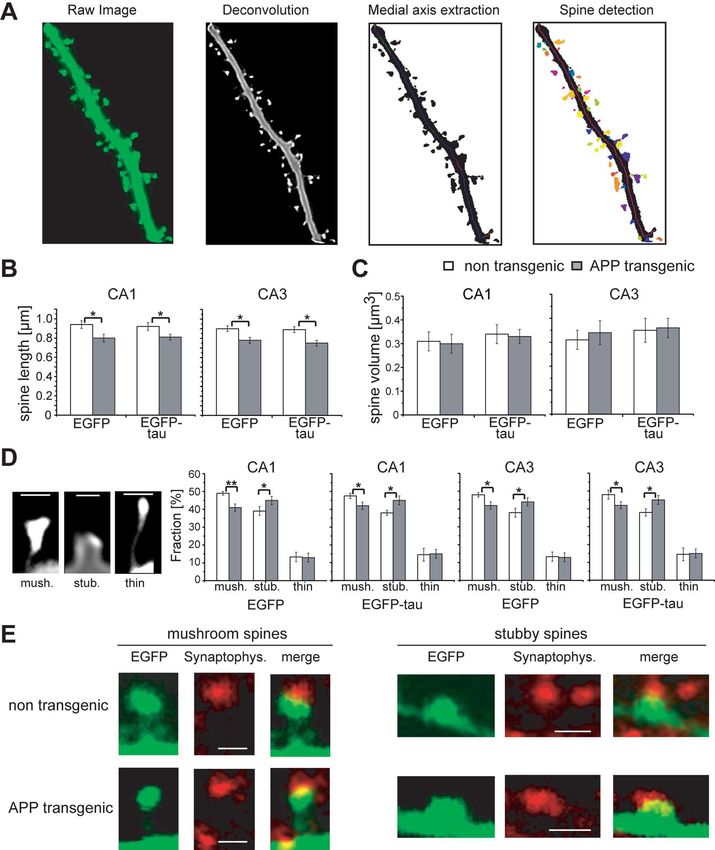
Figure 8. Schematic representation showing the proposed pathways that mediate spine
the HS fraction. In nontransgenic controls, R406W tau and P301L tau is increased in the insol-
pathology and tau-dependent cell death. The formation of A is central, since blocking A
uble fraction to 54 –57% compared with wt tau (42%). On the APP transgenic background the
production by treatment with the ␥-secretase inhibitor DAPT abolishes spine changes and the
insoluble tau fraction is decreased for all constructs (33–39%). Equal amounts of lysates from
induction of tau toxicity. NMDAR activity is required for both induction of spine alterations and
each extraction step were loaded and stained with Tau-5 antibody against total tau.
A-induced tau toxicity, since blocking NMDAR activity with CPP abolishes both pathologies.
A but not tau causes loss of spines, reduction of spine length, and alterations in spine shape, as
The solubility profiles of wt, R406W, and P301L tau do not evidenced by a shift from mushroom to stubby spines. In contrast, A alone is not neurotoxic
correlate with toxicity but requires tau to induce cell death. GSK-3 is activated by A and participates in both
To determine whether the toxicity of the different tau constructs cascades but calcineurin is operating only in mediating spine changes. Within spines, cal-
correlates with potential tau aggregation, the solubility profiles of cineurin is upstream of GSK-3, since calcineurin inhibition mimics the effect of the NMDAR
wt tau, R406W tau, and P301L tau were analyzed by a sequential antagonist CPP. In the soma, A activates GSK-3 independent of calcineurin, which is essen-
extraction protocol using buffers of increasing stringency (Fig. tial for the induction of wt tau toxicity, since blocking GSK-3 activity abolishes cell death
caused by wt tau in APP transgenic cultures, whereas calcineurin inhibition has no effect on cell
7). With all constructs and at every condition, tau was not de-
survival. In contrast, induction of R406W tau toxicity by A is GSK-3 independent, suggesting
tected in the FA fraction, implicating the absence of highly insol- that A affects R406W tau by a different mechanism. Continuous lines show direct effects, and
uble tau species. Compared with wt tau, both FTDP-17 mutants dashed lines show indirect effects with potential intermediate steps.
showed a slightly reduced solubility on a nontransgenic back-
ground (wt tau, 58%; R406W tau, 45%; P301L tau, 42% in the
soluble fraction), which was mainly evident by an increased (Blanchard et al., 2003), spine loss was induced by soluble A in
amount of tau in the SDS fraction. Interestingly, on an APP trans- our experiments. It was previously reported that addition of sol-
genic background, solubility of all constructs was increased by uble A to slice cultures resulted in increased spine length but did
10% for wt tau and by 16 and 22% for R406W tau and P301L tau, not affect spine head diameter (Shrestha et al., 2006). However, a
respectively. Thus, R406W tau and P301L tau do not show dif- detailed analysis of changes in spine types caused by A had not
ferences in their aggregation propensities, although they strongly been performed. In nontransgenic cultures, algorithm-based cal-
differ in their toxic properties in the presence and absence of A culation revealed ⬃50% mushroom, 40% stubby, and 10% thin
as described above (Fig. 5). This indicates that the solubility of wt spines. These numbers closely reflect the distribution in similar
tau, R406W tau, and P301L tau does not correlate with toxicity in cultures that have been evaluated manually by measurements of
our experiments. spine length and head diameter (Zagrebelsky et al., 2005). We
observed a reduced percentage of mushroom spines and an in-
Discussion crease in the fraction of stubby spines in neurons from APP trans-
We have established an ex vivo model of AD using organotypic genic mice. The change in spine shape was also observed in
hippocampal slice cultures from APPSDL transgenic mice in com- dendritic subregions in which no spine loss occurred. This indi-
bination with Sindbis virus-mediated expression of fluorescent cates a transition from mushroom to stubby spines but no selec-
labeled tau constructs. Detailed spine analysis was performed by tive loss of mushroom spines. The morphological change is
algorithm-based evaluation of high-resolution confocal images associated with a reduction in mean spine length, suggesting that
of dendritic segments of CA1 and CA3 pyramidal neurons. Neu- A causes spine retraction. Interestingly, spine volume was not
ronal survival was determined by live imaging. This approach affected. Staining of presynaptic boutons against synaptophysin
permits analysis of the relation between cell death and synaptic showed that mushroom and stubby spines from nontransgenic
changes and the interaction between A and tau pathology. In and APP transgenic animals were in close apposition with synap-
addition, it permits determination of signal transduction mech- tophysin dots. While this is suggestive for the presence of func-
anisms that are involved in each of these pathways in an experi- tional synapses also after the change in spine shape it cannot be
mentally well accessible system. excluded that the strength of synaptic transmission is affected.
Previously it has been shown that senile plaques and A oli- Cell survival was determined by live imaging of infected CA3
gomers reduce spine density in vivo and in slice cultures (Tack- pyramidal neurons. We did not observe a difference between
enberg et al., 2009). In agreement with this, we observed that nontransgenic and APPSDL transgenic cultures after expression of
spine density was strongly reduced in APPSDL transgenic cultures. EGFP indicating that A alone is not neurotoxic. In contrast, we
Interestingly, spines were not affected by tau expression. Spine observed massive neurodegeneration after expression of EGFP-wt
loss was abolished in the presence of the ␥-secretase inhibitor tau in APP transgenic cultures compared with nontransgenic
DAPT, suggesting that the effect on spines was due to A. Since controls, which was prevented by DAPT. This indicates that tau is
APPSDL mice do not develop plaques before the age of 18 months essential for A-induced neurodegeneration, which is in agreementTackenberg and Brandt • Spine Changes and Cell Death Induced by A and Tau J. Neurosci., November 18, 2009 • 29(46):14439 –14450 • 14449
with studies using dissociated hippocampal cultures (Rapoport et mediating spine changes in APPSDL cultures. We found that treat-
al., 2002). This raises the question by which mechanism A ment with NMDAR antagonist CPP, calcineurin inhibitor FK-
induces tau toxicity. We showed that phosphorylation of wt 506, and GSK-3 inhibitor TDZD abolished A-mediated spine
tau is increased on APP transgenic background at the PHF-1 loss. Since the effect of FK-506 closely resembled the effect of CPP
site (phosphorylated S396 and S404), an epitope that is among including a reduction of spine number in controls, GSK-3 ap-
others phosphorylated by GSK-3 (Shahani and Brandt, 2002). In pears to be downstream of calcineurin, which is consistent with
agreement, expression levels of GSK-3 were increased in APP the finding that GSK-3 activity is regulated, among others, by
transgenic cultures. Increased expression or activation of several tau calcineurin (Lee et al., 2005). Thus, A induces a cascade involv-
kinases including GSK-3 has already been reported in AD ing NMDAR, calcineurin, and GSK-3 activation, which alters
(Blurton-Jones and Laferla, 2006). Mutation of 10 of the major neuronal connectivity as observed in AD. It has been suggested
phosphorylation sites to alanine to prevent phosphorylation abol- that A influences spines by mimicking an LTD-like partial
ished increased toxicity in the presence of A. In turn, PHP tau in blockade of NMDARs followed by activation of cofilin and cal-
which the same sites were mutated to glutamate to mimic a perma- cineurin which finally leads to degradation of the actin cytoskel-
nent hyperphosphorylation showed toxicity even on a nontrans- eton within the spine (Shankar et al., 2007). A complete blockade
genic background supporting that increased phosphorylation at of NMDAR activity by NMDAR antagonist CPP may therefore
disease-relevant sites can confer toxicity to tau. prevent the induction of downstream cascades and protect
Many mouse models for AD have been developed by express- against A-induced spine alterations.
ing tau with FTDP-17 mutations in combination with mutated We could show for the first time that the induction of tau
APP or PS1. However, no FAD cases were reported in which tau toxicity by A is also NMDAR dependent since blockade of
is mutated. Thus, it is important to compare the effect of A on NMDARs abolished cell death mediated by wt tau or R406W tau
wt tau and FTDP-17 tau mutants to determine whether these in APP transgenic cultures. In contrast to the cascade causing
provide a valid model for AD pathology. We determined the spine loss, the induction of tau toxicity is not calcineurin depen-
behavior of two FTDP-17 mutants that have been frequently dent. Blocking GSK-3 prevented toxicity of wt tau but not
used. We observed that R406W tau is more toxic than wt tau on a R406W tau on APP transgenic background. This is in agreement
nontransgenic background and becomes highly toxic in the pres- with our finding that phosphorylation of R406W tau is not
ence of A. In contrast, P301L tau did not show any toxicity increased at the PHF-1 site in APP transgenic cultures and
neither in nontransgenic controls nor on an APP background. In further supports that wt tau and R406W tau are differentially
contrast to wt tau, the increased toxicity of R406W tau in APP affected by A.
transgenic slices is not paralleled by increased phosphorylation at Although both spine alterations and cell death are mediated
the PHF-1 site, suggesting that the mechanism how A confers by NMDARs, the downstream cascades substantially differ, since
toxicity to tau is different for wt tau and R406W tau. This makes a blockade of calcineurin prevented spine loss but not cell death.
it questionable to use FTDP-17 tau mutants in combination with We hypothesize that both cascades occur in different cellular
APP or A as a model for neurodegeneration in AD. compartments. A may cause spine changes by inducing a cas-
It is unknown whether soluble or aggregated tau causes neu- cade involving NMDARs, calcineurin, and GSK-3 within the
rodegeneration. We determined the aggregation propensity of wt spine itself while inducing a calcineurin-independent cascade in-
tau, R406W tau, and P301L tau in the presence or absence of A. volving NMDARs and GSK3 at the soma, which causes wt tau
Interestingly, A increased the solubility of all constructs. R406W phosphorylation and cell death.
tau and P301L tau did not show differences in their aggregation
propensity although they strongly differ in their toxic properties References
in the presence and absence of A. Thus, our data indicate that Blanchard V, Moussaoui S, Czech C, Touchet N, Bonici B, Planche M, Canton
T, Jedidi I, Gohin M, Wirths O, Bayer TA, Langui D, Duyckaerts C, Tremp
the solubility of wt tau, R406W tau, and P301L tau does not G, Pradier L (2003) Time sequence of maturation of dystrophic neurites
correlate with toxicity and suggests that tau-dependent neurode- associated with Abeta deposits in APP/PS1 transgenic mice. Exp Neurol
generation occurs in the absence of major tau aggregates. This is 184:247–263.
in agreement with the observation that tau-dependent neurode- Blurton-Jones M, Laferla FM (2006) Pathways by which Abeta facilitates tau
generation occurred in a Drosophila model without NFT forma- pathology. Curr Alzheimer Res 3:437– 448.
tion (Wittmann et al., 2001) and that in a zebrafish model Bourne J, Harris KM (2007) Do thin spines learn to be mushroom spines
that remember? Curr Opin Neurobiol 17:381–386.
degeneration preceded tangle formation (Paquet et al., 2009). In
Braak H, Braak E (1991) Neuropathological stageing of Alzheimer-related
a mouse model, neuron number stabilized and memory recov- changes. Acta Neuropathol 82:239 –259.
ered after tau suppression despite continuous accumulation of Chen P, Gu Z, Liu W, Yan Z (2007) Glycogen synthase kinase 3 regulates
NFTs (Santacruz et al., 2005). N-methyl-D-aspartate receptor channel trafficking and function in cor-
Our approach permits to investigate the signal transduction tical neurons. Mol Pharmacol 72:40 –51.
pathways involved in mediating both spine changes and cell Dovey HF, John V, Anderson JP, Chen LZ, de Saint Andrieu P, Fang LY,
Freedman SB, Folmer B, Goldbach E, Holsztynska EJ, Hu KL, Johnson-
death (Fig. 8). We show that blocking A production by treat-
Wood KL, Kennedy SL, Kholodenko D, Knops JE, Latimer LH, Lee M,
ment with the ␥-secretase inhibitor DAPT abolished both spine Liao Z, Lieberburg IM, Motter RN, et al. (2001) Functional gamma-
changes and the induction of tau toxicity. This indicates that the secretase inhibitors reduce beta-amyloid peptide levels in brain. J Neuro-
generation of A is upstream of both processes, which supports chem 76:173–181.
the amyloid cascade hypothesis. It has been shown previously Ehrengruber MU, Lundstrom K, Schweitzer C, Heuss C, Schlesinger S,
that soluble A oligomers can bind to or near NMDARs (Lacor et Gähwiler BH (1999) Recombinant Semliki Forest virus and Sindbis
al., 2007). Shankar et al. (2007) have shown that blockade of virus efficiently infect neurons in hippocampal slice cultures. Proc
Natl Acad Sci U S A 96:7041–7046.
NMDARs abolished spine loss, which had been induced by the Eidenmüller J, Fath T, Maas T, Pool M, Sontag E, Brandt R (2001)
acute addition of soluble A. A induced LTD via activation of Phosphorylation-mimicking glutamate clusters in the proline-rich region
calcineurin and GSK-3 (Li et al., 2009). This raises the question are sufficient to simulate the functional deficiencies of hyperphosphory-
whether NMDARs, calcineurin, and GSK-3 are also involved in lated tau protein. Biochem J 357:759 –767.You can also read