Airway Management Guidelines for Patients with Known or Suspected COVID-19 Infection
←
→
Page content transcription
If your browser does not render page correctly, please read the page content below
Airway Management Guidelines for Patients with Known or Suspected
COVID-19 Infection
Emergency Medicine: George Kovacs (gkovacs@dal.ca)
Anesthesia: J Adam Law (jlaw@dal.ca)
Critical Care & Anesthesia: Tobias Witter (tobias.witter@nshealth.ca)
Infection Prevention and Control: Ian Davis (Ian.Davis@nshealth.ca)

 Disclaimer:
 Many of the publications upon which this document is based are considered ‘low level’ evidence, i.e.,
 based on expert opinion and consensus. However, we feel that the benefit of providing this guidance
 exceeds the risk. These recommendations should not be considered mandatory or standard of care in
 the management of the COVID-19 patient: the clinician’s professional judgement must be applied to
 every situation.

Updates to this version (V 4.2 January 23, 2021):
o Recommendations provided in this document were based on a focused review of the literature including
   existing/updated consensus guidelines published since the last update. A select number of references
   have been added to the document.
o Where the literature yielded inconsistent and/or conflicting recommendations, additional expert opinion
   was sought from local resources (NS Health IPAC) and international airway management experts.
o A jurisdictional scan and review of existing evidence on various oxygen delivery methods was conducted
   by NS Health Infection Prevention and Control (IPAC). This review formed the basis on which oxygen
   delivery methods should or shouldn’t be considered aerosol-generating medical procedures (AGMP).
o The EMS section has been removed.
o Further educational support material including videos have been embedded.
o At risk patients with escalating oxygen requirements should be placed in a resuscitation-capable negative
   pressure environment with staff wearing appropriate PPE for any necessary.
o The RSI Algorithm was updated emphasizing the importance of provider experience with chosen
   equipment.
o Ventilation by face mask or supraglottic device should be used as required for re-oxygenating patients
   who are desaturating. Ventilation provided by BVM with an attached filter and PEEP valve should be
   guided by waveform capnography.

Definitions for the purpose of this document:
High flow nasal cannula (HFNC): Support device able of delivering high flows (up to 70 l/min depending on
the device) of heated humidified oxygen concentrations via special nasal cannulas (e.g. AirVo, Vapotherm).
Continuous Positive Airway Pressure (CPAP): Any device or adjunct, able to facilitate a continuously elevated
airway pressure with the intent to improve oxygenation.
Biphasic positive airway pressure (BiPAP): A ventilation form alternating between two airway pressures,
allowing for spontaneous breathing on both levels.

1. Background
Despite significant improvements in our understanding and management of patients infected with SARS-
CoV-2, ‘the risk of respiratory failure requiring critical care support in patients with COVID-19 is significant’.1
During the first wave, complexity and fear of the unknown created significant additional challenges for those
tasked with identifying and caring for this new patient population. There remain many unknowns, however
the common path for those severely ill patients is a presentation of acute hypoxemic respiratory failure.

Version 4.2. January 23, 2021                                                                                         1
Acute care provider teams are familiar with managing patients with this presentation. It is clear however,
that while the principles are similar, pre-existing airway management guidelines cannot simply be applied to
those with COVID-19 acute hypoxemic respiratory failure. Numerous COVID-19 airway management
guidelines have been published to address these differences with variable levels of detail and as stated in our
disclaimer remain for the most part supported by either poor quality studies and/or expert opinion 2. Despite
the ongoing gaps in evidence, acute care provider teams have been adapting their practices to local clinical
environments as we all continue to ascend a learning curve in preparing for the arrival, transfer of care and
management of these patients. Both the patient and provider teams are at risk during airway management of
COVID-19 cases. While patient safety continues to be a goal, safety of the staff caring for the patient is
equally important3. Caring for COVID-19 patient safely requires meticulous attention to detail using a
common, standardized and rehearsed approach to airway management.

1.1 Context
In this document, recommendations for management of the COVID-19 patient relate to (a) efficacy of the
recommended treatment modality; (b) its risk to staff safety and (c) resource availability.

1.2 Sources for the document
Material in this document is based on the following sources:
    • Published evidence and recommendations;
    • COVID-19 Hub for Nova Scotia Health Team Members and Physicians;
    • Internal Nova Scotia Health (NSH) multi-disciplinary consensus meetings;
    • Contact with clinicians working in ‘hot zones’ where surges have occurred;
    • Expert opinion from authors of existing published international COVID-19 Consensus Guidelines.
This continues to be a living document, to be updated as more information becomes available or as guidance
needs to be altered. Although many additional articles were reviewed for this update, few would meet a
level of evidence to be included.

2. Principles

General:
   • When airway management of the COVID-19 patient is indicated, provider safety must be ensured.
   • When tracheal intubation is indicated, rapid achievement of first attempt success should be a
        primary goal.

Infection control:
    • For any potentially aerosol-generating medical procedures (AGMPs), including tracheal intubation or
         positive pressure ventilation in the non-intubated patient, airborne/droplet/contact precautions
         should be used by all providers, informed by local infection prevention and control
         recommendations (NSH COVID-19 Hub: Acute Medicine)
    • A resuscitation capable ‘ready bed’ should be designated in an airborne infection isolation room
         (AIIR), with a separate preparatory space (‘antechamber’). If an AIIR is not available, the intubation
         should occur in a single room with the doors closed.

Airway Management Providers:
    • Acute care physicians should train and prepare for intubating COVID-19 patients.
    • The most skilled airway management provider available should perform tracheal intubation.

Version 4.2. January 23, 2021                                                                                 2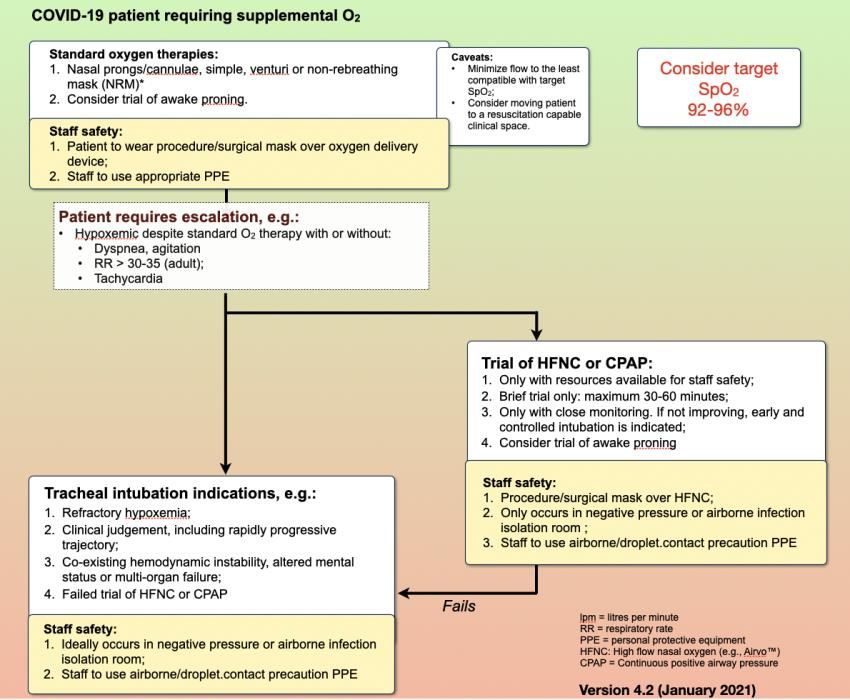
Team:
    •      Clinicians often feel a sense of urgency in managing hypoxemic patients. Airway providers and
           teams should slow down to ensure their own safety as they prepare to manage known or suspected
           COVID-19 patients.
      •    The number of primary team members at the bedside should be limited (e.g., to 3: primary airway
           provider, airway support provider and clinical support provider).
      •    Providers and teams should engage in regular practice of the necessary procedures, including PPE
           donning and doffing and the approach to airway management.

Equipment:
    • Required airway equipment and medications should be prepared and available prior to entry into the
       designated ready room.
    • Video laryngoscopy should be used as the primary approach for tracheal intubation.

Approach:
   • Rapid sequence intubation should be used to facilitate first-pass intubation success and prevent
       patient coughing or gagging.
   • If an awake intubation is considered necessary, e.g., due to unfavourable airway anatomy, this
       should only be done by an expert provider, trained to perform the approach in COVID-19 patients,
       using airborne/droplet/contact precautions.

Communication:
   • Team briefings should occur prior to entering the ready room and immediately prior to initiating
      airway management.
   • Checklists and/or other visual aids should be used to prepare for tracheal intubation;
   • No transports of COVID-19 patients should occur without direct communication with the receiving
      providers/teams.
   • The ability for team members in the room to communicate with the individuals outside the room
      should be tested and practiced (e.g., with use of writing tablets, small walkie-talkies, etc.)
   • Closed loop communication should be used by the team.

3. Suggested Management

3.1 Preamble:
SARS-CoV-2 is a novel virus presenting a new and unique constellation of symptoms and signs. Optimal
management is still unknown. However, the following generalizations apply to these patients’ respiratory
support, including tracheal intubation:
     • If possible, establish ‘do-not-intubate’ or ‘do-not-resuscitate’ status prior to tracheal intubation of
         any, but especially older or co-morbid patients with COVID-19 related respiratory failure.
     • Use of HFNC or CPAP may be considered, in selected cases in an attempt to avoid the need for
         tracheal intubation.4
     • However, in COVID-19 patients in hypoxemic respiratory failure with a worsening trajectory despite
         escalating oxygenation measures (including the use of HFNC or CPAP), tracheal intubation must not
         be delayed, so that it can occur under controlled, non-emergency conditions.
     • Staff safety is paramount, so that use of oxygen delivery and ventilatory support modalities that are
         considered to be at risk of aerosol generation should occur in an AIIR with staff using
         airborne/droplet/contact precautions.

Version 4.2. January 23, 2021                                                                                3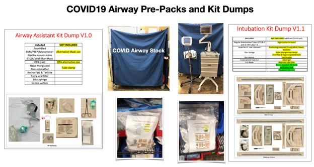
•    Regardless of delivery modality, whenever possible, the patient requiring oxygen supplementation
           should be closely monitored for deterioration in a resuscitation capable room with adequate oxygen
           sources, suction, monitoring capability and room for staff to safely move and have 360 0 access to
           the patient. If needed, escalation to the next level should occur in a timely fashion. When tracheal
           intubation is indicated, it should occur in a controlled manner with close attention to staff safety.
     •     Some patients with COVID-19 who have respiratory failure can present with a profound degree of
           hypoxemia, yet seemingly few symptoms (the ‘silent’ or ‘happy’ hypoxemic patient): speaking in full
           sentences, without significant dyspnea and with normal mentation. These patients should be
           considered for supplemental oxygen that includes HFNC or CPAP.
     •     Others may have a more classic presentation of hypoxemia with tachypnea and high work of
           breathing, hypercarbia and tachycardia or with other signs such as confusion or hypotension. This
           population will likely require early tracheal intubation.
     •     Optimal oxygen goals are unknown, but a target SpO2 of 92-96% should be considered 5. If below
           92%, initiation or escalation of oxygen therapy should be considered. An SpO 2 above 96% should be
           avoided by reducing the oxygen flow rate or de-escalation to a delivery modality with less potential
           for aerosol generation. Going forward, lower oxygen targets may be established for this disease.
     •     The COVID-19 patient in hypoxemic respiratory failure sometimes responds well to proning – both
           with or without tracheal intubation. The non-intubated patient can be supplemented by modalities
           that range from standard nasal prongs/cannulae to CPAP. However, on resumption of the supine
           position, the patient may again desaturate. The team should be ready for this eventuality (NSH
           COVID-19 Hub: Prone Positioning in Acute Care Setting).
     •     As few published studies currently address the degree of aerosol generation from HFNC or CPAP or
           the resulting extent of risk to staff, the term ‘consider’ will be used when a recommendation is made
           for the use of these modalities. However, the recommendations in this document are taking this
           uncertainty into account and with appropriate use of PPE and environmental considerations around
           airborne/droplet/contact precautions, health care providers will be protected if they use these
           modalities (NSH COVID19 Hub: Oxygen Delivery Review).

3.2 Supplemental oxygenation before the need for tracheal intubation is identified.

3.2.1 Note to reader:
The current evidence is sparse and often contradictory regarding the ‘safety’ of a particular oxygen flow
rate or delivery modality to the health care provider. In many cases, evidence is based on ‘bench’ (e.g.,
manikin) studies. The following statements can be made:
    • An oxygen flow rate of < 6 liters per minute (lpm), while still causing some aerosol dispersion, is
         generally considered acceptable for staff wearing droplet/contact precaution level PPE.
    • Regardless of device used, the clinician should strive to use the lowest oxygen flow rate that is
         compatible with adequate patient oxygenation.
    • Aerosol/droplet dispersion appears to be substantially lessened by applying a surgical or procedure
         mask over modalities such as standard nasal cannulae or HFNC.

3.2.2 For the patient with known or suspected COVID-19 in the Emergency Department (ED) or inpatient
       unit – initial oxygen supplementation options:

          o    O2 nasal prongs/cannulae at flows < 6 lpm
                       Droplet and contact precautions should be used.
                       An AIIR is not required, as flow rates ≤ 6 lpm are not considered an AGMP
                       Standard nasal cannulae are generally well-tolerated at flows < 6 lpm.

Version 4.2. January 23, 2021                                                                                  4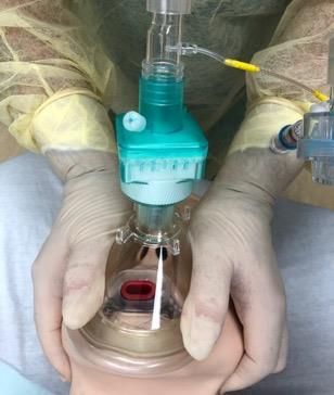
The patient should wear a standard procedure or surgical mask applied over the nasal
                          cannulae to help limit droplet spread.
                          A trial of lateral positioning or self-proning can occur with the patient wearing nasal
                          prongs/cannulae. (NSH COVID-19 Hub: Prone Positioning in Acute Care Setting)
                          There is no convincing evidence that flow rates by nasal prong/cannula  30-35 (adult);
                          - Tachycardia;
                          - Agitation;
                          - Accessory muscle use; paradoxical chest/abdomen movement;
                     Worsening PaO2/FiO2 ratio.
                     Increasing PaCO2.
                     Rapidly progressive disease trajectory or other clinical judgement.
                     Other standard indication for tracheal indication, e.g., failure to protect the airway or
                     obstructing airway pathology, hemodynamic instability, sepsis, multi-organ failure.
         o Tracheal intubation is considered an aerosol-generating medical procedure and should ideally be
              performed in an AIIR.

3.3.2 Available literature and general expert consensus is that the use of HFNC or CPAP can prevent an
intubation in a significant proportion of patients with COVID-19, and even if intubation cannot be avoided, it
will allow for the HCPs to prepare and ready for the intubation. It is important to recognize that HFNC and
CPAP are acknowledged as being potentially aerosol-generating6, 7.

Version 4.2. January 23, 2021                                                                                       5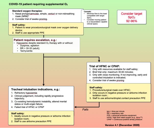
HFNC including Airvo™; Optiflow™ or Vapotherm™ deliver humidified oxygen at flow rates of 40-70
          lpm:
          o Should be considered as potentially aerosol-generating and ideally used in an AIIR. If an AIIR is
              not available then treatment should be performed in a single room with the door closed.
          o Staff caring for the patient using HFNC should use airborne/droplet/contact precautions.
          o To help reduce aerosolization potential, consider using FiO 2 1.0 and reducing flows to the lowest
              needed to achieve target oxygen goal (e.g., SpO2 92-96%), as permitted by hospital supplies of
              oxygen.
          o A trial of HFNC in the prone position can be considered in the cooperative awake, spontaneously
              breathing patient.
          o If HFNC are used, the patient should be closely monitored in an appropriate setting (e.g. ICU, ED,
              IMCU) for deterioration, and if not responding favourably within a 30-60 minute trial period,
              should proceed to a controlled COVID-19 protected tracheal intubation.

          CPAP. If HFNC is not available, CPAP can be delivered by a variety of devices, e.g.:
          o By a non-invasive ventilation device.
          o By a commercial flow-dependent CPAP mask with a viral filter. This includes the Rescuer®
             Emergency CPAP System 8700 (NSH COVID-19 Hub: Acute Medicine-CPAP Rescuer). This has
             been distributed around the province as a resource for empiric use when escalation of oxygen
             therapy is required but local resources do not include traditional CPAP or HFNC devices. It can
             be used to temporize, for example, pending the arrival of a transport crew able to perform
             tracheal intubation.
          o CPAP can be delivered by applying nasal prongs at 5-10 lpm and applying an overlying cuffed
             mask attached to viral filter, catheter mount, bag-valve mask device with PEEP valve (10 cm H20)
             with oxygen running at a flow of 15 lpm (Figure 1).

          Figure 1: BVM/PEEP/manometer, flexible mount, waveform CO 2 connector, viral filter, mask (the
          flexible mount shown in the above figure has a suction port which should be closed AND cannot be
          used for suctioning)

Version 4.2. January 23, 2021                                                                                6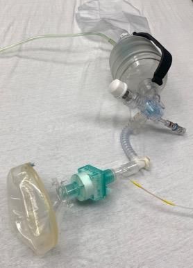
o    All CPAP generating options should be considered as potentially aerosol-generating and ideally
               used in an AIIR. Staff caring for the patient using CPAP should use airborne/droplet/contact
               precautions.
          o    To help reduce aerosolization potential, consider starting with the least supporting pressure
               (e.g., CPAP 8-10 cm H20) consistent with adequate SpO2 (e.g., 92-96%).
          o    A trial of CPAP in the prone position can be considered in the cooperative, awake, spontaneously
               breathing patient.
          o    If CPAP is used, the patient should be closely monitored for deterioration, and if not responding
               favourably within a 30-60 minute trial period, should proceed to a controlled COVID-19
               protected tracheal intubation.

          BiPAP is not generally recommended for support of the COVID-19 patient in hypoxemic respiratory
          failure.8, 9

          If the patient responds well to support with HFNC or CPAP, that modality can be continued with
          ongoing close observation for tiring or deterioration. With or without a trial of HFNC or CPAP, when
          required, tracheal intubation should occur before it is an emergency. Controlled tracheal intubation
          before the patient decompensates will minimize the potential for risk to staff due to breaches of PPE
          donning protocols.

3.3.3 Tracheal intubation after a failed trial of or CPAP:
       o Failure to respond to the trial of HFNC or CPAP is an indication for tracheal intubation. Response
           should be monitored by attention to parameters such as respiratory rate 10 and ROX index
           ([SpO2/FiO2]/RR)4, 11, 12.

          Figure 2: Suggested COVID-19 escalation of oxygenation pathway (for larger image see Appendix 1)

Version 4.2. January 23, 2021                                                                                  7
4. COVID-19 Protected Tracheal intubation of the COVID-19 patient: recommendations
4.1 Location:
Tracheal intubation of the patient with COVID-19 should ideally occur in an AIIR.1, 13, 14 However, the risks of
transporting a critically ill patient to another location for tracheal intubation must be weighed against the
benefits. Regardless of location, ideally, the chosen area should be resuscitation capable, providing staff with
enough room to move safely while caring for an acutely ill patient. Providers should strive to perform in-situ
simulation in these areas.
4.2 Approach:
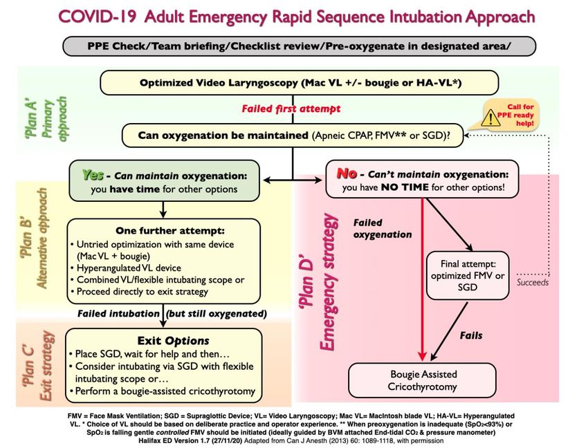
Rapid sequence intubation (RSI) is the default approach for managing these patients, so that the risk of
aerosolized droplet production by coughing or gagging can be minimized. For every case, a ‘double set-up’
RSI is recommended, with the location of the cricothyroid membrane marked and prepped for rescue
cricothyrotomy (Appendix 2: RSI visual aid). Video laryngoscopy is recommended to facilitate tracheal
intubation and maintain distance away from the patient’s face. If used, awake intubation should only be
performed by a provider very familiar with the procedure and its application to the patient with COVID-19.
4.3 Equipment Preparation:
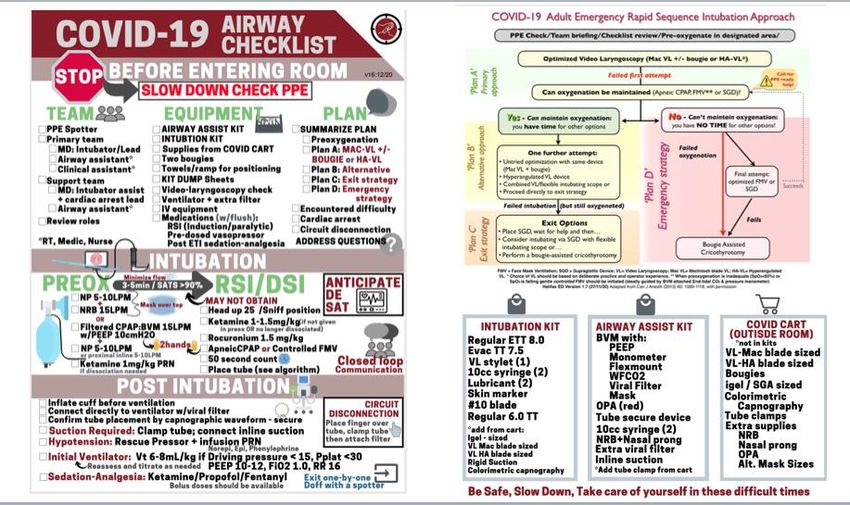
Providers are encouraged to create compact kits that can be taken into the room (e.g., Appendix 3) to avoid
contamination of other equipment. It is assumed that clinicians using the airway equipment described below
are experienced and have practiced the chosen procedures individually and with their teams while wearing
the appropriate PPE. Core equipment should include:
• Suction: open rigid and closed in-line tracheal suction setups.
• The bag-valve mask (BVM) device should be fitted with PEEP valve, cuffed face mask, viral filter and
waveform CO2 and pressure monometer. Adding a flexible catheter mount provides an easier range of
motion for BVM setup to help minimize the risk of accidental disconnection (Figure 1). Connections must
be secure.
• Waveform capnography provides breath to breath ventilation feedback and the appearance of a square
trace is reassuring evidence of a quality mask seal in the spontaneously breathing patient and in those
requiring rescue ventilation.
• A pressure manometer along with waveform capnography provide important user feedback that may
help avoid over-ventilating (high rates, pressures and volumes) patients.
• Oropharyngeal airway (with alternative sizes).
• Regular nasal prongs.
• Intubation devices: Regardless of which video laryngoscope (VL) is chosen, clinicians should be
experienced with its use and deliberately practice to ensure a high first pass success rate without serious
adverse events.
o We recommend use of a video laryngoscope that supports use of single-use (disposable) blades.
For a first attempt, we recommend use of a Macintosh-shaped blade and routine use of a
tracheal tube introducer (‘bougie’)15. There are slight differences between Macintosh-shaped
blades and clinicians should be very familiar with their chosen device (Macintosh VL Video).
Examples of video laryngoscopes include:
Storz C-MAC® S with single-use Macintosh 3 or 4 blades;
GlideScope ® Spectrum™ with single use [Macintosh-shaped] DVM 3 or 4 blades;
McGrath Mac with single-use Mac size 3 or 4 blades.
o Hyper-angulated video laryngoscope blade options are available for all of the foregoing. These
can be used for the patient with anticipated difficult Macintosh laryngoscopy, for a second
intubation attempt after proven difficult Macintosh laryngoscopy (if the patient is still
oxygenated), or alternatively, for first attempt use due to clinician preference if skilled with the
Version 4.2. January 23, 2021 8device. Use of an ‘out-of-package’ bougie is NOT recommended when using a hyper-angulated
VL device (Hyper-angulated VL Video). Examples of hyper-angulated blade VLs include:
Storz C-MAC® S with single-use D-blade;
GlideScope ® Spectrum™ with single-use LoPro S3 or S4 blade;
McGrath™ Mac with X blade.
o Other Options:
A high quality single-use direct laryngoscope should be available in case the VL system
fails.
If available, a single-use flexible intubation endoscope may be valuable in experienced
hands, for example, used through a supraglottic device, e.g., in the event of difficulty
achieving tracheal intubation using standard video laryngoscopy.
• Tracheal tubes:
o Evac (i.e., including a subglottic suction port) tubes should be considered as first choice unless a
difficult tracheal intubation is predicted. If an Evac tube is chosen, for an average sized adult,
decrease the size by 0.5 mm internal diameter (ID).
o If difficult intubation is predicted, a conventional tracheal tube or a Parker Flex-tip tracheal tube
is recommended: size 7.0-7.5 mm ID for an adult female; 7.5-8.0 for an adult male.
o 10 ml syringe for cuff inflation.
o Use a familiar commercial tracheal tube securing device (no tape).
• Tracheal tube adjuncts:
o It is recommended that a bougie be considered for all tracheal intubations facilitated by a video-
enabled or traditional direct Macintosh blade15. As previously noted there are some differences
between Macintosh blades and clinicians should be experienced with their device to ensure a
high first pass success rate.
o If a hyper-angulated blade video laryngoscope blade is chosen for use, a conventional or Parker
Flex-tip tracheal tube is suggested, appropriately shaped to a 60 to 70-degree distal bend with a
rigid or semi-rigid stylet.
o In experienced hands, a bougie that maintains a pre-shaped curve or is steerable may be used
with a hyper-angulated video laryngoscope.
• Tracheal tube confirmation:
o As previously stated, waveform capnography is strongly recommended and should be attached
proximal to the viral filter. Seeing six complete breaths with sustained amplitude confirms
correct tracheal tube placement.
o A colorimetric CO2 detection device (capnometry) is an alternate if waveform capnography is not
available.
o A complete absence of CO2 must not be ascribed to peri-intubation arrest or low-flow state:
rather, esophageal intubation must be excluded.
• Supraglottic airway device for rescue:
o A supraglottic airway device (SGD) should be selected based on the patient’s weight. A second
SGD, one size smaller or a different type should also be available.
o The type of device should be based on provider familiarity and ease of placement and should
ideally support endoscopically guided (with flexible intubation endoscope) tracheal intubation if
needed. A ‘second generation’ device that is designed to obtain a better seal and also features
an esophageal drainage port is ideal.
o The EMS i-gel® with a passive oxygenation port is the currently recommended device for use in
our Provincial emergency departments; other devices such as the King LTS-DTM have a high first
Version 4.2. January 23, 2021 9pass success rate in trained hands, however the SGD will not support an endoscopically guided
intubation through the device (Pending document: NSH COVID-19 Hub: Acute Medicine).
• Cricothyrotomy equipment:
o Bougie, #10 scalpel blade and a 6.0 tracheal tube; pack of sterile gauze.
• Vascular access with two IVs should be in place. If not yet established, supplies for IV and intra-osseous
(I-O) access should be available.
4.4 Pharmacologic preparation:
Drugs should be drawn up and clearly labelled outside the room:
• Ketamine 1.0-1.5 mg/kg. Providers may choose to decrease the dose of ketamine for patients with a
shock index of >1 (HR/SBP) by 25-50%.
• An induction sedative-hypnotic other than ketamine can be used, guided by provider familiarity and
preference;
• Rocuronium 1.5 mg/kg;
• Succinylcholine 1.5 mg/kg can be used as an alternative to rocuronium.
• IM ketamine in the 50 mg/ml concentration should be available for behavior control, if needed.
• Push dose pressors should be drawn up in advance, labelled and ready to administer (e.g., epinephrine or
phenylephrine).
• A norepinephrine drip should be available before RSI, to be started at a dose of 0.1mcg/kg/min, as
necessary.
• Post intubation sedation and analgesia (bolus and infusion) should be readied. Choice of sedative should
be governed chiefly by provider familiarity.
• 20-cc syringe of saline flush solution.
4.5 Personal Protective Equipment (PPE):
PPE for all team members involved in tracheal intubation of the COVID-19 patient should be for
airborne/droplet/contact precautions. Please consult with your local infection control authorities regarding
exact PPE recommendations. A checklist should be used for PPE donning and doffing (Monitor updates on
NSH COVID-19 Hub: Acute Medicine).
• SLOW DOWN: Regardless of the urgency to proceed with the intubation, take the time to don (and later
doff) PPE safely. Designate one team member to be a ‘checker’ or coach. A 360-degree review of each
team member should occur before entering the room.
• Peripheral vision, fogging and glare from PPE visors may cause challenges. Be prepared for this by having
practiced with the equipment during simulation exercises beforehand.
4.6 Team Briefing outside room:
No matter their experience level, providers will be anxious about airway management for patients with
COVID-19. These patients are critically ill and are physiologically compromised. Based on physiologically
similar cohorts, post-intubation cardiac arrest may occur in up to 2-3% of critically ill patients.16, 17 Regardless,
the team must be reminded that their safety is the foremost priority.
• Outside the room, the team leader should be identified. This individual may or may not be the primary
airway provider.
• Primary team identified, for example:
o Primary airway provider;
o Airway support provider (e.g., RT, medic);
Version 4.2. January 23, 2021 10o Clinical support provider (e.g., nurse).
• Support team (if available) is identified with PPE donned in anteroom after the primary team has entered
the patient’s room, for example:
o Second provider as intubation support/cardiac arrest lead;
o Airway support provider (e.g., RT, medic or nurse) as a ‘runner’ who can easily identify the
requested equipment and pass it into the room.
• Articulate the plan to the team:
o RSI as the default approach;
o Preoxygenation strategy;
o Review your plan for difficulty if encountered using the RSI visual aid (e.g., Appendix 2);
o Assign specific airway roles for those inside the room (e.g., timing, bougie assistant, two-handed
BVM ventilation);
o The plan for confirmation of tracheal intubation using capnography or capnometry;
o The plan for transfer to mechanical ventilation, including how to do planned circuit
disconnections, if required;
o The plan for cardiac arrest;
o Invite questions from the team.
4.7 Inside the room:
Preparation should be guided by a checklist with which the team has trained and become familiar (e.g.,
Appendix 4):
• Monitors applied to patient:
o Standard SpO2, ECG, non-invasive blood pressure (on the opposite arm to that to be used for
medication administration, cycled at intervals of no less than 2 minutes);
o Waveform CO2 monitoring should begin with preoxygenation. Anything other than a square
waveform may be indicative of a poor seal on the patient’s face.
• Patient positioning:
o The patient should be positioned in a back up (close to sitting or position of comfort) if
hemodynamics permit. The bed can be transitioned to a flatter (but still somewhat back up or
reverse Trendelenburg) position as the patient loses consciousness with RSI.
o The head and neck should be positioned in the standard ‘sniff’ position.
o Obese patients should be ramped (using blankets or a dedicated positioning insert) to also achieve
ear-to-sternum ‘sniff’ positioning.
• Good free-flowing vascular access should be confirmed.
• A tracheal tube clamp and extra viral filter should be readily available should a circuit disconnection
occur.
• Airway exam: evaluation of the patient’s airway anatomy should occur. Dentures removed.
• Final briefing and review of cognitive aid (e.g., algorithm, Appendix 4) with any changes to the plan based
on airway exam findings.
• Marking the cricothyroid membrane by palpation or facilitated by ultrasound should be considered for all
patients as part of the ‘double set-up’. This will not necessarily represent a point of entry but serves as a
landmark for the initial vertical incision as part of a bougie assisted cricothyrotomy (Video link).
4.8 Pre-oxygenation:
Patients with COVID-19 being intubated for respiratory failure can be expected to desaturate rapidly with the
onset of apnea during RSI. Thus, optimized pre-oxygenation is important. Once again, recognizing the need
Version 4.2. January 23, 2021 11to minimize the potential for aerosol generation during the process, options for pre-oxygenation include the
following:
4.8.1 Continue the existing oxygen delivery modality for the pre-oxygenation phase:
o The patient requiring tracheal intubation shortly after arrival by EMS may already be receiving
CPAP by cuffed facemask system with viral filter and straps.
o Or, EMS may have used a standard BVM CPAP system applied to the patient: facemask-viral
filter-BVM with PEEP valve. This may also be continued into the pre-oxygenation phase.
o An inpatient may have been receiving HFNC or CPAP or in rare cases, BiPAP: any of these
modalities can simply be continued for the pre-oxygenation phase prior to RSI.
Notwithstanding, transferring the patient on HFNC or CPAP to standard cuffed mask
and BVM device for pre-oxygenation provides the opportunity to confirm the size of
mask is that needed for a good seal, before the potential need for positive pressure
ventilation.
As previously indicated, these modalities should ideally be administered in an AIIR
with staff using airborne/droplet/contact precautions.
4.8.2 Change from nasal prongs or non-rebreathing mask to a more effective pre-oxygenation modality:
o The patient currently receiving oxygen supplementation by standard nasal prongs or NRFM
proceeding directly to RSI will need to be transitioned to a more effective means of pre-
oxygenation.
o In some cases, ketamine administration may be required to facilitate tolerance of pre-
oxygenation techniques (‘delayed sequence intubation’)18, 19.
o One option (e.g., Figure 2c) to provide some CPAP during pre-oxygenation is a bag-valve mask
(BVM) flowing at 15 lpm with PEEP valve (10cm H20) and viral filter, placed over nasal prongs
flowing at 10 lpm. The need for additional flow administered by nasal prongs follows from the
significant degradation of flow (often by 50% or more) through many disposable BVMs (CPAP
pre-oxygenation video).
o It is important that the BVM set-up has an integrated connector to enable monitoring of
waveform CO2. A good seal during pre-oxygenation should ideally be confirmed by the presence
of a square waveform capnographic trace in the spontaneously breathing patient.
o Providing gentle manually assisted ventilations (pressure support) in tachypneic spontaneously
breathing patients poses an additional risk of aerosolization when asynchronous breaths are
delivered with a poor mask seal. This is not recommended.
o If available (e.g., in the operating room), end-tidal oxygen readings can be used to monitor efficacy
of pre-oxygenation efforts.
o Regardless of the setting or pre-oxygenation technique, the goal in the pre-oxygenation phase is
to achieve an SpO2 > 90%, if feasible.
4.9 RSI and tracheal intubation should follow:
• Adequate time (e.g., 45 seconds for succinylcholine; 60 seconds for rocuronium) must be given for the
complete onset of neuromuscular blockade to minimize the possibility of coughing or gagging with
airway instrumentation.
• Pre-oxygenation as described above can be continued after loss of consciousness while awaiting the
onset of neuromuscular blockade, with the addition of an oral airway and jaw thrust using a V-E grip (use
of thumbs and thenar eminence to hold mask on the face, while lifting the mandible using the middle
and ring fingers placed behind the angle of the mandible e.g., Figure 3; Video).3
Version 4.2. January 23, 2021 12Figure 5: Two-handed mask hold with a thumbs forward / thenar eminence (‘V-E’) grip and jaw thrust
• Positive pressure bag-mask ventilation while awaiting the onset of neuromuscular blockade is ideally
avoided but if elected, should be done with a V-E grip, and keeping insufflation pressures well below 20
cm H20, with attention to maintaining a good seal.
• The timing and placement of an OPA as the patient transitions to an apneic state must consider the
patient's ability to tolerate the device and the fact that removal of the mask during preoxygenation will
lead to loss of recruitment (CPAP effect).
• Flow to the BVM and nasal prongs should be turned off when the mask is removed for laryngoscopy to
avoid potential contamination from the mask.
• The clinician should stand straight during laryngoscopy, using indirect viewing via the video screen for
both laryngoscopy and intubation.
• Despite good visualization on the video laryngoscope screen, it is critical that the user maintain
awareness of what is happening when the blade, bougie or tracheal tube enter the mouth.
• Use of a bougie should be considered to facilitate all tracheal intubations using Macintosh
videolaryngoscopy15 (VL) as previously discussed. If used, it must be extracted carefully (without flicking
secretions into the room) and discarded in a trash bin next to the bed.
• The tube should be advanced until the cuff has disappeared 2-cm below the cords: auscultation to rule
out endobronchial intubation will be difficult and is not advised.
• The cuff of the tracheal tube must be inflated prior to initiation of positive pressure ventilation.
• The BVM or ventilator circuit with viral filter should be attached to the proximal end of the tracheal tube
AND oxygen flow resumed.
• Sustained waveform capnography should be confirmed.
• Blood pressure should be reassessed.
• The tube should be firmly secured using a commercial securing device.
5. Failed first attempt at tracheal intubation (refer to algorithm, Appendix 2).
5.1 Re-oxygenation:
If the first attempt at laryngoscopy and intubation fails, these patients will very likely desaturate before a
second attempt. If needed, attempts to reoxygenate the patient must occur in a controlled manner. Options
include:
o Gentle face mask ventilation with OPA, 2-handed mask hold for a good seal and low tidal volumes; bag-
valve mask (BVM) flowing at 15 lpm with PEEP valve (10cm H 20) and viral filter, placed over nasal prongs
flowing at 10 lpm.
o Placing a second-generation SGD can be considered for both re-oxygenation and as a potential exit
strategy (preferably one that supports flexible endoscopic intubation).
Version 4.2. January 23, 2021 13o These two techniques are the preferred strategy as waveform capnography feedback will help guide the
effectiveness of these re-oxygenation strategies, often before saturations improve.
o Apneic CPAP: Place an OPA. Re-apply the filtered BVM system, again with PEEP of 10 cm H 20 at a flow of
15 lpm over nasal prongs 10 lpm without manual assistance. This strategy may be in-effective as the
patients saturation will already be declining and there will be no waveform capnography feedback in
this scenario. Active FMV described above should be the default strategy in the re-oxygenation scenario.
5.2 Further attempts at tracheal intubation after a failed first attempt:
o A further attempt at tracheal intubation can occur in the still-adequately oxygenated patient.
o A second attempt can occur with an optimized technique with the original device, use of a different
device (e.g., hyper-angulated videolaryngoscope), or use of a different operator.
o Flexible endoscopic intubation through an intubating SGD is an option for the clinician skilled in the
technique in the still-adequately oxygenated patient.
5.3 Failed tracheal intubation in the still-oxygenated patient
o Even if still adequately oxygenated, failure to intubate the COVID-19 patient in respiratory failure after a
maximum of three attempts should prompt strong consideration for performing a surgical airway, as
awakening the patient is unlikely to be a viable option.
o If not done to this point, an SGD should be placed while equipment and/or personnel to perform surgical
airway are obtained, with attention to minimizing insufflation pressure and leak.
o The ‘exit strategy’ surgical airway should be performed in a timely fashion in the still-oxygenated patient
to help minimize how long positive pressure ventilation must occur by face mask ventilation or a SGD. It
will ideally occur by an open surgical technique (e.g., scalpel/bougie cricothyrotomy) by the most
experienced clinician available to perform the procedure.
o Any ongoing attempts at positive pressure ventilation by face mask or SGD ventilation during
cricothyrotomy should be discontinued just before the cricothyroid membrane is incised, to avoid risk of
aerosolization via the incision.
5.4 ‘Can’t intubate, can’t oxygenate’
o Distinct from the foregoing ‘exit strategy’ situation whereby surgical airway must occur in a timely
fashion, emergency cricothyrotomy must be performed immediately if a ‘can’t intubate, can’t oxygenate’
(CICO) situation occurs.
o The CICO situation is defined by the failure of at least one attempt at all of tracheal intubation, optimized
face mask ventilation and SGD ventilation, with current or imminent hypoxemia.
o If CICO occurs, scalpel/bougie-assisted cricothyrotomy should proceed immediately by the most qualified
individual already present (Bougie assisted cricothyrotomy video).
6. Post-intubation management:
o Ongoing sedation and analgesia should be addressed given the expected duration of pharmacologic
paralysis with high-dose rocuronium;
o If intubated outside an intensive care setting, consideration should be given to maintaining
pharmacologic paralysis with ongoing sedation during transportation to the setting of the patient’s final
disposition. This may help avoid accidental extubation or coughing and bucking during transfer.
o Initial ventilator settings should be consistent with a lung protection ventilation strategy, e.g., tidal
volume 6 ml/kg predicted body weight; plateau pressure < 30 cm H2O; RR 25/minute; PEEP 8-10 cm H2O;
FiO2 1.0, titrated down rapidly as permitted to maintain SpO2 92-96% thereafter. Tidal volume should be
reassessed and adjusted to keep the driving pressure (difference between PEEP and plateau pressure)
below 15 cmH20.20
o Ongoing hypoxemia post intubation can be addressed with options that include increasing FiO 2 if not
already at 1.0; recruitment maneuver to re-recruit alveoli collapsed during the intubation process (e.g.
Version 4.2. January 23, 2021 14inspiratory hold at 40cm H2O x 10-15 seconds), maintaining pharmacologic paralysis and sedation. If
despite these measures, the patient does not improve, placing the patient in prone position is strongly
recommended.
o Consideration should be given to placing invasive vascular access in the same setting as tracheal
intubation, e.g., arterial line + central venous access.
o Consideration should be given to placing a nasogastric tube.
o Inline suction should be used. To place this, a circuit disconnection may be required, as follows:
o Circuit disconnects should be minimized:
If needed, they should ideally occur proximal to the viral filter.
Ventilation should be discontinued beforehand, ideally at end-expiration.
If disconnection is occurring distal to the filter (i.e., between tracheal tube and filter), first clamp the
tube and also ensure that positive pressure ventilation from the ventilator or BVM has temporarily
been suspended. After reconnection and tube de-clamping, confirm successful resumption of
ventilation with waveform capnography.
The tracheal tube of a spontaneously breathing patient should be clamped only VERY briefly, for fear
of development of negative pressure pulmonary edema.
7. Extubation of the trachea
Extubation of the patient with COVID-19 may not occur for some days but may have to be addressed by ICU
staff. Similarly, Anesthesia staff caring for suspected or known COVID-19 positive patients undergoing urgent
or emergency surgical procedures will need to extubate the patient. This is a similarly high-risk time with the
potential for aerosolization of patient secretions due to cough. The following precautions should occur:
o Airborne/droplet/contact precautions.
o Minimize staff in the room.
o Consideration can be given to transfer of the patient to an AIIR for extubation.
o A clear plastic sheet can be transiently placed in front of the patient’s face during extubation, to help
limit droplet spread with any coughing that occurs immediately after extubation;
o Further efforts to avoid droplet/aerosol spread with cough after extubation might include:
Early application of the mask used for preoxygenation, reattached to the circuit distal to
the filter;
Early application of a simple or non-rebreathing face mask;
Nasal prongs/cannulae with application of an overlying procedure or surgical mask.
o Use of an airway exchange catheter as a placeholder, (as may be done for the patient who was difficult to
intubate) should not occur, to minimize the possibility of cough.
8. Cardiac arrest and Protected Code Blue1, 8
8.1 Peri-intubation cardiac arrest in the patient with COVID-19 may relate to the combination of profound
hypoxemia, medications and reduction in venous return from the onset of positive pressure ventilation after
intubation. These patients requiring tracheal intubation are at increased risk of peri-intubation cardiac arrest
given their degree of hypoxemia and apnea intolerance during RSI. Return of spontaneous circulation (ROSC)
in these patients can generally be accomplished with re-oxygenation, together with measures to support
blood pressure and cardiac output. Resuscitation from arrest occurring in the context of tracheal intubation
for the COVID-19 patient would involve staff already wearing airborne/droplet/contact precautions. Beyond
this, standard resuscitation considerations apply, with the exception of ensuring that the cuff of the
endotracheal tube is well-inflated, so that the intra-thoracic pressure generated with chest compressions (if
needed) does not allow air to escape past an underinflated cuff.
Version 4.2. January 23, 2021 158.2 Cardiac arrest occurring in the non-intubated patient on the ward or in the ICU unfortunately has a
poor outcome21. Please see NSH COVID-19 Hub: Code Blue Guiding Principles.
9. Recommended resources
1. Oxygen Delivery to Suspected or Positive COVID-19 Patients Rapid Review - Jurisdictional Scan and
Evidence Synthesis – Updated Dec. 16, 2020 (NSH COVID19 Hub: Oxygen Delivery Review).
2. Consensus statement: Safe airway Society principles of airway management and tracheal intubation
specific to the COVID-19 adult patient group. Medical Journal of Australia pre-print (open access).
Available at: https://www.mja.com.au/journal/2020/212/10/consensus-statement-safe-airway-society-
principles-airway-management-and
3. Royal College of Anaesthetists COVID-19 Airway Management Principles https://icmanaesthesiacovid-
19.org
4. Training and airway management videos related to management of the COVID-19 patient will be
available at https://AIMEairway.ca and should be considered non-proprietary open access materials.
5. Coronavirus disease 2019 (COVI-19) from Life In The Fastlane (LITFL): updated weekly
(https://litfl.com/coronavirus-disease-2019-covid-19/)
6. Aligning difficult airway guidelines with the anesthetic COVID-19 guidelines to develop a COVID-19
difficult airway strategy: a narrative review. Wong-P and Lim-WY. J. Anesth 2020,34(6):924-943.
7. Airway management guidance for the endemic phase of COVID-19. Cook-TM et al. Anaesthesia 2020 doi
10.1111/anae.15253
8. Consensus guidelines for managing the airway in patients with COVID-19. Cook-TM et al. Anaesthesia
2020, 75, 785-799.
9. COVID-19 Treatment Guidelines Panel. Coronavirus Disease 2019 (COVID-19) Treatment Guidelines.
National Institutes of Health. Available at https://www.covid19treatmentguidelines.nih.gov/. Accessed
[December 8,2020]
10. Personal protective equipment during the coronavirus disease (COVID) 2019 pandemic – a narrative
review. Anaesthesia 2020 doi: 10.111/anae.15071.
11. Respiratory support of adult patients with COVID-19. Whittle-JS et al. JACEP Open 2020 doi:
10.1002/emp2.12071.
Procedural Video Support Materials:
1. COVID19 Mac Video Laryngoscopy
2. COVID19 Hyperangulated Video Laryngoscopy
3. 2-handed mask application/ventilation: Use the V-E grip as part of an aggressive jaw thrust
4. CPAP Preoxygenation
5. Emergency Cricothyrotomy (emergency Front of Neck Airway)
10. References:
1. Wax RS, Christian MD. Practical recommendations for critical care and anesthesiology teams caring
for novel coronavirus (2019-nCoV) patients. Can J Anaesth. 2020.
2. Wong P, Lim WY. Aligning difficult airway guidelines with the anesthetic COVID-19 guidelines to
develop a COVID-19 difficult airway strategy: a narrative review. J Anesth. 2020; 34(6): 924-43.
Version 4.2. January 23, 2021 163. El-Boghdadly K, Wong DJN, Owen R, Neuman MD, Pocock S, Carlisle JB, et al. Risks to healthcare
workers following tracheal intubation of patients with COVID-19: a prospective international
multicentre cohort study. Anaesthesia. 2020; 75(11): 1437-47.
4. Ricard JD, Roca O, Lemiale V, Corley A, Braunlich J, Jones P, et al. Use of nasal high flow oxygen
during acute respiratory failure. Intensive Care Med. 2020; 46(12): 2238-47.
5. Alhazzani W, Moller MH, Arabi YM, Loeb M, Gong MN, Fan E, et al. Surviving Sepsis Campaign:
guidelines on the management of critically ill adults with Coronavirus Disease 2019 (COVID-19).
Intensive Care Med. 2020.
6. Hui DS, Chow BK, Lo T, Tsang OTY, Ko FW, Ng SS, et al. Exhaled air dispersion during high-flow nasal
cannula therapy versus CPAP via different masks. Eur Respir J. 2019; 53(4).
7. Haymet A, Bassi GL, Fraser JF. Airborne spread of SARS-CoV-2 while using high-flow nasal cannula
oxygen therapy: myth or reality? Intensive Care Med. 2020; 46(12): 2248-51.
8. NHS England. Guidance for the role and use of non-invasive respiratory support in adult patients with
coronavirus (confirmed or suspected). 2020 [updated March 26, 2020March 31, 2020]; Version
2:[Available from: https://www.england.nhs.uk/coronavirus/wp-
content/uploads/sites/52/2020/03/CLEARED_Specialty-guide_-NIV-respiratory-support-and-
coronavirus-v2-26-March-003.pdf.
9. Alraddadi BM, Qushmaq I, Al-Hameed FM, Mandourah Y, Almekhlafi GA, Jose J, et al. Noninvasive
ventilation in critically ill patients with the Middle East respiratory syndrome. Influenza Other Respir
Viruses. 2019; 13(4): 382-90.
10. Blez D, Soulier A, Bonnet F, Gayat E, Garnier M. Monitoring of high-flow nasal cannula for SARS-CoV-
2 severe pneumonia: less is more, better look at respiratory rate. Intensive Care Med. 2020; 46(11):
2094-95.
11. Roca O, Caralt B, Messika J, Samper M, Sztrymf B, Hernandez G, et al. An Index Combining
Respiratory Rate and Oxygenation to Predict Outcome of Nasal High-Flow Therapy. Am J Respir Crit
Care Med. 2019; 199(11): 1368-76.
12. Roca O, Messika J, Caralt B, Garcia-de-Acilu M, Sztrymf B, Ricard JD, et al. Predicting success of high-
flow nasal cannula in pneumonia patients with hypoxemic respiratory failure: The utility of the ROX
index. J Crit Care. 2016; 35: 200-5.
13. Murthy S, Gomersall CD, Fowler RA. Care for Critically Ill Patients With COVID-19. JAMA. 2020.
14. Brewster DJ, Chrimes NC, Do T. Consensus statement: Safe Airway Society principles of airway
management and tracheal intubation specific to the COVID-19 adult patient group. The Medical
Journal of Australia. 2020; ((preprint)).
15. Driver BE, Prekker ME, Klein LR, Reardon RF, Miner JR, Fagerstrom ET, et al. Effect of Use of a Bougie
vs Endotracheal Tube and Stylet on First-Attempt Intubation Success Among Patients With Difficult
Airways Undergoing Emergency Intubation: A Randomized Clinical Trial. JAMA. 2018; 319(21): 2179-
89.
16. Wardi G, Villar J, Nguyen T, Vyas A, Pokrajac N, Minokadeh A, et al. Factors and outcomes associated
with inpatient cardiac arrest following emergent endotracheal intubation. Resuscitation. 2017; 121:
76-80.
17. Janz DR, Semler MW, Joffe AM, Casey JD, Lentz RJ, deBoisblanc BP, et al. A Multicenter Randomized
Trial of a Checklist for Endotracheal Intubation of Critically Ill Adults. Chest. 2018; 153(4): 816-24.
18. Castro de Oliveira BM, de Souza RLP. Advantages of Delayed Sequence Intubation in Selected
Patients With COVID-19. Anesth Analg. 2020; 131(2): e133-e34.
19. Cook TM, El-Boghdadly K, McGuire B, McNarry AF, Patel A, Higgs A. Consensus guidelines for
managing the airway in patients with COVID-19: Guidelines from the Difficult Airway Society, the
Association of Anaesthetists the Intensive Care Society, the Faculty of Intensive Care Medicine and
the Royal College of Anaesthetists. Anaesthesia. 2020; 75(6): 785-99.
20. Amato MB, Meade MO, Slutsky AS, Brochard L, Costa EL, Schoenfeld DA, et al. Driving pressure and
survival in the acute respiratory distress syndrome. N Engl J Med. 2015; 372(8): 747-55.
Version 4.2. January 23, 2021 1721. Shao F, Xu S, Ma X, Xu Z, Lyu J, Ng M, et al. In-hospital cardiac arrest outcomes among patients with
COVID-19 pneumonia in Wuhan, China. Resuscitation. 2020.
Version 4.2. January 23, 2021 1811. Appendices Appendix 1: Suggested Escalation of Oxygenation Pathway Version 4.2. January 23, 2021 19
Appendix 2: RSI algorithm visual aid

Version 4.2. January 23, 2021          20
Appendix 3: Sample Airway Pre-packs and Kit Dumps

Version 4.2. January 23, 2021                       21
Appendix 4: Sample COVID-19 Airway Checklist

Version 4.2. January 23, 2021                  22
Version 4.2. January 23, 2021 23
You can also read