Left atrial fibrosis: an essential hallmark in chronic mitral regurgitation
←
→
Page content transcription
If your browser does not render page correctly, please read the page content below

Romanian Journal of Cardiology | Vol. 31, No. 1, 2021

REVIEW

Left atrial fibrosis: an essential hallmark in chronic
mitral regurgitation
Maria Concetta Pastore1, Giulia Elena Mandoli1, Aleksander Dokollari2, Gianluigi Bisleri3, Matteo Lisia1,4,
Luna Cavigli1, Flavio D’Ascenzi1, Marta Focardi1, Matteo Cameli1

Abstract: Chronic mitral regurgitation (MR) is the second valvular heart disease for incidence, which worsening severity
gradually affects all cardiac chambers and leads to poor outcome if untreated. The recent development of minimally inva-
sive surgical techniques and percutaneous intervention has reduced the operative risk, allowing a more confident referral
of these patients for intervention. Therefore, there is a growing need of reliable markers to select the best therapeutic
strategies and to identify the optimal timing for intervention. Myocardial fibrosis (MF) gradually occurs as a result of left
atrial and ventricular (LA and LV) remodeling due to MR pressure and volume overload. It has been identified as an index
of clinical outcome and arrhythmic risk in patients with MR. Particularly, the assessment of LA fibrosis not only allows to
define different MR etiology, but also was associated with prognosis and atrial fibrillation (AF) burden. Nowadays, noninva-
sive estimation of MF is possible through the use of advanced imaging modalities, particularly cardiac magnetic resonance
and speckle tracking echocardiography. This review discusses the role of LA fibrosis as a diagnostic and prognostic marker
in patients with MR and its quantification by noninvasive multimodality cardiac imaging.
Keywords: left atrial, fibrosis, mitral regurgitation, speckle tracking, cardiac magnetic resonance.

Rezumat: Regurgitarea mitrală cronică (MR) este a doua boală cardiacă valvulară ca incidenţă, care se agravează progresiv
si afectează treptat toate cavitatile cardiace cu prognostic sever în cazul netratării acesteia. Dezvoltarea recentă a tehnicilor
chirurgicale minim invazive şi a intervenţiilor percutanate a redus riscul operator, permiţând o selectie mai sigură a acestor
pacienţi pentru intervenţie. Prin urmare, este nevoie de markeri fiabili pentru a selecta cele mai bune strategii terapeutice
şi pentru a identifica momentul optim pentru intervenţie. Fibroza miocardică (MF) apare treptat ca urmare a remodelării
atrialei şi ventriculare stângi (LA şi VS) din cauza presiunii MR şi a supraîncărcării de volum. A fost identificat ca un indice
de prognostic clinic şi al riscului aritmic la pacienţii cu MR. În special, evaluarea fibrozei LA nu numai că permite definirea
diferită a etiologiei MR, dar a fost asociată şi cu prognosticul şi evoluţia fibrilaţiei atriale (FA). În zilele noastre, estimarea
neinvazivă a MF este posibilă prin utilizarea unor modalităţi avansate de imagistică, în special rezonanţa magnetică cardiacă
şi ecocardiografia speckle tracking. Acest review discută rolul fibrozei LA ca marker de diagnostic şi prognostic la pacienţii
cu MR şi cuantificarea acesteia prin imagistica cardiacă multimodală neinvazivă.
Cuvinte cheie: atriala stângă, fibroză, insuficienţă mitrală, speckle tracking, rezonanţă magnetică cardiacă.

INTRODUCTION                                                              treated, in terms of worse survival, quality of life and
Chronic mitral regurgitation (MR) is the second most                      increased burden of atrial fibrillation (AF) and heart
common valvular heart disease, due to primary (or-                        failure (HF) symptoms1,2.
ganic) or secondary (functional e.g. post-ischemic or                         The advances in MR treatment, with the develop-
dilative cardiomyopathy) etiology involving the mitral                    ment of minimally invasive surgical techniques and
valve (MV). It is characterized by progressive worse-                     percutaneous intervention, have led to a reduction in
ning of severity and poor long-term prognosis if un-                      the operative risk parallel to a more confident referral

1
  Department of Medical Biotechnologies, Division of Cardiology,            Contact address:
  University of Siena, Italy                                              Matteo Cameli, MD PhD
2
  Division of Cardiac Surgery, „St. Michael” Hospital, Toronto, ON,       Department of Medical Biotechnologies, Division of Cardiology,
  Canada                                                                  University of Siena, Viale Bracci 1, Siena, Italy.
3
  Cardiac Surgery Innovation Laboratory, „Queen’s” University, Kingston   E-mail: matteo.cameli@yahoo.com
  Division of Cardiac Surgery, University of Toronto, Toronto, ON,
  Canada
4
  Department of Cardiovascular Diseases – AUSL Romagna, Division of
  Cardiology, „Santa Maria delle Croci” Hospital, Ravenna, Italy

36
Romanian Journal of Cardiology                                                                    Maria Concetta Pastore et al.
Vol. 31, No. 1, 2021                                                             Left atrial fibrosis in mitral regurgitation

of these patients for intervention3. Therefore, there        and other extracellular matrix (ECM) proteins9. Since
is a growing need of reliable markers to accurately          ECM surrounds myocytes and vasculature cells as a
assess the optimal timing of surgical/percutaneous in-       scaffold and controls biochemical signals, its expansion
tervention.                                                  causes excessive LA fibrosis with myofibril lysis and
   Myocardial fibrosis (MF) is a maladaptive remode-         loss of intercellular junctions.
ling process which gradually occurs in cardiac cham-            This process makes electric impulse propagation
bers in response to different pathologic conditions          between atrial myocytes slower and favors re-entry
causing direct or indirect cardiac injury. These inclu-      circuits with the occurrence of AF10. On the other
de chronic MR, where MF could develop before the             hand, when contractile and elastic fibers are replaced
appearance of symptoms and other criteria for sur-           by fibrotic tissue, LA gradually loses its compliance,
gery2. It is the result of excessive activation of cardiac   becoming uncapable to sustain chronic volume over-
fibroblasts with high extracellular matrix deposition        load, as in MR. This will reflect to pulmonary circu-
which leads to the lysis of myofibril and cardiomyo-         lation and finally right heart, with severe prognostic
cytes. Although the gold standard technique for the          consequences if left untreated11.
detection of MF still remains myocardial biopsy, other
                                                             LA fibrosis in acute vs chronic mitral
imaging techniques have recently been focused on the
                                                             regurgitation
evaluation of MF4,5 not only for the study of ischemic
                                                             Acute mitral regurgitation (MR) is a medical and sur-
or non-ischemic cardiomyopathies, but also for a furt-
                                                             gical emergency which usually presents with severe
her characterization of cardiac damage and arrhythmic
                                                             decompensated HF. This is due to the sudden pre-
burden in patients with MR.
                                                             ssure and volume load imposed on the LA. Physical
   This review aims to highlight the usefulness of left
                                                             examination is often inconclusive because of the se-
atrial (LA) fibrosis as a key element for diagnostic and
                                                             verely deteriorated hemodynamic conditions leading
prognostic evaluation in patients with MR and to pro-
                                                             to a soft or absent murmur. In fact, it is often misdia-
vide information on its quantification by noninvasive
                                                             gnosed as acute pulmonary disease in the emergency
multimodality cardiac imaging.
                                                             department12.
Pathophysiologic background of LA fibrosis                      Thus, in doubtful cases, echocardiography should
MF arises as an adaptive process of remodeling; how-         be performed as first-line exam for differential diagno-
ever, it often ends up with the distortion of myo-           sis; however, if unknown MR is found, one should de-
cardial architecture and the loss of myocytes, either        termine if the trigger of hemodynamic deterioration is
by apoptosis or necrosis, with severe impairment of          acute MR or if there was a pre-existing MR. First of all,
contractile function6. This could be primary, deriving       normal dimensions of cardiac chambers and eccentric
from genetic or non-genetic causes (including dilated,       jets suggest an acute etiology of the mitral disease;
hypertrophic and arrhythmogenic cardiomyopathies)            however, these are non-specific signs. Obviously, the
or secondary to myocardial damage as in myocarditis,         presence of endocarditis or papillary muscle head/ten-
valvular heart disease or myocardial infarction.             dineae chordae rupture will be conclusive, but if the-
   The LA has an essential role both in guaranteeing a       se signs are absent, indirect but more specific indices
correct left ventricular (LV) filling during each diastole   should be investigated. The assessment of LA fibrosis
and in preserving pulmonary veins from volume and            may be of help in these cases, since, due to the rapidity
pressure overload. Because of its peculiar anatomy and       of the raise in LV filling pressures in acute severe MR,
thin walls, LA is extremely sensitive to internal and ex-    there is not enough time for LA remodeling for adap-
ternal stressors. In case of sustained atrial tachycardia,   tion to the new hemodynamic conditions, and hence,
such as chronic AF, or pressure-and volume overload,         the high intracardiac pressures reflect backwards lea-
as in HF or chronic MR, the diastolic dysfunction and        ding to pulmonary edema12. Therefore, the evaluation
the increase in LV filling pressures are responsible for     of presence/absence (or high/low grades) of LA fibro-
LA maladaptive remodeling and fibrosis, which chroni-        sis with quick and reliable techniques could be para-
cally lead to LA enlargement and lower distensibility7,8.    mount to solve any doubt about acute or preexisting
   Particularly, a transformation of the wall environ-       MR also in the emergency setting. Advanced echo-
ment, with the activation of inflammatory cells and          cardiographic techniques, such as speckle-tracking
cytokines, takes place, causing a marked proliferation       echocardiography (STE), could offer a noninvasive and
of cardiac fibroblasts and the production of collagen        easy assessment of the grade of LA fibrosis in these

                                                                                                                           37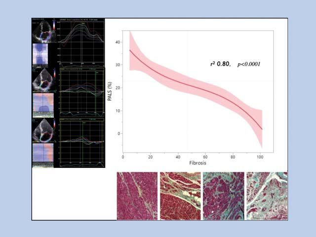
Maria Concetta Pastore et al.                                                             Romanian Journal of Cardiology
Left atrial fibrosis in mitral regurgitation                                                        Vol. 31, No. 1, 2021

cases, since it is performed using common 2D-scale          in ischemic cardiomyopathy and of gradual fibrosis in
echocardiographic images. However, even though its          case of dilated cardiomyopathy), while LA gradually
predictive role on LA fibrosis in chronic MR has been       develops MF as a consequence of newly established
established, there is a lack of evidence on its use to      chronic MR19. Finally, patients with „atrial functional
assess LA fibrosis in acute MR.                             MR” will already have a certain grade of LA fibrosis
   On the other hand, in chronic MR several degrees         when MR occurs, according to the preexistence and
of MF according to the etiology and stage of mitral         duration of AF itself20,21. This will chronically increase,
disease can be found. As concerning LA, this could be       and LV fibrosis will occur later as in primary MR, but
either associated with LA enlargement („LA structural       all the above mentioned steps will be anticipated due
remodeling”, characterized by considerable LA dilata-       to an already-damaged LA.
tion) or not („LA functional remodeling”, characteri-           Accordingly, in a biopsy study, Foglieni et al. found
zed by subtle ultrastructural modifications, leading to     early cellular and interstitial alterations in LA tissue
impaired function and pro-arrhythmic state without          in patients with chronic MR and sinus rhythm which
overt LA dilatation)13. In fact, previous models showed     were analogous to patients with AF, and their grade
the existence of several LA ultrastructural changes in      was increasingly higher in patients with LA dilatation22.
relation to LA dilation due to chronic MR14. Moreo-             These considerations overall suggest that the study
ver, Mary-Rabine et al. used intra-operative myocardi-      of left heart fibrosis as additional element in patients
al biopsy to prove an impaired contraction of the atria     with MR of unknown or mixed classification would
in patients with MR undergoing cardiac surgery due          help us to correctly define the etiology and the stage
to myolysis and an imbalance in collagen synthesis and      of the disease based on the grade of MF involving the
degradation15.                                              LA and the LV, and to identify subtle cardiac dysfuncti-
                                                            on before the development of symptoms. This would
LA fibrosis in primary vs secondary mitral
                                                            have important prognostic and therapeutic implica-
regurgitation
                                                            tions; thus, more evidence is warranted to support
Primary or „organic” MR is due to intrinsic valvular
                                                            these hypotheses. Figure 1 attempts to represent the
disease, whereas secondary or „functional” MR is ca-
                                                            expectable findings as concerns MF of the left heart
used by regional and/or global LV remodeling without
                                                            chambers according to the respective time of establi-
structural abnormalities of the mitral valve, or by func-
                                                            shment based on different etiologies of chronic MR.
tional remodeling of the LA due to chronic AF, the
so-called „atrial functional MR”16.                         Prognostic role of LA fibrosis in chronic mitral
   These etiologies differ in the timing of establish-      regurgitation
ment of MF in the two left heart chambers. However,         The assessment of LA fibrosis has been associated
the important thing is that MF of both chambers could       with the presence of heart disease and arrhythmias,
occur before the appearance of symptoms, and that           including congestive HF and AF23,24.
these structural changes of the myocardium may be              Daccarett et al. investigated the link between pro-
associated with subtle functional abnormalities17.          gressive LA remodeling and prognosis, showing the
   In particular, according to the natural history of the   correlation between stroke and high levels of LA fi-
disease, the LA is the first affected chamber from pri-     brosis detected by delayed enhancement CMR in pa-
mary MR overload, consequently, it is the first cham-       tients with AF25.
ber to develop maladaptive remodeling and fibrosis.            Interestingly, Kitkungvan et al. studied the presence
The LV is usually involved in the advanced phases of        of MF detected by cardiac magnetic resonance (CMR)
the disease, when LA is no more capable to compen-          in 424 patients with primary MR (229 of whom with
sate for volume overload and dilates; as a result, LV       MV prolapse), founding that diffuse interstitial fibrosis
is overwhelmed by higher filling blood volumes and          was associated with MR severity, regardless of pri-
pressures as well and gradually remodels, providing a       mary MR etiology, and that its extent was indepen-
considerably higher risk of malignant arrhythmias (es-      dently associated with symptoms related to MR and
pecially in patients with MV prolapse18).                   clinical events26.
   Conversely, in patients with secondary „functional”         LA strain by STE has shown to be a reliable marker
MR due to the traditionally known etiologies of LV          atrial fibrosis. Thus, some Authors studied its poten-
ischemia or dilatation, the LV is the target chamber        tial prognostic role in patients with MR, reaching inte-
for myocardial injury and fibrosis (in terms of a “scar”    resting results. In fact, a reduced LA strain has shown

38
Romanian Journal of Cardiology Maria Concetta Pastore et al.
Vol. 31, No. 1, 2021 Left atrial fibrosis in mitral regurgitation
Figure 1. Graphic representation of the different timing of myocardial fibrosis of the left atrium and the left ventricle according to mitral regurgitation
(MR) etiology (primary or „organic”, secondary or “functional”, divided into ischemic, dilated cardiomyopathy, and atrial functional MR) and the increasing
severity of MR; the onset of considerable MR in considered as zero point of reference. AF, atrial fibrillation; DCM, dilated cardiomyopathy; LA, left atrium;
LV, left ventricle; MR, mitral regurgitation.
to predict poor clinical outcome, in terms of survival and myocardial biopsy, describing that atrial fibrosis
and HF hospitalization, in patients with moderate and increases parallel to the duration of MR, with non-uni-
severe MR 10 (as discussed below). It has also been form degrees across different areas of the atria. Then,
described as an important predictor of the develop- they hypothesized that the dispersion of atrial fibro-
ment of paroxysmal and permanent AF in patients sis may contribute to increased susceptibility to AF
with MR, since the presence atrial fibrosis is gradually by influencing the conduction velocity rather than the
related to the increased risk of developing AF. effective refractory period28.
Moreover, AF onset causes further LA remodeling,
LA fibrosis and atrial fibrillation burden
and the progressive increase of LA fibrosis favors the
To date, the association between MR and AF deve-
conversion to a permanent AF form29. In fact, AF per-
lopment has been well-established. This comes as a
sistence has been shown to correlate also with ECM
consequence of LA remodeling and fibrosis, which is
composition and volume30.
responsible for localized wall regions of conduction
In a multicenter prospective observational study of
slowing, with higher conduction heterogeneity provi-
persistent versus paroxysmal AF, LA fibrosis was clas-
ding a substrate for AF or other supraventricular ar-
sified into 4 severity stages based on its extent at CMR
rhythmias27.
imaging („Utah stages”: 30%). The inclusion of LA fibrosis quantification in a
pig models with the use of electroanatomic mapping
39Maria Concetta Pastore et al. Romanian Journal of Cardiology
Left atrial fibrosis in mitral regurgitation Vol. 31, No. 1, 2021
predictive model improved the ability to predict AF LA strain measurement is performed on previously
recurrence (C-statistic increase from 0.65 to 0.69)31. acquired two-dimensional grey-scale echocardiogra-
Therefore, the prevention of atrial fibrosis would phic images, obtained using conventional two-dimen-
be essential to prevent AF onset and progression, and sional gray-scale echocardiography, during a brief bre-
the identification of advanced stages of fibrosis can ath hold and stable ECG recording, using dedicated
guide the choice of the best therapeutic strategy. LA apical views (frame rate required: 60-80 fps). The
Of note, Kuppahally et al. have demonstrated an in- operator can manually trace the LA endocardium in
verse relationship between the grade of fibrosis mea- both four- and two-chamber (if available) views by
sured by CMR late gadolinium enhancement and LA a point-and-click approach; the system will automa-
strain by STE, particularly in patients with persistent tically identify an endocardial region of interest of 6
AF compared to paroxysmal forms32. segments, that can be manually adjusted in width and
LA strain has been described as an important index shape. After acceptance, the software generates the
for the development of paroxysmal and permanent AF longitudinal strain curves corresponding to the defor-
caused by MF, as it strongly affects LA deformation mation of all segments together with average curve
properties quantified by STE33. It also proved to be which is representative of the phases of global LA de-
associated with AF occurrence and to be progres- formation all over the cardiac cycle.
sively more impaired in patients with MR and more The application of STE to characterize atrial functi-
episodes of paroxysmal AF34; moreover, it was also on allows to quantify reservoir, conduit and contracti-
associated with the development of post-operative AF le function. The most used index is peak atrial longitu-
in patients undergoing MV surgery35. dinal strain (PALS), a marker of LA reservoir function,
This was confirmed by Leung et al., showing that pa- with normality cut-off values of 39%38, calculated using
tients with persistent AF have longer total atrial con- QRS as reference for its slightly higher feasibility39,40.
duction time (PA-TDI) and worse LA reservoir strain The presence of fibrosis of LA walls causes a rele-
compared with patients with paroxysmal AF and con- vant decrease of PALS as a sign of reduced compliance
trols, suggesting increasing burden of fibrosis and LA of the atrium, and which has shown to anticipate the
structural remodeling in the progression of AF36. The- presence of atrial dilation41,42. Recent studies have de-
se findings suggest that STE could have a role in the monstrated the value of LA strain analysis, especially
identification of patients with MR and higher extent of a reduction of PALS, to predict LA wall fibrosis with a
MF who are prone to develop AF. good correlation with CMR32 and to stratify the risk of
stroke in patients with AF33.
Speckle tracking echocardiography for
LA strain has shown a great utility in patients with
indirect imaging of LA fibrosis
MR as potential marker of MF and of clinical outco-
The extent of adaptive or maladaptive remodeling of
me43. Particularly, in a recent prospective study we
atrial chambers can be indirectly quantified through
have shown a strong independent association of global
echocardiographic measures such as atrial emptying
PALS with the extent of LA fibrosis (Figure 2) assessed
fraction, transmitral flow, pulmonary veins veloci-
by biopsy specimens in patients with severe MR un-
ties, and tissue doppler analysis. However, most of
dergoing cardiac surgery; moreover, global PALS was
these measures have some pitfalls. Speckle tracking
an independent marker of post-operative clinical (car-
analysis has emerged in the last years as a more relia-
diovascular events) and functional outcome (NYHA
ble method which could overcome these limitations, class-Borg scale)44. Furthermore, in another biopsy
allowing an angle-independent and objective quantifi- study conducted in a similar cohort we showed a
cation of myocardial deformation in different diseases. stepwise reduction of global PALS and a close nega-
STE has proved to indirectly assess the presence of tive correlation between global and LA fibrosis grade
MF through the analysis of intrinsic myocardial dyna- which was superior to LA indexed volume, LA ejecti-
mics: as MF causes abnormal endocardial thickening on fraction, and E/E’ ratio. Among these indices, global
by an increase in myocardial stiffness, this could be PALS showed the best diagnostic accuracy to detect
observed as a reduction of myocardial strain in each LA fibrosis (area under the curve 0.89)45.
component of deformation analysis (i.e. longitudinal, Also, Her et al. showed that LA global strain was
circumferential, radial) for both LV and LA cham- significantly correlated with the degree of LA fibrosis
bers4,37. assessed by histopathology (r=-0.55, pRomanian Journal of Cardiology Maria Concetta Pastore et al.
Vol. 31, No. 1, 2021 Left atrial fibrosis in mitral regurgitation
Figure 2. Correlation of peak atrial longitudinal strain (PALS) by speckle tracking echocardiography with the extent of left atrial fibrosis invasively estimated
by biopsy specimens in patients undergoing cardiac surgery for severe mitral regurgitation48.
rhythm, presence of rheumatic heart disease and type All these evidence supports the introduction of
of predominant MV disease (B=-1.37, 95% confidence LA strain as a noninvasive and easy-to-use marker of
interval -2.32 ;-0.41, p=0.006)46. MF to include in the daily evaluation of patients with
As concerns prognosis, global PALS reduction chronic MR to guide therapeutic strategies. However,
showed to be predictive of clinical outcome in pati- these promising results require bigger studies to be
ents with moderate and severe MR34,47. Moreover, a generalized. Moreover, some limitation of LA strain
recent study showed that PALS during exercise was by STE should be kept in mind, such as its dependance
related to all-cause mortality and HF hospitalization on loading conditions and image quality, and the lack
in 196 with primary or secondary MR: it showed how of disease-specific reference values.
exercise PALS could be used as a marker of LA re-
Cardiac magnetic resonance imaging for LA
serve, which is somewhat preserved in primary MR,
fibrosis assessment
unlike secondary MR with similar severity degrees,
CMR is considered the noninvasive gold standard
that showed worse LA function during exercise, cor-
method to assess MF, though limited by high costs and
responding to lower exercise performance (measured
low availability. CMR allows the assessment of reac-
by cardiopulmonary test parameters) and to worse
tive fibrosis, with the use of T1 mapping techniques,
clinical outcome48.
and replacement fibrosis, with the use of late gadoli-
PALS has also proved a great utility during pre-sur-
nium enhancement (LGE). In addition, functional con-
gical evaluation of primary MR for the prediction of
sequences of MF can be evaluated with myocardial
postoperative LA reverse remodeling, also following
tagging and feature tracking CMR, which assess the
MitraClip procedure, over conventional LV and LA
active myocardial deformation (strain). Several studies
echocardiographic indices49.
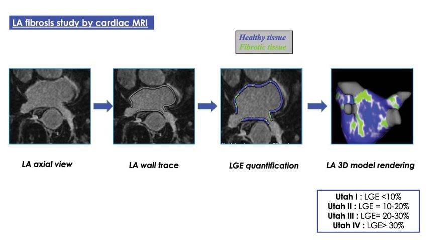
41Maria Concetta Pastore et al. Romanian Journal of Cardiology Left atrial fibrosis in mitral regurgitation Vol. 31, No. 1, 2021 proved that CMR techniques may be more sensitive In patients with MR, CMR complements echocardi- than the conventional measures to detect these struc- ography for the study of MF, that is also a risk marker tural and functional impairment in patients with severe for arrhythmic complications52. Particularly, an impor- MR5. tant application of CMR is the estimation of arrhyth- LGE CMR is considered the reference parameter mic risk in patients with MV prolapse based of the to quantify myocardial replacement fibrosis and scar. quantification of MF53-55. Interestingly, Basso et al. later The increased ECV and decreased capillary density of demonstrated that arrhythmic MV prolapse was asso- the fibrous tissue result in increased volume of distri- ciated with fibrosis not only at the level of the papillary bution and prolonged wash-out of gadolinium in com- muscle but also within the infero-basal wall in a series parison to the normal myocardium. With the increase of patients who experienced sudden cardiac death or of interstitial fibrosis, longer native T1 values (without presented with complex ventricular arrhythmias and the use of gadolinium contrast) are observed while the without significant MR56, suggesting an independent post-contrast T1 values become shorter. By combi- fibrotic role of the disease regardless of the degree ning them, myocardial ECV fraction can be computed, of MR. which quantifies the ECM space and is a marker of As concerning LA, its study with CMR could be myocardial fibrosis (since collagen I is the main compo- operated with high-resolution LGE scans, with accu- nent of the ECM)50. These measures are considered of rate rendering of LA wall. For 3D visualization of LA added value over LGE, allowing to quantify the degree fibrosis based on the relative LGE signal intensity, a of fibrosis and, particularly, to detect diffuse interstiti- specific color coding may be used: healthy tissue is al fibrosis, which is often associated with early stages blue, whereas any tissue with LGE is green and yellow. of the disease. However, these are not fully specific: Additionally, a color lookup-table mask may be used abnormal myocardial ECV fraction can be observed in to better differentiate enhanced and non-enhanced infiltrative diseases (i.e. amyloidosis) and edema, while tissue. The grade of LA fibrosis is then classified as native T1 values impairment are observed also in iron one of the established 4 Utah stages, on the basis of deposition and diffuse fat infiltration51. Furthermore, LA wall enhancement as a percentage of the total LA standardization of CMR T1 mapping techniques across wall volume: stage I=LA fibrosis
Romanian Journal of Cardiology                                                                          Maria Concetta Pastore et al.
Vol. 31, No. 1, 2021                                                                   Left atrial fibrosis in mitral regurgitation

gure 4)57. This approach is mostly used in patients          should always be consciously evaluated in the overall
undergoing catheter ablation; particularly, LA remo-         clinical context.
deling, measured by LGE-MRI, has shown to predict
clinical outcome and AF recurrence after AF catheter         CONCLUSIONS
ablation58-60.                                               The evaluation of MF of both LA and LV could offer
   However, CMR low availability, its expense, and           important information in patients with MR.
relative contraindication for patients with implantable         In particular, the study of LA fibrosis using advan-
medical devices (e.g. pacemakers), still limit its current   ced imaging techniques allows an early detection of
clinical use in daily practice. Moreover, the evaluation     LA damage in chronic MR, providing additive value for
of fibrosis with CMR for the LA remains time consu-          the diagnostic and prognostic assessment in order to
ming and challenging for the LA due to its thin-walled       guide the management of these patients. The choice
structure (2-3 fold thinner that LV wall). For this rea-     of imaging modality should be tailored to the speci-
son, poor evidence currently exists on the study of LA       fic patient and clinical indication, also considering the
fibrosis in patients with MR.                                availability of each technique.
Cardiac computed tomography
The use of cardiac computed tomography (CT) in               Conflict of interest: none declared.
patients with MR with limited acoustic windows has           References
been described in the literature. Indeed, CT offers          1.  Monteagudo Ruiz JM, Galderisi M, Buonauro A, Badano L, Aruta P,
                                                                 Swaans MJ, Sanchis L, Saraste A, Monaghan M, Theodoropoulos KC,
better spatial resolution with around 84.6% sensitivity          Papitsas M, Liel-Cohen N, Kobal S, Bervar M, Berlot B, Filippatos
and 100% specificity for assessment of MV abnorma-               G, Ikonomidis I, Katsanos S, Tanner FC, Cassani D, Faletra FF, Leo
lities61. Moreover, it offers a simultaneous evaluation          LA, Martinez A, Matabuena J, Grande-Trillo A, Alonso-Rodriguez D,
                                                                 Mesa D, Gonzalez-Alujas T, Sitges M, Carrasco-Chinchilla F, Li CH,
of coronary artery disease, therefore it could be pre-           Fernandez-Golfin C, Zamorano JL. Overview of mitral regurgitation
cious in case of double indication for the study of MR           in Europe: results from the European Registry of mitral regurgitation
and coronary arteries. However, radiation exposure,              (EuMiClip). Eur Heart J Cardiovasc Imaging. 2018;19(5):503-507.
                                                             2. Baumgartner H, Falk V, Bax JJ, De Bonis M, Hamm C, Holm PJ, Iung
poor temporal resolution and limited ability to exami-           B, Lancellotti P, Lansac E, Rodriguez Muñoz D, Rosenhek R, Sjögren
ne each MV leaflet are some of the important limitati-           J, Tornos Mas P, Vahanian A, Walther T, Wendler O, Windecker S,
                                                                 Zamorano JL; ESC Scientific Document Group. 2017 ESC/EACTS
ons of cardiac CT for the diagnostic evaluation of MR.           Guidelines for the management of valvular heart disease. Eur Heart
In addition, its accuracy is not comparable to that of           J. 2017;38(36):2739-2791.
CMR for the detection of MF.                                 3. Harb SC, Griffin BP. Mitral Valve Disease: a Comprehensive Review.
                                                                 Curr Cardiol Rep. 2017;19(8):73.
Clinical applications of LA fibrosis assessment              4. Cameli M, Mondillo S, Righini FM, Lisi M, Dokollari A, Lindqvist P,
                                                                 Maccherini M, Henein M. Left Ventricular Deformation and Myo-
LA fibrosis is a pivotal element in patients with MR,            cardial Fibrosis in Patients With Advanced Heart Failure Requiring
and its routine assessment would be useful to enhance            Transplantation. J Card Fail. 2016;22(11):901-907.
the comprehension of the severity of cardiac damage          5. Podlesnikar T, Delgado V, Bax JJ. Cardiovascular magnetic resonance
                                                                 imaging to assess myocardial fibrosis in valvular heart disease. Int J
consequent to MR, thus providing information on its              Cardiovasc Imaging. 2018;34(1):97-112.
etiology and aiding MR grading, and the accuracy in          6. D’Ascenzi F, Anselmi F, Focardi M, Mondillo S. Atrial Enlargement in
                                                                 the Athlete’s Heart: Assessment of Atrial Function May Help Distin-
the prediction of arrhythmic risk and clinical outco-            guish Adaptive from Pathologic Remodeling. J Am Soc Echocardiogr.
me. This would lead to a more accurate evaluation of             2018;31(2):148-157.
these patients and, through the application of nonin-        7. Burstein B, Nattel S. Atrial fibrosis: mechanisms and clinical rele-
                                                                 vance in atrial fibrillation. J Am Coll Cardiol. 2008;51:802-9.
vasive imaging modalities, to an improvement of the          8. Cameli M, Pastore MC, Henein MY, Mondillo S. The left atrium and
algorithms of surgical referral, e.g. including PALS or          the right ventricle: two supporting chambers to the failing left ven-
LGE quantification to the criteria for surgery. How-             tricle. Heart Fail Rev. 2019;24(5):661-669.
                                                             9. Travers JG, Kamal FA, Robbins J, Yutzey KE, Blaxall BC. Cardiac
ever, one should bear in mind that atrial structural             Fibrosis: The Fibroblast Awakens. Circulation research. 2016;118:
remodeling is multifactorial, thus MR could represent            1021-40.
                                                             10. Cameli M, Incampo E, Mondillo S. Left atrial deformation: Useful in-
one of the multiple factors concurring in atrial fibro-          dex for early detection of cardiac damage in chronic mitral regurgita-
sis. In fact, the presence of cardiovascular risk factors        tion. Int J Cardiol Heart Vasc. 2017;17:17-22.
(hypertension, diabetes mellitus), of atrial involvement     11. Dziadzko V, Clavel MA, Dziadzko M, Medina-Inojosa JR, Michelena
                                                                 H, Maalouf J, Nkomo V, Thapa P, Enriquez-Sarano M. Outcome and
in different cardiomyopathies (hypertrophic cardi-               undertreatment of mitral regurgitation: a community cohort study.
omyopathy, dilated cardiomyopathy, amyloidosis), all             Lancet. 2018;391(10124):960-969.
can contribute, beyond the associated MR, to atrial          12. Watanabe N. Acute mitral regurgitation. Heart. 2019;105(9):671-
                                                                 677.
fibrosis occurrence. Therefore, the imaging findings
                                                                                                                                   43
Maria Concetta Pastore et al. Romanian Journal of Cardiology
Left atrial fibrosis in mitral regurgitation Vol. 31, No. 1, 2021
13. Thomas L, Abhayaratna WP. Left Atrial Reverse Remodeling: Mech- 29. Dzeshka MS, Lip GY, Snezhitskiy V, Shantsila E. Cardiac Fibrosis in
anisms, Evaluation, and Clinical Significance. JACC Cardiovasc Imag- Patients With Atrial Fibrillation: Mechanisms and Clinical Implica-
ing. 2017;10(1):65-77. tions. J Am Coll Cardiol. 2015;66(8):943-59.
14. Verheule S, Wilson E, Everett T IV, Shanbhag S, Golden C, Olgin J. 30. Gulati A, Jabbour A, Ismail TF et al. Association of fibrosis with mor-
Alterations in atrial electrophysiology and tissue structure in a ca- tality and sudden cardiac death in patients with non-ischemic dilated
nine model of chronic atrial dilatation due to mitral regurgitation. cardiomyopathy. Jama. 2013;309:896-908.
Circulation 2003;107:2615e2622 31. Marrouche NF, Wilber D, Hindricks G, Jais P, Akoum N, Marchlinski
15. Mary-Rabine L, Albert A, Pharm TD, Hordof A, Fenoglio JJ, Malm JR, F, Kholmovski E, Burgon N, Hu N, Mont L, Deneke T, Duytschae-
Rosen MR. The relationship of human atrial cellular electrophysiolo- ver M, Neumann T, Mansour M, Mahnkopf C, Herweg B, Daoud E,
gy to clinical function and ultrastructure. Circ Res 1983;52: 188e199 Wissner E, Bansmann P, Brachmann J. Association of atrial tissue fi-
16. Kagiyama N, Mondillo S, Yoshida K, Mandoli GE, Cameli M. Subtypes brosis identified by delayed enhancement MRI and atrial fibrillation
of Atrial Functional Mitral Regurgitation: Imaging Insights Into Their catheter ablation: the DECAAF study. JAMA. 2014;311(5):498-506.
Mechanisms and Therapeutic Implications. JACC Cardiovasc Imag- 32. Kuppahally SS, Akoum N, Burgon NS et al. Left atrial strain and
ing. 2020;13(3):820-835. strain rate in patients with paroxysmal and persistent atrial fibril-
17. Mihaila S, Muraru D, Miglioranza MH, Piasentini E, Aruta P, Cucchini lation: relationship to left atrial structural remodeling detected by
U, Iliceto S, Vinereanu D, Badano LP. Relationship between mitral delayed-enhancement MRI. Circulation Cardiovascular imaging.
annulus function and mitral regurgitation severity and left atrial re- 2010;3:231-9.
modelling in patients with primary mitral regurgitation. Eur Heart J 33. Cameli M, Mandoli GE, Loiacono F, Sparla S, Iardino E, Mondillo S.
Cardiovasc Imaging. 2016;17(8):918-29. Left atrial strain: A useful index in atrial fibrillation. Int J Cardiol;
18. Kitkungvan D, Nabi F, Kim RJ, Bonow RO, Khan MA, Xu J, Little SH, 220:208-13.
Quinones MA, Lawrie GM, Zoghbi WA, Shah DJ. Myocardial Fibro- 34. Cameli M, Lisi M, Righini FM, Focardi M, Alfieri O, Mondillo S. Left
sis in Patients With Primary Mitral Regurgitation With and Without atrial speckle tracking analysis in patients with mitral insufficiency
Prolapse. J Am Coll Cardiol. 2018;72(8):823-834. and history of paroxysmal atrial fibrillation. Int J Cardiovasc Imaging.
19. Sharma H, Liu B, Mahmoud-Elsayed H, Myerson SG, Steeds RP. Mul- 2012;28(7):1663-70.
timodality Imaging in Secondary Mitral Regurgitation. Front Cardio- 35. Candan O, Ozdemir N, Aung SM, et al. Left atrial longitudinal strain
vasc Med. 2020 Dec 22;7:546279. doi: 10.3389/fcvm.2020.546279. parameters predict postoperative persistent atrial fibrillation follow-
PMID: 33415127; PMCID: PMC7782243. ing mitral valve surgery: a speckle tracking echocardiography study.
20. Muraru D, Guta AC, Ochoa-Jimenez RC, Bartos D, Aruta P, Mi- Echocardiography 2013; 30:1061e8.
haila S, Popescu BA, Iliceto S, Basso C, Badano LP. Functional Re- 36. Leung M, Abou R, van Rosendael PJ, van der Bijl P, van Wijngaarden
gurgitation of Atrioventricular Valves and Atrial Fibrillation: An Elu- SE, Regeer MV, Podlesnikar T, Ajmone Marsan N, Leung DY, Del-
sive Pathophysiological Link Deserving Further Attention. J Am Soc gado V, Bax JJ. Relation of Echocardiographic Markers of Left Atrial
Echocardiogr. 2020;33(1):42-53. Fibrosis to Atrial Fibrillation Burden. Am J Cardiol. 2018;122(4):584-
21. Callegari S, Macchi E, Monaco R, Magnani L, Tafuni A, Croci S, Nica- 591.
stro M, Garrapa V, Banchini A, Becchi G, Corradini E, Goldoni M, 37. Cameli M, Mondillo S, Galderisi M, Mandoli GE, Ballo P, Nistri S,
Rocchio F, Sala R, Benussi S, Ferrara D, Alfieri O, Corradi D. Clini- Capo V, D’Ascenzi F, D’Andrea A, Esposito R, Gallina S, Montisci
copathological Bird’s-Eye View of Left Atrial Myocardial Fibrosis in R, Novo G, Rossi A, Mele D, Agricola E. L’ecocardiografia speck-
121 Patients With Persistent Atrial Fibrillation: Developing Architec- le tracking: roadmap per la misurazione e l’utilizzo clinico [Speckle
ture and Main Cellular Players. Circ Arrhythm Electrophysiol. 2020; tracking echocardiography: a practical guide]. G Ital Cardiol (Rome).
13(7):e007588. 2017;18:253-269.
22. Foglieni C, Rusconi R, Mantione ME, Fragasso G, Alfieri O, Maisano 38. Pathan F, D’Elia N, Nolan MT, Marwick TH, Negishi K. Normal
F. Early left atrial tissue features in patients with chronic mitral re- Ranges of Left Atrial Strain by Speckle-Tracking Echocardiography:
gurgitation and sinus rhythm: Alterations of not remodeled left atria. A Systematic Review and Meta-Analysis. J Am Soc Echocardiogr.
Int J Cardiol. 2016;219:433-8. 2017;30(1):59-70.e8.
23. Pastore MC, Mandoli GE, Aboumarie HS, Santoro C, Bandera F, 39. Badano LP, Kolias TJ, Muraru D, Abraham TP, Aurigemma G, Ed-
D’Andrea A, Benfari G, Esposito R, Evola V, Sorrentino R, Cameli vardsen T, D’Hooge J, Donal E, Fraser AG, Marwick T, Mertens L,
P, Valente S, Mondillo S, Galderisi M, Cameli M; Working Group Popescu BA, Sengupta PP, Lancellotti P, Thomas JD, Voigt JU; In-
of Echocardiography of the Italian Society of Cardiology. Basic and dustry representatives; Reviewers: This document was reviewed by
advanced echocardiography in advanced heart failure: an overview. members of the 2016–2018 EACVI Scientific Documents Commit-
Heart Fail Rev. 2020;25(6):937-948. tee. Standardization of left atrial, right ventricular, and right atrial de-
24. Pellman J, Lyon RC, Sheikh F. Extracellular matrix remodelling in formation imaging using two-dimensional speckle tracking echocar-
atrial fibrosis: mechanisms and implications in atrial fibrillation. J Mol diography: a consensus document of the EACVI/ASE/Industry Task
Cell Cardiol 2010;48:461e467. Force to standardize deformation imaging. Eur Heart J Cardiovasc
25. Daccarett M, Badger TJ, Akoum N, Burgon NS, Mahnkopf C, Ver- Imaging. 2018;19(6):591-600. doi: 10.1093/ehjci/jey042. Erratum in:
gara G, Kholmovski E, McGann CJ, Parker D, Brachmann J, MacLeod Eur Heart J Cardiovasc Imaging. 2018;19(7):830-833.
RS, Marrouche NF. Association of left atrial fibrosis by delayed-en- 40. Cameli M, Miglioranza MH, Magne J, Mandoli GE, Benfari G, Ancona
hancement magnetic resonance imaging and the risk of stroke in pa- R, Sibilio G, Reskovic Luksic V, Dejan D, Griseli L, Van De Heyning
tients with atrial fibrillation. J Am Coll Cardiol 2011;57:831e838. CM, Mortelmans P, Michalski B, Kupczynska K, Di Giannuario G,
26. Kitkungvan D, Yang EY, El Tallawi KC, Nagueh SF, Nabi F, Khan MA, Devito F, Dulgheru R, Ilardi F, Salustri A, Abushahba G, Morrone D,
Nguyen DT, Graviss EA, Lawrie GM, Zoghbi WA, Bonow RO, Qui- Fabiani I, Penicka M, Katbeh A, Sammarco G, Esposito R, Santoro C,
nones MA, Shah DJ. Extracellular Volume in Primary Mitral Regur- Pastore MC, Comenale Pinto S, Kalinin A, Pičkure Ž, Ažman Juvan
gitation. JACC Cardiovasc Imaging. 2020:S1936-878X(20)30917-7. K, Zupan Mežnar A, Coisne A, Coppin A, Opris MM, Nistor DO,
doi: 10.1016/j.jcmg.2020.10.010. Paakkanen R, Biering-Sørensen T, Olsen FJ, Lapinskas T, Vaškelyté JJ,
27. Li D, Fareh S, Leung TK, Nattel S. Promotion of atrial fibrillation by Galian-Gay L, Casas G, Motoc AI, Papadopoulos CH, Loizos S, Ágos-
heart failure in dogs: atrial remodeling of a different sort. Circulation. ton G, Szabó I, Hristova K, Tsonev SN, Galli E, Vinereanu D, Mihaila
1999;100:87-95. Baldea S, Muraru D, Mondillo S, Donal E, Galderisi M, Cosyns B, Ed-
28. Li B, Luo F, Luo X, Li B, Qi L, Zhang D, Tang Y. Effects of atrial fibro- vardsen T, Popescu BA. Multicentric Atrial Strain COmparison be-
sis induced by mitral regurgitation on atrial electrophysiology and tween Two Different Modalities: MASCOT HIT Study. Diagnostics
susceptibility to atrial fibrillation in pigs. Cardiovasc Pathol. 2019; (Basel). 2020 Nov 13;10(11):946. doi: 10.3390/diagnostics10110946.
40:32-40.
44Romanian Journal of Cardiology Maria Concetta Pastore et al.
Vol. 31, No. 1, 2021 Left atrial fibrosis in mitral regurgitation
41. Mandoli GE, Sisti N, Mondillo S, Cameli M. Left atrial strain in left 51. Moon JC, Messroghli DR, Kellman P, Piechnik SK, Robson MD,
ventricular diastolic dysfunction: have we finally found the missing Ugander M, Gatehouse PD, Arai AE, Friedrich MG, Neubauer S,
piece of the puzzle? Heart Fail Rev. 2020 May;25(3):409-417. doi: Schulz-Menger J, Schelbert EB; Society for Cardiovascular Magnetic
10.1007/s10741-019-09889-9. PMID: 31773504. Resonance Imaging; Cardiovascular Magnetic Resonance Working
42. D›Ascenzi F, Cameli M, Henein M, Iadanza A, Reccia R, Lisi M, Curci Group of the European Society of Cardiology. Myocardial T1 map-
V, Sinicropi G, Torrisi A, Pierli C, Mondillo S. Left atrial remodel- ping and extracellular volume quantification: a Society for Cardiovas-
ling in patients undergoing transcatheter aortic valve implantation: cular Magnetic Resonance (SCMR) and CMR Working Group of the
a speckle-tracking prospective, longitudinal study. Int J Cardiovasc European Society of Cardiology consensus statement. J Cardiovasc
Imaging. 2013;29(8):1717-24. Magn Reson. 2013;15(1):92.
43. Pastore MC, De Carli G, Mandoli GE, D’Ascenzi F, Focardi M, Con- 52. Disertori M, Rigoni M, Pace N et al. Myocardial Fibrosis Assessment
torni F, Mondillo S, Cameli M. The prognostic role of speckle track- by LGE Is a Powerful Predictor of Ventricular Tachyarrhythmias in
ing echocardiography in clinical practice: evidence and reference val- Ischemic and Nonischemic LV Dysfunction: A Meta-Analysis. JACC
ues from the literature. Heart Fail Rev. doi: 10.1007/s10741-020- Cardiovascular imaging. 2016;9:1046-55. (43–46).
09945-9. 53. Parwani P, Avierinos JF, Levine RA, Delling FN. Mitral Valve Pro-
44. Mandoli GE, Pastore MC, Benfari G, Bisleri G, Maccherini M, Lisi lapse: Multimodality Imaging and Genetic Insights. Prog Cardiovasc
G, Cameli P, Lisi M, Dokollari A, Carrucola C, Vigna M, Montesi G, Dis. 2017;60(3):361-369
Valente S, Mondillo S, Cameli M. Left atrial strain as a pre-operative 54. Han Y, Peters DC, Salton CJ, Bzymek D, Nezafat R, Goddu B, Kiss-
prognostic marker for patients with severe mitral regurgitation. Int J inger KV, Zimetbaum PJ, Manning WJ, Yeon SB. Cardiovascular mag-
Cardiol. 2021;324:139-145. netic resonance characterization of mitral valve prolapse. JACC Car-
45. Cameli M, Lisi M, Righini FM, Massoni A, Natali BM, Focardi M, Tac- diovasc Imaging. 2008;1(3):294-303
chini D, Geyer A, Curci V, Di Tommaso C, Lisi G, Maccherini M, 55. Perazzolo Marra M, Basso C, De Lazzari M, Rizzo S, Cipriani A,
Chiavarelli M, Massetti M, Tanganelli P, Mondillo S. Usefulness of Giorgi B, Lacognata C, Rigato I, Migliore F, Pilichou K, Cacciavillani
atrial deformation analysis to predict left atrial fibrosis and endo- L, Bertaglia E, Frigo AC, Bauce B, Corrado D, Thiene G, Iliceto S.
cardial thickness in patients undergoing mitral valve operations for Morphofunctional Abnormalities of Mitral Annulus and Arrhythmic
severe mitral regurgitation secondary to mitral valve prolapse. Am J Mitral Valve Prolapse. Circ Cardiovasc Imaging. 2016;9(8):e005030.
Cardiol. 2013;111(4):595-601 56. Basso C, Perazzolo Marra M, Rizzo S, De Lazzari M, Giorgi B, Cipri-
46. Her AY, Choi EY, Shim CY, Song BW, Lee S, Ha JW, Rim SJ, Hwang ani A, Frigo AC, Rigato I, Migliore F, Pilichou K, Bertaglia E, Cac-
KC, Chang BC, Chung N. Prediction of left atrial fibrosis with speck- ciavillani L, Bauce B, Corrado D, Thiene G, Iliceto S. Arrhythmic
le tracking echocardiography in mitral valve disease: a comparative Mitral Valve Prolapse and Sudden Cardiac Death. Circulation. 2015;
study with histopathology. Korean Circ J. 2012;42(5):311-8. 132(7):556-66.
47. Cameli M, Pastore MC, Righini FM, Mandoli GE, D’Ascenzi F, Lisi M, 57. Siebermair J, Kholmovski EG, Marrouche N. Assessment of Left
Nistor D, Sparla S, Curci V, Di Tommaso C, Marino F, Stricagnoli Atrial Fibrosis by Late Gadolinium Enhancement Magnetic Reso-
M, Mondillo S. Prognostic value of left atrial strain in patients with nance Imaging: Methodology and Clinical Implications. JACC Clin
moderate asymptomatic mitral regurgitation. Int J Cardiovasc Imag- Electrophysiol. 2017;3(8):791-802.
ing. 2019;35(9):1597-1604. 58. Marrouche NF, Wilber D, Hindricks G, et al.Association of atrial tis-
48. Sugimoto T, Bandera F, Generati G, Alfonzetti E, Barletta M, Losi- suefibrosis identified bydelayed enhancement MRI and atrialfibrilla-
to M, Labate V, Rovida M, Caracciolo M, Pappone C, Ciconte G, tioncatheter ablation: the DECAAF study. JAMA 2014;311:498–506.
Guazzi M. Left Atrial Dynamics During Exercise in Mitral Regurgita- 59. McGann C, Akoum N, Patel A, et al. Atrialfibrillation ablation out-
tion of Primary and Secondary Origin: Pathophysiological Insights by come is predicted by leftatrial remodeling on MRI. Circ Arrhythm
Exercise Echocardiography Combined With Gas Exchange Analysis. Electro-physiol 2014;7:23–30.
JACC Cardiovasc Imaging. 2020;13(1 Pt 1):25-40. 60 Khurram IM, Habibi M, Gucuk Ipek E, Chrispin J, Yang E, Fukumoto
49. Toprak C, Kahveci G, Kilicgedik A, Pala S, Kirma C, Tabakci MM, K, Dewire J, Spragg DD, Marine JE, Berger RD, Ashikaga H, Rickard
Inanir M, Esen AM. Left atrial remodeling in patients undergoing per- J, Zhang Y, Zipunnikov V, Zimmerman SL, Calkins H, Nazarian S. Left
cutaneous mitral valve repair with the MitraClip system: an advanced Atrial LGE and Arrhythmia Recurrence Following Pulmonary Vein
echocardiography study. Echocardiography. 2016;33(10):1504-1511. Isolation for Paroxysmal and Persistent AF. JACC Cardiovasc Imag-
50. Miller CA, Naish JH, Bishop P, Coutts G, Clark D, Zhao S, Ray SG, ing. 2016;9(2):142-8.
Yonan N, Williams SG, Flett AS, Moon JC, Greiser A, Parker GJ, 61. Ghosh N, Al-Shehri H, Chan K, Mesana T, Chan V, Chen L, Yam
Schmitt M. Comprehensive validation of cardiovascular magnetic Y, Chow BJ. Characterization of mitral valve prolapse with cardiac
resonance techniques for the assessment of myocardial extracellular computed tomography: comparison to echocardiographic and intra-
volume. Circ Cardiovasc Imaging. 2013;6(3):373-83. operative findings. Int J Cardiovasc Imaging. 2012;28(4):855-63.
45You can also read