Consensus Recommendations for the Use of 18F- FDG PET as an Indicator of Therapeutic Response in Patients in National Cancer Institute Trials
←
→
Page content transcription
If your browser does not render page correctly, please read the page content below
SPECIAL         CONTRIBUTION

Consensus Recommendations for the Use of 18F-
FDG PET as an Indicator of Therapeutic Response
in Patients in National Cancer Institute Trials
Lalitha K. Shankar1, John M. Hoffman2, Steve Bacharach3, Michael M. Graham4, Joel Karp5, Adriaan A. Lammertsma6,
Steven Larson7, David A. Mankoff8, Barry A. Siegel9, Annick Van den Abbeele10, Jeffrey Yap10, and Daniel Sullivan1
1Cancer Imaging Program, National Cancer Institute, National Institutes of Health, Bethesda, Maryland; 2Division of Nuclear Medicine,

Huntsman Cancer Institute, University of Utah School of Medicine, Salt Lake City, Utah; 3Department of Radiology, University of
California, San Francisco, California; 4Division of Nuclear Medicine, Department of Radiology, University of Iowa, Iowa City, Iowa;
5Division of Nuclear Medicine, Department of Radiology, University of Pennsylvania, Philadelphia, Pennsylvania; 6Department of

Nuclear Medicine and PET Research, VU University Medical Centre, Amsterdam, The Netherlands; 7Department of Radiology,
Memorial Sloan-Kettering Cancer Center, New York, New York; 8Division of Nuclear Medicine, University of Washington, Seattle,
Washington; 9Mallinckrodt Institute of Radiology, St. Louis, Missouri; and 10Department of Radiology, Dana-Farber Cancer Institute,
Harvard Medical School, Boston, Massachusetts

M       any therapeutic clinical trials have proposed using a
measure of metabolic change to assess therapeutic response
                                                                           same time, it has become equally clear that the potential of
                                                                           18F-FDG PET as such a tool will not be achieved unless

                                                                           standard protocols are developed so that data can be accu-
rather than relying on conventional anatomic measurements                  mulated and compared across multiple clinical sites. Today,
of changes in tumor size on CT or MRI. PET assessment of                   the methods of obtaining 18F-FDG PET scans and assessing
                                                                           18F-FDG metabolism and uptake vary.
changes in 18F-FDG uptake by tumors is gaining accep-
tance as such a measure.                                                      To provide such guidance and to help standardize the
   Despite the increasing use of 18F-FDG PET as a bio-                     acquisition and interpretation of 18F-FDG PET images in
marker for predicting therapeutic response, there are no                   clinical trials sponsored by the National Cancer Institute
widely accepted standardized protocols for using 18F-FDG                   (NCI), the Cancer Imaging Program of the NCI convened a
PET as a tool for assessing response to therapy, nor are                   workshop on January 10211, 2005, in Washington, DC, at
there validated criteria for judging response using 18F-FDG                which the current status of 18F-FDG PET technology and
PET. The European PET community, working with the                          clinical experience—both in diagnosis and in monitoring
European Organization for Research and Treatment of Can-                   therapeutic response—was reviewed. The participants fo-
cer, initiated a project to begin defining response criteria for           cused on patient preparation, image acquisition, image re-
PET and published their preliminary consensus recommen-                    construction, quantitative and semiquantitative image analysis,
dations in 1999 (1). The European Organization for Re-                     quality assurance, reproducibility, and other parameters im-
search and Treatment of Cancer continues to accumulate                     portant in 18F-FDG PET studies before and after a therapeutic
data in order to more carefully assess the role of 18F-FDG                 intervention. Their discussions were based on the existing
PET in measuring therapeutic response.                                     medical literature and on their own expertise.
   18F-FDG PET has become a common imaging modality                           This document represents the outcome of those deliber-
in oncology, primarily as a result of the widespread avail-                ations. We intend that it serve as the recommended set of
ability of PET instruments, an accumulation of clinical                    procedures for the acquisition and analysis of 18F-FDG
data, and the gradual expansion of oncology indications                    PET scans of patients participating in NCI-sponsored
that Medicare will reimburse. With this increasing clinical                diagnostic and therapeutic clinical trials. We hope that
experience, it is becoming clear that 18F-FDG PET may                      these guidelines will help bring about a future in which
                                                                           18F-FDG PET can provide an early metabolic assessment of
have an important role as a surrogate endpoint for assessing
the clinical efficacy of novel oncologic therapies. At the                 therapeutic response.

                                                                           IMAGE ANALYSIS AND UPTAKE QUANTIFICATION
  Received Mar. 1, 2006; revision accepted Mar. 10, 2006.
  For correspondence or reprints contact: Lalitha K. Shankar, MD, Cancer     18F-FDG   is a marker of metabolic activity in a variety of
Imaging Program, National Cancer Institute, 6130 Executive Blvd., Room
6048, Bethesda, MD 20892-7412.
                                                                           tissues and tumors (2). Most malignant tissues have in-
  E-mail: shankarl@mail.nih.gov                                            creased 18F-FDG uptake associated with an increased rate

                                                                                    NCI PET GUIDELINES • Shankar et al.             1059
of glycolysis and of glucose transport. Warburg first de- A major issue in monitoring tumor response by determin-
scribed this fundamental aberration of malignant cells in ing 18F-FDG uptake is that the uptake depends on the time of
the 1930s (3), and more recently, several groups have measurement. An important aspect of oncologic 18F-FDG
described the specific cellular mechanisms associated with PET is whole-body imaging, because it assesses the entire
glucose uptake in malignant tissue (4–6). The increase in body for malignant disease. However, whole-body imaging
18F-FDG uptake noted in malignant tissue is related in a complicates semiquantitative and quantitative techniques
complex manner to the proliferative activity of malignant because various parts of the body are imaged at different
tissue and to the number of viable tumor cells (7–9). For times after the injection of 18F-FDG. In reality, whole-body
these reasons, investigators have postulated that alterations images are a composite of static images obtained sequen-
and changes in 18F-FDG uptake after treatment of cancer tially beginning at some defined time after the injection of
should reflect the cellular response to the treatment, likely 18F-FDG. The static images are typically obtained over a
including effects such as changes in the number of viable defined period and often are corrected for attenuation using a
tumor cells and altered cellular proliferation. However, a separate transmission scan. When used to assess the response
complex mix of different cellular processes determines the of tumors to therapy, whole-body 18F-FDG PET must be
rate of glucose metabolism. The precise mechanism by attenuation corrected to allow for accurate kinetic analysis or
which alterations in these cellular processes with cancer semiquantitative measurement.
treatment lead to changes in 18F-FDG uptake is incom- The standardized uptake value (SUV) is the semiquan-
pletely understood and may be different for different tumor titative method most commonly used to determine 18F-FDG
types and different treatments. uptake in attenuation-corrected PET images. With this
Numerous approaches have been used to assess 18F-FDG technique, the tumor 18F-FDG concentration is normalized
uptake in malignant tissue. There are 3 broad categories: to the amount of injected activity and total volume of
visual interpretation and estimation of relative uptake, distribution. Numerous indices have been used to represent
assessment of uptake over a defined time using semiquan- the volume of distribution, such as body weight, lean body
titative methods, and assessment of uptake from the time of mass, and body surface area (10). Another variable incor-
injection to a defined endpoint using kinetic analysis. Table porated into the SUV equation is normalization for the
1 provides an overview of the various methods of assessing serum glucose concentration. When corrected only for body
tumor 18F-FDG uptake and their advantages and disadvan- weight, SUV does not take into account the relatively lower
tages. Each method has been shown to have clinical utility 18F-FDG accumulation in fatty tissues (11). Normalization
but has been applied rigorously in only a few trials assessing to body surface area or lean body mass potentially reduces
response to therapy. the effect of weight loss (which may occur during therapy)
Visual assessment, the easiest method, is subjective and on subsequent SUV determinations. Lean body mass may
not suitable for clinical trials in which a more objective be the better method because of the availability of sex-
quantitative measure is desirable, barring the uncommon specific corrections (12).
occurrence of a complete response to therapy. Visual as- Full kinetic modeling has been used infrequently for the
sessment is based on a comparison of 18F-FDG uptake in evaluation of malignancy in clinical practice because of the
tumor with 18F-FDG uptake in surrounding tissue, either of complexity of such an approach, including patient compli-
which may show the effects of a therapeutic intervention on ance issues and the requirement for arterial blood sampling
subsequent scans. or dynamic imaging of a blood-pool structure to obtain a
TABLE 1
Methods of Assessing 18F-FDG Uptake (1)
Method Advantage Disadvantage Dependency
Visual Static/whole-body imaging Subjectivity Uptake time
No need for blood sampling Chance of threshold variation between readers Blood glucose concentration
Short scan times Low statistics Partial-volume effects
6 Attenuation correction Single snapshot of dynamic process
Dependency on background activity
SUV Static/whole-body imaging Numerous methods of calculation Uptake time
Semiquantitative analysis Low statistics Blood glucose concentration
No need for blood sampling Single snapshot of dynamic process Body weight
Ease of computation Need for attenuation correction Partial-volume effects
Inaccuracy in detecting small changes
Kinetic Dynamic data acquisition Need for input function (arterial preferred) Partial-volume effects
Quantitative analysis Complexity of computation Quality of input function
Low dependency on imaging time
1060 THE JOURNAL OF NUCLEAR MEDICINE • Vol. 47 • No. 6 • June 2006precise input function (13). The advantages of a full kinetic function, investigators have used various surrogate ap-
quantitative analysis, however, are that it yields an absolute proaches, including dynamic scanning over the heart or a
rate for 18F-FDG metabolism, is independent of imaging major artery. In addition, techniques have been developed
time, and provides insight into various components of for arterializing venous blood. However, these are fraught
glucose metabolism such as transport and phosphorylation. with technical difficulties, particularly in patients with poor
Early in the development of PET, absolute quantitative venous access, as is typical in patients with cancer. Several
techniques were commonly used for the brain because a ‘‘simplified kinetic’’ methods have been proposed and
dynamic image set of the nonmoving brain could be represent a compromise between full kinetic analysis and
obtained easily. Even in this context, however, the tech- simple static imaging (20–22). These methods might prove
nique was not widespread as a clinical tool because of its useful in monitoring changes in 18F-FDG metabolism with
complexity, the time involved, and the need for arterial therapy but, to date, have not been widely tested.
blood sampling (14). A tremendous amount of research has A major difficulty with whole-body 18F-FDG PET is that
gone into defining the specific rate constants, lumped the patient may have numerous lesions, including both the
constant, and other parameters of 18F-FDG quantitation in primary tumor and metastatic lesions, spread throughout
brain tissue (15). A critical component in determining the the body. 18F-FDG uptake into both primary and metastatic
absolute metabolic rate for glucose from 18F-FDG studies is tumors, as well as into other body tissues, is a dynamic
the lumped constant, because it reflects the difference process that peaks and plateaus at various time points de-
between the affinities of 18F-FDG and glucose for transport pendent on the tumor tissue kinetics for 18F-FDG uptake,
and subsequent phosphorylation (16). Some studies have the method by which the patient is prepared for the study,
suggested that the lumped constant varies considerably in and other unknown variables. Therefore, it is extremely
tumors, leading many investigators to describe the 18F-FDG important that in serial examinations the target lesion or
metabolic rate instead of the glucose metabolic rate in 18F- lesions be imaged at exactly the same time after injection of
FDG PET studies of tumors. the tracer. An 18F-FDG uptake period of at least 60 min
Other techniques were eventually developed to determine is generally considered most appropriate for patients with
parameters of interest in studies of neurologic disorders, malignancy, and this period was used in most of the
including graphical or Patlak analysis for irreversibly trapped published clinical studies. However, uptake in the tumors
tracers (17,18). Because 18F-FDG is an irreversibly trapped of some patients evaluated with dynamic imaging may not
metabolic tracer, the influx rate constant can be determined peak or plateau until 90 or 120 min, or longer, after tracer
from a graphical approach without the nonlinear optimiza- injection. Therefore, in a given patient, image acquisitions
tion inherent in the full kinetic approach. As in kinetic should commence at exactly the same time after injection
analysis, however, graphical analysis requires dynamic scan- of 18F-FDG and the sequences should be of exactly the
ning and determination of the blood time–activity curve, same time and length to ensure that each component static
possibly by arterial blood sampling. This technique has been image of the whole-body image is obtained similarly.
extrapolated from brain imaging and used in tumor imaging Figures 1 and 2 show the tissue time–activity relationships
when a tumor is evaluated over a defined period. Whole-body
imaging is difficult with this technique because dynamic
tissue time–activity data are required for each specific loca-
tion or tumor (19). The potential value in absolute quantita-
tive PET studies is the ability to determine metabolic rate and
the greater robustness of the approach to variations that may
affect semiquantitative studies, such as the time from injec-
tion to scanning.
In the full kinetic approach, the study reflects transport
and phosphorylation of 18F-FDG in both normal and ma-
lignant tissues. It is obvious that these approaches, both
absolute quantitation with dynamic imaging and Patlak
analysis, will be burdensome and difficult to implement
routinely in patients with cancer or, indeed, in large phase
II and phase III clinical trials. One advantage of 18F-FDG
PET is the ability to easily image whole-body distribution
of the tracer and look for new metastatic lesions. This
advantage would be compromised with the full kinetic and FIGURE 1. Tissue time–activity curves for 10 patients with
Patlak approaches, which require monitoring of arterial solitary pulmonary nodules imaged over time with dynamic
18F-FDG plasma concentration and, consequently, can be emission PET (23). Lesions were identified, ROI analysis
performed, and SUV determined. 18F-FDG uptake plateaued
difficult for patients and PET center personnel. To avoid at various times after injection. Reprinted with permission from
placing an arterial catheter to obtain the arterial input the Society of Nuclear Medicine.
NCI PET GUIDELINES • Shankar et al. 1061a reconstructed image resolution of approximately 5–10
mm. However, this may be altered depending on the fil-
tering applied before, during, or after reconstruction and on
the reconstruction and display matrix sizes (27). It has been
amply demonstrated that measuring objects of less than 2
times the resolution of the scanner results in varying and
possibly significant partial-volume effects. Unfortunately,
partial-volume correction is often complicated and laborious.
Nevertheless, partial-volume correction has been shown to
improve the diagnostic accuracy of SUV measurements
(28,29). This point is critical, because many therapeutic
interventions reduce the size of the tumor. In the absence of
partial-volume correction, 18F-FDG uptake in small tumors
will be underestimated.
Scanner Quality Control
PET scanners should routinely be assessed for quantitative
integrity and stability by being tested using various imaging
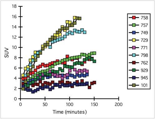
FIGURE 2. Tissue time–activity curves for 16 patients. Static protocols on a standard phantom. For SUV measurements,
PET was performed at 1, 3, and 5 h after injection of 18F-FDG, this assessment should include a comparison against a dose
and activity in lesions was determined. 18F-FDG uptake calibrator to ensure accuracy; that is, a comparison of the
plateaued at various times after injection and, in several lesions, absolute activity measured, versus the measured injected,
was still increasing even at 5 h after injection. (Courtesy of should be performed. This comparison is particularly impor-
Karen Kurdziel.)
tant after software or hardware upgrades.
of various lesions and the resultant variability in SUV Region-of-Interest (ROI) Determination
determination as a function of time. If uptake is still in- Tumors are extremely heterogeneous and contain necrotic
creasing, significant variability in SUV determination is tissue, cystic components, and fibrous elements, as well as
possible unless the patient is imaged at the same time on all malignant tumor cells. This heterogeneity becomes a critical
sequential studies used to assess response (23,24). Studies issue after therapy begins and the malignant or other com-
have shown that in metabolically active tumors, SUV can ponents of the ‘‘tumor’’ mass change. Drawing of the ROI by
change significantly over the course of 10–15 min (25,26 ). hand or by edge-finding techniques is typical for defining the
Comparisons of various kinetic modeling and semiquan- tumor boundaries. Both methods appear to work well as long
titative techniques show a good correlation between abso- as they are performed in the same, systematic manner on
lute quantitative metabolic rate and SUV normalized to serial examinations. Threshold-determination or edge-finding
body weight, lean body mass, or body surface area. Many algorithms are accurate and can be applied with less subjec-
members of the working group expressed a preference for tive interaction from the technician or physician determining
normalizing to lean body mass, but existing data did not the ROI. It should be obvious, however, that the same
warrant a unanimous preference for normalizing to lean approach must be applied systematically and uniformly
body mass over other parameters. Given the complexities of across all patients and across all sequential tumor measure-
conducting kinetic analysis, the working group concluded ments. Another factor that may confound ROI determination
that a reasonable approach for large phase II or III clinical is the partial-volume effect already mentioned. Both the
trials would be semiquantitative analysis (i.e., measurement mean value within the ROI and the maximum value (re-
of SUV normalized to either lean body mass or body sur- flecting the most metabolically active region) within the ROI
face area). If there is a perceived need to obtain the absolute may have clinical importance and should be reported.
quantitative metabolic rate or more detailed information
on 18F-FDG kinetics in a protocol evaluating therapeutic Blood Glucose Level
response (e.g., for new therapies that may affect tracer The concentration of circulating glucose can signifi-
delivery to the tumor and limit uptake), this need could be cantly affect 18F-FDG uptake by tumors, and various
more easily addressed in the setting of a single-institution groups have reported schemes for correcting the 18F-FDG
phase I or early phase II study. SUV for the circulating plasma glucose concentration. The
working group agreed that in patients with a plasma glu-
cose concentration within the reference range, SUV deter-
FACTORS AFFECTING UPTAKE DETERMINATION minations are not appreciably related to serum plasma glucose
Partial-Volume Effects concentration (9,30). The working group also agreed that
Partial-volume effects secondary to scanner resolution in patients with a high serum glucose concentration, the
are an important technical factor. Most PET scanners have problems with administration of insulin would diminish the
1062 THE JOURNAL OF NUCLEAR MEDICINE • Vol. 47 • No. 6 • June 2006accuracy of SUV determination by altering the biodistribu- for the following morning. For 18F-FDG PET studies
tion of 18F-FDG, especially in insulin-sensitive tissue such performed in the afternoon, a light breakfast with minimal
as muscle, myocardium, fat, and gut. In such patients, the carbohydrate-containing foods is acceptable. However, pa-
18F-FDG PET study should be rescheduled, and adjustments tients should fast for at least 4 h after finishing that meal.
to diet and medications be made if necessary, so that the While fasting, patients should consume at least two to three
fasting blood glucose concentration can be brought down to 355-mL (12-oz) glasses of water to ensure adequate hy-
an acceptable range at the time of 18F-FDG injection, that dration.
is, 1502200 mg/dL or less. The working group also agreed When patients arrive at the PET facility, their height and
that diabetic patients should not be excluded from clinical weight should be measured and recorded. Venous serum
trials but that such patients should be scanned early in the glucose should be measured to determine whether the
morning before the first meal and that the doses of insulin concentration is within the reference range (,120 mg/dL
and hypoglycemic medication should be titrated appropri- for nondiabetic patients and 1502200 mg/dL for diabetic
ately the night before and morning of the study. Before patients). Before injection of the 18F-FDG tracer, patients
scheduling an 18F-FDG PET study, diabetic patients should should be asked to urinate to minimize the possibility that
test their ability to maintain reasonable plasma glucose they will need to move during the 18F-FDG uptake phase.
levels after fasting, while avoiding insulin close to the time If the serum glucose concentration is greater than 200
that 18F-FDG would be administered. mg/dL, the study should be rescheduled. The glucose con-
centration should be measured consistently and accurately
RECOMMENDATIONS OF WORKSHOP PANEL across all patients, preferably by a credentialed clinical lab-
These recommendations are summarized in Table 2. oratory. Insulin should not be used to adjust the blood glu-
cose at the time of the imaging procedure.
Patient Preparation A medical history should be obtained from patients. Any
Patient preparation is critical to the quality of 18F-FDG history of previous treatment with radiation, chemotherapy,
PET, both as a diagnostic test and as an assessment of or other experimental therapeutics, and when these thera-
therapeutic response. The following are recommendations pies were performed and completed, should be documented.
to ensure consistency of data across institutions, as well as In particular, the use of medications that may affect the
in the same patient in serial 18F-FDG PET studies: uptake or biodistribution of 18F-FDG, such as marrow-
Patients should avoid strenuous exercise for 24 h before stimulating cytokines or steroids, should be noted. These
the 18F-FDG PET study to minimize uptake of the radio- data are important in assessing the interval from the com-
tracer in muscles. pletion of a certain therapy to the time of the 18F-FDG PET
Patients should, as much as possible, be on a low- study to ensure that all relevant confounding clinical issues
carbohydrate diet for 24 h before the study. are identified.
Patients should fast for a minimum of 4 h before re- Adequate hydration is important in 18F-FDG PET to
ceiving the injection of 18F-FDG. In general, patients ensure excretion of 18F-FDG from background tissue. If
should not eat anything after midnight if a study is planned possible, patients should drink 500 mL of water after
TABLE 2
Recommendations of Workshop Panel
Parameter Recommendation
Patient preparation Patients fast overnight for morning scan or 4 h for afternoon scan. Venous serum glucose concentration is
measured before injection (,120 mg/dL for nondiabetic patients and 150–200 mg/dL for diabetic patients).
Diabetic patients are scanned in morning after overnight fast and before first use of medication.
Patients are well hydrated and, if possible, drink 500 mL of water after injection and before scanning.
For renal/pelvic imaging, furosemide (20–40 mg) may be given 10–15 min after 18F-FDG injection,
or urinary catheter may be used.
All medications being taken by patients are recorded.
Diazepam or other mild sedative may be used at clinician’s discretion to decrease uptake in muscle.
PET timing Pretreatment and posttreatment scans are acquired.
Pretreatment scans are acquired as close to start of therapy as possible (preferably ,2 wk).
Posttreatment scans are acquired no sooner than 2 wk after end of chemotherapy to avoid transient
increases or decreases. Timing is determined by endpoint being assessed.
Timing of scans after changes due to radiotherapy needs further investigation.
Whole-body imaging begins 60 6 10 min after injection of 18F-FDG.
Attenuation Attenuation correction is used. No standard procedure has yet been recommended. Procedure chosen is
correction documented.
18F-FDG dose No standard dose has yet been recommended. Doses of 370–740 MBq (10–20 mCi) are appropriate. Dose
injected is documented.
NCI PET GUIDELINES • Shankar et al. 1063injection and before scanning. Depending on the type of Whole-body acquisition is important because it allows for
study performed and the area of clinical concern, a urinary sampling of all areas of interest and can assess whether new
(e.g., Foley) catheter may be required to ensure adequate lesions have appeared and, thus, the possibility that disease
visualization of pelvic structures. If the patient is to be has progressed. Whole-body acquisitions can be in either
catheterized for the imaging study, the bladder catheter 2- or 3-dimensional mode with attenuation correction, but a
should be placed before the 18F-FDG injection. In other consistent method should be chosen for all serial scanning
instances (or in addition to Foley catheter placement), for of an individual patient throughout the clinical trial. The
specific imaging of the pelvis or kidney region, intravenous use of CT in combined PET/CT scanners is strongly encour-
administration of a diuretic, such as furosemide, 20–40 mg, aged to provide anatomic registration for PET data.
may be required. The diuretic should be administered ap- The whole-body acquisition should sample from the angle
proximately 10–15 min after injection of the 18F-FDG to of the jaw to the level of the mid thigh. Because several target
allow time for the drug to clear 18F-FDG from the renal lesions may be identified on the initial 18F-FDG PET study or
collecting system and for patients to void before being on anatomic imaging studies, it is critical that for a given
placed on the scanner. If there are no medical contraindi- patient, all subsequent 18F-FDG PET studies be performed
cations, patients requiring clearance of the urinary back- identically to the first to ensure the quantitative integrity of
ground activity should receive 250–500 mL of intravenous the data and the validity of comparisons. For example, if the
saline (not dextrose-containing solutions) during the 18F- patient is scanned from the head to the thighs in the baseline
FDG uptake period to ensure adequate hydration. scan, subsequent scans should also be started at the head and
Patients should be placed in a comfortable position, extend to the thighs. The parameters for the timing of both
either supine or semirecumbent, in a dimly lit, quiet room. emission and transmission acquisitions vary from one patient
The room should be kept warm to avoid shivering and other to another depending on the size of the patient, the PET
temperature effects that may increase muscular or fat camera used, and the amount of 18F-FDG injected. There-
uptake. A large-bore intravenous line (21 gauge or greater) fore, the timing of the acquisitions cannot be standardized.
should be placed in an arm or hand vein contralateral to any However, the times at which target lesions are imaged after
known site of disease. 18F-FDG injection should be as close as possible to those
The dose of 18F-FDG should be 5.18–7.77 MBq (0.14– used on the baseline or previous study. It is strongly encour-
0.21 mCi) per kilogram of body weight, with a typical aged that serial studies to evaluate therapeutic response be
range of 370–740 MBq (10–20 mCi). This amount may done in exactly the same way, at the same institution, on the
need to be adjusted for a 3-dimensional brain acquisition. same type of camera, and using the same dose, imaging
The exact times at which the dose is calibrated and the times, acquisition parameters, and reconstruction parame-
injection given should be recorded to permit correction of ters.
the administered dose for radioactive decay. In addition, the Patients with head and neck malignancies may require
dose remaining in the tubing or syringe, or that spilled more extensive imaging of the head. Some patients (e.g.,
during injection, should be recorded. The injection should those with malignant melanoma or sarcoma) may require
be performed through an intravenous catheter using a slow imaging of the lower extremities. Patients with brain tu-
infusion over 1–2 min. mors require imaging of the whole brain, typically using
The administration of a sedative, such as diazepam, is at either 1 or 2 acquisitions and bed positions depending on
the discretion of the clinician. A sedative can facilitate the field of view of the PET camera.
muscle relaxation and reduce 18F-FDG uptake in muscle
and brown fat—particularly important for patients who are PET Timing Relative to Prior Therapy
extremely anxious or for whom the area of interest is the Insufficient data are available on the optimal interval from
head and neck. In patients with a history of or a suspicion of completion of therapy to imaging with 18F-FDG PET. Nev-
head and neck tumors, a benzodiazepine or similar seda- ertheless, the working group recommends that the complete
tive, if not medically contraindicated, should be adminis- treatment history of the patient be documented, particularly
tered orally or intravenously approximately 30 min before the use of supportive therapies such as bone marrow expan-
injection of the 18F-FDG to ensure a degree of relaxation of sion drugs and the recent use of corticosteroids. Pretreatment
the neck muscles. The amount and timing of the sedative scanning is generally critical to assess subsequent response.
should be documented. The timing of posttreatment scanning depends on numerous
Whole-body imaging should begin 60 6 10 min (mean 6 variables, including correlative studies, whether a complete
SD) after injection. clinical response variable is under consideration, the ex-
pected responsiveness of the tumor type to the therapy being
Image Acquisition and Reconstruction used, and the endpoints of the study.
Because the specifications of PET cameras are variable Currently available information supports the recommen-
and manufacturer specific, every attempt should be made to dation that posttreatment imaging be performed 2 wk after
use the same scanner (ideally at the same center) or same the end of a specific chemotherapy cycle. The exact timing
scanner model for serial scanning of the same patient. may depend on the frequency and duration of therapy. It is
1064 THE JOURNAL OF NUCLEAR MEDICINE • Vol. 47 • No. 6 • June 2006postulated that the transient and nondurable alterations in superior to the other. Each clinical trial should set a
18F-FDG uptake that may occur in tumors during the immediate- protocol calling for all SUV calculations to be done the
posttreatment period will be minimized using this approach. same way.
A specific understanding of the basic biology of the tumor In addition, the SUV of a reference organ or tissue not
from previous clinical and preclinical studies may help one involved in the neoplastic process should be measured after
determine the optimal posttreatment time point. each scan to help ensure that SUV changes in tumors are
Data on the treatment interval after the completion of related either to treatment response or to disease progression.
radiotherapy are less clear. Acute inflammatory changes
with subsequent alterations in 18F-FDG uptake in both ROI Determination
tumor and surrounding tissue have been documented (31). Tumors can be of various sizes and of various heteroge-
Newer radiation therapies such as g-knife and focal high- neities. Accurate and reproducible determination of the
dose radiation appear to enhance inflammatory reactions, ROI will be critical for determining SUV. With therapy,
and thus confound the interpretation of 18F-FDG PET alterations in the pattern of heterogeneity and in 18F-FDG
scans, in patients studied within a short period after com- uptake may occur and must be considered when one is
pleting these therapies (32). Many investigators recommend drawing or determining the ROI. On the pretreatment scan,
a delay of 628 wk or longer after radiation therapy before the identified target lesion should be the most visible and
performing the posttreatment 18F-FDG PET study (33). easily defined lesion. The mean SUV of the region and the
Although further study may be required to arrive at an maximum pixel SUV should be determined and recorded.
appropriate interval for scanning after completion of radi- No prescribed methodologies for determining regions of
ation therapy, a longer wait clearly helps in distinguishing interest have been validated. Thresholding techniques or
inflammatory response from viable residual tumor. freehand drawing are typically used. No specific recom-
mendations on either of these approaches can be made. The
Image Analysis choice of method will depend on the technical support staff,
The working group agreed that there is no single optimal expertise, and image-processing capabilities of an individ-
method for analysis of 18F-FDG PET whole-body images in ual PET center. However, in each clinical trial, the same
oncology but that there can be standardized protocols for ROI technique should be specified (e.g., whether to include
use in a particular clinical trial. The working group recom- necrotic areas) and used in subsequent 18F-FDG PET
mended that phase I trials use either full or partial kinetic studies to ensure quantitative consistency. Quantitative mea-
analysis, such as Patlak analysis, if deemed necessary, surements of mean and maximum tumor ROI counts per
along with semiquantitative SUV analysis based on lean pixel, calibrated as mBq/L (mCi/L), should be obtained
body mass and body surface area. The reason for recom- with the scanner. The consensus of the working group was
mending that SUVs be calculated on the basis of both lean that maximum or ‘‘peak’’ approaches are the most robust
body mass and body surface area is to develop a body of and reproducible and that the maximum SUV and mean
data to determine whether they are equivalent or whether SUV of each tumor should be recorded. The panel strongly
one is better than the other. It is also critical that before a encouraged further cooperative studies, including work
particular clinical trial begins, the method of ROI determi- with camera manufacturers, to improve reproducibility and
nation be agreed on and specified in the protocol. standardization between centers by developing more stan-
Of obvious importance is that whole-body 18F-FDG PET dard and automated methods of defining regions.
provides information additional to that obtained from stan- During the course of treatment, the extent and shape of
dard anatomic imaging studies such as CTor MRI. Therefore, an imaged tumor might change. Documentation of either an
it is also critical that the whole-body 18F-FDG PET study be increase or a decrease in dimensions or a change in shape is
interpreted carefully and reported as a clinical study would be recommended.
reported to ensure that new lesions are identified. This care As discussed, partial-volume effects on determinations of
18F-FDG uptake may be significant. If a significant de-
will be critical in the development of subsequent response
criteria. SUV should be determined in order to assess the 18F- crease in tumor size is evident from anatomic imaging
FDG uptake and define the response in target lesions of studies (which are typically available throughout therapy),
interest. Image reconstruction parameters depend on the PET this information should be documented because subsequent
scanner and other variables. Filters, image reconstruction analysis may require partial-volume corrections of the 18F-
techniques and parameters, and application of the attenuation FDG PET data. Further data analysis and research are
map must be consistent across all scanning of a given patient. required to better define how the assessment of response
The exact timing of image acquisition for each target lesion is can be adjusted to account for partial-volume effects, tumor
critical and must be kept constant on all subsequent studies of heterogeneity, and other confounding variables.
a given patient.
SUV should be determined for all target lesions and CONCLUSION
should be calculated consistently on the basis of either lean 18F-FDG PET has gained acceptance as a valuable clinical
body mass or body surface area. No data indicate that one is tool for detecting, staging, and managing disease. It is now
NCI PET GUIDELINES • Shankar et al. 1065clear that 18F-FDG PET can also be an important tool for 14. Hawkins RA, Phelps ME, Huang SC. Effects of temporal sampling, glucose
metabolic rates, and disruptions of the blood-brain barrier on the FDG model
assessing therapeutic efficacy in large, multicenter clinical with and without a vascular compartment: studies in human brain tumors with
trials, but only with the application of standard protocols. PET. J Cereb Blood Flow Metab. 1986;6:170–183.
Currently, there is no one best methodology for obtaining or 15. Wienhard K. Measurement of glucose consumption using [(18)F]fluoro-
deoxyglucose. Methods. 2002;27:218–225.
analyzing 18F-FDG PET scans, nor is there one agreed-on 16. Spence AM, Muzi M, Graham MM, et al. Glucose metabolism in human
standard for judging the significance of a response seen on malignant gliomas measured quantitatively with PET, 1-[C-11]glucose and FDG:
18F-FDG PET. Enacting these recommendations to develop analysis of the FDG lumped constant. J Nucl Med. 1998;39:440–448.
17. Patlak CS, Blasberg RG, Fenstermacher JD. Graphical evaluation of blood-to-
standard protocols for NCI-sponsored clinical trials should brain transfer constants from multiple-time uptake data. J Cereb Blood Flow
go a long way toward determining when and for what Metab. 1983;3:1–7.
indications 18F-FDG PET can serve as a surrogate measure 18. Patlak CS, Blasberg RG. Graphical evaluation of blood-to-brain transfer
constants from multiple-time uptake data: generalizations. J Cereb Blood Flow
of therapeutic efficacy. The result should be shorter clinical Metab. 1985;5:584–590.
trials and improved therapy for patients with cancer. 19. Minn H, Zasadny KR, Quint LE, Wahl RL. Lung cancer: reproducibility of
quantitative measurements for evaluating 2-[F-18]-fluoro-2-deoxy-D-glucose
uptake at PET. Radiology. 1995;196:167–173.
20. Hunter GJ, Hamberg LM, Alpert NM, Choi NC, Fischman AJ. Simplified
REFERENCES
measurement of deoxyglucose utilization rate. J Nucl Med. 1996;37:950–955.
1. Young H, Baum R, Cremerius U, et al. Measurement of clinical and subclinical 21. Graham MM, Peterson LM, Hayward RM. Comparison of simplified quantitative
tumour response using [18F]-fluorodeoxyglucose and positron emission tomog- analyses of FDG uptake. Nucl Med Biol. 2000;27:647–655.
raphy: review and 1999 EORTC recommendations. European Organization for 22. Sundaram SK, Freedman NM, Carrasquillo JA, et al. Simplified kinetic analysis
Research and Treatment of Cancer (EORTC) PET Study Group. Eur J Cancer. of tumor 18F-FDG uptake: a dynamic approach. J Nucl Med. 2004;45:1328–
1999;35:1773–1782. 1333.
2. Kubota K. From tumor biology to clinical Pet: a review of positron emission 23. Lowe VJ, Delong DM, Hoffman JM, Coleman RE. Optimum scanning protocol
tomography (PET) in oncology. Ann Nucl Med. 2001;15:471–486. for FDG-PET evaluation of pulmonary malignancy. J Nucl Med. 1995;36:
3. Warburg O. The Metabolism of Tumors. London, U.K.: Constable Press; 1930. 883–887.
4. Bos R, van Der Hoeven JJ, van der WE, et al. Biologic correlates of 24. Freedman NM, Sundaram SK, Kurdziel K, et al. Comparison of SUV and Patlak
(18)fluorodeoxyglucose uptake in human breast cancer measured by positron slope for monitoring of cancer therapy using serial PET scans. Eur J Nucl Med
emission tomography. J Clin Oncol. 2002;20:379–387. Mol Imaging. 2003;30:46–53.
5. Maschauer S, Prante O, Hoffmann M, Deichen JT, Kuwert T. Characterization of 25. Thie JA, Hubner KF, Smith GT. Optimizing imaging time for improved
18F-FDG uptake in human endothelial cells in vitro. J Nucl Med. 2004;45:455–460.
performance in oncology PET studies. Mol Imaging Biol. 2002;4:238–244.
6. Avril N. GLUT1 expression in tissue and (18)F-FDG uptake. J Nucl Med. 26. Beaulieu S, Kinahan P, Tseng J, et al. SUV varies with time after injection in
2004;45:930–932. (18)F-FDG PET of breast cancer: characterization and method to adjust for time
7. Higashi K, Clavo AC, Wahl RL. Does FDG uptake measure proliferative activity differences. J Nucl Med. 2003;44:1044–1050.
of human cancer cells? In vitro comparison with DNA flow cytometry and 27. Videen TO, Dunford-Shore JE, Diringer MN, Powers WJ. Correction for partial
tritiated thymidine uptake. J Nucl Med. 1993;34:414–419. volume effects in regional blood flow measurements adjacent to hematomas
8. Haberkorn U, Strauss LG, Reisser C, et al. Glucose uptake, perfusion, and cell in humans with intracerebral hemorrhage: implementation and validation.
proliferation in head and neck tumors: relation of positron emission tomography J Comput Assist Tomogr. 1999;23:248–256.
to flow cytometry. J Nucl Med. 1991;32:1548–1555. 28. Hickeson M, Yun M, Matthies A, et al. Use of a corrected standardized
9. Vesselle H, Schmidt RA, Pugsley JM, et al. Lung cancer proliferation correlates uptake value based on the lesion size on CT permits accurate characterization
with [F-18]fluorodeoxyglucose uptake by positron emission tomography. Clin of lung nodules on FDG-PET. Eur J Nucl Med Mol Imaging. 2002;29:
Cancer Res. 2000;6:3837–3844. 1639–1647.
10. Stahl A, Ott K, Schwaiger M, Weber WA. Comparison of different SUV-based 29. Boellaard R, Krak NC, Hoekstra OS, Lammertsma AA. Effects of noise, image
methods for monitoring cytotoxic therapy with FDG PET. Eur J Nucl Med Mol resolution, and ROI definition on the accuracy of standard uptake values: a sim-
Imaging. 2004;31:1471–1478. ulation study. J Nucl Med. 2004;45:1519–1527.
11. Wahl RL, Henry CA, Ethier SP. Serum glucose: effects on tumor and normal 30. Thie JA, Smith GT, Hubner KF. 2-Deoxy-2-[F-18]fluoro-D-glucose-positron
tissue accumulation of 2-[F-18]-fluoro-2-deoxy-D-glucose in rodents with mam- emission tomography sensitivity to serum glucose: a survey and diagnostic appli-
mary carcinoma. Radiology. 1992;183:643–647. cations. Mol Imaging Biol. 2005;7:361–368.
12. Sugawara Y, Zasadny KR, Neuhoff AW, Wahl RL. Reevaluation of the 31. Castellucci P, Zinzani P, Nanni C, et al. 18F-FDG PET early after radiotherapy in
standardized uptake value for FDG: variations with body weight and methods lymphoma patients. Cancer Biother Radiopharm. 2004;19:606–612.
for correction. Radiology. 1999;213:521–525. 32. Plowman PN. Stereotactic radiosurgery. VIII. The classification of postradiation
13. Weber WA, Ziegler SI, Thodtmann R, Hanauske AR, Schwaiger M. Reproduc- reactions. Br J Neurosurg. 1999;13:256–264.
ibility of metabolic measurements in malignant tumors using FDG PET. J Nucl 33. Avril NE, Weber WA. Monitoring response to treatment in patients utilizing PET.
Med. 1999;40:1771–1777. Radiol Clin North Am. 2005;43:189–204.
1066 THE JOURNAL OF NUCLEAR MEDICINE • Vol. 47 • No. 6 • June 2006You can also read