CHAPTER 5. Innovations on butterfly wings: co-option of insect embryonic and wing patterning genes in eyespot formation
←
→
Page content transcription
If your browser does not render page correctly, please read the page content below
CHAPTER 5. Innovations on butterfly wings: co-option of
insect embryonic and wing patterning genes in eyespot
formation

Suzanne V. Saenko, Paul M. Brakefield and Patrícia Beldade

Co-option of conserved genes and genetic pathways seems to play a fundamental
role in morphological evolution. How are ancestral gene networks redeployed in
the development of novel structures? How do modifications of these networks
produce changes in phenotype? The eyespots of Bicyclus anynana butterflies
provide a study system where these questions can be addressed in an integrative
way. Eyespots are formed around inductive organizers called foci. Focal cells
produce a morphogen the levels of which establish the colour of surrounding
scales in pupal wings. The number and position of foci are determined during the
last larval stage. Here we focused on several conserved genetic pathways which
are fundamental to insect embryonic or wing development and are potentially
redeployed in formation of eyespots. We characterized expression patterns of
candidate eyespot genes and attempted to manipulate their function. Our study
established a relationship between eyespot focal determination and expression of
the Hox gene Antennapedia and suggested that this conserved transcription factor
might be the first key regulator of eyespot formation. This illustrates that even
highly conserved Hox genes can be co-opted to perform additional functions
during animal development. Furthermore, we observed unexpected patterns of the
putative eyespot morphogen wingless and its antisense transcripts in pupal wings
and suggested their potential role for fine-tuning of gene expression. We also
described unforeseen differences in the expression of Hedgehog pathway genes
in B. anynana and Junonia coenia. These results suggest that the genetic
mechanisms underlying eyespot formation in these butterflies might have
diversified substantially.

                                                                              83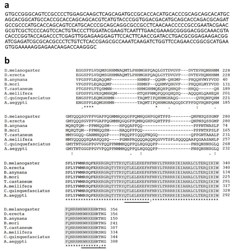
INTRODUCTION
The origin and diversification of novel morphological traits has always fascinated
biologists and laymen alike, and is now a key research theme in evolutionary
developmental biology, or evo-devo (Wagner & Lynch 2009). Novelties seem to
arise through co-option of conserved developmental toolkit genes (Ganfornia &
Sanchez 1999; True & Carroll 2002), although recent studies also implicated
taxonomically-restricted genes in the generation of lineage-specific traits
(reviewed in Khalturin et al. 2009). The relative importance of new and
conserved genes and the molecular changes that produce novelty-specific gene
networks must be studied in a broad range of taxa in order to understand general
principles about the evolution of novel traits.
       The extremely diverse butterfly wing patterns are generated by the
arrangement of pigmented scales and have no obvious homology to wing patterns
of other insects. This makes them an ideal system to study the genetic and
developmental mechanisms underlying the origin of novelties. Particularly
eyespots have emerged as a promising evo-devo model used to investigate how
novel characters arise (Keys et al. 1999; Brunetti et al. 2001; Saenko et al. 2008)
and diversify (Allen et al. 2008; Beldade, French & Brakefield 2008; Monteiro
2008). Eyespots have a clear adaptive value (e.g. Olofsson et al. 2010; Robertson
& Monteiro 2005) and show extreme intra- and interspecific variation in
morphology (Nijhout 1991). These pattern elements are formed by the action of
multiple gene networks which regulate pigment biosynthetic pathways in the
developing scales (reviewed in Beldade & Saenko 2009), and changes in the
organization of these networks ultimately yield the observed eyespot diversity. A
more complete account of eyespot evolution awaits a detailed understanding of
how ancestral gene networks are co-opted in the development of wing patterns
(see Monteiro & Podlaha 2009), and of how modifications of the eyespot
patterning network under the influence of natural and sexual selection produce
the observed changes in the phenotype.
       Until now, studies of eyespot development have focused mainly on two
species of the family Nymphalidae, Bicyclus anynana and Junonia coenia
(Beldade & Brakefield 2002; McMillan, Monteiro & Kapan 2002). Classical
surgical manipulations have shed light on some of the cellular interactions
underlying eyespot formation (Nijhout 1980; French & Brakefield 1992, 1995),
and gene expression studies implicated a number of conserved genetic pathways
that play essential roles in insect embryonic and wing development, and have
been co-opted in eyespot formation (reviewed in Beldade & Saenko 2009).
Eyespot development begins at the end of the larval stage when several wing-
patterning genes are upregulated in positions of the wing epidermis
corresponding to the centers, or foci, of adult eyespots. During

                                                                                 84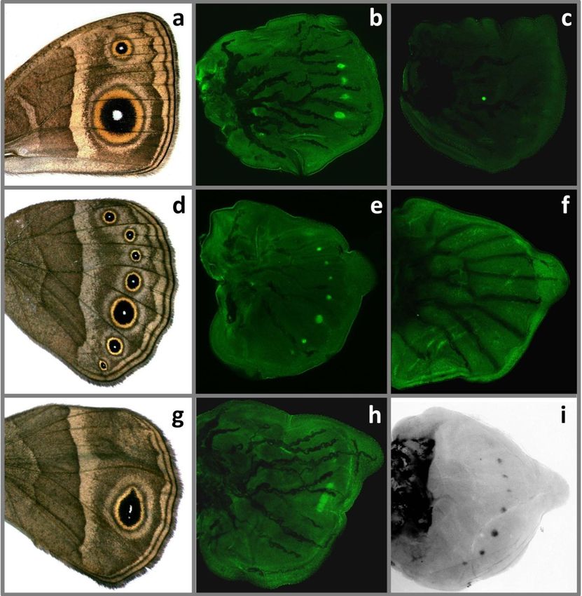
the early pupal stage, the cells of these foci provide positional information, in the
form of one or several signaling molecules that diffuse throughout the epidermis
and define cellular territories corresponding to the different colour rings of the
adult eyespot. Epidermal cells respond to these morphogen(s) in a concentration-
dependent manner by activating different pigment biosynthesis pathways and
subsequently by producing wing scales of different colours. The Hedgehog (Hh)
signaling pathway, together with the transcription factors Spalt (Sal), Engrailed
(En) and Distal-less (Dll), and Notch (N) receptor, have been implicated in the
process of eyespot determination (Carroll et al. 1994; Brakefield et al. 1996;
Keys et al. 1999; Reed & Serfas 2004). The signaling molecules Wingless (Wg)
and Decapentaplegic (Dpp) have both been proposed as candidate eyespot
morphogens (Monteiro et al. 2006). All these genes were suggested by studies of
their expression patterns, and functional tests of their role are yet to be
performed.
       Here we characterized expression patterns of key players in several
conserved genetic pathways which are fundamental to insect embryonic or wing
development and are potentially redeployed in different stages of eyespot
formation. First, the Hh signaling pathway was implicated in eyespot focus
determination in J. coenia larval wings, where genes encoding the Hh ligand, its
receptor Patched (Ptc) and the transcription factor Cubitus interruptus (Ci) are
expressed around or in eyespot foci (Keys et al. 1999). Based on gene expression
data from this and other experiments, and on the available information about
genetic interactions in Drosophila melanogaster wing imaginal discs, Evans and
Marcus (2006) proposed two models of the genetic regulatory hierarchy for the
eyespot focus determination, and suggested that several predictions of these
models could be tested experimentally in B. anynana wild-type and Cyclops
mutant butterflies (Marcus & Evans 2008). Next, the Wg pathway has been
implicated in the process of focal signaling by Monteiro et al. (2006), who
detected Wg protein in eyespot centres of early pupal wings with the antibody
developed against human Wnt-1 protein. One of the known targets of Wg
signaling in Drosophila wing imaginal discs, Dll (Neumann & Cohen 1997a), is
expressed in the area around eyespot foci in pupal wings of B. anynana and other
butterflies (Brakefield et al. 1996; Brunetti et al. 2001). This upregulation of Dll
might be a response to a concentration gradient of Wg, which makes this
molecule a good candidate for an eyespot morphogen that is secreted from
eyespot centres and induces a cellular response in the surrounding epidermis
(Neumann & Cohen 1997b). Moreover, our comparative analysis of embryonic
defects in pleiotropic mutants with disturbed eyespot size and/or colour
composition suggests that these mutations might have occurred in a segment
polarity gene, probably a negative regulator of the Wg signaling pathway (see
Chapter 3).
                                                                                   85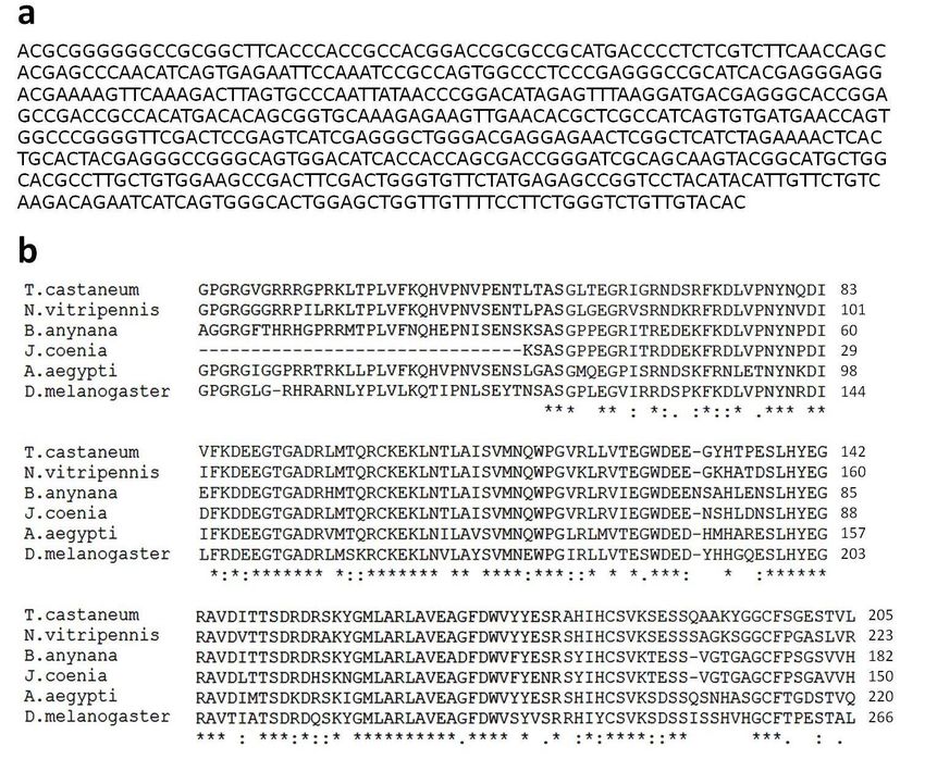
To test the predictions of the models proposed by Marcus & Evans, and to
further investigate the role of Wg in eyespot formation, we studied the spatio-
temporal expression patterns of genes encoding the Hh and Wg ligands and their
respective receptors, Ptc and Frizzled (Fz), in developing wings of B. anynana. In
addition, we examined expression patterns of the Hox genes Antennapedia (Antp)
and Ultrabithorax (Ubx), two highly conserved transcription factors involved in
arthropod embryonic patterning (Hughes & Kaufman 2002). Ubx also controls
insect hindwing identity, including wing scale morphology and colour pattern
elements in butterflies (Lewis et al. 1999; Weatherbee et al. 1999), but no Hox
gene has been demonstrated to be involved in eyespot determination. Finally, we
attempted to inhibit the functions of two transcription factors implicated in
eyespot development via RNAi or morpholino injections.

MATERIAL AND METHODS
Biological material
B. anynana wild-type (WT) and mutant Cyclops stocks were reared in standard
laboratory conditions at 27oC (cf. Brakefield, Beldade & Zwaan 2009). Pupation
times were recorded using time-lapse photography with 30 minute intervals.
Larvae and pupae were anesthetized in ice-cold PBS and their wings dissected
using fine foreceps and scissors. For larval wing discs, we used the staging
system based on wing vein and tracheal development: stage 0 – wing disc as
found after the last molt; 0.5 – vein lacunae are visible; 1 – anterior trachea
extend into vein lacunae; 1.5 – posterior trachea extend into vein lacunae; 2 –
most trachea reach border lacuna; 2.5 – trachea extend into border lacuna; 3 –
trachea form continuous line in border lacuna (cf. Reed, Chen & Nijhout 2007).

Cloning of B. anynana Antp and hh homologues
Total RNA was extracted from embryos and wings dissected out of last instar
larvae and 1 – 4 days old pupae. RNA extractions were done with Trizol
(Invitrogen) according to manufacturer’s instructions. Samples were run on an
agarose gel to verify RNA quality and treated with DNase (Ambion) to remove
traces of genomic DNA. For degenerate PCR, cDNA was prepared using Reverse
Transcription System (Promega). The SMARTer rapid amplification of cDNA
ends (RACE) Amplification Kit (Clontech) was used for cDNA preparation and
5’-RACE PCR.
       To clone the Antp gene in B. anynana, protein sequences of seven insects
were aligned using ClustalW (Larkin et al. 2007), and degenerate primers were
designed in the highly conserved homeobox region (Fig. 1) using CODEHOP
(Rose et al. 1998). A 159-bp fragment of Antp was amplified with primers 5’-

                                                                                86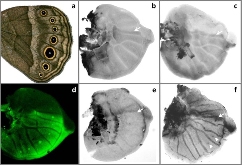
CAGACCCTGGAGCTGGAGAARGARTTYCAYT and 5’-
GCCCTTGGTCTTGTTCTCCTTYTTCCAYTTC from embryonic cDNA using
the following touchdown PCR conditions: denaturing at 95°C for 2 min; 18
cycles at 95°C for 30 s, 58 – 50°C (decreasing by 4°C per 6 cycles) for 30 s,
72°C for 1.5 min; 40 cycles at 95°C for 30 s, 48°C for 30 s, 72°C for 1.5 min;
final extension at 72°C for 5 min. The product was cloned into the pGEM®T-
Easy vector (Promega) and sequenced with vector primers M13F and M13R. The
sequence obtained was then used to design gene-specific primers for 5’RACE
PCR. The first-round PCR was performed on larval forewing cDNA with primer
5’-GATTTGGCGCTCGGTGAGACAGAGG using the following conditions: 5
cycles at 94°C for 30 s, 72°C for 3.5 min; 5 cycles at 94°C for 30 s, 70°C for 30
s, 72°C for 3.5 min; 25 cycles at 94°C for 30 s, 68°C for 30 s, 72°C for 3.5 min.
This product was then used as a template in the second-round PCR with internal
5’- CCGCGTCAGGTATCGGTTGAAGTGG primer, carried out for 22 cycles
(94°C for 30 s, 68°C for 30 s, 72°C for 3.5 min). The 700-bp product was excised
from an agarose gel, cleaned with Wizard SV Gel and PCR Clean-Up System
(Promega), cloned into the pCRII®-TOPO vector (Invitrogen), amplified and
sequenced with vector primers M13F and M13R.
       An available sequence of a 339-bp fragment of B. anynana hh homologue
(Arjen van ‘t Hof, pers. comm.) was used to design gene-specific primers for
5’RACE PCR. The first amplification with primer 5’-
GCTCCAGTGCCCACTGATGATTCTG was carried out for 25 cycles of 94°C
for 30 s, 68°C for 30 s, 72°C for 3 min and a final extension at 72°C for 10 min,
and was followed by the second-round PCR with internal primer 5’-
ACACTGATGGCGAGCGTGTTCAACT, performed under the same conditions.
The ~250 bp product was cloned and sequenced in the same way as the Antp
product, and aligned with the already available sequence. All sequences were
edited in BioEdit and aligned against their insect homologues in NCBI BLAST
(http://www.ncbi.nlm.nih.gov/BLAST/). ExPASy’s translation tool
(http://www.expasy.ch/tools/dna.html) was used to obtain the translations of the
nucleotide sequences and ClustalW (2.0.12) multiple alignment tool
(http://www.ebi.ac.uk/Tools/clustalw2) to produce the protein alignments. The
nucleotide sequences of Antp and hedgehog have been deposited to GenBank
(HQ020406, HQ020407).

In situ hybridizations
The fragments of B. anynana genes encoding Wg and Hh ligands and their
receptors were amplified from embryonic cDNA with the following primers: wg
(AY218276.1) 5’-GTCATGATGCCCAATACCG/5’-
GCAGTTGCATCGTTCCACTA; fz 5’-TGCCCATTTACACATCAGGA/5’-
GTATGTGGTTCCTGGCTGCT; ptc (HQ020408) 5’-
                                                                               87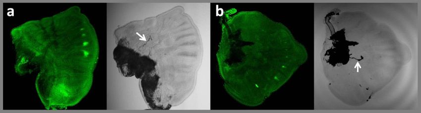
CGTACTTGATGCTTGGCAGA/5’-GCCAGTAGAACGCTCAGTCC; hh 5’-
TGACCCCTCTCGTCTTCAAC/5’-AAAACAACCAGCTCCAGTGC. The
products were cloned into pCRII®-TOPO dual-promoter vector using the TOPO
TA cloning kit (Invitrogen). These vectors, as well as the plasmid containing
159-bp fragment of Antp, were used for synthesis of in situ hybridization
digoxygenin-labeled sense and antisense riboprobes. Plasmids were extracted
from bacterial colonies with QIAprep Spin Miniprep Kit (QIAGEN) and used as
templates for PCR reactions with gene-specific and vector primers M13F and
M13R. The amplified products were cleaned with Wizard SV Gel and PCR
Clean-Up System (Promega) and used for SP6 or T7 transcription with DIG RNA
labeling mix (Roche Applied Science). All probes were run on an agarose gel and
measured with NanoDrop spectrophotometer (Thermo Scientific) to assess their
quality and concentration. Whole mount in situ hybridizations of larval and pupal
wings were done according to the protocols described in Brakefield, Beldade &
Zwaan (2009). Sense probes were used as negative controls.

Immunohistochemistry
Antibody stainings of larval and pupal wings were performed as described in
Brakefield, Beldade & Zwaan (2009). The following antibodies were used:
monoclonal mouse anti-Antp 4C3 and 8C11 (Condie, Mustard & Brower 1991)
[dilution 1:50], anti-Notch C17.9C6 (Fehon et al. 1990) and anti-Ubx/Abd-A
FP6.87 (Kelsh et al. 1994) [1:20], anti-En/Inv 4F11 (Patel et al. 1989) [1:50],
polyclonal rabbit anti-Dll (Panganiban et al. 1995) [1:200] and anti-Sal (de Celis,
Barrio & Kafatos 1999) [1:500]. Primary antibodies were detected with Alexa
Fluor 488 anti-mouse and Texas Red anti-rabbit secondary antibodies (Molecular
Probes) [1:200]. Images were collected on a BioRad MRC 1024 ES and Zeiss
Imager M1 laser scanning confocal microscopes. For the time series experiment
(Fig. 7), right wings of 70 larvae were co-stained with anti-Antp 4C3 and anti-Sal
antibodies, and left wings with anti-Notch and anti-Dll antibodies.

Antp and Dll knock-down experiments
For the production of a template for in vitro transcription, PCR with vector-
specific M13F and M13R primers was performed on a pCRII®-TOPO vector
containing a 450-bp Antp insert and the promotor sequences for the T7 and SP6
polymerases. After cleaning with Wizard SV Gel and PCR Clean-Up System
(Promega), 1 µg of the PCR product was used as a template for transcription
reactions with T7 and SP6 enzymes. Sense and antisense transcripts were
synthesized using the MEGAscript® RNAi Kit (Ambion), according to the
manufacturer’s instructions, and annealed at room temperature to obtain double-
stranded RNA (dsRNA). After purification in sterile 0.5xPBS, the quality of
dsRNA was verified on an agarose gel, and its concentration measured with
                                                                                 88
NanoDrop spectrophotometer (Thermo Scientific). Injections into the dorsal part
of the thorax of B. anynana larvae were performed under a dissection microscope
using 5 µl syringe (Hamilton). We injected 3 and 5 µl of dsRNA at concentration
366 ng/µl into 4th (N = 13) and 5th (N = 10) instar larvae, respectively. Three
days later, each individual was injected again with 5 µl of Antp dsRNA.
A transcription-blocking vivo-morpholino (5’-
AGGCCCAAAGGGATCTGAGAAACTC) for the B. anynana Dll gene
(AF404825.1) was obtained from GeneTools, LLC (OR, USA). This morpholino
is complementary to a 25 bp sequence situated 45 bp upstream of the start-codon
in the Dll mRNA. Upon binding to its target sequence, it should block the
translation and thus prevent the synthesis of Dll protein. The morpholino was
diluted in sterile water [1:5] to obtain 0.1 nmole/µl solution, and delivered into
larvae in two ways – via injections in the heamolymph or via food. Injections
were done in in the dorsal part of the thorax, one day before or after the last larval
molt (here referred to as 4th and 5th instar, respectively). Fourth instar larvae
were injected with 1.5 – 2.5 µl (N = 40); last instars with 2.5 - 3.0 µl on the first
and the third day (N = 20). Similar volumes of sterile water were injected as a
control.
For the delivery of the morpholino via larval food, 1% of Tween was added
to the morpholino 0.1 nmole/µl solution, 10 µl of which was then applied on
maize leaf pieces of approximately 4 cm length. Newly molted 5th instar larvae
(N = 25) were placed in small petri dishes with treated maize leaves and kept
there until all leaves were eaten. The treatment was repeated two and seven days
later. As a control, maize leaves covered with 1% Tween in water were fed to
larvae (N = 25) in a similar way. Larvae from all experiments were reared
through to adulthood and the eclosed butterflies were examined for
morphological aberrations.
RESULTS AND DISCUSSION
B. anynana Antp and hh homologues
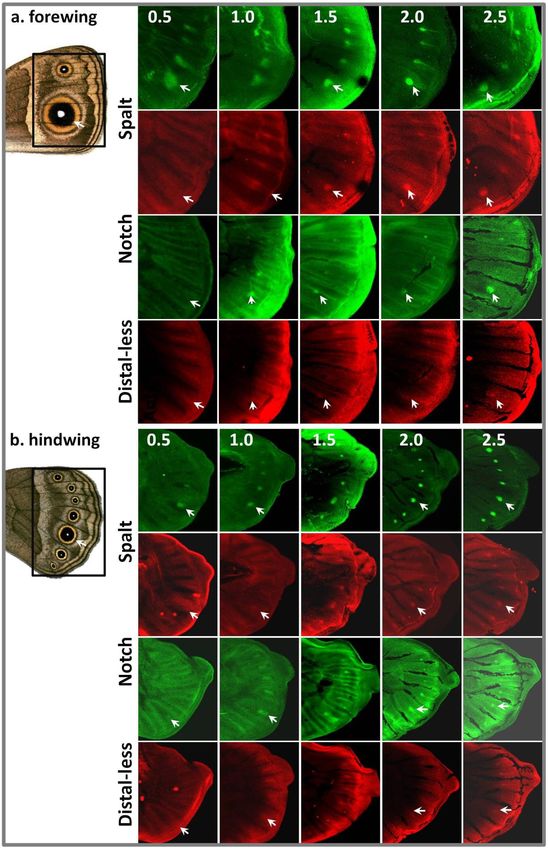
Sequence analysis of the 450-bp fragment of B. anynana Antp obtained in this
study revealed that this partial cds encodes 150 amino acids (AA) and shares 95%
AA identity with Antp protein of another lepidopteran, the silkworm Bombyx
mori. It also shows a high degree of similarity to its homologs in other insects,
mainly for the homeobox domain (Fig. 1). A 339-bp fragment of B. anynana hh
was extended to 548 bp in 5’direction. The corresponding 182 AA product is
closely similar to Hh proteins of other insects, and shares 93% AA identity with
Hh of J. coenia, another Nymphalid butterfly (Fig. 2). The fragments obtained in
this study thus represent B. anynana orthologues of Antp and hh genes.
89Figure 1. B. anynana Antp homologue.
a. Partial cds (450 bp) obtained by degenerate and 5’RACE PCR. b. A CLUSTAL W multiple
sequence alignment of the predicted B. anynana Antp amino acid sequence with the
corresponding fragments of Antp proteins of other insects. Numbers on the right show
sequence position of amino acids. Sequence identities are marked with (*), conserved
substitutions with (:) and semi-conserved substitutions with (.) (cf. ClustalW programme). The
following protein sequences [GenBank accession numbers] were used: Bombyx mori
[NP_001037319.1], Drosophila melanogaster [NP_996171.1], Drosophila erecta
[XP_001979126.1], Tribolium castaneum [NP_001034505.1], Apis mellifera
[NP_001011571.1], Culex quinquefasciatus [XP_001869455.1], Aedes aegypti
[XP_001660496.1]. The grey box corresponds to the homeobox region, and underlined amino
acids – to sites of degenerate primer design.
90Figure 2. B. anynana hh homologue.
a. Partial cds (548 bp) obtained by 5’RACE PCR. b. A CLUSTAL W multiple sequence
alignment of the predicted B. anynana Hh amino acid sequence with the corresponding
fragments of Hh proteins of other insects. Numbers on the right show sequence position of
amino acids. Sequence identities are marked with (*), conserved substitutions with (:) and
semi-conserved substitutions with (.) (cf. ClustalW programme). The following protein
sequences [GenBank accession numbers] were used: Junonia coenia [AAD08931.1],
Drosophila melanogaster [AAA16458.1], Tribolium castaneum [NP_001107837.1], Aedes
aegypti [XP_001657979.1], Nasonia vitripennis [XP_001605475.1].
No evidence for Hh signalling in eyespot centres in B. anynana larval wings
In situ hybridizations were performed to determine the spatial distribution of hh
and ptc transcripts in the wing discs of B. anynana WT larvae. As in D.
melanogaster (Lee et al. 1992) and other insects including the butterfly J. coenia
(Keys et al. 1999), hh mRNA was detected in the posterior compartments of all
44 early and mid-fifth instar wings examined (Fig. 3b), and in 32 out of 48 wings
of late-fifth instars (Fig. 3c). Detection of hh mRNA presumably failed in the
other 16 wings which were covered by rigid peripodial membrane that may not
be permeable to the probe. Transcript of the Hh receptor-encoding gene ptc was
found along the antero-posterior compartment boundary and in vein lacunae in all
9150 mid- and late-fifth instar larvae wing discs examined (Fig. 3e), also
resembling patterns found in D. melanogaster (Phillips et al. 1990) and J. coenia
(Keys et al. 1999). Higher levels of ptc mRNA were only occasionally detected
in the regions corresponding to centres of some eyespots (Fig. 3f), but hh
transcripts were never seen in the cells flanking eyespot foci. Stainings with
control sense probes produced no patterns.
The absence of hh transcripts in eyespots of B. anynana prevented us from
testing the predictions of models proposed by Marcus & Evans (2008) for
eyespot focus determination. We found that expression patterns of Hh pathways
genes differed in some aspects from those described for larval wings of J. coenia,
where hh transcripts were found around, and ptc mRNA in the regions
corresponding to future eyespot foci (Keys et al. 1999). The conserved function
of Hh signaling in insect wing (Tabata & Kornberg 1994) was evident in both
species, as was clear from hh upregulation in the posterior compartment and high
levels of ptc transcription in the cells just anterior to the antero-posterior
boundary. However, the putative role for this pathway in eyespot focus
determination as suggested by studies in J. coenia was not confirmed in B.
anynana. Both Hh signal transducer Ci and its target En were detected in eyespot
foci of J. coenia (Keys et al. 1999; Reed, Chen & Nijhout 2007), and in all
eyespot foci of B. anynana (Fig. 3d; see also Keys et al. 1999). The absence of
the Hh ligand and its receptor in the eyespot field in one of the species is thus
very remarkable, and suggests that activation of Ci and en in eyespot foci might
be Hh-independent in B. anynana. The genetic pathways underlying eyespot
development in these nymphalids may differ substantially. This emphasizes the
necessity of studies of complete genetic pathways in multiple organisms, both at
the levels of gene expression and of gene function.
Wg as a putative morphogen: sense and antisense transcripts in pupal wings
The spatial patterns of wg mRNA were examined in pupal wings at 12 – 18 hours
post pupation (hpp, N = 60). In the hindwings, wg transcripts were detected in
eyespot foci in pupal wings at 14 – 18 hpp (Fig. 4b,d). Unexpectedly, stainings
with the control sense probe on the opposite side wings of the same individuals
produced similar results (Fig. 4c,d). To check for the possibility that these
patterns are due to unspecific binding of the sense probe, we performed in situ
hybridizations in larval wings. Consistent with previous findings (Carroll et al.
1994), wg mRNA was detected in the wing margin, and only with the anti-sense
probe (Fig. 4i,j). This suggests that the control probe hybridizes in a specific
fashion to transcripts complementary to wg mRNA, which are present in pupal,
but not in larval wings. Expression patterns of wg sense and anti-sense transcripts
in the forewings were more complex than those observed in the hindwings. At 14
92Figure 3. Expression patterns of hh, ptc and en in larval wing discs.
a. Representative image of B. anynana hindwing with seven marginal eyespots. b. hh mRNA is
detected in the posterior wing compartment in mid-5th instar larval wings (stage 1, arrows
point to the antero-posterior boundary) and c. in late 5th instar larval wings (stage 2.5). d. en is
upregulated in the posterior compartment and in the centres of all eyespots. e. ptc is expressed
along the antero-posterior boundary (arrows) in mid-5th instar wings, and also in some eyespot
foci (arrowheads) in the late 5th instar wing discs (f).
hpp, both wg mRNA and its complementary transcript were detected in eyespot
foci (Fig. 4e). In older pupal wings (15 - 18 hpp), additional ring-like patterns of
wg mRNA, but not of the anti-sense transcript, were found around forewing
eyespot foci at a time when both had already disappeared from the centres of
posterior forewing eyespots (Fig. 4f-h).
These results are consistent with findings of Monteiro and colleagues
(2006) who detected Wg protein in eyespot foci at 10 – 16 hpp. They suggest that
Wg may indeed be (one of) the morphogen(s) produced in eyespot centres.
However, wg mRNA processing, and thus protein levels, might be modulated by
its anti-sense transcript in a highly specific way. Recent findings have revealed
that expression of complementary anti-sense transcripts in a tissue-specific
manner is a widespread phenomenon, and that they not only regulate the
expression levels and processing of the sense transcripts, but can also silence
their transcription (Lapidot & Pilpel 2006; Werner & Sayer 2009). Anti-sense
transcripts may, therefore, be essential to the fine-tuning of specific genes. It is
unclear if both wg transcripts are expressed in exactly the same cells in B.
anynana pupal wings, and how they interact, but it is possible that translation
93Figure 4. Expression patterns of wg and fz.
a. Ventral side of a B. anynana wild-type female; white squares indicate sections of the wings
shown in panels b, c, e – g, k, and l. Pupal hindwings stained with wg anti-sense (b) and sense
(c) probes at 14 – 18 hpp show similar patterns in eyespot foci (enlargement of 5th hindwing
eyespot in d). In early pupal forewings (14 hpp), wg sense (e) and anti-sense transcripts
(pattern identical to that in e) were detected in eyespot foci (arrows). At later stage (15 – 18
hpp) wg mRNA (f) was found in and around anterior eyespot focus, but not in posterior
eyespot focus (arrows). Anti-sense transcripts (g) were detected in anterior eyespot foci, but
not in a surrounding ring (enlargement of the anterior eyespot area in h). Control stainings in
last instar larval wing discs produced a pattern in the distal margin with anti-sense probe (i),
but not with sense probe (j). Visualization of fz produced similar patterns with anti-sense (k)
and sense probes (l).
from the sense strand starts slightly earlier than that from the complementary
strand. This might be sufficient to produce Wg protein, and could be involved in
controlling its levels in a very precise manner.
Analysis of a signal transduction pathway requires examination of all its
components (i.e. ligand, receptors etc.) in an integrated way. In situ
hybridizations with probes against the B. anynana homologue of fz, a gene
94encoding one of the Wg receptors (Bhanot et al. 1999), produced identical
patterns in all pupal wings examined (N = 30; Fig. 4k,l). fz mRNA and the anti-
sense transcripts were detected throughout the wing epidermis at very low levels,
but the distribution of these transcripts was seemingly not cellular. This suggests
that B. anynana fz gene is not expressed in early pupal wings or is expressed at
very low levels (as in Drosophila, Park et al. 1994), and that it is not essential for
Wg signal transduction in this type of tissue. Another receptor of this family
might fulfill this role (e.g. four Frizzled proteins have been identified in flies, two
of which are redundant, see Bhat 1998; Bhanot et al. 1999).
Antp is associated with position, number and shape of eyespot foci
The spatial patterns of Antp expression, as examined in the wing discs of ‘wild-
type’B. anynana final instar larvae with antibodies 4C3 and 8C11 (Fig. 5b,e) and
with the riboprobe against mRNA (Fig. 5i), produced similar patterns and
revealed a strong association between the position and number of future eyespot
centres, and regions with upregulated levels of this Hox gene. Both mRNA and
protein were detected in the presumptive centres of the seven hindwing eyespots,
and in all four potential eyespot foci on the forewing. Usually, B. anynana
forewings bear two eyespots (Fig. 5a), but extra eyespots in the intermediate
positions are typical for the Spotty mutant (Brakefield & French 1993) and are
found occasionally in ‘wild-type’stock butterflies and in lines selected for large
eyespots (Monteiro, Brakefield & French 1994; Beldade & Brakefield 2003).
Upregulated levels of Antp may indicate wing positions which are competent to
produce eyespots, but not necessarily doing that. Moreover, expression pattern of
this Hox gene perfectly correlated with the shape and position of the single
elliptical eyespot in larval hindwings of the venation mutant Cyclops (Fig. 5g,h).
Stainings with the anti-Ubx FP6.87 antibody showed high levels of
expression of the target gene in the hindwing, but not in the forewing tissue (Fig.
5c,f). This was consistent with the previously described function of Ubx in
determination of dorsal appendages on the third thoracic segment (Weatherbee et
al. 1999; Tomoyasu, Wheeler & Denell 2005). We found no clear evidence for
the upregulation of this Hox gene in the presumptive eyespot fields in larval
wings of B. anynana. In 5 out of 30 individuals that were examined, the antibody
was, however, detected at slightly higher levels in the centres of future eyespots.
It is not impossible that this antibody binds unspecifically to Antp protein, which
is present in eyespot foci at high levels. Since this anti-Ubx antibody recognizes
the homeodomain bearing region of both Ubx and Abdominal-A proteins in
Drosophila (Kelsh et al. 1994) it might weakly bind to the equivalent region of
Antp and produce faint patterns in the eyespot regions.
95Figure 5. Expression patterns of the Hox genes Antp and Ubx in larval wings.
Representative images of adult B. anynana a. forewing and d. hindwing. In wild-type larvae,
Antp protein was detected in eyespot foci (b and e), while Ubx was absent from the forewing
(c) and expressed ubiquitously in the hindwing (f). In the Cyclops mutant, typically one
elongated eyespot is present on the adult hindwing (g), and Antp is found in a stripe that
corresponds to a single eyespot focus (h). Patterns of Antp mRNA detected with the riboprobe
(i) were similar to those detected with the antibodies (compare to e).
Antp upregulation is the earliest event in the process of eyespot development
Stainings with the 4C3 antibody in the wing discs of young last instar larvae (1 –
2 days after the last molt; wing developmental stage 0 - 1) revealed that
upregulation of this Hox gene in eyespot centres occurs very early in the final
instar before the trachea become extended in the vein lacunae (stage 0.5, Fig. 6).
In 21 out of 26 larvae examined at this stage, Antp protein was already visible in
eyespot centres. Vein lacunae and short tracheoles were discernable in all these
wings. The finding that Antp is upregulated early in eyespot focal cells suggests
that it may be involved in the process of focus determination.
96Figure 6. Expression patterns of Antp at early larval stages.
Representative images of a. a forewing (left – immunostaining with 4C3 antibody, right – light
microscopy image of the same wing) and b. a hindwing at stage 0.5, characterized by presence
of few discernable vein lacunae and small tracheoles (arrows) extending from basal tracheal
mass. At this stage, Antp protein is already detected in four eyespot centres on the forewing,
and in most eyespot foci on the hindwing.
To establish the relationship between the spatio-temporal expression of
Antp and other genes previously implicated in eyespot determination (see Chapter
1), we produced co-stainings with antibodies that recognize Sal, N and Dll
proteins (Fig. 7). Expression patterns of these genes in final-instar wing discs
were consistent with those described previously for B. anynana (Carroll et al.
1994; Monteiro et al. 2006) and J. coenia (Reed, Chen & Nijhout 2007). Namely,
N and Dll proteins were detected in the intervein stripes in the early last-instar
wings, before tracheal extension (stages 0.5 – 1), and became concentrated in
discrete focal patterns at later stages. Just as in J. coenia, Sal was initially
upregulated in broad intervenous bands, and subsequently became expressed only
in the focal cells of the future eyespots. We found that Antp upregulation in
eyespot foci preceded that of N, which has been described previously as the
earliest known event in the process of eyespot determination (Reed & Serfas
2004). At stage 0.5, Antp was already present in four eyespot centres on the
forewing and in 5-7 of the hindwing eyespot foci, while N was expressed broadly
in intervenous regions without any noticable upregulation in the presumptive
focal cells (Fig. 7). During the following stages, Antp became strongly
upregulated in the centres of those wing cells that will produce eyespots (i.e. two
on the forewing and seven on the hindwing) and faded in the intermediate
forewing cells.
Our analysis of Antp expression in B. anynana larval wings revealed that
the upregulation of this Hox gene in eyespot centres is the earliest event
associated with eyespot development described to date. Not only does Antp
appear in eyespot foci before N, Dll or Sal, but it is also upregulated in discrete
focal patterns directly, while other genes are also expressed in ‘non-eyespot’
areas, such as wing margin and intervein bands (Fig. 7). How can this result be
integrated with the current knowledge about genetic mechanisms of eyespot
focus determination? In the model that takes into account expression pattern data
from B. anynana and J. coenia, and the knowledge of gene interactions from D.
97Figure 7. Spatio-temporal patterns of expression of Antp, sal, N and Dll in larval wings.
Representative images of forewing (a) and hindwing (b) sections are shown at wing
developmental stages 0.5 - 2.5 (arrows point to the eyespot indicated on the adult wing
images). Upregulation of Antp in eyespot centres is followed by that of sal, N and Dll.
98melanogaster, eyespot formation is initiated in the areas of wing epidermis with
the lowest levels of N downregulation by unknown repressors diffusing from the
wing margin and veins (Evans & Marcus 2006). Stable levels of N in
presumptive eyespot focal cells lead to upregulation of Dll and, subsequently, to
activation of other ‘eyespot’ genes. We propose that a similar mechanism (i.e.
diffusion of wing vein/margin signalling molecules) might exist that activates
expression of Antp in a subset of wing cells very early during wing development,
and that Antp and N control eyespot focus determination in B. anynana. Antp is
one of the highly conserved Hox proteins required for specification of embryonic
segment and appendage identity (Hughes & Kaufman 2002). It determines the
identity of thoracic segments (Struhl 1982; Schneuwly, Klemenz & Gehring
1987) during Drosophila embryogenesis. It is also expressed in the leg discs
during the larval stage where it represses genes required for antennal
development (Casares & Mann 1998), thereby acting as the key selector gene
responsible for leg identity (Emerald & Cohen 2004). Antp is also involved in
insect haematopoiesis (Crozatier & Meister 2007) and nervous system
development (Sprecher et al. 2004; Rogulja-Ortmann, Renner & Technau 2008),
and in silk gland development in the silkworm Bombyx mori (Nagata et al. 1996).
Interestingly, ectopic expression of Antp combined with activation of N signaling
is capable of inducing leg and wing development via upregulation of the genes
Dll and vestigial in D. melanogaster (Kurata et al. 2000). It is possible that a
similar type of interaction between Antp, N and Dll takes place in the developing
B. anynana wings leading to initiation of the eyespot-inducing cascade. This
study represents the first finding of a role of Antp in the development of adult
colour patterns. If functional tests could confirm that this Hox gene regulates
eyespot determination in butterfly wings, it may become the first example of its
novel, lineage-specific regulatory function.
No visible effects of Dll and Antp knock-down on adult morphology
To establish a functional relationship between eyespot determination and
upregulation of Antp and Dll in eyespot foci, we attempted to prevent the
translation of these genes in the developing larval wings by means of RNA
interference (RNAi) with dsRNA against Antp mRNA, or translation-blocking
Dll vivo-morpholino. In total, we injected between 2.9 and 3.7 µg of Antp
dsRNA in the late 4th and early 5th instar larvae, respectively. All 23 injected
animals developed normally, and no effect on eyespot pattern or any other aspect
of morphology was observed in the adults. Feeding of larvae with Dll morpholino
(3 nmole per individual, N = 25), as well as injections in the 4th instar larvae
(0.15 nmole per individual, N = 40), did not have any noticeable effect on
developmental time, morphology or wing colour pattern. Injections in the early
995th instars (0.6 nmole per individual, N = 20) did not produce any obvious effects
on leg or wing morphology, or on eyespot pattern, except shortened antennae in
two individuals.
It is likely that both injections of dsRNA and morpholino, as well as
application of the latter via food, failed to inhibit the translation of the target
genes. Unfortunately, very little succes has been achieved with RNAi in
Lepidoptera, in particular with the attempts to downregulate genes involved in
wing patterning by means of dsRNA injections (see Monteiro & Prudic 2010, but
also Masumoto, Yaginuma & Niimi 2009), and the reasons for this are unclear.
Moreover, morpholinos have been widely and succesfully applied in vertebrate
research, but only a few studies were published that made use of morpholino-
mediated inhibition of gene translation in arthropods, mostly during
embryogenesis (e.g. Bucher & Klinger 2004; Ozhan-Kizil, Havemann &
Gerberding 2009). In the future, a transgenic approach might be applied more
successfully to functional studies of candidate genes for eyespot formation
(Ramos & Monteiro 2007), and on the dissection of molecular and genetic
mechanisms underlying co-option of such conserved genes in the evolution of
novel traits.
CONCLUDING REMARKS
We examined the spatio-temporal expression patterns of genes encoding Wg and
Hh signaling molecules, their respective receptors Fz and Ptc, and the Hox
proteins Antp and Ubx in the developing wings of B. anynana butterflies.
Expression of wg during the signaling stage of eyespot morphogenesis was
consistent with its role as eyespot morphogen (Monteiro et al. 2006), but also
suggested some unexpected aspects of its potential regulation by anti-sense
transcripts. Our study also implicated Antp in eyespot determination and
provided the first evidence for a Hox protein being associated with an adult insect
colour pattern element. Moreover, it revealed unforeseen differences in hh and
ptc expression patterns between B. anynana and J. coenia butterflies and
suggested that the genetic mechanisms underlying nymphalid eyespot formation
might have diversified substantially. Thus, the evolution of eyespot development
may have led to a surprising level of variability in underlying molecular
mechanisms, as the ecological potentials of such novelties were exploited in
morphological and behavioural diversification. These results emphasize the
importance of a comparative analysis of eyespot development in a broad range of
butterfly species. They also make it obvious that functional analysis of candidate
genes, expressed in suggestive patterns during eyespot formation, is a critical step
in the study of eyespot evolution. Since a series of studies have suggested an
100association of increasing numbers of genes with the determination of adult colour
pattern, it is crucial to confirm that these genes are indeed involved in pattern
formation. Only then will it be possible to explore how the gene networks
generating eyespot patterns have originated and evolved. Unfortunately, our
knock-down experiments for two such genes failed to establish a relationship
between observed expression patterns and eyespot phenotype.
Acknowledgements
The 4C3, 8C11 and C17.9C6 monoclonal antibodies were obtained from the
Developmental Studies Hybridoma Bank. We thank Sean Carroll, Nipam Patel and
Rosa Barrio for the antibodies they kindly provided; Gerda Lamers for help with the
confocal microscopy; Arnaud Martin for help with in situ hibridizations; Arjen van‘t
Hof and Adelina Jeronimo for providing the sequences and help with cloning of Antp
and hh; and Maurijn van der Zee for fruitful discussion and suggestions. This work was
supported by grants to PB from the Dutch Science Foundation NWO (VIDI
864.08.010) and the Portuguese Foundation for Science and Technology (PTDC/BIA-
BDE/65295/2006).
101REFERENCES
Allen CE, Beldade P, Zwaan BJ & Brakefield PM (2008) Differences in the selection
response of serially repeated color pattern characters: standing variation,
development, and evolution. BMC Evol Biol. 8, 94.
Beldade P & Brakefield PM (2002) The genetics and evo-devo of butterfly wing
patterns. Nat Rev Genet. 3, 442-452.
Beldade P & Brakefield PM (2003) Concerted evolution and developmental integration
in modular butterfly wing patterns. Evol Dev. 5, 169-79.
Beldade P, French V & Brakefield PM (2008) Developmental and genetic mechanisms
for evolutionary diversification of serial repeats: eyespot size in Bicyclus anynana
butterflies. J Exp Zool B (Mol Dev Evol). 310, 191-201.
Beldade P & Saenko SV (2009) Evolutionary and developmental genetics of butterfly
wing patterns: focus on Bicyclus anynana eyespots. In Molecular Biology and
Genetics of the Lepidoptera (Contemporary Topics in Entomology), M.T. Goldsmith
& F. Marec (eds). pp. 89-104. CRC Press.
Beldade P, Saenko SV, Pul N & Long AD (2009) A gene-based linkage map for
Bicyclus anynana butterflies allows for a comprehensive analysis of synteny with the
lepidopteran reference genome. PLoS Genet. 5, e1000366.
Bhanot P, Fish M, Jemison JA, Nusse R, Nathans J & Cadigan KM (1999) Frizzled and
DFrizzled-2 function as redundant receptors for wingless during Drosophila
embryonic development. Development 126, 4175--4186.
Bhat KM (1998) Frizzled and Frizzled2 play a partially redundant role in Wingless
signaling and have similar requirements to Wingless in neurogenesis. Cell 95, 1027-
1036.
Brakefield PM & French V (1993) Butterfly wing patterns: developmental mechanisms
and evolutionary change. Acta Biotheor. 41, 447-468.
Brakefield PM, et al (1996) Development, plasticity and evolution of butterfly eyespot
patterns. Nature 384, 236-242.
Brakefield PM, Beldade P & Zwaan BJ (2009) The African Butterfly Bicyclus anynana:
A Model for Evolutionary Genetics and Evolutionary Developmental Biology. In
Emerging Model Organisms: A Laboratory Manual, Volume 1, R.R. Behringer, A.D.
Johnson & R.E. Krumlauf (eds). Cold Spring Harbor Laboratory Press.
Brunetti CR, Selegue JE, Monteiro A, French V, Brakefield PM & Carroll SB (2001)
The generation and diversification of butterfly eyespot colour patterns. Curr Biol. 11,
1578-1585.
Bucher G & Klinger M (2004) Divergent segmentation mechanism in the short germ
insect Tribolium revealed by giant expression and function. Development 131, 1729-
40.
Carroll SB, Gates J, Keys DN, Paddock SW, Panganiban GE, Selegue JE & Williams
JA (1994) Pattern formation and eyespot determination in butterfly wings. Science
265, 109-114.
Casares F & Mann RS (1998) Control of antennal versus leg development in
Drosophila. Nature 392, 723– 726.
102Condie JM, Mustard JA & Brower DL (1991) Generation of anti-Antennapedia
monoclonal antibodies and Antennapedia protein expression in imaginal discs.
Drosophila Info Serv. 70, 52-54.
Crozatier M & Meister M (2007) Drosophila haematopoiesis. Cell Microbiol. 9, 1117-
26.
de Celis JF, Barrio R & Kafatos FC (1999) Regulation of the spalt/spalt-related gene
complex and its function during sensory organ development in the Drosophila
thorax. Development 126, 2653-62.
Emerald BS & Cohen SM (2004) Spatial and temporal regulation of the homeotic
selector gene Antennapedia is required for the establishment of leg identity in
Drosophila. Dev Biol. 267, 462-72.
Evans TM & Marcus JM (2006) A simulation study of the genetic regulatory hierarchy
for butterfly eyespot focus determination. Evol Dev. 8, 273-283.
Fehon RG, Kooh PJ, Rebay I, Regan CL, Xu T, Muskavitch MAT & Artavanis-
Tsakonas S (1990) Molecular interactions between the protein products of the
neurogenic loci Notch and Delta, two EGF-homologous genes in Drosophila. Cell
61, 523-534.
French V & Brakefield PM (1992) The development of eyespot patterns on butterfly
wings: morphogen sources or sinks? Development 116, 103-109.
French V & Brakefield PM (1995) Eyespot development on butterfly wings: the focal
signal. Dev Biol. 168, 112-123.
Ganfornina MD & Sánchez D (1999) Generation of evolutionary novelty by functional
shift. Bioessays 21, 432-9.
Hughes CL & Kaufman TC (2002) Hox genes and the evolution of the arthropod body
plan. Evol Dev. 4, 459-99.
Kelsh R, Weinzierl RO, White RA & Akam M (1994) Homeotic gene expression in the
locust Schistocerca: an antibody that detects conserved epitopes in Ultrabithorax and
abdominal-A proteins. Dev Genet. 15, 19-31.
Keys DN, et al (1999) Recruitment of a hedgehog regulatory circuit in butterfly eyespot
evolution. Science 283, 532-534.
Khalturin K, Hemmrich G, Fraune S, Augustin R & Bosch TC (2009) More than just
orphans: are taxonomically-restricted genes important in evolution? Trends Genet.
25, 404-13.
Kurata S, Go MJ, Artavanis-Tsakonas S & Gehring WJ (2000) Notch signaling and the
determination of appendage identity. Proc Natl Acad Sci U S A. 97, 2117-22.
Lapidot M & Pilpel Y (2006) Genome-wide natural antisense transcription: coupling its
regulation to its different regulatory mechanisms. EMBO Rep. 7, 1216-1222.
Larkin MA, et al (2007) ClustalW and ClustalX version 2. Bioinformatics 23, 2947-
2948.
Lee JJ, von Kessler DP, Parks S & Beachy PA (1992) Secretion and localized
transcription suggest a role in positional signaling for products of the segmentation
gene hedgehog. Cell 71, 33-50.
Lewis DL, et al (1999) Ectopic gene expression and homeotic transformations in
arthropods using recombinant Sindbis viruses. Curr Biol. 9, 1279-87.
103Marcus JM & Evans TM (2008) A simulation study of mutations in the genetic
regulatory hierarchy for butterfly eyespot focus determination. Biosystems 93, 250-
255.
Masumoto M, Yaginuma T & Niimi T (2009) Functional analysis of Ultrabithorax in
the silkworm, Bombyx mori, using RNAi. Dev Genes Evol. 219, 437-44.
McMillan WO, Monteiro A & Kapan DD (2002) Development and evolution on the
wing. Trends Ecol Evol. 17, 125-133.
Monteiro A (2008) Alternative models for the evolution of eyespots and of serial
homology on lepidopteran wings. Bioessays 30, 358-366.
Monteiro A, Brakefield PM & FrenchV (1994) The evolutionary genetics and
developmental basis of wing pattern variation in the butterfly Bicyclus anynana.
Evolution 48, 1147-1157.
Monteiro A & Podlaha O (2009) Wings, horns, and butterfly eyespots: How do
complex traits evolve? PLoS Biol. 7, e1000037.
Monteiro A, Glaser G, Stockslager S, Glansdorp N & Ramos D (2006) Comparative
insights into questions of lepidopteran wing pattern homology. BMC Dev Biol. 6, 52-
65.
Monteiro A & Prudic KL (2010) Multiple approaches to study color pattern evolution
in butterflies. Trends Evol Biol. 2, e2.
Nagata T, et al (1996) Developmental expression of the Bombyx Antennapedia
homologue and homeotic changes in the Nc mutant. Genes Cells. 1, 555-68.
Neumann CJ & Cohen SM (1997a) Long-range action of Wingless organizes the
dorsal-ventral axis of the Drosophila wing. Development 124, 871-880.
Neumann CJ & Cohen SM (1997b) Morphogens and pattern formation. Bioessays 19,
721-729.
Nijhout HF (1980) Pattern formation on Lepidopteran wings: determination of an
eyespot. Dev Biol. 80, 267-274.
Nijhout HF (1991) The development and evolution of butterfly wing patterns.
Smithsonian Institution Press: Washington.
Olofsson M, Vallin A, Jakobsson S & Wiklund C (2010) Marginal eyespots on butterfly
wings deflect bird attacks under low light intensities with UV wavelengths. PLoS
One. 5, e10798.
Ozhan-Kizil G, Havemann J & Gerberding M (2009) Germ cells in the crustacean
Parhyale hawaiensis depend on Vasa protein for their maintenance but not for their
formation. Dev Biol. 327, 230-9.
Panganiban G, Sebring A, Nagy L & Carroll SB (1995) The development of crustacean
limbs and the evolution of arthropods. Science 270, 1363–1366.
Park WJ, Liu J & Adler PN (1994) frizzled gene expression and development of tissue
polarity in the Drosophila wing. Dev Genet. 15, 383--389.
Patel NH, Martin-Blanco E, Coleman KG, Poole SJ, Ellis MC, Kornberg T.B. &
Goodman, C.S. (1989) Expression of engrailed protein in arthropods, annelids, and
chordates. Cell 58, 955-968.
104Phillips, R.G., Roberts, I.J., Ingham, P.W. & Whittle, J.R. (1990) The Drosophila
segment polarity gene patched is involved in a position-signalling mechanism in
imaginal discs. Development 110, 105-14.
Ramos, D.M. & Monteiro, A. (2007) Transgenic approaches to study wing color pattern
development in Lepidoptera. Mol Biosyst. 3, 530-5.
Reed R.D., Chen, P.H. & Nijhout, H.F. (2007) Cryptic variation in butterfly eyespot
development: the importance of sample size in gene expression studies. Evol Dev. 9,
2-9.
Reed, R.D. & Serfas, M.S. (2004) Butterfly wing pattern evolution is associated with
changes in a Notch/Distal-less temporal pattern formation process. Curr Biol. 14,
1159-1166.
Robertson, K.A. & Monteiro, A. (2005) Female Bicyclus anynana butterflies choose
males on the basis of their dorsal UV-reflective eyespot pupils. Proc R Soc B. 272,
1541-1546.
Rogulja-Ortmann, A., Renner, S. & Technau, G.M. (2008) Antagonistic roles for
Ultrabithorax and Antennapedia in regulating segment-specific apoptosis of
differentiated motoneurons in the Drosophila embryonic central nervous system.
Development 135, 3435-45.
Rose, T.M., Schultz, E.R., Henikoff, J.G., Pietrokovski, S., McCallum, C.M. &
Henikoff, S. (1998) Consensus-degenerate hybrid oligonucleotide primers for
amplification of distantly related sequences. Nucleic Acids Res. 26, 1628-35.
Saenko, S.V., French, V., Brakefield, P.M. & Beldade, P. (2008) Conserved
developmental processes and the formation of evolutionary novelties: examples from
butterfly wings. Phil Trans R Soc B. 363, 1549-1555.
Schneuwly, S., Klemenz, R. & Gehring, W.J. (1987) Redesigning the body plan of
Drosophila by ectopic expression of the homoeotic gene Antennapedia. Nature 325,
816-8.
Sprecher, S.G., Müller, M., Kammermeier, L., Miller, D.F, Kaufman, T.C., Reichert, H.
& Hirth, F. (2004) Hox gene cross-regulatory interactions in the embryonic brain of
Drosophila. Mech Dev. 121, 527-36.
Struhl, G. (1982) Genes controlling segmental specification in the Drosophila thorax.
Proc Natl Acad Sci U S A. 79, 7380-4.
Tabata, T. & Kornberg, T.B. (1994) Hedgehog is a signaling protein with a key role in
patterning Drosophila imaginal discs. Cell 76, 89-102.
Tomoyasu, Y., Wheeler, S.R. & Denell, R.E. (2005) Ultrabithorax is required for
membranous wing identity in the beetle Tribolium castaneum. Nature 433, 643-7.
True, J. & Carroll, S.B. (2002) Gene co-option in physiological and morphological
evolution. Ann Rev Cell Dev Biol. 18, 53-80.
Wagner, G.P. & Lynch, V.J. (2009) Evolutionary novelties. Curr Biol. 20, R48-52.
Weatherbee, S.D., Nijhout, H.F., Grunert, L.W., Halder, G., Galant, R., Selegue, J. &
Carroll, S.B. (1999) Ultrabithorax function in butterfly wings and the evolution of
insect wing patterns. Curr Biol. 9, 109-15.
Werner, A. & Sayer, J.A. (2009) Naturally occurring antisense RNA: function and
mechanisms of action. Curr Opin Nephrol Hypertens. 18, 343-349.
105106
You can also read