Osteogenesis Imperfecta in Two Finnish Lapphund Puppies
←
→
Page content transcription
If your browser does not render page correctly, please read the page content below
Veterinary Medicine: Research and Reports Dovepress
open access to scientific and medical research
Open Access Full Text Article
C A S E R E P O RT
Osteogenesis Imperfecta in Two Finnish Lapphund
Puppies
Ming Yi Tse 1 Abstract: Two 8-week-old Finnish Lapphund dogs presented with pain on manipulation,
Ian R Porter 2 abnormal long bone conformation, retrognathism, and stunted growth compared to their litter
Elena Demeter 3 mates. Multiple long bone fractures were evident on radiographs. Clinical pathology showed
Erica Behling-Kelly 4 an atypically normal serum alkaline phosphatase activity for dogs this age. Due to poor
quality of life, the dogs were humanely euthanized and subjected to a complete necropsy. On
Joseph J Wakshlag 2
necropsy, all bones were soft and easily broken. Histologic examination revealed that the
Andrew D Miller 3
secondary spongiosa was diminished with abnormal bony trabeculae embedded in abundant
1
City University of Hong Kong, Jockey loose vascular stroma. No Haversian canals were observed and the cortices contained
Club College of Veterinary Medicine and
Life Sciences, Hong Kong, 999077, abundant woven bone separated by fibrovascular tissue consistent with the diagnosis of
People’s Republic of China; 2Cornell osteogenesis imperfecta (OI). Inbreeding of the sire and female offspring led to
University College of Veterinary a suspicion of recessive inheritance and the particular genetic collagen disorder remains to
Medicine, Department of Clinical
Sciences, Ithaca, NY, 14850, USA; be identified in this breed.
3
Cornell University College of Veterinary Keywords: osteogensis imperfecta; OI, dog, histopathology
Medicine, Department of Biomedical
Sciences, Section of Anatomic Pathology,
Ithaca, NY, 14853, USA; 4Cornell
University College of Veterinary
Medicine, Department of Population Introduction
Medicine, Ithaca, NY, 14853, USA
Osteogenesis imperfecta (OI), also known as brittle bone disease, is a heritable
genetic collagen disorder that leads to clinical signs including skeletal fragility,
skeletal deformity, joint laxity, and scoliosis1 This disorder is associated with
defective connective tissues and bone formation, and can also affect extraskeletal
tissues and organs.2–4 Extraskeletal presentations include hypercalciuria, blue/grey
sclera, hearing loss, dentinogenesis imperfecta, and aortic root dilation.5–8 In milder
cases, musculoskeletal clinical signs such as joint pain, fractures, back pain, and
tendon ruptures are common in humans.1,5–8 Other OI manifestations include
neurologic conditions such as macrocephaly, hydrocephalus, and basilar
invagination.9–11 In human medicine, OI is most often characterized by bone
fragility and phenotypic differentiation including blue/greyish discoloration of the
sclera and dentinogenesis imperfecta are considered when classifying type I to IV
OI.12,13
Given the great variety of pathological conditions that can interfere with the
physiological development in the animal skeleton, researchers in early 1970 estab
Correspondence: Joseph J Wakshlag lished a nosology and classification system of skeletal disorders in humans to make
Department of Clinical Sciences, Cornell definitive diagnosis and prognosis possible.14 As of 2019, the system had a collection
University College of Veterinary
Medicine, Clinical Sciences C3-536, of 461 genetic disorders of the skeleton classified, involving 437 genes in 42 groups.15
Ithaca, NY, 14853, USA OI belongs to group 25 of this classification system, together with other bone density
Tel +1 607 253-4389
Email Jw37@cornell.edu disorders. In humans, the majority (more than 90%) of OI cases are the result of
Veterinary Medicine: Research and Reports 2021:12 177–185 177
© 2021 Tse et al. This work is published and licensed by Dove Medical Press Limited. The full terms of this license are available at https://www.dovepress.com/terms.php
and incorporate the Creative Commons Attribution – Non Commercial (unported, v3.0) License (http://creativecommons.org/licenses/by-nc/3.0/). By accessing the work
you hereby accept the Terms. Non-commercial uses of the work are permitted without any further permission from Dove Medical Press Limited, provided the work is properly attributed. For
permission for commercial use of this work, please see paragraphs 4.2 and 5 of our Terms (https://www.dovepress.com/terms.php).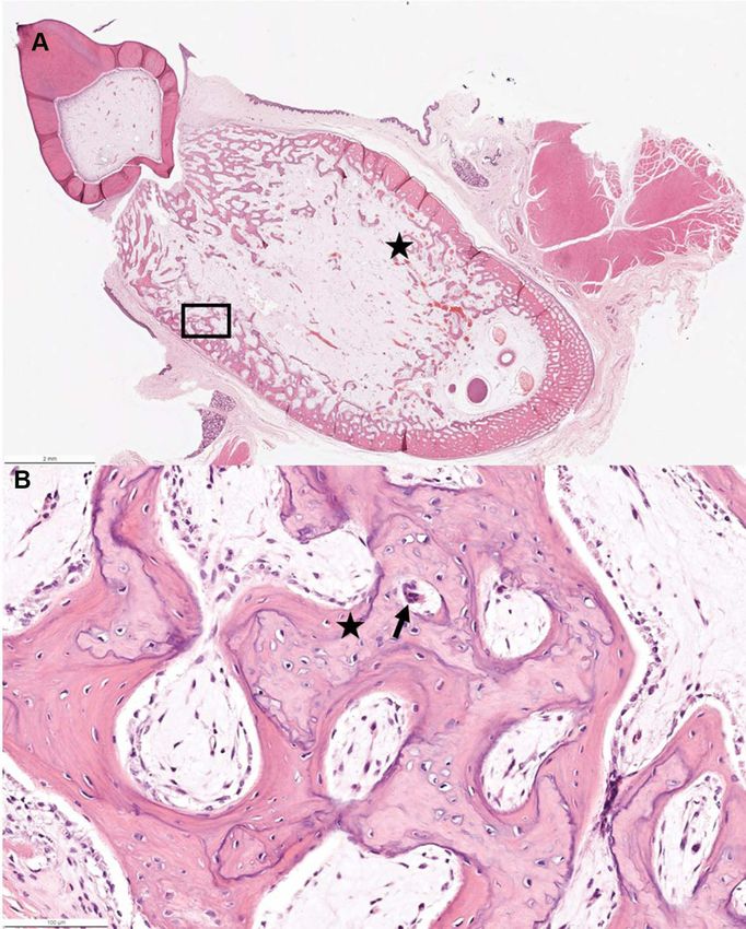
Tse et al Dovepress
autosomal dominant mutations in genes that encode type abnormal radius curvus of the forelimbs, being smaller than
I collagen: COL1A1 (collagen type I alpha 1 chain) and litter mates (weights of normal litter mates could not be
COL1A2 (collagen type I alpha 2 chain).16 obtained), and displaying poor mobility. The puppies were
In animals, OI has been reported in domestic species such part of a litter of six that resulted from a breeding between
as dogs, cats, cattle, and sheep as well as captive tigers.17–28 a sire and female offspring of that sire from a previous litter.
OI is uncommon in veterinary medicine with a majority of On presentation to our clinic, radiographic confirmation of
cases being diagnosed primarily through histopathologic abnormal conformation and folding fractures of multiple
examination with more current reports delving into genetic long bones were identified. Following clinical evaluation
causes. Current, animal model and naturally occurring dis that included a physical examination, complete blood counts,
eases involved in the diagnosis of OI are included in the serum biochemistry, and thyroid hormone testing the puppies
University of Sydney Online Mendelian Inherited Database were humanely euthanized due to poor quality of life and
as a reference for all reported incidences with identified submitted for necropsy and histopathology examination.
genetic causes (https://www.omia.org/home/). In published
reports of dogs, OI is described in the Dachshund, Chow Clinical Summary
Chow, Golden Retriever, Beagle, Collie, Norwegian At three weeks of age the owner identified that the two
Elkhound, Bedlington Terrier, and Lagotto Romagnolo and aforementioned puppies were significantly smaller and less
in one case of a mixed breed litter; with the hallmark findings mobile than the 4 healthier litter mates (Figure 1). It was also
being orthopedic dysfunction leading to minimal trauma or noted by the owner that the teeth appeared to be delayed in
non-traumatic fractures.22–28 In some of these cases the bone eruption from the upper and lower arcade. According to the
pathology is limited to a single long bone, while in other owner the puppies were eating well, showed normal defeca
cases as described in Dachshunds, Collies, and Beagles; tion and urination patterns, and were in good body condi
newborn puppies develop multiple non-traumatic long bone tion, although stunted in growth compared to litter mates. At
fractures and often have delayed growth curves compared to that time based on limited telemedicine examination due to
healthy litter mates and potential hypodontia.22–28 The dis the coronavirus pandemic, a presumed inherited musculos
order can be autosomal dominant or recessive and genetic keletal or neurologic abnormality was postulated.
aberrations have been identified as the SERPINH1 mutation At 8 weeks of age the puppies were admitted to the
in the Dachshund, and the COL1A2 mutation in the Chow Cornell University Hospital for Animals. Historically, the
Chow and Lagotto Romagnolo.25,26,28 puppies continued to grow, but were smaller than litter
This case report describes OI in two 8-week-old male mates with obvious limb deformities in the forelimbs and
Finnish Lapphund puppies that were referred to the Cornell hypomobility. Puppy one weighed 1.2 kg on presentation,
University Hospital for Animals with an initial complaint of while puppy two weighed 1.6 kg, with no significant
Figure 1 Picture showing the stunted growth of puppy one compared to litter mates at 3 weeks of age.
178 https://doi.org/10.2147/VMRR.S308418 Veterinary Medicine: Research and Reports 2021:12
DovePress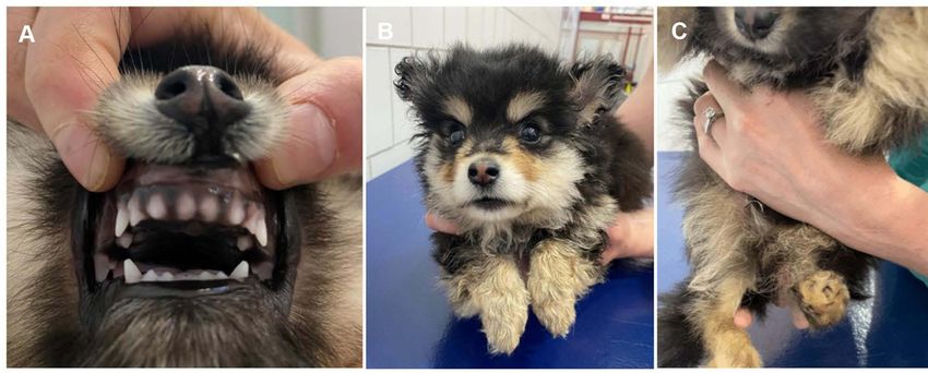
Dovepress Tse et al
Figure 2 (A) Picture showing the normal orientation of teeth but abnormal eruption observed in upper arcade of puppy two at 8 weeks of age. (B) Picture displaying the
carpal valgus orientation of puppy one at 8 weeks of age. (C) Picture displaying the varus deformity on the left hindlimb and contracture of the right hindlimb in puppy two at
8 weeks of age.
findings in their vital parameters on physical exam. During an abnormal finding in young puppies since bone growth
oral examination mild hypodontia and retrognathism was is expected to contribute to a higher serum activity of this
identified in both dogs (Figure 2A). Both puppies dis enzyme.29–31 Both puppies had an increased serum crea
played valgus deformities of the carpus and a limited tine kinase activity which can be normal in puppies of this
range of motion at the elbow and shoulder bilaterally age,29 but may also be elevated due to presumed progres
(Figure 2B). There was profound muscle atrophy in the sive muscle damage. Puppy one displayed an increased
forelimbs and the hindlimbs. On hindlimb manipulation, serum lactate dehydrogenase activity (Table 2).
there was pain during hip extension and knee flexion with
limited range of motion, and puppy two appeared to dis Table 1 Complete Blood Count for Puppy 1 and Puppy 2 at 8
Weeks of Age
play a varus orientation of the right hindlimb with con
tracture of the right hindlimb (Figure 2C). Neurologic Complete Blood Count Puppy 1 Puppy 2 Ref. Range
evaluation found delayed proprioception in all limbs that Hematocrit (%) 34 34 41–58
was deemed to be pain related. Hemoglobin (g/dL) 10.6 10.2 14.1–20.1
Both dogs were intravenously sedated using dexmede Red Blood Cells (mil/uL) 4.6 4.9 5.7–8.5
Mean Corpuscle Volume (fL) 74 69 64–76
tomidine (5 ug/kg) and butorphanol (0.2 mg/kg) for radio
Mean Corpuscle Hemoglobin (pg) 23 21 21–26
graphy and computed tomography imaging. Immediately Mean Corpuscle Hemoglobin conc. 31 30 33–36
following imaging blood was collected for complete blood (g/dL)
counts, serum biochemistry and thyroid hormone screening. Red Cell Distribution and Width (%) 12.6 13.7 10.6–14.3
Reticulocyte Count (%) 2.2 1.6 0.2–1.5
Absolute Reticulocyte Count 102 77.3 11.0–92.0
Clinical Pathology Findings (thou/uL)
Nucleated Red Blood Cells (/100 0 0 0–1
The complete blood count revealed a low red blood cell
WBC)
count and hemoglobin concentration, mild increase in White Blood Cells (thou/uL) 12.7 14.7 5.7–14.2
reticulocytes, and a mild mature neutrophilia in relation Segmented Neutrophils (thou/uL) 10.4 12.2 2.7–9.4
ship to adult reference ranges (Table 1). These findings are Lymphocytes (thou/uL) 1.2 1.2 0.9–4.7
Monocytes (thou/uL) 0.9 0.7 0.1–1.3
documented as normal variation in healthy puppies of
Eosinophils (thou/uL) 0.3 0.1 0.1–2.1
similar age.29–31 The prototypical age-associated biochem Basophils (thou/uL) 0 0 0.0–0.1
ical findings include a mild panhypoproteinemia and Platelets (thou/uL) 616 690 186–545
hyperphosphatemia. Serum alkaline phosphatase activity Mean Platelet Volume (fL) 9.8 11.1 8.4–14.1
was within the adult reference range, and is considered Note: Bolded values are outside of the reference range.
Veterinary Medicine: Research and Reports 2021:12 https://doi.org/10.2147/VMRR.S308418
179
DovePress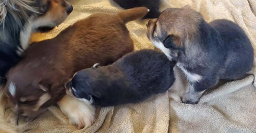
Tse et al Dovepress
Table 2 Serum Biochemistry, Total Serum Thyroid Hormone cared for under the best practices of veterinary care and
(Total T4) and Free Serum Thyroid Hormone (Free T4) Results within the guidelines for the Cornell University Hospital
for Puppy 1 and Puppy 2 at 8 Weeks of Age.
for Animals. The owner signed informed consent regard
Serum Biochemistry Puppy 1 Puppy 2 Ref. ing the euthanasia and provided consent for pathologic
Range examination of tissue to confirm the presumptive diagnosis
Sodium (mEq/L) 141 145 143–150 of OI with permission for presentation and/or publication.
Potassium (mEq/L) 5.8 5.1 4.1–5.4 As these were not research animals, and were client
Chloride (mEq/L) 108 108 106–114 owned, these dogs were exempt from any institutional
Bicarbonate (mEq/L) 23 24 14–24
animal use and care committee approval oversight.
Anion Gap 17 17 16–27
Urea Nitrogen (mg/dL) 12 9 9–26
Creatinine (mg/dL) 0.2 0.2 0.6–1.4 Pathologic Findings
Calcium (mg/dL) 10.3 10.7 9.4–11.1 At necropsy, upon manipulation, all examined bones were
Phosphate (mg/dL) 5.7 6.5 2.7–5.4 brittle and easily fractured when applying minimal force.
Magnesium (mEq/L) 1.9 1.9 1.5–2.1 Light microscopy evaluation of representative sections of
Total Protein (g/dL) 3.8 4 5.5–7.2
the mandible, femur, ribs, humerus, scapula, and entire
Albumin (g/dL) 1.5 2.7 3.2–4.1
skull at the mid-nasal cavity level were performed for
Globulin (g/dL) 1.5 1.3 1.9–3.7
Glucose (mg/dL) 90 105 68–104 both puppies, in addition to all major organs.
Alanine Aminotransferase (U/L) 17 28 17–95 In all sections of femur, humerus, ribs examined the
Aspartate Aminotransferase 34 37 18–56 secondary spongiosa was lacking with the remaining bony
(U/L) trabeculae having frequent reversal lines, rare osteocytes,
Alkaline Phosphatase (U/L) 86 88 7–115
and lined by a layer of small osteoblasts that sometimes
Gamma Glutamyltransferase 3 4 0–8
contained a cytoplasmic vacuole and frequent osteoclasts.
(U/L)
Total Bilirubin (mg/dL) 0 0 0.0–0.2 Loose fibrovascular stroma was present between the tra
Direct Bilirubin (mg/dL) 0 0 0.0–0.1 beculae. In all sections of bone no Haversian canals were
Indirect Bilirubin (mg/dL) 0 0 0.0–0.1 noted in any of the cortices. The cortices had thin, irregu
Amylase (U/L) 405 332 322–1310 lar, woven bone separated by fibrovascular tissue
Lipase (U/L) 224 81 15–228
(Figure 4) and the periosteum was absent (Figures 4 and
Cholesterol (mg/dL) 244 202 136–392
Creatine Kinase (U/L) 592 751 64–314
5). Examination of tooth morphology was normal with
Lactate Dehydrogenase (U/L) 412 369 24–388 subjective thinning of the dentin layer (Figure 5).
Iron (ug/dL) 185 132 97–263
Total T4 (ug/dL) 2.1 2.4 1.5–3.0 Discussion
Free T4 (ng/dL) 1.5 1.1 0.7–2.5 In this study, we clinically and pathologically diagnosed
Note: Bolded values are outside of the reference range. two 8-weeks-old Finnish Lapphund puppies of the same
litter with OI. These two puppies started showing OI
clinical signs within the first 3 weeks of life, similar to
Radiographic Findings the OI cases described in Golden Retrievers, Collies,
Diffusely there was generalized decrease in bone opacity Beagles, Bedlington Terrier, Dachshunds and Lagotto
(diffuse osteopenia) throughout the skeleton of both pup Romagnolos.17,22–25,27,29,32,33 On clinical presentation,
pies identified by the board certified radiologist (IP). The the puppies had valgus orientation of the carpus and one
cortices of all long bones were severely diminished and also had varus orientation on the right hindlimb. There was
multiple “folding” fractures were present with minimal limited range of motion on manipulation and contracture
callus formation (Figure 3A–C). Differential diagnosis of the limbs, a finding commonly observed in human
based on the radiographs included both skeletal dysplasia cases; however, similar perinatal limb deformities can
and metabolic bone disease. When considered with the also be associated with soft tissue or ligamentous
clinical history, a skeletal dysplasia such as OI was con laxity.34 Joint range of motion differs in various manifes
sidered most likely. Due to the multiple fractures and tations of OI, with most human cases presenting with
skeletal deformities associated with the poor prognosis hypomobility as observed in OI type III patients, with
the owner elected humane euthanasia. The dogs were hypermobility observed in OI type I.35 The finding of
180 https://doi.org/10.2147/VMRR.S308418 Veterinary Medicine: Research and Reports 2021:12
DovePressDovepress Tse et al
Figure 3 (A and B) Fractures are present in both the left and right femur (*), the left tibia (folding fracture), questionably the right tibia (>) of puppy two. (C) Folding
fractures are present in both the left and right humerus (*) and radius (>), and deform the shape of the limbs. Note the severe diffuse osteopenia, most severe in the long
bones where there is severe cortical thinning, and in the facial bones of puppy five.
Veterinary Medicine: Research and Reports 2021:12 https://doi.org/10.2147/VMRR.S308418
181
DovePressTse et al Dovepress
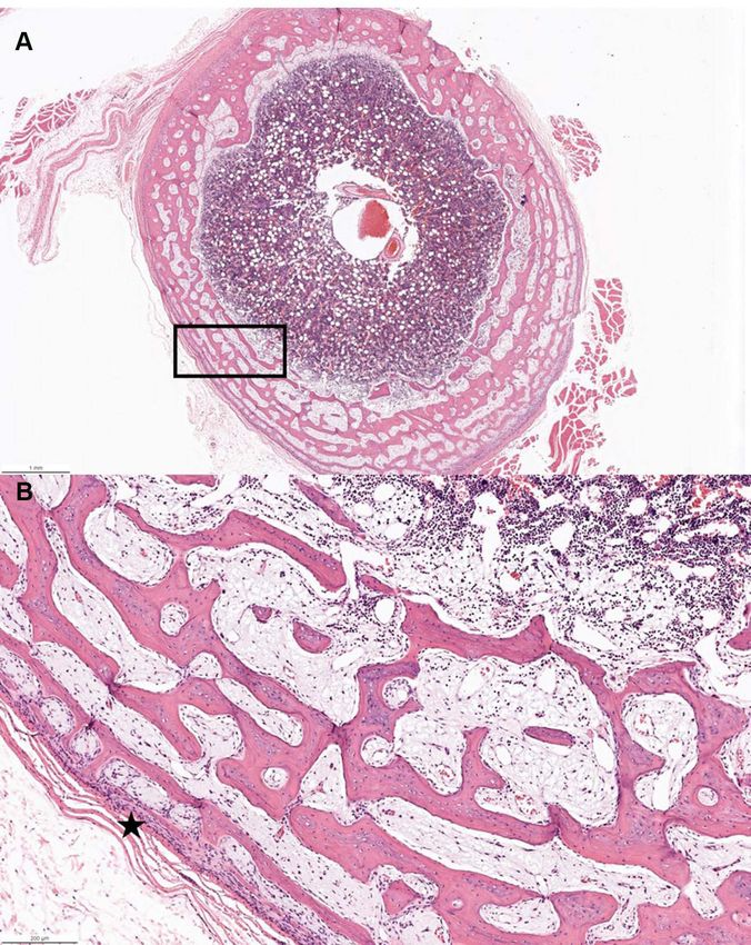
Figure 4 Femur hematoxylin and eosin staining. (A) Transverse section showing marked cortical thinning with abrupt transition from bone to muscle and subjectively
hypercellular bone marrow with a black rectangle to indicate where panel B is examined at higher power (B) Higher magnification of rectangular area showing inapparent
periosteum and markedly thin cortex (*).
profound muscle atrophy in all four extremities in both bone histopathology. Evident osteopenia with
puppies has also been observed in children.36 In addition, minimal to absent cortices which lacked Haversian
OI in children has also been associated with altered struc canals and displayed areas of dense fibrovascular
tural and mechanical properties of deformed tendons and stroma, which is typical of this disorder. In addition,
ligaments.36 Although we could elicit a pain response as there was delayed dentition with the dentin layer
reported in other juvenile forms of the disease, pain being subjectively thin, similar to the OI identified in
responses appear to be variable and may be related to the Dachshunds and other breeds.17,28,36–38 Retrognathism,
extent of the pathological fractures depending on the identified in both puppies, is rare among canine OI
case.17,37 case reports, but not uncommon in humans, suggesting
In addition to the pathological fractures evident on a possible difference in presentation in Finnish
radiographs, the puppies in this report had abnormal Lapphund dogs.39,40
182 https://doi.org/10.2147/VMRR.S308418 Veterinary Medicine: Research and Reports 2021:12
DovePressDovepress Tse et al
Figure 5 Mandible hematoxylin and eosin staining. (A) Traverse section revealing diminished dentin layer of the tooth and marked thinning of cortical bone with paucity of
bony trabeculae (*) with a black rectangle to indicate where panel B is examined at higher power (B) Higher magnification of rectangular area showing irregular bone with
reversal lines (*), lined by small osteoblasts and frequent osteoclasts (arrow).
Complete blood cell count, serum chemistry and thyr Other potential abnormalities in both puppies were
oid hormone testing was performed to assess any abnorm decreases in albumin concentration and rises in creatine
alities that might be related to this disorder, and most kinase activity. Prior literature suggests total protein can
strikingly there was no rise in alkaline phosphatase be low in puppies of this age, so the finding of low
enzyme activity typical of puppies undergoing rapid bone albumin may be an age-related physiological change; how
turnover, while serum phosphorus was elevated which is ever, it is not well documented, except for Labradors and
typically observed in growing dogs.29–31 Red blood cell Schnauzers who show gradual increases from 8–26 weeks
counts and hemoglobin concentrations were low which is of age.31 The finding of elevated serum creatine kinase
commonly observed in puppies due to the rapid turnover activity is likely to be an age-related finding; however,
of red blood cells containing fetal hemoglobin isoforms to progressive muscle damage associated with the chronic
adult isoforms leading to a normal physiological anemia.29 pathological fractures and prolonged recumbency may
Veterinary Medicine: Research and Reports 2021:12 https://doi.org/10.2147/VMRR.S308418
183
DovePressTse et al Dovepress
also be contributing to this elevation. One puppy had an article or revising it critically for important intellectual
elevated lactate dehydrogenase as a marker of anoxic or content; agreed to submit to the current journal; gave
inflamed tissue. The same puppy showed a mild elevation final approval of the version to be published; and agree
in neutrophils suggestive of systemic inflammation, which to be accountable for all aspects of the work.
could be related to the mild elevation in lactate
dehydrogenase.
Disclosure
The parents of the puppies were healthy reproducing
The authors report no conflicts of interest in this work.
adults. The pedigree of the puppies was consistent with
a monogenic autosomal recessive inheritance and the ratio
of the affected Finnish Lapphund puppies from the pre References
sumed carrier × carrier breeding in light of the father to 1. Arponen H, Mäkitie O, Waltimo-Sirén J. Association between joint
hypermobility, scoliosis, and cranial base anomalies in paediatric
daughter breeding in this particular case. However, given Osteogenesis imperfecta patients: a retrospective cross-sectional
the small number of puppies within the litter, other modes study. BMC Musculoskelet Disord. 2014;15:428. doi:10.1186/1471-
of inheritance cannot be ruled out. OI in dogs is reported 2474-15-428
2. Kang H, Aryal ACS, Marini JC. Osteogenesis imperfecta: new genes
to be caused by autosomal dominant and recessive modes reveal novel mechanisms in bone dysplasia. Transl Res.
of inheritance with gene mutations in COL1A1 in a Golden 2017;181:27–48. doi:10.1016/j.trsl.2016.11.005
3. Lim J, Grafe I, Alexander S, Lee B. Genetic causes and mechanisms
Retriever,41 COL1A2 in a beagle and a Chow Chow,28,29 of osteogenesis imperfecta. Bone. 2017;102:40–49. doi:10.1016/j.
and SERPINH1 in Dachshunds.42 COL1A1 and COL1A2 bone.2017.02.004
code for α1 and α2 collagen chains, associated with the 4. Marini JC, Forlino A, Bachinger HP, et al. Osteogeneis imperfect.
Nat Rev Dis Primers. 2017;3:17058–17072. doi:10.1038/nrdp.20
production, folding, and maintenance of the structure and 17.52
secretion of type 1 collagen.43 SERPINH1 encodes for heat 5. Monti E, Mottes M, Fraschini P, et al. Current and emerging treat
ments for the management of osteogenesis imperfecta. Ther Clin Risk
shock protein 47 (HSP47), an essential collagen chaperone Manag. 2010;6:367–381. doi:10.2147/tcrm.s5932
protein which aids in appropriate folding of collagens and 6. Lindahl K, Langdahl B, Ljunggren Ö, et al. Treatment of osteogen
stabilizes collagen triple helices.26 In both the Golden esis imperfecta in adults. Eur J Endocrinol. 2014;71:R79–90.
doi:10.1530/EJE-14-0017
Retriever and Beagle there was no pedigree information 7. Lamanna A, Fayers T, Clarke S, et al. Valvular and aortic diseases in
to fully understand the COL1A1 and COL1A2 mutations osteogenesis imperfecta. Heart Lung Circ. 2013;22:801–810.
doi:10.1016/j.hlc.2013.05.640
identified, respectively.23,41 The SERPINH1 mutation 8. McKiernan FE. Musculoskeletal manifestations of mild osteogenesis
observed in German Dachshunds,42 involved similar F1 imperfecta in the adult. Osteoporos Int. 2005;16:1698–1702.
to F2 breeding characteristics and clinical presentations doi:10.1007/s00198-005-1905-5
9. Charnas LR, Marini JC. Communicating hydrocephalus, basilar inva
are similar to the puppies in this case report suggesting gination, and other neurologic features in osteogenesis imperfecta.
this may be a fruitful area for future research. At this time Neurology. 1993;43:2603–2608. doi:10.1212/WNL.43.12.2603
10. Tsipouras P, Barabas G, Matthews WS. Neurologic correlates of
we have not elucidated the potential mutations or modes of osteogenesis imperfecta. Arch Neuro. 1986;43:150–152. doi:10.10
inheritance in the Finnish Lapphund. 01/archneur.1986.00520020044016
Based on our pathologic and radiographic findings, the 11. Sasaki-Adams D, Kulkarni A, Rutka J, Dirks P, Taylor M, Drake JM.
Neurosurgical implications of osteogenesis imperfecta in children:
Finnish Lapphund is another identified breed with OI. report of 4 cases. J Neurosurg Ped. 2008;1:229–236. doi:10.3171/
Further genetic studies are warranted since communication PED/2008/1/3/229
12. Tauer JT, Robinson ME, Rauch F. Osteogenesis imperfecta: new
with the breed group has identified other European bred perspectives from clinical and translational research. JBMR Plus.
dogs with OI. Clinical presentation as early as 3 weeks of 2019;3(8):e10174. doi:10.1002/jbm4.10174
age can be suggestive of OI and clinical pathology find 13. Biggin A, Munns CF. Osteogenesis imperfecta: diagnosis and
treatment. Curr Osteoporos Rep. 2014;12:279–288. doi:10.1007/
ings are typical for puppies of this age with the only s11914-014-0225-0
abnormal finding related to bone metabolism being 14. Warman ML, Cormier-Daire V, Hall C, et al. Nosology and classifi
cation of genetic skeletal disorders: 2010 revision. Am J Med Gen
a normal alkaline phosphatase activity which could be Part A. 2011;155:943–968. doi:10.1002/ajmg.a.33909
a prognostic indicator for suspicion of OI. Veterinary 15. Mortier GR, Cohn DH, Cormier-Daire V, et al. Nosology and classi
fication of genetic skeletal disorders: 2019 revision. Am J Med Gen
community awareness is prudent for further investigation
Part A. 2019;179:2393–2419. doi:10.1002/ajmg.a.61366
into the genetics of this disorder. 16. Rauch F, Glorieux FH. Osteogenesis imperfecta. Lancet.
Author contribution: All authors made substantial con 2004;363:1377–1385. doi:10.1016/S0140-6736(04)16051-0
17. Seeliger F, Leeb T, Peters M, et al. Osteogenesis Imperfecta in Two
tributions to conception and design, acquisition of data, or Litters of Dachshunds. Vet Path. 2003;40:530–539. doi:10.1354/
analysis and interpretation of data; took part in drafting the vp.40-5-530
184 https://doi.org/10.2147/VMRR.S308418 Veterinary Medicine: Research and Reports 2021:12
DovePressDovepress Tse et al
18. Scott PP, McKusich VA, McKusick AB. The nature of osteogenesis 31. Brenten T, Morris PJ, Salt C, et al. Age-associated and
imperfecta in cats: evidence that the disorder is primarily nutritional, breed-associated variations in haematological and biochemical vari
not genetic, and therefore not analogous to the disease in man. J Bone ables in young Labrador retriever and miniature schnauzer dogs. Vet
Joint Surg. 1963;45:125–134. doi:10.2106/00004623-196345010- Rec Open. 2016;3:e000166. doi:10.1136/vetreco-2015-000166
00010 32. Dammrich K. Osteoporoses in young animals. Pathol Vet.
19. Jensen PT, Rasmussen PG, Basse A. Congenital osteogenesis imper 1967;4:435–463. doi:10.1177/030098586700400502
fecta in Charollais cattle. Nordisk Veterinaermedi. 1976;28:304–308. 33. Rowe DW, Shapiro JR. Osteogenesis imperfecta. In: Avioli LV,
20. Arthur DG, Thompson KG, Swarbrick P. Lethal osteogenesis imper Krane SM, editors. Metabolic Bone Disease. 3rd ed. London, UK:
fecta and skin fragility in newborn New Zealand Romney lambs. NZ Academic Press; 1998:651–695.
Vet J. 1992;40:112–116. doi:10.1080/00480169.1992.35712 34. Wilson DA. Angular Limb Deformity. In: Clinical Veterinary Advisor
21. Horvath SA, Francesetti FL, Riveros SV. Treatment of imperfect the Horse. St. Louis, Missouri, Estados Unidos: Elsevier/Saunders;
osteogenesis in a tiger (Panthera tigris). Avances En Ciencias 2013:30–31.
Veterinarias (Chile). 1986;1:49–51. 35. Engelbert RH, Graaf YV, Empelen RV, et al. Osteogenesis Imperfecta
22. Campbell BG, Wootton JA, Krook L, DeMarco J, Minor RR. Clinical in Childhood: impairment and Disability. Pediatrics. 1997;99
signs and diagnosis of osteogenesis imperfecta in three dogs. J Am (E3):1–7. doi:10.1542/peds.99.2.e3
Vet Med Assoc. 1997;211:183–187. 36. Veilleux LN, Trejo P, Rauch F. Muscle abnormalities in osteogenesis
23. Lettow E, Dammrich K. Clinic and pathology of osteogenesis imper imperfecta. J Musculoskel Neuro Inter. 2017;17:1–7.
fecta in young dogs. Zbl Vet Med. 1960;7:936–966. 37. Costa PP, Custodio J, Ebina FS, et al. Osteogenesis imperfect in an
24. Dehaan JJ, Peck JN, Campbell B, et al. Osteogenesis imperfecta in young pinscher dog. Acta Scientiae Veterinariae. 2018;46
three dogs from a single litter. Vet Comp Ortho Traum. (Supplement).
2000;13:23–27. doi:10.1055/s-0038-1632625 38. Gold R, Pool RR, Edwards EE. Osteogenesis and dentinogenesis
25. Quist EM, Doan R, Pool RR, et al. Identification of a candidate imperfecta in a four-month-old English mastiff. Vet Rec Case Rep.
mutation in the COL1A2 gene of a chow chow with osteogenesis 2019;7(3):e000835. doi:10.1136/vetreccr-2019-000835
imperfecta. J Heredity. 2017;109:308–314. doi:10.1093/jhered/ 39. Aizenbud D, Peled M, Figueroa AA. A combined orthodontic and
esx074 surgical approach in osteogenesis imperfecta and severe class iii
26. Lindert U, Weis MA, Rai J, et al. Molecular consequences of the
malocclusion: case report. J Oral Maxillofac Surg. 2008;66:
SERPINH1/HSP47 mutation in the dachshund natural model of
1045–1053. doi:10.1016/j.joms.2008.01.010
osteogenesis imperfecta. J Biol Chem. 2015;290(29):17679–17689.
40. Marshall C, Lopez J, Crookes L, Pollitt RC, Balasubramanian M.
doi:10.1074/jbc.M115.661025
A novel homozygous variant in SERPINH1 associated with a severe,
27. Campbell BG, Wootton JA, Macleod JN, Minor RR. Canine
lethal presentation of osteogenesis imperfecta with hydranencephaly.
COL1A2 mutation resulting in c-terminal truncation of Pro-α2(I)
Gene. 2016;95:49–52. doi:10.1016/j.gene.2016.09.035
and severe osteogenesis imperfecta. J Bone Min Res. 2001;16:
41. Campbell BG, Wooton JA, MacLeod JN, Minor RR. Sequence of
1147–1153. doi:10.1359/jbmr.2001.16.6.1147
normal canine COL1A1 and identification of a heterozygous alpha1
28. Letko A, Zdora I, Hitzler V, et al.A de novo in-frame duplication in
(I) collagen Gly208Ala nutation in a severe case of osteogenesis
the COL 1A2 gene in a Lagotto Romagnolo dog with osteogenesis
imperfecta. Anim Genet. 2019;(50):786–787. doi:10.1111/age.12843 impferfecta. Arch Biochem Biophys. 2000;384:37–46. doi:10.1006/
29. Rosset E, Rannou B, Casseleux G, Chalvet-Monfray K, Buff S. Age- abbi.2000.2099
related changes in biochemical and hematologic variables in Borzoi 42. Drogemuller C, Becker D, Brunner A, et al. A missense mutation in
and Beagle puppies from birth to 8 weeks. Vet Clin Pathol. the SERPINH1 gene in Dachshunds with osteogenesis imperfecta.
2012;41:272–282. doi:10.1111/j.1939-165X.2012.00415.x PLoS Genet. 2009;5:e1000579. doi:10.1371/journal.pgen.1000579
30. Harper EJ, Hackett RM, Wilkinson J, Heaton PR. Age-related varia 43. Craig LE, Dittmer KE, Thompson KG. Bone and Joints. In:
tions in hematologic and plasma biochemical test results in Beagles Maxie MG, editor. Jubb, Kennedy, and Palmer. Pathology of
and Labrador Retrievers. J Am Vet Med Assoc. 2003;223:1436–1442. Domestic Animals. 6th ed. Missouri: Elsevier; 2016:157–163.
doi:10.2460/javma.2003.223.1436
Veterinary Medicine: Research and Reports Dovepress
Publish your work in this journal
Veterinary Medicine: Research and Reports is an international, peer- and includes a very quick and fair peer-review system. Visit
reviewed, open access journal publishing original research, case http://www.dovepress.com/testimonials.php to read real quotes from
reports, editorials, reviews and commentaries on all areas of veterinary published authors.
medicine. The manuscript management system is completely online
Submit your manuscript here: http://www.dovepress.com/veterinary-medicine-research-and-reports-journal
Veterinary Medicine: Research and Reports 2021:12 DovePress 185You can also read