Juvenile Idiopathic Arthritis in Harlequin Ichthyosis, a Rare Combination or the Clinical Spectrum of the Disease? Report of a Child Treated With ...
←
→
Page content transcription
If your browser does not render page correctly, please read the page content below
Juvenile Idiopathic Arthritis in Harlequin Ichthyosis,
a Rare Combination or the Clinical Spectrum of the
Disease? Report of a Child Treated With Etanercept
and Review of the Literature
Francesco Baldo
Fondazione IRCCS Cà Granda Ospedale Maggiore Policlinico
Michela Brena
Fondazione IRCCS Cà Granda Ospedale Maggiore Policlinico
Simone Carbogno
Fondazione IRCCS Cà Granda Ospedale Maggiore Policlinico
Francesca Minoia
Fondazione IRCCS Cà Granda Ospedale Maggiore Policlinico
Stefani Lanni
Fondazione IRCCS Cà Granda Ospedale Maggiore Policlinico
Sophie Guez
Fondazione IRCCS Cà Granda Ospedale Maggiore Policlinico
Antonella Petaccia
Fondazione IRCCS Cà Granda Ospedale Maggiore Policlinico
Carlo Agostoni
Fondazione IRCCS Cà Granda Ospedale Maggiore Policlinico
Rolando Cimaz
ASST Gaetano Pini: Azienda Socio Sanitaria Territoriale Gaetano Pini
Giovanni Filocamo ( giovanni.filocamo@policlinico.mi.it )
Pediatric Rheumatology, Pediatric Medium Intensity Care Unit Fondazione IRCCS Cà Granda, Ospedale
Maggiore Policlinico Clinica De Marchi Via della Commenda, 9 20122 Milano https://orcid.org/0000-
0002-9638-6512
Case Report
Keywords: Juvenile Idiopathic Arthritis, Harlequin Ichthyosis, Intra-articular corticosteroids Injection,
Etanercept
Posted Date: October 7th, 2020
Page 1/15DOI: https://doi.org/10.21203/rs.3.rs-84750/v1
License: This work is licensed under a Creative Commons Attribution 4.0 International License.
Read Full License
Version of Record: A version of this preprint was published at Pediatric Rheumatology on June 3rd, 2021.
See the published version at https://doi.org/10.1186/s12969-021-00571-9.
Page 2/15Abstract
Background. Harlequin ichthyosis (HI) is the most severe phenotype of autosomal recessive congenital
ichthyosis. Juvenile Idiopathic Arthritis (JIA) represents a heterogenous group of disorders all sharing the
clinical manifestation of chronic arthritis. Association of HI and chronic arthritis has been reported in few
cases.
Case presentation. We report the case of a child HI who developed a severe form of chronic polyarthritis
during the first years of life, treated with repeated multiple joint injections, methotrexate and etanercept
with good response and any adverse events.
Conclusion. The case reported, followed by a review of the literature, could suggest the possibility that the
presence of this peculiar severe polyarthritis with early onset and FR / ANA negativity, could be part of the
clinical manifestation of Harlequin Ichthyosis instead that a rare combination of two diseases as
reported by previous papers.
Introduction
Harlequin ichthyosis (HI) is the most severe phenotype of autosomal recessive congenital ichthyosis, a
rare heterogeneous group of recessively inherited ichthyosis which encompasses a wide range of clinical
phenotypes. [1,2]
Harlequin Ichthyosis (HI) is due to homozygous nonsense mutations in the ABCA12 (ATP binding
cassette subfamily A member 12) gene.[3] Newborns with HI present with a distinct clinical appearance
encased in a dense, armor-like skin separated by polygonal deep erythematous fissures that simulate the
traditional costume of a harlequin. Facial features are distorted by severe ectropion, eclabium, flattened
nose, and rudimentary ears. [1,4,5] The skin rigidity can restrict respiratory movements, the hands and
feet are ischemic, often with associated poor developed digits with claw-like appearance and osseous
reabsorption; flexion deformity of the limb joints is common. Developmental delay is frequently described
as well.[6] Skin barrier function is markedly impaired, which can lead to hypernatremic dehydration,
impaired thermoregulation, increased metabolic demands, and increased risk of respiratory dysfunction
and infection, which may cause premature death within the first days to weeks of life.[7] Historically,
infants with HI did not survive beyond the neonatal period; however, prolonged survival has been
achieved by intensive supportive measures, emollients and, in some cases, oral administration of
systemic retinoids. The cuirass of the survivors fades within 2-3 months and they subsequently develop
an erythematous, scaly, very severe ichthyotic pattern with ectropion, abnormal external ears and
alopecia. [8] ABCA12 encodes for the ATP-Binding Cassette A12, a membrane associated keratinocyte-
specific protein whose function is to transfer specific lipids into the lamellar granules, which are then
processed and secreted into the stratum corneum (SC) to form lipid lamellae.[8] ABCA12 mutations lead
to disorders in the distribution and transport of glycosylceramides and decreased levels of
hydroxyceramides, one of the main components of the lipid barrier in the SC. ABCA12 deficiency results
Page 3/15in hyperkeratosis and premature terminal differentiation of keratinocytes, as well as lack of
desquamation of the corneocytes, due to transport defects of specific proteases, such as callicrein 5 and
cathepsin D.[9-12] The type of ABCA12 mutation has a major impact on the severity of the disease, which
is due to homozygous nonsense mutations with absent or minimal residual ABCA12 function. [3]
Juvenile Idiopathic Arthritis (JIA) is the most common chronic rheumatic disease of childhood. It
represents a heterogenous group of disorders all sharing the clinical manifestation of chronic arthritis.
[13] Different disease subtypes are classically recognized, ranking from systemic arthritis, in which
systemic manifestation are present, to other forms that are mainly characterized by joint involvement
(oligoarticular and polyarticular forms), to spondyloarthropathy-like forms (enthesitis-related arthritis and
psoriatic arthritis).[14] Patients with positivity for anti-nuclear antibodies (ANA) are at risk to develop
chronic anterior non-infectious uveitis.[15] Nonsteroidal anti-inflammatory drugs (NSAIDs), intra-articular
corticosteroids injections (IACI), systemic steroids and conventional and biologic disease-modifying
antirheumatic drugs (DMARDs) are well-known medications used to treat different subtypes of JIA.[16,17]
Due to the rare nature of both diseases, association of HI and chronic arthritis has been reported in few
other cases. We report the case of a child born with HI who developed a severe form of chronic
polyarthritis from 4 years of age. Furthermore, we offer a literature review on this topic.
Case Report
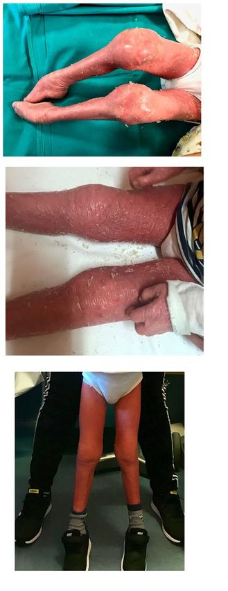
A 7-year-old boy with HI (homozygous mutation c.541 C>T in ABCA12 gene) presented to our pediatric
rheumatology clinic due to pain, swelling and joint stiffness. In the first years of life, he suffered from
recurrent sepsis, caused by different bacteria such as Staphilococcus aureus and Stenotrophomonas
maltophilia. At the time of the examination, he had flexion contractures of fingers and of large joints of
the lower limbs (Figure 1a). He showed severe developmental delay, and he had periodical ear-nose-and-
throat, ophthalmological and dermatological follow-up. The child was on treatment with systemic
retinoids from birth.
The mother reported a 3 year-history of chronic polyarthritis involving all the large joints with severe
limitation on motion, particularly of both ankles, and causing inability to walk. Laboratory investigations
were normal except for the presence of microcytic anemia and a mild increase in C-reactive protein (CRP).
ANA, anti-extractable nuclear antigen (ENA), anti-cyclic citrullinated peptide (CCP) antibodies and
rheumatoid factor (RF) were negative. An ophthalmology examination did not show uveitis.
According to the International League of Associations for Rheumatology (ILAR) classification, [18] the
clinical features were consistent with polyarticular RF negative JIA . The boy underwent multiple IACI
(triamcinolone hexacetonide, for large joints and methylprednisolone acetate for small joints). A second
line treatment with oral methotrexate was also started. (Figure 1b)
Three months later, owing to the presence of persistently active joint disease to the ankles, IACI were
repeated in active joints.
Page 4/15However, he did not reach complete remission of arthritis. Therefore, eight months after the beginning of
methotrexate, a third IACI was performed and a Tumor Necrosis Factor antagonist was started
(etanercept), which allowed to reach a status of minimal disease activity two months later.[19]
Appropriate physical therapy was started, and articular functional ability improved in the following
months, allowing the child to walk again. Improvement of skin erythema was observed as well. Eighteen
months after beginning etanercept treatment, the patient was only in minimal disease activity; improved
articular functional ability has been achieved and no medication side effects were reported (Figure 1c).
Methotrexate was tapered to every other week administration.
Case Review
Development of JIA in HI patients has already been described. Information about family history, genetics
and treatment of the different cases is shown in Table 1.
The first case of HI and arthritis was reported by Chan et al. [20], who described a female newborn of
2250 g, delivered at 37 weeks of gestational age who showed classical HI appearance at birth and was
admitted to neonatal intensive care unit (NICU), treated with intravenous and topical antibiotics for skin
infections. Oral etretinate at a dose of 1 mg/kg/day was initiated at the age of 36 days (replaced with
acitretin at 7 years of age). At 6 years, she developed swelling of the wrists and ankles as well as of small
joints of the hands, with radiologic evidence of erosive arthritis. She received prednisolone, and ibuprofen
at a dose of 30 mg/kg till she was 11 years of age, when it was replaced by rofecoxib. At 9 years of age,
because of inadequate disease control, methotrexate (MTX) was added to therapy, which led to
improvement in erythroderma as reported by parents.
Auriti et al. described two other cases.[21] The first one was male, delivered via caesarean section (CS) at
35 weeks because of preterm prelabour rupture of membranes (pPROM), with a birth weight of 2300 g
and admitted to NICU. Retinoid treatment was started in the first day of life and stopped after 10 days.
During his early life, he needed multiple red blood cells and platelet transfusions due to severe anemia,
and several intravenous antibiotic courses for recurrent sepsis. At 3 months of life, he developed severe
knee swelling, which was, the first time, thought to be due to septic arthritis, but never completely resolved
after antibiotic treatment. Laboratory tests showed mild increase in CRP and erythrocyte sedimentation
rate (ESR); both ANA and RF were negative. He presented multiple flares of knee arthritis, unsuccessfully
treated with oral NSAIDs. At the age of two he underwent IACI, started physiotherapy and achieved
complete remission.
The second case was a male, born late preterm, delivered via CS. At birth, he showed persistent
bradycardia, hypotonia and generalized cyanosis, and needed a 30" cardiopulmonary resuscitation. He
was born showing collodion baby feature with thickened and fissured skin, flexion contractures of upper
and lower limbs and collodion like membrane all over the body. He was admitted to NICU, and developed
Enterococcus faecalis sepsis and bronchitis. Acitretin treatment was started in early life for 1 month, then
Page 5/15stopped because of liver toxicity. He was also given physiotherapy rehabilitation program for congenital
contractures.
At 2 years of life, he developed severe hands, right knee, elbows, wrists and tibiotalar, subtalar and
talonavicular joint arthritis associated with diffuse tenosynovitis of fingers, wrists, ankles. The child lost
walking capabilities because of severe polyarthritis. Elevation of white blood cells, eosinophils, IgE, CRP
and ESR, while RF was negative. The child was first treated with ibuprofen and then received multiple IACI
and weekly MTX. Clinical and radiological resolution was achieved, except for persistent tenosynovitis of
both ankles.
Clement et al. described another case of a term male newborn with classical HI appearance at birth, who
received treatment with acitretin. At 10 years, he developed severe arthritis in multiple joints. Blood tests
showed microcytic hypochromic anemia and raised CRP levels; ENA, anti-double strained DNA and RF
were negative. Initially, he received NSAIDs without clinical improvement, then he was shifted to
Etoricoxib with temporary response. The patient was then given MTX and Etanercept at 25 mg/week with
improvement in mobility, but he needed total hip arthroplasty by the age of 17. [22] He was then given
adalimumab, MTX and leflunomide, but reported worsening of the cutaneous disease with cracks and
increased skin infections with all previous DMARDs and biologics.
In a case series of 45 patients with molecular diagnosis of HI, Rajpopat et al. [6] report two more cases of
children with signs of arthritis: one showed bone erosions on radiographs, the other intermittent swelling
of the knee. The first has probably more recently been described also by Raghuvanushi et al. [23]: the
patient was referred to the musculoskeletal service because of a long-term condition of painful elbows,
shoulders, fingers, and locking of wrists. Conventional radiography documented destructive alterations of
wrists with subluxation of proximal row of carpal bones. A magnetic resonance imaging (MRI) scan of
shoulders revealed rotator cuff tendinopathy and small joint effusion without synovitis. The patient was
seronegative and is managed with IACI and physical therapy rehabilitation. No further details are
available for the second case.
Discussion
The association of HI and arthritis has been reported in literature, although to date few cases have been
described. It is unclear whether the concomitant arthritis can be regarded as JIA or whether it represents a
different entity with unique features to considered as part of HI itself. The longer survival of HI patients
may increase the number of patients who may develop arthritis throughout the disease course, and will
increase the probability to better understand in the future the best management of joint involvement
In the published cases, arthritis often showed sometimes a rather aggressive course, leading to joint
erosions and even to early joint replacement. Most patients showed early arthritis onset. Acute phase
reactants were usually only slightly increased, while ANA, FR and ENA were found to be negative.
Response to IACI was generally good, although temporary, and response to conventional and biologic
DMARS was variable.
Page 6/15In JIA immune cells, including T and B lymphocytes, infiltrate the synovial membrane of inflamed joints,
suggesting that the adaptive immune system is involved in the pathogenesis of the disease. [24] Recently,
a particular T lymphocyte population, Th17 cells, has been found to be crucial in JIA pathogenesis:
tumor necrosis factor (TNF)-α inhibitors are the main biological drugs used in JIA and interfere with these
cells, giving an explanation of efficacy of etanercept treatment in JIA. [25]
Harlequin Ichthyosis is characterized by a profound dysregulation of lipid secretion into the stratum
corneum and skin barrier function. Currently, therapy is aimed to treatment of scaling, using keratolytics
such as urea,[26] or long-term retinoids, [27] which may negatively affect bone density, [28] and can
generate skin erosion and thinning leading to further epidermal barrier function impairment. [29]
Retinoids have also been shown to cause arthropathy, but this typically presents in the axial skeleton as
ossification along the anterior longitudinal ligament and as pelvic hyperostosis.
As already found in other dermatoses such as atopic dermatitis and psoriasis, cytokine dysregulation and
barrier impairment are both factors that underpin the disease. According to that, psoriasis-like immune
dysregulation and lipid alterations seems to characterize the ichthyoses. [30] In different ichthyoses, high
levels of IL-17 and TNF-α cytokine was documented. [31,32] This is accompanied by increased systemic
and skin-homing T-cell activation and multicytokine polarization, with IL-17/IL-22 polarization
predominance. [33] Ustekinumab, a monoclonal antibody directed against IL-23, was able to reduce skin
erythema, scaling, and Trans Epidermal Water Loss (TEWL), a skin barrier functional index, in two
patients with an ichthyotic syndrome. [31] To investigate the therapeutic potential of IL-17 targeting
drugs, a clinical trial of secukinumab (anti-IL17-antibody) in patients with ichthyoses (NCT03041038), is
currently ongoing.
Cutaneous and systemic immune inflammation has been well studied and successfully treated in other
skin diseases such as psoriasis and atopic dermatitis. [33] However, inflammation in ichthyosis is little
considered. In psoriasis, biologic treatment with IL-17 antagonist is reported as highly effective in
reversing the inflammation and the cutaneous disease. [31] Of interest, the role of anti- TNF-α treatment in
psoriasis and psoriatic arthritis is already well documented. [34]
Conventional DMARDs in patients with HI and chronic inflammatory arthritis have already been used
without significant side effects. In a single case report worsening of erythroderma and increased
cutaneous infections were reported, with poor disease control even using MTX and a biologic agent in
combination.[22] In our patient, the severity of arthritis required the association of conventional and
biological DMARDs with improvement of arthritis and decrease of erythema, without any infectious
events reported at 18 months follow-up, nor any other adverse events.
The role of other proinflammatory cytokines and other biology treatments in ichthyosis have been
proposed. Interleukine (IL)1 alpha is constitutively expressed in the upper epidermis [35,36], as is its
receptor [37], and the soluble decoy receptor (IL1RA) [38,39], and a fine balance in expression of IL1 alpha
and IL1RA has to be granted in healthy skin. [40] Some studies suggest that up-regulation of IL-1 is
common to all Autosomal Recessive Congenital Ichthyosis, and the entity of this upregulation is related
Page 7/15to clinical severity. An in vitro study on disease-mimic organotypic cultures treated with IL-1 receptor
antagonist was beneficial on hyperkeratosis in a dose-dependent fashion. [41]
JIA is now thought to be a multifactorial pathology, in which genetic susceptibility meets an
environmental trigger, leading to an uncontrolled response toward putative self- antigens. [42]
Some of the environmental triggers that have been studied as risk factors for the development of arthritis
are frequently reported in HI patients.[43] Infections during the first year of life were associated with
increased risks for seronegative JIA.[44] Extensive antibiotic use is also common in the early life of these
patients. Antibiotics were associated with JIA development in a large pediatric population, and could play
a role in JIA pathogenesis by acting through microbiome disruption. [45] Prematurity and caesarian-
section delivery are frequent in patients with HI. Some studies have proposed that unlabored C-sections
may slightly increase the risk of JIA due to changes in newborn microbiota and immune response. [44,46]
Conclusion
Few cases of HI have been reported and survival through the early life is still a hard challenge for families
and clinicians alike. However with increasing in survival of patients with HI, further disease related
complications, leading to a decrease in patients’ quality of life may become more frequent.
In our patient HI has been found to be associated to highly aggressive, erosive arthritis and this is in
keeping with the cases previously reported in literature. Although the exact pathogenetic mechanism
underlying HI-associate arthritis is still to be clarified, a prominent role of inflammation has been
demonstrated in HI. This can also drive the choice of a common therapeutic target for both arthritis and
some features of the cutaneous disease in patients with HI itself.
No resolutive therapy exists, to date, for HI, even if experimental gene therapies are in preclinical
development. [47] Together with conventional therapies, a role of anti-inflammatory drugs, and especially
of biologic therapy in patients with severe congenital forms of ichthyosis may be investigated.
List Of Abbreviations
ANA: anti-nuclear antibodies
CCP: cyclic citrullinated peptide
CRP: C Reactive Proteine
CS: caesarean section
DMARDs: disease-modifying antirheumatic drugs
ENA: anti-extractable nuclear antigen
Page 8/15ESR: erythrocyte sedimentation rate
HI: Harlequin ichthyosis
IACI: intra-articular corticosteroids injections
IL: Interleukine
ILAR: International League of Associations for Rheumatology
JIA: Juvenile Idiopathic Arthritis
MRI: magnetic resonance imaging
MTX: methotrexate
NICU: neonatal intensive care unit
NSAIDs: Nonsteroidal anti-inflammatory drugs
pPROM: preterm prelabour rupture of membranes
RF: rheumatoid factor
SC: stratum corneum
Declarations
Ethics approval: N/A
Consent to participate: N/A
Consent for publication: Consent for publication was obtained from the child parents.
Availability of data and materials
The datasets used during the current study and the informed consent for publication are available from
the corresponding author on reasonable request.
Competing interests
The authors declare that they have no competing interests
Funding
N/A
Page 9/15Authors' contributions
FB and SC contributed to acquisition of the data and draft the manuscript, MB and FM contributed to the
revision of the literature and draft the manuscript, SG and MB contributed to the clinical and
dermatological management of the patient, SL and GF contributed to performing, analyzing and
documenting the intraarticular joint injections and therapeutic decision; AP, SG, CA, and RC performed
important contribution to the revision of the manuscript. All authors approved the final submitted version
of the manuscript.
References
1. Glick JB, Craiglow BG, Choate KA, et al. Improved Management of Harlequin Ichthyosis with
advances in neonatal intensive care. Pediatrics. 2017;139:e20161003.
2. Tadini G, Brena M, Gelmetti C, Pezzani L (2015) Ichthyoses. Atlas of Genodermatoses
3. Kelsell DP, Norgett EE, Unsworth H, et al. Mutations in ABCA12 underlie the severe congenital skin
disease harlequin ichthyosis. Am J Hum Genet. 2005;76(5):794-803. doi:10.1086/429844
4. Vahlquist A, Fischer J, Törmä H. Inherited Nonsyndromic Ichthyoses: An Update on Pathophysiology,
Diagnosis and Treatment. Am J Clin Dermatol. 2018;19(1):51-66. doi:10.1007/s40257-017-0313-x
5. Rodríguez-Pazos L, Ginarte M, Vega A, Toribio J. Autosomal recessive congenital ichthyosis. Actas
Dermosifiliogr. 2013;104(4):270-284. doi:10.1016/j.adengl.2011.11.021
6. Rajpopat S, Moss C, Mellerio J, et al. Harlequin ichthyosis: a review of clinical and molecular findings
in 45 cases. Arch Dermatol. 2011;147(6):681-686. doi:10.1001/archdermatol.2011.9
7. Harvey HB, Shaw MG, Morrell DS. Perinatal management of harlequin ichthyosis: a case report and
literature review. J Perinatol. 2010;30(1):66-72. doi:10.1038/jp.2009.100
8. Sakai K, Akiyama M, Sugiyama-Nakagiri Y, McMillan JR, Sawamura D, Shimizu H. Localization of
ABCA12 from Golgi apparatus to lamellar granules in human upper epidermal keratinocytes. Exp
Dermatol. 2007;16(11):920-926. doi:10.1111/j.1600-0625.2007.00614.x
9. Zuo Y, Zhuang DZ, Han R, et al. ABCA12 maintains the epidermal lipid permeability barrier by
facilitating formation of ceramide linoleic esters. J Biol Chem. 2008;283(52):36624-36635.
doi:10.1074/jbc.M807377200
10. Zhang L, Ferreyros M, Feng W, et al. Defects in Stratum Corneum Desquamation Are the Predominant
Effect of Impaired ABCA12 Function in a Novel Mouse Model of Harlequin Ichthyosis. PLoS One.
2016;11(8):e0161465. Published 2016 Aug 23. doi:10.1371/journal.pone.0161465
11. Elias PM, Williams ML, Holleran WM, Jiang YJ, Schmuth M. Pathogenesis of permeability barrier
abnormalities in the ichthyoses: inherited disorders of lipid metabolism. J Lipid Res. 2008;49(4):697-
714. doi:10.1194/jlr.R800002-JLR200
12. Milner ME, O'Guin WM, Holbrook KA, Dale BA. Abnormal lamellar granules in harlequin ichthyosis. J
Invest Dermatol. 1992;99(6):824-829. doi:10.1111/1523-1747.ep12614791
Page 10/1513. Ravelli A, Martini A. Juvenile idiopathic arthritis. Lancet. 2007;369(9563):767-778.
doi:10.1016/S0140-6736(07)60363-8
14. Prakken B, Albani S, Martini A. Juvenile idiopathic arthritis. Lancet. 2011;377(9783):2138-2149.
doi:10.1016/S0140-6736(11)60244-4
15. Vastert SJ, van Wijk R, D'Urbano LE, et al. Mutations in the perforin gene can be linked to
macrophage activation syndrome in patients with systemic onset juvenile idiopathic arthritis.
Rheumatology (Oxford). 2010;49(3):441-449. doi:10.1093/rheumatology/kep418
16. Giancane G, Consolaro A, Lanni S, Davì S, Schiappapietra B, Ravelli A. Juvenile Idiopathic Arthritis:
Diagnosis and Treatment. Rheumatol Ther. 2016;3(2):187-207. doi:10.1007/s40744-016-0040-4
17. Cimaz R, Maioli G, Calabrese G. Current and emerging biologics for the treatment of juvenile
idiopathic arthritis. Expert Opin Biol Ther. 2020;20(7):725-740. doi:10.1080/14712598.2020.1733524
18. Petty RE, Southwood TR, Manners P, et al. International League of Associations for Rheumatology
classification of juvenile idiopathic arthritis: second revision, Edmonton, 2001. J Rheumatol.
2004;31(2):390-392.
19. Consolaro A, Bracciolini G, Ruperto N, et al. Remission, minimal disease activity, and acceptable
symptom state in juvenile idiopathic arthritis: defining criteria based on the juvenile arthritis disease
activity score. Arthritis Rheum. 2012;64(7):2366-2374. doi:10.1002/art.34373
20. Chan YC, Tay YK, Tan LK, Happle R, Giam YC. Harlequin ichthyosis in association with
hypothyroidism and juvenile rheumatoid arthritis. Pediatr Dermatol. 2003;20(5):421-426.
doi:10.1046/j.1525-1470.2003.20511.x
21. Auriti C, Rotunno R, Diociaiuti A, et al. Juvenile idiopathic arthritis in infants with Harlequin
Ichthyosis: two cases report and literature review. Ital J Pediatr. 2020;46(1):44. Published 2020 Apr
15. doi:10.1186/s13052-020-0817-5
22. Clement SA, Burrows NP, Sansome A, Hazleman BL, Ostör AJ. Harlequin ichthyosis and juvenile
idiopathic arthritis: a rare combination. Clin Rheumatol. 2007;26(3):460-462. doi:10.1007/s10067-
006-0475-z
23. Raghuvanshi S, Pinnel J, Potter T. Harlequin Ichthyosis and Inflammatory Arthritis: Case Reports of a
very Rare Combination. Rheumatology, Volume 54, Issue suppl_1, 1 April 2015, Page i55.
doi:org/10.1093/rheumatology/kev088.017
24. Murray KJ, Luyrink L, Grom AA, et al. Immunohistological characteristics of T cell infiltrates in
different forms of childhood onset chronic arthritis. J Rheumatol. 1996;23(12):2116-2124.
25. Maggi L, Mazzoni A, Cimaz R, Liotta F, Annunziato F, Cosmi L. Th17 and Th1 Lymphocytes in
Oligoarticular Juvenile Idiopathic Arthritis. Front Immunol. 2019;10:450. Published 2019 Mar 14.
doi:10.3389/fimmu.2019.00450
26. Küster W, Bohnsack K, Rippke F, Upmeyer HJ, Groll S, Traupe H. Efficacy of urea therapy in children
with ichthyosis. A multicenter randomized, placebo-controlled, double-blind, semilateral study.
Dermatology. 1998;196(2):217-222. doi:10.1159/000017902
Page 11/1527. Lacour M, Mehta-Nikhar B, Atherton DJ, Harper JI. An appraisal of acitretin therapy in children with
inherited disorders of keratinization. Br J Dermatol. 1996;134(6):1023-1029.
28. Rood MJ, Lavrijsen SP, Huizinga TW. Acitretin-related ossification. J Rheumatol. 2007;34(4):837-838.
29. Elias PM, Fritsch PO, Lampe M, et al. Retinoid effects on epidermal structure, differentiation, and
permeability. Lab Invest. 1981;44(6):531-540.
30. Malik K, He H, Huynh TN, et al. Ichthyosis molecular fingerprinting shows profound TH17 skewing
and a unique barrier genomic signature. J Allergy Clin Immunol. 2019;143(2):604-618.
doi:10.1016/j.jaci.2018.03.021
31. Paller AS, Renert-Yuval Y, Suprun M, et al. An IL-17-dominant immune profile is shared across the
major orphan forms of ichthyosis. J Allergy Clin Immunol. 2017;139(1):152-165.
doi:10.1016/j.jaci.2016.07.019
32. Paller AS. Profiling immune expression to consider repurposing therapeutics for the ichthyoses. J
Invest Dermatol. 2019;139(3):535-540. doi:10.1016/j.jid.2018.08.027
33. Czarnowicki T, He H, Leonard A, et al. The major orphan forms of ichthyosis are characterized by
systemic T-cell activation and Th-17/Tc-17/Th-22/Tc-22 polarization in blood. J Invest Dermatol.
2018;138(10):2157-2167. doi:10.1016/j.jid.2018.03.1523
34. Smith CH, Yiu ZZN, Bale T, et al. British Association of Dermatologists guidelines for biologic therapy
for psoriasis 2020: a rapid update [published online ahead of print, 2020 Mar 18]. Br J Dermatol.
2020;10.1111/bjd.19039. doi:10.1111/bjd.19039
35. Camp R, Fincham N, Ross J, Bird C, Gearing A. Potent inflammatory properties in human skin of
interleukin-1 alpha-like material isolated from normal skin. J Invest Dermatol. 1990;94(6):735-741.
doi:10.1111/1523-1747.ep12874591
36. Camp RD, Fincham NJ, Ross JS, Bacon KB, Gearing AJ. Leukocyte chemoattractant cytokines of the
epidermis. J Invest Dermatol. 1990;95(6 Suppl):108S-110S. doi:10.1111/1523-1747.ep12874966
37. Groves RW, Sherman L, Mizutani H, Dower SK, Kupper TS. Detection of interleukin-1 receptors in
human epidermis. Induction of the type II receptor after organ culture and in psoriasis. Am J Pathol.
1994;145(5):1048-1056.
38. Gruaz-Chatellard D, Baumberger C, Saurat JH, Dayer JM. Interleukin 1 receptor antagonist in human
epidermis and cultured keratinocytes. FEBS Lett. 1991;294(1-2):137-140. doi:10.1016/0014-
5793(91)81360-k
39. Hammerberg C, Arend WP, Fisher GJ, et al. Interleukin-1 receptor antagonist in normal and psoriatic
epidermis. J Clin Invest. 1992;90(2):571-583. doi:10.1172/JCI115896
40. Wood LC, Elias PM, Sequeira-Martin SM, Grunfeld C, Feingold KR. Occlusion lowers cytokine mRNA
levels in essential fatty acid-deficient and normal mouse epidermis, but not after acute barrier
disruption. J Invest Dermatol. 1994;103(6):834-838. doi:10.1111/1523-1747.ep12413597
41. O'Shaughnessy RF, Choudhary I, Harper JI. Interleukin-1 alpha blockade prevents hyperkeratosis in an
in vitro model of lamellar ichthyosis. Hum Mol Genet. 2010;19(13):2594-2605.
doi:10.1093/hmg/ddq145 5
Page 12/1542. Glass DN, Giannini EH. Juvenile rheumatoid arthritis as a complex genetic trait. Arthritis Rheum.
1999;42(11):2261-2268. doi:10.1002/1529-0131(199911)42:113.0.CO;2-P
43. Horton DB, Shenoi S. Review of environmental factors and juvenile idiopathic arthritis. Open Access
Rheumatol. 2019;11:253-267. Published 2019 Nov 6. doi:10.2147/OARRR.S165916
44. Carlens C, Jacobsson L, Brandt L, Cnattingius S, Stephansson O, Askling J. Perinatal characteristics,
early life infections and later risk of rheumatoid arthritis and juvenile idiopathic arthritis. Ann Rheum
Dis. 2009;68(7):1159-1164. doi:10.1136/ard.2008.089342
45. Horton DB, Scott FI, Haynes K, et al. Antibiotic exposure and Juvenile Idiopathic Arthritis: a case-
control study. Pediatrics. 2015;136(2):e333-e343. doi:10.1542/peds.2015-0036
46. Kristensen K, Henriksen L. Cesarean section and disease associated with immune function. J Allergy
Clin Immunol. 2016;137(2):587-590. doi:10.1016/j.jaci.2015.07.040
47. Gorell E, Nguyen N, Lane A, Siprashvili Z. Gene therapy for skin diseases. Cold Spring Harb Perspect
Med 2014;4:a015149. org/10.1101/cshperspect.a015149
Tables
Table 1: Summary of genetic results and treatment reported in cases of JIA associated with HI
Page 13/15Parents Genetics Treatment
Auriti et al, unrelated compoud NSAIDs (ineffective); intra articular triamcinolone
(2020) Case heterozygosity hexacetonide 1 mg/kg) at 2 years
1 [21] c.4036delG and
c.7444C>T
genomic variants in
the ABCA12 gene
Auriti et al, unrelated compound Ibuprofen (effective, but relapse at
(2020) Case heterozygosity discontinuation); repeated IACI and weekly MTX,
2 [21] c.224T>A, at 2 months: clinical and radiological resolution
c6610C>T, of all joint except both ankles because of
C164G>A c.346G>T anterolateral compartments tendon synovitis
in ABCA12 gene;
variant c.817G>A in
heterozygosity in
TGM1 gene
Clement et al unknown ABCA12 mutation NSAIDs (ineffective); Etoricoxib (temporarily
(2007) [22]; effective); MTX (temporarily effective)
Rajpopat et Etanercept; total Hip arthroplasty by the age of
al (2011) [6]; 17* (see Rajpopat et al Arch. Dermatol, June
Raghuvanshi 2011);subsequently tried on adalimumab, MTX,
S et al then leflunomide. Reported worsening of skin
(2015) [23] condition and increased skin infections
Rajpopat et unknown Unknown NSAIDs for 1 year
al (2011) [6],
Raghuvanshi unknown Unknown Intensive physiotherapy, splints and intra-
S, et al articular injections.
(2015) [23],
Rajpopat et
al (2011) [6]
Chan et al unrelated Unknown Prednisolone. Ibuprofen was started
(2003) [20] concurrently, and maintained until she was 11
years of age, then replaced by rofecoxib. MTX
was added at the age of 9 years.
Abbreviations: NSAIDs (Nonsteroidal anti-inflammatory drug); IACI (Intra-Articular Corticosteroids
Injection); MTX (Methotrexate).
Figures
Page 14/15Figure 1
Severe arthritis in a 4 years old boy affected by Harlequin Ichthyosis. 1a. At visit 0 in our Clinic 1b. After 2
months from the first multiple intraarticular steroids injection 1c. After 8 months from the beginning of
etanercept.
Page 15/15You can also read