CAVERNOUS neurocartography - THE TUNNELS AND PASSAGES INTO THE CRANIUM - NANSIG
←
→
Page content transcription
If your browser does not render page correctly, please read the page content below

neurocartography                             FEBRUARY 2021, VOL. 1, NO. 2

CAVERNOUS
THE TUNNELS AND PASSAGES
INTO THE CRANIUM

CAVERNOUS      NEUROSURGICAL      ACCESS
SINUS          ENGINES            POINT
Exploring      Cranial drilling   A window into
intracranial   machines           the brain
caves

N      A      N     S     I    G
Established in 2010, NANSIG
allows medical students and
junior doctors to gain relevant
experience in Neurology and
Neurosurgery. As the largest
group of its kind, we are proud
to work in partnership with the
Society of British Neurosurgeons
and the Association of British
Neurologists.
FEBRUARY
2021
VOLUME 1,
NO. 2
neurocartography
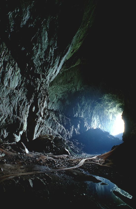
OUT OF THE DARK
Stranded miles deep in the Tham
Luang cave complex. In June 2018,
twelve members of the Wild Boars
football team and their coach become
trapped by monsoon flash-floods whilst
exploring the cave. With no navigation
or imaging technology to locate the
boys, nor viable drilling options
suitable for traversing this
subterranean maze, rescuers dive
through treacherous underwater
chambers. All were rescued alive and
well. Reaching targets deep within
complex enclosed systems requires
meticulous planning and
multidisciplinary teamwork. Technical
expertise, spatial awareness of the
environment, logistical efficiency, and
absolute dedication from both the
rescuer and the rescued are paramount.
CAVERNOUS SINUS
4 Neuroanatomy up-close
Exploring intracranial caves
FROM FLINT TO MACHINE
7 History of trepanation
Development of an ancient
surgical art through the millennia
CRANIAL PERFORATORS
9 Neurosurgical engines
How modern-day neurosurgeons
drill holes in heads
ACCESS POINT
11 Neurological surgery
Planning the operative approach
before knife touches skin
SECOND EDITION
Do you like caving or do you have
DECIMAL, DESTINY, & HAMLET claustrophobia? The cranium houses the
13 Decompressive hemicraniectomy brain, which communicates with the outer
Rationalising trepanation world via a series of intricate tunnels and
passages. Though relatively well protected
in the enclosed box of the skull, cerebral
lesions and elevations in intracranial
pressure mean that there is no easy way out
or in. Decompress or herniate. Survive or
succumb.
neurocartography

    The flooded Tham Luang caves were unforgiving. The narrowest sections
    of the cave reach 15 inches in height, enough space for three mars bars
    stacked end-to-end. These choke points are panic-inducing in even the
    most experienced of divers. In such high-pressure scenarios when the
    diver must escort each boy underwater for 4 hours to reach safety, any
    moment of panic would prove fatal for both the rescuer and the boy. As
    such, each of the boys were sedated with intramuscular ketamine, which
    would allow the boys to maintain their own airways with minimal
    myocardial depression. Atropine was also given for its anti-muscarinic
    effects to minimise respiratory secretions.

    This was a fight against a time. The rescue had to be done before the the
    impending monsoon floods. The Tham Luang cave system drains and
    percolates immense quantities of water in the Northern Chiang Rai
    province. As complex as subterranean caves are, the intracranial cavity,
    particularly the base of skull regions, pose a formidable challenge to
    navigate. Subterranean caverns develop over millions of years through
    mother nature's hydrostatic and chemical mechanisms. Contrastingly,
    the gross anatomical structure of the brain and the cranial passages
    connecting it to the outer world develop over months through our mother
    and father's genetic instructions.

    The neural tracts interconnecting different regions of the brain provide
    an infinitely possible number of configurations. Though tractographic
    DWI scans exists, our exploration and understanding of axonal tracts still
    remain limited. The trajectory of cranial nerves are also mind-boggling.
    Take cranial nerve IV, the Trochlear nerve. It is the longest intracranial
    nerve (also contested by the Abducens nerve), originating from the dorsal
    aspect of the brainstem, tunnelling through the cavernous sinus, passing
    between the superior cerebellar and posterior cerebral arteries of the
    Circle of Willis and innervating the superior oblique muscle via the
    superior orbital fissure. It is often the first cranial nerve to go when the
    intra-cranial pressure rises, and this is precisely because of its
    mesmerisingly long intracranial course (read up on the Monro-Kellie
    doctrine on the mechanisms underlying this).

4
CONTENTS OF THE CAVERN

                                                                             Oculomotor nerve
                                                                             Trochlear nerve
                                                                             Ophthalmic nerve
                                                                             Maxillary nerve
                                                                             Carotid (ICA)
                                                                             Abducens nerve
                                                                             Trochlear nerve

                                                                      The cavernous sinus is a paired dural venous sinus
                                                                      located in the middle cranial fossa. It plays a major
                                                                      role in the venous drainage of external facial vessels
                                                                      into the internal jugular vein and communicates
                                                                      multiple cranial nerves. It is also the only structure
                                                                      in the human body whereby an artery (the internal
                                                                      carotid artery) tunnels through a venous structure.
                                                                      No one quite knows why the internal carotid artery
                                                                      does this, but some postulate that this has a role in
                                                                      thermoregulation as the warm arterial blood is
                                                                      cooled by the surrounding venous system.

                                                                      Having good spatial awareness of structures within
                                                                      the cavernous sinus has significant clinical
                                                                      implications. The cavernous sinus drains blood
                                                                      from the superficial facial veins via the superior and
                                                                      inferior ophthalmic veins. As such, it is possible to
                                                                      get infectious or thrombotic emboli from external
                                                                      facial regions. This can lead to a condition known as
                                                                      cavernous sinus thrombosis. The cavernous sinus is
                                                                      compact, measuring approximately 1cm x 2cm in
                                                                      dimension and lesions here can disrupt cranial
                                                                      nerves III, IV, V1, V2, and VI. Cranial nerve VI
                                                                      (Abducens nerve) is often the first nerve to go in
                                                                      cavernous sinus thrombosis as this nerve tunnels in
                                                                      the middle of the sinus whereas the other cranial
                                                                      nerves are are located in the lateral wall.

                                                                      Lesions in the pituitary gland are often accessed via
                                                                      the trans-sphenoidal route. De-bulking of such
                                                                      tumours must be done with care - drill laterally
                                                                      either side and you may disrupt the cavernous sinus
                                                                      and the internal carotid artery. Venture too far
                                                                      superiorly and the patient may be rendered blind
                                                                      from damage to the optic chiasm.

In certain cases, surgical options can be unfeasible or even
dangerous for reaching specific intracranial targets. Some lesions
are simply too deep or eloquent structures may obstruct the path.
Instead of drilling a route through the skull, interventional
radiologists use existing paths within the cranium. They make
incisions in the femoral artery and traverse up the arterial system
to coil intra-cerebral aneurysms from the inside. Fancy a cave dive
or an arterial swim?

                                                                           FEBRUARY 2021 | NANSIG.ORG
neurocartography

6         FEBRUARY 2021 | NANSIG.ORG

neurocartography

THE CRANIAL TRAUMA HYPOTHESIS WAS PROPOSED BY VERANO AND
ANDRUSHKO THROUGH THEIR EXCAVATION OF SKULL SPECIMENS FROM
INCAN BURIAL SITES. THE MAJORITY OF TREPANATIONS WERE FOUND ON
THE LEFT FRONTOTEMPORAL REGIONS. FROM THIS OBSERVATION, THEY
SUGGESTED THAT TREPANATION WAS COMMONLY PERFORMED FOR
TREATING COMBAT HEAD WOUNDS. THE FACT THAT MOST WARRIORS WERE
RIGHT-HANDED AND THE OVER-REPRESENTATION OF MALES IN THEIR
SAMPLE FURTHER REINFORCED THEIR HYPOTHESIS.

neurocartography
              howcased in a display of human archeology at the American

    S
              Museum of Natural History, New York, you will find Squier’s
              Skull. Physicians and anatomists are uniquely fascinated with
              skulls for their elaborate bony features serving its role in
              housing some of the most delicate structures of the human
body. However, you would not come to marvel at Squier’s skull for its
normal anatomy – it is difficult to ignore the unusual 15mm by 17mm
rectangle opening on the frontal bone. This was the mark of a deliberate
procedure. Trepanation, from the Greek trypanon, is a surgical procedure
meaning to bore a hole in the skull. Various techniques have been
described, including scrapping, cutting or chipping the bone to gain access
to internal structures. Trepanation has been a historical, controversial and
sometimes spiritual procedure.

Squier’s skull was discovered during an exhibition in Peru in 1865 by
Ephraim Squier. When presented to scholars in the US, the skull
evidenced surgical knowledge and skills not thought possible by ancient
indigenous races and provoked enormous interest. Despite popular belief,
humans have been carving holes in skulls as far back as 10,000 BC with the
use of flints! The procedure was not distinct to Peru. Remarkably similar
findings in skulls have been uncovered across Europe, Africa, the
Americas, Pacific Islands and Asia. The motivations for trepanation have
been diverse, and not always clear. From elevating depressed skulls
fractures received in battle, to the relief of headaches or evil spirits
believed to be causing seizures or mental illness.

Surprisingly, not all cases of trepanation were disasters (despite some
worrying motives!) - evidence of bone healing around the site of the
procedure has been shown in discovered skulls. However, success may
only have been apparent for days to months; a significant challenge was
the risk of infections. Some steps were taken to minimise these risks: the
Incajs used balsam and cinnamic acid at the entry site, whilst the Tolai
people of Papua New Guinea (1800s) practiced with single-use apparatus
and irrigated wounds with sterile coconut water. This was truly ahead of
the time, as Germ theory did not appear in western medicine until the
mid-late 19th century. DOI: 10.3171/2014.2.FOCUS13548

                                                                                           ippocrates offered the first documentation of trepanation for its

                                                                               H
                                                                                           therapeutic effects. His translated work ‘On Wounds to the Head’
                                                                                           enriched the future generation of surgeons through methodology
                                                                                           and the rationalisation of surgery. Hippocrates stated the great
                                                                                           importance of history taking to understand the injury and its
                                                                             mechanism, before deciding if surgery was needed. For instance, trepanation
                                                                             of ‘dented’ fractures occurring from battle, was deemed beneficial to prevent
                                                                             the accumulation of blood and subsequent brain compression. Hippocrates
                                                                             also detailed areas of weaker bone and warned surgeons against trepanning at
                                                                             the suture lines – essential for preserving the dural sinuses needed for normal
                                                                             blood flow throughout the brain.

                                                                             The surgical procedure has since evolved, and techniques of craniotomy
                                                                             emerged in the early 19th century. The American, Wilhelm Wagner described
                                                                             trepanation as the ‘removal of a piece of the intact bony calvarium’. Carving in
                                                                             the shape of an Omega symbol, Wagner could create a bone flap! Allowing him
                                                                             to open and close the entry into the skull. This was later influential in
                                                                             neurosurgeon George Heuer’s approach (frontotemporal craniotomy) to
                                                                             visualise the optic chiasm and reach tumours and aneurysms.

                                                                             Heuer’s frontotemporal craniotomy remains in practice as it helps to optimise
                                                                             brain exposure but minimise potential damage of brain retraction for certain
                                                                             procedures. In addition, with the availability of modern-day technologies, like
                                                                             high-resolution MRIs and 3D printing, clinicians can now map the pathways
                                                                             from skull to brain for the least invasive route and provide a greater insight for
                                                                             what lies ahead. From Heuer’s approach, new cerebrovascular surgical
                                                                             techniques emerged, a notable example includes the clipping of aneurysms,
                                                                             devised from Walter Dandy of John Hopkins – a life-saving technique
                                                                             benefiting many people across the world today.

                                                                             Trepanation opened the window for brain surgery (metaphorically and
                                                                             literally). We continue to appreciate and explore its history, helping us to
                                                                             uncover the earliest roots from which neurosurgery has stemmed.
                                                                             DOI: 10.1002/ajpa.20836

8                                                                                                            FEBRUARY 2021 | NANSIG.ORG
neurocartography
                                                                                  John Lewis (1838)
                                         CRANK DRILLS

                                                                                             Leonardo Gigli (1893)
                                                              WIRE SAWS

    Neolithic ancestor
       The "Flint"

                                                                         CIRCULAR SAWS                                           Fedor Krause (1897)

                                                                                                                    George Harrington (1846)
                                                                                   DENTAL ENGINES

                                     SURGICAL ENGINES                                           OSTEOTOMES             Borchardt (1906)

    The                                                                                                                                    Raney (1941)

    Neurosurgical                                                                                            PNEUMATIC
                                                                                                                 DRILLS

    Engine
    Teeth. Enamel. Dental crowns. We can trace the origins of                                               CRANIAL                          (1980s)
    modern day cranial perforators, the bread and butter of                                                 PERFORATORS
    neurosurgery, to the pioneering works of dentists in the
    18th century. Mechanical drills turned by hand cranks
    were widely used in the early 19th century. The first motor-
    driven dental engine was invented by George Harrington
    in 1846, and over generations it has been adopted and
    refined by other surgical specialties to cut through bone.

                he earliest trepanned skulls are discovered in the pre-Incan      highly vascular scalp and the drilling speed of surgical engines such as

      T         regions of what is now modern day Peru. These specimens
                date back to 8000BC. Neolithic techniques involved
                sharpened flints scraping away at skin and cranium.
                                                                                  osteotomes were difficult to control. Harvey Cushing opted for the Gigli
                                                                                  saw, commenting that "an operator who persists in taking dangerous
                                                                                  corners at high speed will be the cause of a serious or fatal accident
                                                                                  some day, whether he is driving an automobile or opening a skull".
     Trepanation is the oldest known surgical procedure for which we have
     archaeological evidence. Though our ancestors often had mythical or          The traditional Gigli saw grew in popularity among surgeons, owing to
     spiritual indications for craniotomy, they nonetheless bore successful       its simplicity and lower risk of injury to the underlying dura and brain.
     holes in heads. Advancements in cerebral localisation, antisepsis, and       However, these too proved to be tedious, frequently jamming or
     surgical techniques, led to the rationalisation of craniotomy for specific   breaking. Invented in the late 20th century, cranial perforators are
     clinical indications (i.e. intracranial lesions, haematoma evacuation,       widely used in modern practice. These drills rotate at speeds of up to
     depressed skull fractures). Access into the intracranial cavity poses a      1000rpm and are equipped with clutch systems that detects subtle
     peculiar challenge. Hammer and chisel exert too much vertical inertial       changes in pressure to prevent plunging of the drill into the underlying
     force on the brain. Circular saws caused excessive bleeding from the         cortex. DOI: 10.1097/00006123-199101000-00017.

9                                                                                                             FEBRUARY 2021 | NANSIG.ORG
neurocartography

‘The physician must be able to tell the antecedents,
    know the present, and foretell the future'
                 __________
               Of the Epidemics,
          HIPPOCRATES (400 B.C.)
FEBRUARY
2021
VOLUME 1,
NO. 2
neurocartography
NOSE PICKING
Neurosurgeons at the Zucker School
of Medicine, Northwell, perform an
endoscopic trans-sphenoidal pituitary
tumour removal. Life-saving. Now
that's a legitimate excuse for picking
your nose!
rom the pterional to the orbitozygomatic; modern neurosurgery has curated an array
F
of anatomical start-points that allow a surgeon to delve deep into the brain with great
precision, involving the least obstructed route with maximal exposure and minimal
disturbance to normal anatomy. Aiming to reduce mortality associated with
transcranial approaches by eliminating extensive brain retraction, the transsphenoidal
approach was pioneered in the early 1900s. Despite the attractiveness of such a short distance
between the incision and sellar, early attempts at this operation often resulted in meningitis and
poor cosmetic outcome. The transsphenoidal approach today boasts a ‘keyhole’ incision and
maintenance of nasal anatomy to a far greater degree.
Driven by technological advancement and
a better understanding of surgical
neuroanatomy, neurosurgical approaches
once limited to trepanation have evolved
into versatile, minimally-invasive
microsurgeries.
The transsphenoidal approach is The nasal cavity is divided in half by the nasal septum and
challenging, and rests upon good knowledge segmented by superior, middle and inferior nasal
of the anatomical relationships along its conchae. The initial stages of a transsphenoidal approach
course. Neuronavigation, often fluoroscopy, establish an adequate surgical pathway through the nasal
help to define the anatomy of this region. cavity, involving septal mucosal dissection, and lateral
Operating microscopes guide an endoscope dislocation of the middle nasal conchae to widen the
to the back of the sphenoid sinus, where operative window. Sphenoidotomy, the surgical opening
fragments of bone are cut away to access the of the sphenoid sinus- occurs along the sphenoethmoidal
skull base. The tumour is cut into small recess. Close attention to vasculature must be taken as to
pieces by microsurgical instruments before avoid early epistaxis from laceration of the sphenopalatine
resection. artery or its branches. www.neurosurgicalatlas.com
11 FEBRUARY 2021 | NANSIG.ORGKOCHER'S POINT
Anatomical landmarks increase precision and reduce surgical
complication. A core tool in any surgery, they enable one to
orientate and manoeuvre through complex anatomy. Particularly
important in neurosurgery is recognition of eloquent brain that, if
harmed, can pose significant consequences for the patient. Where
there are the means to do so, awake or image-guided procedures
have been used in conjunction with anatomical landmarks to allow
a surgical team to monitor changes in neurological function
throughout the procedure.
Craniometric points are landmarks at the external surface of the
skull that localise crucial intracranial structures, such as sulci and
gyri. They include projections, depressions, sutures and suture
junctions. Despite the rise of neuronavigation technologies,
craniometric points continue to play a pivotal role in operative
planning. Kocher’s point is a craniometric point to determine the
entry site for tools involved in CSF diversion; including external
ventricular drain (EVD), ventriculoperitoneal (VP) shunt and
endoscopic third ventriculostomy (ETV). It is a burr hole 11cm
superior and posterior to the bridge of the nose, and 2-3cm lateral
to the midline (approximately at the mid-pupillary line).
DOI: 10.3171/2014.2.FOCUS13547
12neurocartography
HAMLET
DESTINY
DECIMAL
Indications for a craniectomy in the previous millennia and even up
to a few hundred years ago were often based on pseudoscientific or
outright outrageous reasons to the 21st century clinician. Although
modern medicine is still in its infancy, the advent of evidence-based
medicine has rationalised such neurosurgical procedures and vastly
improved their safety.
ife threatening, space occupying brain oedema
L
occurs in 1-10% of patients with supratentorial
infarcts, commonly arising at the second and fifth
day after stroke onset. Prognosis of these malignant
middle cerebral artery (MCA) infarcts is poor, with
fatality rates of up to 80%. No medical treatment has proven
effective.
There have been anecdotal and case reports, as well as non-
randomised studies suggesting that decompressive surgery
reduces mortality rates in patients with malignant MCA
infarction. Despite consensus that neurosurgical intervention is
life-saving for MCA infarction, there was no concrete data to
support this. Enter, HAMLET, DESTINY, and DECIMAL.
This multi-centre study randomised patients with malignant
MCA infarction into two groups; i) control group with
conservative management; ii) treatment group with
decompressive craniectomy. It found that the mortality rate was
considerably higher in the control group (71%) compared to the
treatment group (22%). Moreover, the number of surviving
patients with severe disability (mRS = 5) remain unchanged for
both the control and treatment group. However, those in the
treatment group were more likely to survive with moderately
severe disability (mRS = 4).
The HAMLET, DESTINY, and DECIMAL study presented
concrete evidence to a procedure initially supported by under-
powered data. It has changed clinical practice; decompressive
hemicraniectomies must now be seriously considered for those
with malignant MCA infarction in those younger than 60 years of
age. However, the following question remains; after a stroke,
would you rather die or survive unable to walk and
independently attend your bodily needs (mRS = 4)?
TO DRILL OR
As clinicians, the decision on performing a
decompressive hemicraniectomy must
therefore be made on an individual patient
NOT TO DRILL?
basis.
DOI:10.1016/S1474- 4422(07)70036-4
13 FEBRUARY 2021 | NANSIG.ORGneurocartography FEBRUARY 2021, VOL. 1, NO. 2
THE UNCHARTED
BETWEEN OUR EARS
EXPLORERS OF THE
BRAIN
DAVID LEE
NHS Grampian AFP
ORLA MANTLE
King's College London
ELLIOT LEE
University of Nottingham
ROSALINE DE KONING
University of Oxford
slee38@qub.ac.ukYou can also read