Human Brain/Cloud Interface - Frontiers
←
→
Page content transcription
If your browser does not render page correctly, please read the page content below
HYPOTHESIS AND THEORY
                                                                                                                                                published: 29 March 2019
                                                                                                                                           doi: 10.3389/fnins.2019.00112

                                            Human Brain/Cloud Interface
                                            Nuno R. B. Martins 1,2* , Amara Angelica 3 , Krishnan Chakravarthy 4,5 , Yuriy Svidinenko 6 ,
                                            Frank J. Boehm 7 , Ioan Opris 8,9 , Mikhail A. Lebedev 10,11,12 , Melanie Swan 13 ,
                                            Steven A. Garan 1,2 , Jeffrey V. Rosenfeld 14,15,16,17 , Tad Hogg 18 and Robert A. Freitas Jr. 18
                                            1
                                              Lawrence Berkeley National Laboratory, Berkeley, CA, United States, 2 Center for Research and Education on Aging
                                            (CREA), University of California, Berkeley, & LBNL, Berkeley, CA, United States, 3 Kurzweil Technologies, Newton, MA,
                                            United States, 4 UC San Diego Health Science, San Diego, CA, United States, 5 VA San Diego Healthcare System, San
                                            Diego, CA, United States, 6 Nanobot Medical Animation Studio, San Diego, CA, United States, 7 NanoApps Medical, Inc.,
                                            Vancouver, BC, Canada, 8 Miami Project to Cure Paralysis, University of Miami, Miami, FL, United States, 9 Department
                                            of Biomedical Engineering, University of Miami, Coral Gables, FL, United States, 10 Center for Neuroengineering, Duke
                                            University, Durham, NC, United States, 11 Center for Bioelectric Interfaces of the Institute for Cognitive Neuroscience of the
                                            National Research University Higher School of Economics, Moscow, Russia, 12 Department of Information and Internet
                                            Technologies of Digital Health Institute, I.M. Sechenov First Moscow State Medical University, Moscow, Russia,
                                            13
                                               Department of Philosophy, Purdue University, West Lafayette, IN, United States, 14 Monash Institute of Medical
                                            Engineering, Monash University, Clayton, VIC, Australia, 15 Department of Neurosurgery, Alfred Hospital, Melbourne, VIC,
                                            Australia, 16 Department of Surgery, Monash University, Clayton, VIC, Australia, 17 Department of Surgery, F. Edward Hébert
                                            School of Medicine, Uniformed Services University of the Health Sciences, Bethesda, MD, United States, 18 Institute for
                                            Molecular Manufacturing, Palo Alto, CA, United States

                                            The Internet comprises a decentralized global system that serves humanity’s collective
                          Edited by:
                                            effort to generate, process, and store data, most of which is handled by the rapidly
                    Hari S. Sharma,         expanding cloud. A stable, secure, real-time system may allow for interfacing the cloud
          Uppsala University, Sweden        with the human brain. One promising strategy for enabling such a system, denoted
                       Reviewed by:         here as a “human brain/cloud interface” (“B/CI”), would be based on technologies
                     Vassiliy Tsytsarev,
 University of Maryland, College Park,      referred to here as “neuralnanorobotics.” Future neuralnanorobotics technologies are
                         United States      anticipated to facilitate accurate diagnoses and eventual cures for the ∼400 conditions
                       Brent Winslow,
     Design Interactive, United States
                                            that affect the human brain. Neuralnanorobotics may also enable a B/CI with controlled
                 *Correspondence:
                                            connectivity between neural activity and external data storage and processing, via
                  Nuno R. B. Martins        the direct monitoring of the brain’s ∼86 × 109 neurons and ∼2 × 10¹⁴ synapses.
               nunomartins@lbl.gov;         Subsequent to navigating the human vasculature, three species of neuralnanorobots
           nunorbmartins@gmail.com
                                            (endoneurobots, gliabots, and synaptobots) could traverse the blood–brain barrier
                   Specialty section:       (BBB), enter the brain parenchyma, ingress into individual human brain cells, and
         This article was submitted to
                                            autoposition themselves at the axon initial segments of neurons (endoneurobots), within
                    Neural Technology,
               a section of the journal     glial cells (gliabots), and in intimate proximity to synapses (synaptobots). They would
            Frontiers in Neuroscience       then wirelessly transmit up to ∼6 × 1016 bits per second of synaptically processed
      Received: 10 September 2018           and encoded human–brain electrical information via auxiliary nanorobotic fiber optics
        Accepted: 30 January 2019
         Published: 29 March 2019
                                            (30 cm3 ) with the capacity to handle up to 1018 bits/sec and provide rapid data
                           Citation:
                                            transfer to a cloud based supercomputer for real-time brain-state monitoring and data
            Martins NRB, Angelica A,        extraction. A neuralnanorobotically enabled human B/CI might serve as a personalized
       Chakravarthy K, Svidinenko Y,
                                            conduit, allowing persons to obtain direct, instantaneous access to virtually any
     Boehm FJ, Opris I, Lebedev MA,
    Swan M, Garan SA, Rosenfeld JV,         facet of cumulative human knowledge. Other anticipated applications include myriad
     Hogg T and Freitas RA Jr (2019)        opportunities to improve education, intelligence, entertainment, traveling, and other
       Human Brain/Cloud Interface.
             Front. Neurosci. 13:112.
                                            interactive experiences. A specialized application might be the capacity to engage
      doi: 10.3389/fnins.2019.00112         in fully immersive experiential/sensory experiences, including what is referred to here

Frontiers in Neuroscience | www.frontiersin.org                                     1                                                March 2019 | Volume 13 | Article 112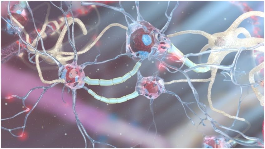
Martins et al.                                                                                                                 Human Brain/Cloud Interface

                                            as “transparent shadowing” (TS). Through TS, individuals might experience episodic
                                            segments of the lives of other willing participants (locally or remote) to, hopefully,
                                            encourage and inspire improved understanding and tolerance among all members of
                                            the human family.
                                            Keywords: brain/cloud interface, brain-computer interface, brain-to-brain interface, brain-machine interface,
                                            transparent shadowing, neuralnanorobots, neuralnanorobotics, nanomedicine

INTRODUCTION                                                                      including, most notably: Parkinson’s and Alzheimer’s (Freitas,
                                                                                  2016), addiction, dementia, epilepsy, and spinal cord disorders
    “We’ll have nanobots that... connect our neocortex                            (NINDS, 2017).
    to a synthetic neocortex in the cloud... Our thinking
    will be a.... biological and non-biological hybrid.”                              Neuralnanorobots are also expected to empower many non-
                               — Ray Kurzweil, TED 2014                           medical paradigm-shifting applications, including significant
                                                                                  human cognitive enhancement, by providing a platform for direct
    There is an incessant drive in medicine toward the                            access to supercomputing storage and processing capabilities and
development of smaller, more capable, efficacious, and cost-                      interfacing with artificial intelligence systems. Since information-
effective devices and systems. The primary driver of this quest                   based technologies are consistently improving their price-
relates to the cellular and sub-cellular genesis of human disease,                performance ratios and functional design at an exponential
at which scale, nanodevices can directly interact and potentially                 rate, it is likely that once they enter clinical practice or
positively influence disease outcomes or prevent them altogether,                 non-medical applications, neuralnanorobotic technologies may
particularly in regard to brain disorders (Kandel et al., 2000,                   work in parallel with powerful artificial intelligence systems,
Kandel, 2001; Zigmond et al., 2014; Chaudhury et al., 2015;                       supercomputing, and advanced molecular manufacturing.
Fornito et al., 2015; Falk et al., 2016). The pursuit of ever                         Furthermore, autonomous nanomedical devices are expected
smaller tools to treat patients is approaching a pivotal juncture                 to be biocompatible, primarily due to their structural materials,
in medical history as advanced nanomedicine — specifically,                       which would enable extended residency within the human
medical nanorobotics — is expected to serve as a dynamic                          body (Freitas, 1999a, 2002, 2003). Medical neuralnanorobots
tool toward addressing most human brain disorders. The goal                       might also be fabricated in sufficient therapeutic quantities
is to finally empower medical professionals to treat diseases                     to treat individual patients, using diamondoid materials, as
at individual cellular and sub-cellular resolution (Freitas, 1998,                these materials may provide the greatest strength, resilience,
1999b, 2003, 2005a,c, 2007, 2016; Morris, 2001; Astier et al., 2005;              and reliability in vivo (Freitas, 2010). An ongoing international
Patel et al., 2006; Park et al., 2007; Popov et al., 2007; Mallouk and            “Nanofactory Collaboration” headed by Robert Freitas and Ralph
Sen, 2009; Martel et al., 2009; Kostarelos, 2010; Mavroides and                   Merkle has the primary objective of constructing the world’s
Ferreira, 2011; Boehm, 2013).                                                     first nanofactory, which will permit the mass manufacture of
    The application of nanorobots to the human brain is                           advanced autonomous diamondoid neuralnanorobots for both
denoted here as “neuralnanorobotics.” This technology may                         medical and non-medical applications (Freitas and Merkle, 2004,
allow for the monitoring, recording, and even manipulation                        2006; Freitas, 2009, 2010).
of many types of brain-related information at cellular and                            It is conceivable that within the next 20–30 years,
organellar levels (Martins et al., 2012, 2015, 2016). Medical                     neuralnanorobotics may be developed to enable a safe,
neuralnanorobots are expected to have the capacity for real-                      secure, instantaneous, real-time interface between the
time, non-destructive monitoring of single-neuron and single-                     human brain and biological and non-biological computing
synapse neuroelectric activity, local neuropeptide traffic, and                   systems, empowering brain-to-brain interfaces (BTBI), brain-
other relevant functional data, while also allowing the acquisition               computer interfaces (BCI), and, in particular, sophisticated
of fundamental structural information from neuron surfaces, to                    brain/cloud interfaces (B/CI). Such human B/CI systems may
enhance the connectome map of a living human brain (Sporns                        dramatically alter human/machine communications, carrying
et al., 2005; Lu et al., 2009; Anderson et al., 2011; Kleinfeld                   the promise of significant human cognitive enhancement
et al., 2011; Seung, 2011; Martins et al., 2012, 2015, 2016).                     (Kurzweil, 2014; Swan, 2016).
Non-destructive neuralnanorobotically mediated whole-brain                            Historically, a fundamental breakthrough toward the
monitoring coupled with single-cell repair capabilities (Freitas,                 possibility of a B/CI was the initial measurement and recording
2007) is anticipated to provide a powerful medical capability to                  of the electrical activity of the brain via EEG in 1924 (Stone and
effectively treat most, or all of the ∼400 known brain disorders,                 Hughes, 2013). At the time, EEG marked a historical advance in
                                                                                  neurologic and psychiatric diagnostic tools, as this technology
Abbreviations: AIS, Axon initial segment; B/CI, brain/cloud interface; BCI,       allowed for the measurement of a variety of cerebral diseases,
brain–computer interface; BMI, brain–machine interface; BTBI, brain-to-           the quantification of deviations induced by different mental
brain interface; EEG, electroencephalography; fMRI, functional magnetic
resonance imaging; FNIRS, functional near-infrared spectroscopy; TS,              states, and detection of oscillatory alpha waves (8–13 Hz), the
transparent shadowing.                                                            so-called “Berger’s wave.” The first EEG measurements required

Frontiers in Neuroscience | www.frontiersin.org                               2                                         March 2019 | Volume 13 | Article 112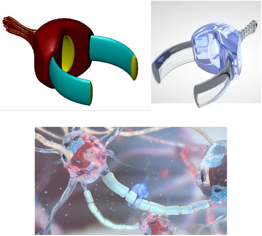
Martins et al.                                                                                                                        Human Brain/Cloud Interface

the insertion of silver wires into the scalps of patients, which                           effort, often over the Internet. For both personal or business
later evolved to silver foils that were adhered to the head.                               applications, the cloud facilitates rapid data access, provides
These rudimentary sensors were initially linked to a Lippmann                              redundancy, and optimizes the global usage of processing
capillary electrometer. However, significantly improved results                            and storage resources while enabling access from virtually
were achieved through the use of a Siemens double-coil                                     any location on the planet. However, the primary challenge
recording galvanometer, which had an electronic resolution of                              for worldwide global cloud-based information processing
0.1 mv (Jung and Berger, 1979).                                                            technologies is the speed of access to the system, or latency.
    The first reported scientific instance of the term “brain–                             For example, the current round-trip latency rate for transatlantic
computer interface” dates to 1973, ∼50 years following the                                 loops between New York and London is ∼90 ms (Verizon,
first EEG recording, when it was envisioned that EEG-reported                              2014). Since there are now more than 4 billion Internet users
brain electrical signals might be employed as data carriers in                             worldwide, its economic impact on the global economy is
human–computer communications. This suggestion assumed                                     increasingly significant. The economic impact of IoT (Internet of
that mental decisions and reactions might be probed by                                     Things) applications alone has been estimated by the McKinsey
electroencephalographic potential fluctuations measured on the                             Global Institute to range from $3.9 to $11.1 trillion per year by
human scalp, and that meaningful EEG phenomena should                                      2025. The global economic impact of cloud-based information
be viewed as a complex structure of elementary wavelets that                               processing over the next few decades may be at least an order
reflected individual cortical events (Vidal, 1973).                                        of magnitude higher once cloud services are combined in
    Currently, invasive1 and non-invasive brain–computer                                   previously unimagined ways, disrupting entire industries (Miraz
interfaces and non-invasive brain-to-brain communication                                   et al., 2015). A neuralnanorobotics-mediated human B/CI,
systems have already been experimentally demonstrated and are                              potentially available within 20–30 years, will require broadband
the subject of serious research worldwide. Once these existing                             Internet access with extremely high upload and download speeds,
technologies have matured, they might provide treatments                                   compared to today’s rates.
for completely paralyzed patients, eventually permitting the                                   Humankind has at its core a potent and ceaseless drive
restoration of movement in paralyzed limbs through the                                     to explore and to challenge itself, to improve its collective
transmission of brain signals to muscles or external prosthetic                            condition by relentlessly probing and pushing boundaries while
devices (Birbaumer, 2006). The first reported direct transmission                          constantly attempting to breach those barriers that tenuously
of information between two human brains without intervention                               separate the possible from the impossible. The notions of
of motor or peripheral sensory systems occurred in 2014,                                   human augmentation and cognitive enhancement are borne
using a brain-to-brain communication technique referred to as                              of these tenets.
“hyperinteraction” (Grau et al., 2014).                                                        This drive includes an incessant quest for exploration and
    The most promising long-term future technology for non-                                a constant desire for social interaction and communication —
destructive, real-time human–brain–computer interfaces and                                 both of which are catalysts for rapidly increasing globalization.
brain-to-brain communications may be neuralnanorobotics                                    Consequently, the development of a non-destructive, real-time
(Martins et al., 2016). Neuralnanorobotics, which is the                                   human B/CI technology may serve as an intimate, personalized
application of medical nanorobots to the human brain, was                                  conduit through which individuals would have instantaneous
first envisaged by Freitas, who proposed the use of nanorobots                             access to virtually any facet of cumulative human knowledge
for direct real-time monitoring of neural traffic from in vivo                             and also the optional specialized capacity to engage in myriad
neurons, as well as the translation of messages to neurons                                 real-time fully immersive experiential and sensory worlds.
(Freitas, 1999b, 2003). Other authors have also envisioned B/CI
and predicted that in the future, humans will have access to
a synthetic non-biological neocortex, which might permit a                                 THE HUMAN BRAIN
direct B/CI. Within the next few decades, neuralnanorobotics
may enable a non-destructive, real-time, ultrahigh-resolution                              The Quantitative Human Brain
interface between the human brain and external computing                                   The human brain comprises a remarkable information storage
platforms such as the “cloud.”                                                             and processing system that possesses an extraordinary
    The term “cloud” refers to cloud computing, an information                             computation-per-volume efficiency, with an average weight
technology (IT) paradigm and a model for enabling ubiquitous                               of 1400 g and a volume of ∼1350 cm3 , contained within
access to shared pools of configurable resources (such as                                  an “average” intracranial volume of ∼1,700 cm3 . A brief
computer networks, servers, storage, applications, and services),                          quantification of the brain’s constituents and operational
that can be rapidly provisioned with minimal management                                    parameters includes ∼1,350 cm3 (∼75%) brain cells, ∼200 cm3
                                                                                           (15%) blood, and up to ∼150 cm3 (10%) of cerebrospinal fluid
1
 For the purposes of this paper, the term “invasive” is defined as a medical               (Rengachary and Ellenbogen, 2005). The raw computational
procedure or device that imparts quantifiable physiological damage (at any level) to       power of the human brain has been estimated to range from
a patient. In the case of the envisaged nanomedically enabled B/CI, the assumption         1013 to 1016 operations/sec (Merkle, 1989). The human brain’s
is that millions of micron-scale nanorobots will be non-invasive — i.e., they may
                                                                                           functional action potential based information is estimated
ingress into a patient and subsequently auto-situate themselves at various sites
within the human brain, with no disruptive functional physiological or experiential        as 5.52 × 1016 bits/sec (Martins et al., 2012), with a brain
effects. Or in some cases, they may be minimally invasive.                                 power output estimated at 15–25 W and a power density of

Frontiers in Neuroscience | www.frontiersin.org                                        3                                       March 2019 | Volume 13 | Article 112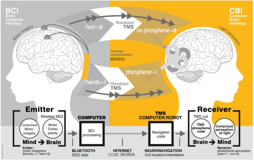
Martins et al.                                                                                                                               Human Brain/Cloud Interface

1.1–1.8 × 10⁴ W/m3 at an operating temperature of 37.3◦ C
(Freitas, 1999b).
   When considering the human brain at the regional level, an
exceptional component is the neocortex (Tables 1, 2), which
has a highly organized neural architecture that encompasses
sensorimotor, cognitive, and emotional domains (Alexander
et al., 1986; Fuster and Bressler, 2012). This cortical structure
consists of mini-columnar and laminar arrangements of
neurons that are linked via afferent and efferent connections
distributed across multiple brain regions (Lorento de Nó,
1938; Mountcastle, 1997; Shepherd and Grillner, 2010;
Opris, 2013; Opris et al., 2011, 2013, 2014, 2015). Cortical
minicolumns consist of chains of pyramidal neurons that are                               FIGURE 1 | Artistic representation of neurons (with blue processes) and glial
surrounded by a “curtain of inhibition” formed by interneurons                            (white) cells. [Image credit: Yuriy Svidinenko, Nanobotmodels Company].
(Szentágothai and Arbib, 1975).
   At the cellular level, the average human brain is estimated
to contain (86.06 ± 8.2) × 109 neurons, with ∼80.2%
(69.03 ± 6.65 × 10⁹ neurons) located in the cerebellum, ∼19%                             brain domains. For example, the glia/neuron ratio of the cerebral
(16.34 ± 2.17 × 10⁹ neurons) located in the cerebral cortex, and                         cortex is 3.72:1 (60.84 billion glia; 16.34 billion neurons) but only
only ∼0.8% (0.69 ± 0.12 × 10⁹ neurons) located throughout the                            0.23:1 (16.04 billion glia; 69.03 billion neurons) in the cerebellum;
rest of the brain (Azevedo et al., 2009). The human cerebellum                           the basal ganglia, diencephalon, and brainstem have a combined
and cerebral cortex together hold the vast majority (99.2%) of                           ratio of 11.35:1 (Azevedo et al., 2009).
brain neurons (Azevedo et al., 2009). Another approximation,                                In addition, synapses, numbering (2.42 ± 0.29) × 1014
based on combining estimates for the different brain regions,                            in the average human brain, are collectively estimated to
produced a similar value of 94.2 ± 11.3 × 10⁹ neurons for the                            process information at spiking rates of (4.31 ± 0.86) × 1015
whole human brain (Martins et al., 2012).                                                spikes/sec, empowering the human brain to process data at
   Glial cells comprise another brain-cell type (Figure 1). The                          (5.52 ± 1.13) × 1016 bits/sec (Martins et al., 2012). Synapses
average number of glial cells in the human brain is estimated                            are elements of the neural network that play a critical role in
to be 84.61 ± 9.83 × 10⁹ (Herculano-Houzel, 2009), with the                              processing information in the brain, being involved in learning,
population of glial cells in the neocortex estimated at from 18.2                        long-term and short-term memory storage and deletion, and
to 38.6 × 10⁹ (Karlsen and Pakkenberg, 2011). The ratio of glia to                       temporal information processing (Black et al., 1990; Bliss and
neurons likely has functional relevance (Nedergaard et al., 2003)                        Collingridge, 1993; Kandel, 2001; Fuhrmann et al., 2002; Lee et al.,
and varies between different brain regions. While the whole-brain                        2008; Holtmaat and Svoboda, 2009; Liu et al., 2012). Synapses
glia/neuron ratio is ∼1:1, there are significant differences between                     are also key effectors for signal transduction and plasticity in
                                                                                         the brain. Proper synapse formation during childhood provides
                                                                                         a substrate for cognition, whereas improper formation or
                                                                                         functionality leads to neuro-developmental disorders including
TABLE 1 | Neocortical measures (Pakkenberg and Gundersen, 1997; Stark et al.,
2007a,b).
                                                                                         mental retardation and autism (Rollenhagen and Lübke, 2006;
                                                                                         Mcallister, 2007; Rollenhagen et al., 2007). Synapse loss, as
             Surface     Thickness         Volume    Neuron number      Neurons          occurs in Alzheimer’s patients, is intimately associated with
              (cm2 )       (mm)             (cm3 )      density         (N, 109 )        cognitive decline (Dekosky and Scheff, 1990; Terry et al., 1991;
                                                       (106 /cm3 )
                                                                                         Scheff and Price, 2006).
Female      1678–1680    2.61–2.74         440–458       43.1–43.8     19.3–19.7
Male        1883–1900    2.72–2.79         517–524       44.0–44.1     22.8–22.9
Humans           1820        2.69           489            44.0           21.5           Processing Units
                                                                                         Structural cellular or sub-cellular elements of the human
                                                                                         brain are considered as information processing units if
TABLE 2 | Enumeration of neurons and synapses in the human neocortex (Tang
et al., 2001; Sandberg and Bostrom, 2008; Karlsen and Pakkenberg, 2011).                 they are involved in significant functional input/output
                                                                                         changes in electrochemically based brain-data storage and/or
Neocortex        Total        Number of Number of          Number of    Glial cell       processing systems.
region        neocortex       synapses   neurons          synapses per number               There is some disagreement in the current scientific literature
             volume (cm3 )      (1012 )   (109 )          neuron (103 )   (109 )
                                                                                         regarding the quantification of this “significance” metric. This
Occipital           69              22.0       3–4.65          4.36          3           incongruity has led various authors to consider different cellular
Parietal           149              41.5       4–6.61          6.33          4           and subcellular structures as fundamental elements of human
Temporal           133              42.0       4–4.80          8.95          5           brain storage and its computation system, encompassing (aside
Frontal            239              58.9       6–7.89          7.54          7           from neurons and synapses): dendritic trees, axons, proteins,
Total              590          164.0          17–23.9         6.93         18           and even neural microtubules (Koch et al., 1983; Bialek, 1993;

Frontiers in Neuroscience | www.frontiersin.org                                      4                                               March 2019 | Volume 13 | Article 112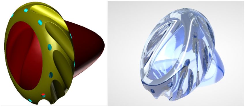
Martins et al.                                                                                                     Human Brain/Cloud Interface

Juusola et al., 1996; Zador, 1998; Manwani and Koch, 2001;                Real-time monitoring of the whole human brain (by
London and Häusser, 2005; Ford, 2010).                                 placing neuralnanorobots within each neuron and nearby
   Estimates for whole-brain electrical data processing rates          synaptic connections to record/transmit data from localized
range from 1.48 × 1011 bits/sec. to a high of 3.2 × 1029               neuron and synapse spiking) may provide redundant
bits/sec (Sandberg and Bostrom, 2008; Martins et al., 2012).           data that might be employed in the development of
The human brain might even have more than 100 times higher             validation protocols.
computational capacity than previously thought, based on the
discovery that dendrites may generate nearly 10 times as many
electrochemical spikes as do neuron soma, and are hybrids that         THE CLOUD
process both analog and digital signals (Moore et al., 2017).
This finding may challenge the long-held belief that spikes            Due to the immense volume of data involved, data transfer to and
in the soma (body of the neuron) are the primary means                 from living human brains and the cloud may likely require the use
through which perception, learning, and memory formation               of supercomputers with artificial intelligence algorithms. Current
occur. Dendrites comprise more than 90% of neural tissue,              von Neumann-based-architecture supercomputers with massive
so knowing that they are much more active than the soma                numbers of processors are either centralized (composed of large
would fundamentally alter our understanding of how the                 numbers of dedicated processors) or distributed (based on a large
brain processes information. As dendrites are ∼100 times               number of discrete computers distributed across a network, such
larger by volume than neuronal bodies, the immense number              as the Internet).
of firing dendritic spikes would suggest that the brain may                One estimate of maximum computational speed required to
indeed possess significantly higher computational power than           handle the electrical data in the human brain is 5.52 × 10¹⁶
earlier estimated.                                                     bits/sec (Martins et al., 2012). Several centralized and distributed
   However, there is currently a consensus that neurons                supercomputers have processing speeds that are significantly
and synapses constitute the fundamental electrochemical                higher than this estimate (Martins et al., 2012). As of November
processing units of the human brain (Gkoupidenis et al., 2017;         2018, the fastest supercomputer worldwide was Summit,
Jackman and Regehr, 2017).                                             developed at the United States Oak Ridge National Laboratory
   The roles of neurons in electrical information processing           (Tennessee), with 122.3 petaflops on the High Performance
include receiving, integrating, generating, and transmitting           Linpack (HPL) benchmark. This computational model may be
action-potential-based information (Koch, 1997; Koch and Segev,        questionable, however, as computers are based on von Neumann
2000; Zhang, 2008). However, several neuronal noise sources            architecture, whereas brain circuits are not; and brains operate
influence the reliability and precision of neuronal signaling,         in a massively parallel manner, whereas computers do not
so stimulus-response functions are sometimes unreliable and            (Nagarajan and Stevens, 2008; Whitworth and Ryu, 2008).
are dissociated from what is being encoded via spike activity              The Internet consists of a decentralized global system,
(Bialek and Rieke, 1992).                                              based on von-Neumann-architecture-based computers and
   The other fundamental consensual processing units of                supercomputers, used for data transfer across processing and
electrochemical information are synapses. Synapses are                 storage units. The global storage capacity of Internet data centers
a core component of the neuron network that process                    in 2018 was 1450 exabytes (Statistica, 2018). Van den Bosch et al.
information and are involved in learning and memory,                   (2016) estimate that the storage capacity of the World Wide Web
with synapse dimensions and morphologies reported as playing           doubles every 3 years, with its computational capacity doubling
a fundamental role in long- and short-term memory storage              every 1.5 years.
and deletion. Synapses are also engaged in signal transduction             However, once brain data is interfaced with supercomputers
and plasticity, ensuring one-way transmission of signals, and          in near real-time, the connection to supercomputers in the
are involved in temporal information processing to allow               cloud will be the ultimate bottleneck between the cloud and
complex system behaviors, along with acting to decelerate              the human brain (Knapp, 2013). This challenge includes, in
electrical signals (Puro et al., 1977; Black et al., 1990; Bliss       particular, the bottleneck of the bandwidth required to transmit
and Collingridge, 1993; Kandel, 2001; Rollenhagen and                  data worldwide. According to one study, “Global Internet
Lübke, 2006; Rollenhagen et al., 2007; IBM, 2008; Lee et al.,          traffic in 2021 will be equivalent to 127 times the volume of
2008; Holtmaat and Svoboda, 2009). The role of synapses                the entire global Internet in 2005. Globally, Internet traffic
as processing units of the human brain is reinforced by the            will reach 30 GB per capita by 2021, up from 10 GB per
results of computational simulation, which indicate that               capita in 2016” (Cisco, 2017). This speed is forcing innovation
the computational power of a network is increased using                to deal with bandwidth constraints. Conventional fiber-optic
dynamic synapses. This suggests that emulation of biological           cables transfer trillions of bits/sec between massive data centers.
synapses is a prerequisite for the development of brain-like           As of October 2018, the average Internet peak connection
computational systems (Maass and Zador, 1999; Fuhrmann                 speed was 189.33 Mbps in Singapore and 100.07 Mbps in
et al., 2002; Kuzum et al., 2012). A recently developed                the United States (Kemp, 2018). Several commercial efforts to
ultra-low-power artificial synapse for neural computing has            increase Internet speeds are presently underway, including the
demonstrated the capacity to provide 500 distinct states               recently built $300 million fiber-optic cable between Oregon,
(Van de Burgt et al., 2017).                                           Japan, and Taiwan. In 2016, much of the world’s Internet traffic

Frontiers in Neuroscience | www.frontiersin.org                    5                                        March 2019 | Volume 13 | Article 112
Martins et al.                                                                                                         Human Brain/Cloud Interface

was transmitted via undersea fiber-optic cables; the 6,600 km-             as a component of a B/CI system, carbon-nanotube-based
long MAREA Facebook/Microsoft-owned cable was estimated                    electrical stimulation would also require a two-way information
to carry 160 Tb/sec of data across the Atlantic Ocean (Hecht,              pathway at single-neuron resolution for neuronal electrochemical
2016). Current commercial 4G networks provide broadband                    information recording.
speeds of up to 100 Mbits/sec. However, United States carriers                 Fluorescing carbon nanodots (synthesized using D-glucose
have stated that they plan to deploy 5G technology in 2020                 and L-aspartic acid) with uniform diameters of 2.28 ± 0.42 nm
that will eventually “bring speeds of around 10 gigabits per               have been employed to target and image C6 glioma cells
second to your phone. That’s more than 600 times faster than               in mouse brains. Excellent biocompatibility, tunable full-
typical 4G speeds of today’s mobile phones, and 10 times                   color emission, and the capacity to freely penetrate the BBB
faster than Google Fiber’s standard home broadband service”                might make fluorescing carbon nanodots viable candidates as
(Finley, 2018).                                                            tagging agents to facilitate the implementation of nanomedical
                                                                           B/CI technologies (Zheng et al., 2015). However, fluorescing
                                                                           carbon nanodots might be problematic, since crossing the
                                                                           BBB is a challenging process for ∼98% of all small molecules
POTENTIAL OF CURRENT                                                       (Pardridge, 2005; Grabrucker et al., 2016). This is primarily
TECHNOLOGIES TOWARD A                                                      due to the BBB forming a dynamic, blood-and-brain-regulated,
BRAIN/CLOUD INTERFACE                                                      strict physical, transport, metabolic, and immunologic barrier
                                                                           while it is permeable to O2 and CO2 and other gaseous
Nanoparticles, Nanotubes, and Nanodots                                     molecules, as well as water and other lipid soluble substances
One promising near-term technology that may enable an                      (Serlin et al., 2015), the barrier is very restrictive to large
interface with brain-based neural networks is magnetoelectric              molecules. However, small peptides may cross the BBB by
nanoparticles, which may be employed to enhance coupling                   either non-specific fluid-phase endocytosis or receptor-mediated
between external magnetic fields and localized electric fields             transcytosis (RMT) mechanisms.
that emanate from neural networks (Yue et al., 2012; Guduru                    Optically based nanotechnologies, including optical imaging
et al., 2015). Magnetoelectric nanoparticles might also induce             methods, have demonstrated valuable applications at the
nanoparticles to traverse the blood–brain barrier (BBB) by                 cellular level. For example, quantum dot fullerenes have been
applying a direct-current magnetic field gradient to the cranial           employed for in vitro and in vivo cellular membrane potential
vault. Magnetoelectric nanoparticles have already been utilized            measurements (Nag et al., 2017).
to control intrinsic fields deep within the mouse brain and
have permitted the coupling of external magnetic fields to
neuronal electric fields. A strategy developed for the delivery            Injectable “Neural Lace”
of nanoparticles to the perineuronal environment is expected               A recently proposed technology for the potential integration
to provide a means to access and eventually stimulate selected             of brain neural networks and computing systems at the
populations of neurons (Freitas, 1999b).                                   microscale is referred to as “neural lace.” This would introduce
    The delivery of nanoparticles into the human brain will indeed         minimally invasive three-dimensional mesh nanoelectronics,
pose a formidable challenge. For intravenous injection, at least           via syringe-injection, into living brain tissue to allow for
90% of nanoparticles have been observed to be sequestered within           continuous monitoring and stimulation of individual neurons
tissues and organs prior to reaching the brain (Calvo et al.,              and neuronal networks. This concept is based on ultraflexible
2001), so intra-arterial injections might be more reliable. Steering       mesh nanoelectronics that permit interfaces with non-planar
nanoparticles to selected brain regions may also be achieved using         topographies. Experimental results have been reported using the
external magnetic fields (Li et al., 2018). Since it has been shown        injection and unfolding of sub-micrometer-thick, centimeter-
that certain customized nanoparticles may damage dopaminergic              scale macroporous mesh nanoelectronics through needles with
and serotoninergic systems, a further detailed analysis of                 diameters as small as 100 µm, which were injected into cavities
the biodistribution and metabolism of nanoparticles will be                with a >90% device yield (Liu et al., 2015). One of the other
required. Further, the risk of infection, inflammatory reactions,          potential applications of syringe-injectable mesh nanoelectronics
potential immunogenicity, cytotoxicity, and tumorigenicity must            is in vivo multiplexed neural network recording.
be effectively addressed prior to the in vivo application of                   Plug-and-play input/output neural interfacing has also been
nanoparticles in humans (Cupaioli et al., 2014).                           achieved using platinum electrodes and silicon nanowire field-
    The use of carbon-nanotube-based electrical stimulation                effect transistors, which exhibited a low interface contact
of targets deep within the brain has been proposed as a                    resistance of ∼3  (Schuhmann et al., 2017). Dai et al. (2018)
novel treatment modality for patients with Parkinson’s disease             also demonstrated “stable integration of mesh nanoelectronics
and other CNS disorders (Srikanth and Kessler, 2012). This                 within brain tissue on at least 1 year scales without evidence
strategy utilizes unidirectional electrical stimulation, which is          of chronic immune response or the glial scarring characteristic
more precise and avoids the surgical risks associated with                 of conventional implants.” This group also showed that the
deep macroelectrode insertion, used with current methods of                activities of individual neurons and localized neural circuits could
deep brain stimulation (Mayberg et al., 2005; Taghva et al.,               be monitored and stimulated over timelines of eight months or
2013) that employ long stereotactically placed quadripolar                 more, for applications such as recording of alterations in the
macroelectrodes through the skull. When intended for use                   activities of specific neurons as the brain ages (Dai et al., 2018).

Frontiers in Neuroscience | www.frontiersin.org                        6                                        March 2019 | Volume 13 | Article 112
Martins et al. Human Brain/Cloud Interface
Neural Dust permitted direct stimulation of nerve cells (Fromherz and
Future human B/CI technologies may preferably require long- Stett, 1995; Offenhausser, 1996; Vassanelli and Fromherz, 1997;
term, self-implanting in vivo neural interface systems, a Schätzthauer and Fromherz, 1998). Currently, nanoelectronics
characteristic that is absent from most current BMI technologies. devices utilizing carbon nanotubes and silicon nanowires can
This means that the system design should balance the size, power, detect and identify neuronal biomolecular chemical secretions
and bandwidth parameters of neural recording systems. A recent and their bioelectrical activities (Veliev, 2016). An array of
proposal capable of bidirectional communication explored the nanowire transistors can detect, stimulate, or inhibit nerve
use of low-power CMOS circuitry coupled with ultrasonic impulses and their propagation along individual neurites
delivery of power and backscatter communications to monitor (Freitas, 1999b; Zeck and Fromherz, 2001; Patolsky et al.,
localized groups of neurons (Seo et al., 2013). The goal was to 2006). To demonstrate experimental minimally invasive neuron
enable scalability in the number of neural recordings from the cytosolic recording of action potentials, a nanotransistor
brain, while providing a path toward a longer-duration BMI. device was placed at the tip of a bent silicon nanowire to
This technology currently employs thousands of independent intracellularly record action potentials (Tian et al., 2010; Duan
free-floating 10–100 µm scale sensor nodes referred to as et al., 2011). Vertically arranged gold nanowire arrays have
“neural dust.” These nodes detect and report local extracellular been used to stimulate and detect electrical activity at the
electrophysiological data, while using a subcranial interrogator nanoscale from simultaneous locations within neurons (Saha
that establishes power and communications links with each of et al., 2008). High-density arrays of nanowire FETs enabled
the neural dust elements. Power transmission is accomplished mapping signals at the subcellular level – a functionality
ultrasonically to enable low-efficiency (7%, 11.6 dB) links, that is not possible with conventional microfabricated devices
yielding ∼500 µW of received power (>107 higher than the (Timko et al., 2010).
∼40 pW EM transmission available at a similar-size scale) In principle, neuralnanorobotics may empower a near-
with a 1 mm2 interrogator, which may eventually provide optimal BCI with long-term biocompatibility by incorporating
∼10 µm sensing nodes. silicon, platinum, iridium, polyesterimide-insulated gold wires,
peptide-coated glassy carbon pins, carbon nanotubes, polymer-
based electrodes, silicon nitride, silicon dioxide, stainless steel,
Brain–Machine Interface (BMI) or nichrome (Niparko et al., 1989a,b; Edell et al., 1992; Yuen
Brain–machine interface technology is currently being pursued and Agnew, 1995; Huber et al., 1998; Malmstrom et al., 1998;
via invasive neural interfaces composed of neural microchip Decharms et al., 1999; Normann et al., 1999;Mattson et al., 2000;
sensor arrays that contain a plurality of electrodes that can detect Kristensen et al., 2001; Parak et al., 2001; Freitas, 2003). Neural
multicellular signals. These are available for several brain areas electrodes can be implanted without producing any detectable
(e.g., visual cortex, motor cortex neuroprosthetics, hippocampus, damage beyond the initial trauma and brief phagocytosis, which
and others) (Berger et al., 2005; BrainGate, 2009). are typically limited to the edges of the electrode insertion
There are currently two different types of BMI systems. pathway (Babb and Kupfer, 1984) (Freitas, 2003). Several types
One type samples the neural activity of a single brain and of neural electrodes are presently employed to interface with
unidirectionally controls an external device (Lebedev, 2014), the brain via cochlear implants at scala tympani electrode
while the other type (sensory BMI) includes sensory feedback arrays, and in potential CNS auditory prostheses, retinal chip
from the device to the brain (O’Doherty et al., 2011). Non- implants, semiconductor-based microphotodiode arrays placed
invasive neural BMI interface strategies include the use of EEG, in the subretinal space, visual cortex microelectrode arrays,
magnetoencephalography (MEG), fMRI (Miyawaki et al., 2008) and other neural implants intended for the mobilization of
and optical strategies, including fNIRS (Naseer and Hong, 2015). paraplegics, phrenic pacing, or cardiac assistance (Haggerty and
One 8-channel EEG signal-capture platform, built around Texas Lusted, 1989; Niparko et al., 1989a,b; Lefurge et al., 1991; Burton
Instruments’ ADS1299 analog front-end integrated circuit, may et al., 1996; Heiduschka and Thanos, 1998; Guenther et al.,
soon be printable at home, thus democratizing low-resolution 1999; Normann et al., 1999; Peachey and Chow, 1999; Kohler
brain-data-extraction technologies (OpenBCI, 2019). et al., 2001; Mayr et al., 2001; Pardue et al., 2001; Shoham
Neurophotonics integrated with prosthetics, which et al., 2001; Freitas, 2003; Mannoor et al., 2013). Each of these
links artificial limbs and peripheral nerves using two-way electrodes interface with very diminutive and specific brain
fiber-optic communications to enable the ability to feel regions, and are always confined to the surface areas of highly
pressure or temperature, is expected to permit high-speed localized domains.
communications between the brain and artificial limbs. Early “neural dust” proposals for providing BCI access
Neuralnanorobots are anticipated to optimize interfaces using to specific human–brain regions (e.g., neocortex) had
advanced touch-sensitive limbs that convey real-time sensory several inherent limitations (Seo et al., 2013). Conversely,
information to amputees, via a direct interface with the brain neuralnanorobotics technologies may possess the appropriate
(Tabot et al., 2013). scale for optimally enabling BCI, exhibiting suitable mobility,
At the cellular level, attempts to achieve a direct junction being minimally invasive, imparting negligible localized tissue
between individual nerve cells and silicon microstructures are damage, and possessing robust monitoring capabilities over
being pursued. Neuron-silicon junctions were spontaneously distinct information channels without requiring conventional
formed using the nerve cells of a mammalian brain, which surgical implantation.
Frontiers in Neuroscience | www.frontiersin.org 7 March 2019 | Volume 13 | Article 112Martins et al. Human Brain/Cloud Interface
Neuralnanorobotics may also be massively distributed, two-dimensional display) this Brainet might be considered as a
whereas surgically introduced neural implants must be rudimentary “super-brain,” where the contributions of individual
positioned in one or several specific locations. These participants gave rise to higher-order operations that were
shortcomings suggest that neuralnanorobotics may be a not performable by each individual alone. Several cooperative
preferred solution to the formidable challenges ahead in the BMI schemes have also been implemented in humans — for
development of B/CI technologies. example, cooperative navigation of a spacecraft (Poli et al., 2013),
cooperatively enabled decision making (Eckstein et al., 2012;
Brain-To-Brain Interface Yuan et al., 2013; Poli et al., 2014), and movement planning
A BTBI involves inducing two distinct brains to directly (Wang and Jung, 2011).
communicate with each other (Pais-Vieira et al., 2015). BTBI A four-brain Brainet system was dubbed an “organic
systems were initially implemented in humans (Figure 2) using computer” for mimicking simple computer-like operations,
non-invasive recordings and brain stimulation. Information was such as information-input retention, in a memory-like buffer
transferred from the sensorimotor cortex of one participant composed of four serially connected rat brains (Pais-Vieira et al.,
(recorded via EEG) to the visual (Grau et al., 2014) or motor 2015). This experimental Brainet system always outperformed
(Rao et al., 2014) cortex of the second participant (delivered via single-brain computation performance, particularly for
transcranial magnetic stimulation, or TMS). discrimination tasks, in which the four brains “voted” to generate
A number of BTBI’s involving different species have also been the response. This comprised an interesting advance toward
recently demonstrated, for example, by linking the brain of a the potential eventual emergence of very complex operations in
human to the spinal cord of an anesthetized rat (Yoo et al., 2013). systems with massive numbers of Brainet participants.
In another example of interspecies BTBI, a human brain guided A three-human BTBI system, called “BrainNet,” has been
the movements of a Madagascar hissing cockroach along an recently developed, which allowed three human subjects to
S-shape track, controlling the cockroach antennae via electrical collaboratively solve a task using non-invasive, direct brain-to-
stimulation (Li and Zhang, 2016). Human brains have also been brain communication (Jiang et al., 2018). Similar to the two-
connected to cell cultures, experimentally demonstrating that human BTBI system, the three-human BTBI system interface
brain activity can control gene expression, using an EEG-based used EEG to record brain signals from the “Senders” and TMS to
BMI to trigger optogenetic stimulation of designer cells, thereby non-invasively deliver information to the brain of the “Receiver.”
mediating their genetic expression (Folcher et al., 2014). The two Senders’ brain signals were decoded using real-time
EEG data analysis, extracting their decisions to rotate, or not
Brainet Systems rotate, a block in a Tetris-like game. These decisions were then
A particularly intriguing application of BTBI technologies, uploaded to the cloud and subsequently downloaded and applied
termed “Brainets,” involve the interfacing and processing of to the Receiver’s brain via magnetic stimulation of the occipital
neuronal signals recorded from multiple brains, to enable cortex. Once this information was received, the Receiver, who
information exchange between interconnected brains (Pais- could not see the game screen, integrated the information and
Vieira et al., 2015) in order to perform cooperative tasks decided to rotate, or not rotate, the block. The experiment was
(Ramakrishnan et al., 2015). While not yet particularly repeated with five groups with an average accuracy of 0.813. Such
sophisticated, recently demonstrated Brainet systems have high reliability supports further research to improve multi-person
already provided several interesting insights, including BTBI systems that empower future cooperative multi-human
verification of potential direct communications between problem solving.
the brains of two rats located on different continents, after the Based on current elementary Brainet implementations, it is
rats had been permanently implanted with microelectrodes in not yet clear if more complex Brainet systems might be employed
the sensorimotor cortex (Pais-Vieira et al., 2013). for high-throughput information transfer between individual
Experiments have tested three different control systems using brains, although improved Brainet performance is expected with
2–3 implanted monkeys that shared BMI-mediated control of a more advanced Brainet operations. With further progress in
virtual arm (Ramakrishnan et al., 2015). The first type of shared- the field, the number of information transfer channels may
control, using two subjects, merged recorded neural signals increase, along with the number of subjects involved in each
to move a virtual arm on a computer screen. The extracted Brainet system. Clinically relevant Brainets that connect patients
brain data were summed and observed to improve performance, with therapists, or healthy to unhealthy individuals, would be a
using noise cancelation. Another system involved two monkeys particularly interesting application.
with partitioned contributions. The first monkey controlled the
X-coordinate of the virtual arm, whereas the second monkey
controlled the Y-coordinate. The overall task performance was Limited Prospects for Current
shown to be improved as each monkey made fewer errors. Techniques
(Interestingly, each monkey brain adapted and responded less Current technological trajectories appear to be converging
to the other coordinate). A third experiment involved three toward the creation of systems that will have the capacity to
animals, which together operated and controlled the virtual arm empower a human B/CI. However, since the human brain
in three dimensions. As the monkeys were unaware that their possesses cellular (neuron) and sub-cellular (synapse) processing
final task was three-dimensional (given that each monkey had a elements, any technology that is capable of establishing a
Frontiers in Neuroscience | www.frontiersin.org 8 March 2019 | Volume 13 | Article 112Martins et al. Human Brain/Cloud Interface
FIGURE 2 | Brain-to-brain interface (BTBI) for information transfer between human subjects. The emitter subject is shown on the left, where sensorimotor cortex
activity was recorded using EEG electrodes. The emitter performed an imagery based binary motor task: imagery of the feet (bit value 0) versus imagery of the hands
(bit value 1). The receiver subject is shown on the right. The TMS coil was positioned differently over the visual cortex for 1 and 0 bit values, and evoked or did not
evoke phosphenes (flashes of light), respectively. An Internet link was used for this brain-to-brain communication. Image reproduced from Grau et al. (2014).
long-term and non-destructive, real-time human interface More specifically, endoneurobots are autonomous neuron-
with the cloud must embody the following capabilities: resident neuralnanorobots that interface with all ∼86 × 109
(1) ultrahigh-resolution mobility, (2) autonomous or semi- human–brain neurons at the AIS to directly monitor and interact
autonomous activity, (3) non-intrusive (ideally, physiologically with action-potential-based electrically processed information.
imperceptible) ingress/egress into/from the human body, and (4) Synaptobots are autonomous neuron-resident neuralnanorobots
supplying sufficient and robust information transfer bandwidth that might employ multiple flexible stalk-mounted nanosensors
for interfacing with external supercomputing systems. Current to interface with each of the ∼2 × 10¹⁴ synapses of the human
techniques, whether in present-day or extrapolated future forms, brain to directly monitor and interact with synaptically processed
appear to be unscalable and incapable of fulfilling all of the and stored information. Gliabots are glia-resident autonomous
temporal or spatial resolution requirements necessary for a neuralnanorobots that are endowed with the capacity to monitor
properly comprehensive fully functional human B/CI. human–brain glial cells and may further serve as supportive
infrastructure elements of the system. Subsequent iterations of an
initial high-speed nanofiber-optic network may also incorporate
wireless transmitters (self-embedded at the periphery of the
NEURALNANOROBOTIC BRAIN/CLOUD human brain or within the skull) configured as an evenly
INTERFACE distributed network that can wirelessly enable an interface with
neurons, axons, and synapses to receive/transmit data from/to
Neuralnanorobotics is expected to provide a non-destructive,
the cloud.
real-time, secure, long-term, and virtually autonomous in vivo
To achieve a safe, reliable, high-performance B/CI system,
system that can realize the first functional human B/CI (Martins
a critical mission requirement is the initial establishment of
et al., 2012, 2015, 2016). Neuralnanorobots could monitor
intimate and stable connections to monitor the electrical firing
relevant functional and structural connectome data, functional-
patterns and waveforms of the ∼86 × 109 neurons and the
action-potential-based electrical information processing that
∼2 × 10¹⁴ synapses of the human brain at a suitable repetition
occurs within synapses and neurons, and synaptic and neuronal
rate (400–800 Hz is the reported average maximum range)
structural changes associated with processing such electrolytic-
(Wilson, 1999; Contreras, 2004). Neuralnanorobots themselves,
based functional data (Seung, 2011). Monitoring the intracellular
and/or other dedicated nanomedical mapping devices, such as an
structural and functional connectome may be enabled by three
envisaged Vascular Cartographic Scanning Nanodevice (VCSN)
classes of neuralnanorobots, introduced here as endoneurobots,
(Domschke and Boehm, 2017) might initially generate an ultra-
synaptobots, and gliabots (Martins et al., 2016). They also
high-resolution connectome map of the human brain. This would
constitute a non-intrusive, self-installed in vivo accessory high-
permit the acquisition and storage of detailed structural and
speed nanofiber-optic network, which has been described
functional connectomic data for each unique individual brain
elsewhere (Freitas, 1999b).
Frontiers in Neuroscience | www.frontiersin.org 9 March 2019 | Volume 13 | Article 112Martins et al. Human Brain/Cloud Interface
and allow for reporting specific spatial coordinates of different
classes of neurons, as well as their typical electrophysiological
spiking pattern behaviors (i.e., regular-spiking, bursting, or fast-
spiking) (Seung, 2011).
For the purposes of a B/CI, interfacing with neuronal
and synaptically processed action-potential-based electrical
brain activity alone (without monitoring chemically based
information) may be sufficient to facilitate robust human
B/CI systems. For example, one recent study has found
that quantum dots can function as voltage-sensitive probes
for real-time visualization of cellular membrane potential
in neurons (Nag et al., 2017). Optical interrogation of
individual cells and organelles with a spatial resolution of
∼100 nm might be enabled through the use of carbon-
nanotube-based endoscopes that project from B/CI nanorobots
(Singhal et al., 2011).
Here, synaptically processed action-potential-based
information is regarded as fundamental information (Fuhrmann
et al., 2002; Shepherd, 2003; Abbott and Regehr, 2004).
Synaptobots would detect virtually all of the synaptically
processed action potentials and their waveforms and report
synaptically processed spikes into the data handling system.
Consequently, neuralnanorobots would assist with the prediction
of neurotransmitter bursts that traverse each synaptic gap. All
these data would be continually processed at sub-millisecond
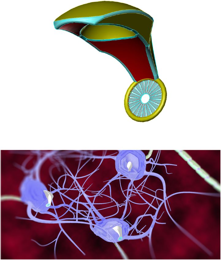
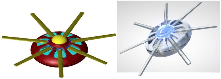
FIGURE 4 | Artistic representations of gliabots, which would self-migrate to
resolution, enabling a virtually real-time data stream between the glial cells and position themselves intracellularly at the most appropriate
human brain and the cloud. intra-glial regions to perform supportive B/CI operations. [Image credits:
(A) Frank Boehm - Nanoapps Medical, Inc. (B) Julia Walker, Department of
Chemical Engineering, Monash University]. (These conceptual illustrations do
Endoneurobots and Gliabots not represent the actual neuralnanorobot design of the gliabots).
Neuralnanorobots might be transdermally injected, after
which they would navigate the vasculature and anchor
to the endothelial cells of the BBB. A 10 µm3 volume
AIS (Martins et al., 2016). Similarly, a 10 µm3 volume of
of endoneurobots (Figure 3) would subsequently egress
gliabots (Figure 4) would egress the bloodstream, enter their
the bloodstream, traverse the BBB by methods that have
respective glial cells, and position themselves intracellularly
been extensively reviewed elsewhere (Freitas, 2016), enter
at the most appropriate intra-glial region, which can vary.
the brain parenchyma, and begin to navigate within the
The synaptobots would also enter the human body via the
neuropil. Subsequently, they would enter the neuron cell
bloodstream, cross the BBB (possibly assisted by auxiliary
soma and position themselves intracellularly within the
transport nanorobots), enter the brain parenchyma, commence
navigation within the neuropil, enter the neuron cell soma,
and then proceed intracellularly into the pre-synaptic or
post-synaptic structure of a synapse.
The synaptobots would reside in the proper monitoring
position within the neurons, in close proximity to presynaptic or
postsynaptic structures. Once in place, these neuralnanorobots
would monitor the action potentials and the structural changes
initiated by the action-potential-based functional data. These
data would be transferred from the synaptobots to corresponding
endoneurobots (in some cases, with communications and other
support from nearby gliabots). Once the data is received
FIGURE 3 | Artistic representation of endoneurobot (left) with diamondoid
by the endoneurobots, it would proceed to the previously
depiction (right). Grooves and orifices might facilitate propulsion within the installed in vivo high-speed nanofiber-optic network, for
neurons. Extendable tendrils could project from a number of these orifices to subsequent transfer to the central units that are responsible
enable stable anchoring and precise post-anchor positioning. [Image credits: for transmitting data to an external supercomputer. The
(left) Frank Boehm - Nanoapps Medical, Inc. and (right) Yuriy Svidinenko -
auxiliary nanofiber-optic network system would provide
Nanobotmodels Company]. (These conceptual illustrations do not literally
represent the actual neuralnanorobot design of the endoneurobots).
essential support for the data that is transmitted by the
endoneurobots and synaptobots, thereby minimizing their
Frontiers in Neuroscience | www.frontiersin.org 10 March 2019 | Volume 13 | Article 112You can also read