Toward a global and reproducible science for brain imaging in neurotrauma: the ENIGMA adult moderate/severe traumatic brain injury working group
←
→
Page content transcription
If your browser does not render page correctly, please read the page content below
Brain Imaging and Behavior
https://doi.org/10.1007/s11682-020-00313-7

 SI: ENIGMA TBI

Toward a global and reproducible science for brain imaging
in neurotrauma: the ENIGMA adult moderate/severe traumatic brain
injury working group
Alexander Olsen 1,2 & Talin Babikian 3,4 & Erin D. Bigler 5,6 & Karen Caeyenberghs 7 & Virginia Conde 1 &
Kristen Dams-O’Connor 8,9 & Ekaterina Dobryakova 10,11 & Helen Genova 10 & Jordan Grafman 12,13 & Asta K. Håberg 14,15 &
Ingrid Heggland 16 & Torgeir Hellstrøm 17 & Cooper B. Hodges 5,18,19 & Andrei Irimia 20,21 & Ruchira M. Jha 22,23,24 &
Paula K. Johnson 5,25 & Vassilis E. Koliatsos 26,27 & Harvey Levin 28,29 & Lucia M. Li 30,31 & Hannah M. Lindsey 5,18,19 &
Abigail Livny 32,33 & Marianne Løvstad 34,35 & John Medaglia 36,37 & David K. Menon 38 & Stefania Mondello 39 &
Martin M. Monti 40,41 & Virginia F.J. Newcombe 38 & Agustin Petroni 1,42,43 & Jennie Ponsford 44,45 & David Sharp 46,47 &
Gershon Spitz 44 & Lars T. Westlye 35,48 & Paul M. Thompson 49,50 & Emily L. Dennis 5,49 & David F. Tate 5,19 &
Elisabeth A. Wilde 5,19,28 & Frank G. Hillary 52,52

# The Author(s) 2020

Abstract
The global burden of mortality and morbidity caused by traumatic brain injury (TBI) is significant, and the heterogeneity of TBI
patients and the relatively small sample sizes of most current neuroimaging studies is a major challenge for scientific advances
and clinical translation. The ENIGMA (Enhancing NeuroImaging Genetics through Meta-Analysis) Adult moderate/severe TBI
(AMS-TBI) working group aims to be a driving force for new discoveries in AMS-TBI by providing researchers world-wide with
an effective framework and platform for large-scale cross-border collaboration and data sharing. Based on the principles of
transparency, rigor, reproducibility and collaboration, we will facilitate the development and dissemination of multiscale and big
data analysis pipelines for harmonized analyses in AMS-TBI using structural and functional neuroimaging in combination with
non-imaging biomarkers, genetics, as well as clinical and behavioral measures. Ultimately, we will offer investigators an
unprecedented opportunity to test important hypotheses about recovery and morbidity in AMS-TBI by taking advantage of
our robust methods for large-scale neuroimaging data analysis. In this consensus statement we outline the working group’s short-
term, intermediate, and long-term goals.

Keywords Brain injury . Radiology . Open Science . Neurodegeneration . Rehabilitation . ENIGMA

Brain injury and the ENIGMA consortium                                  nance imaging (MRI) methods and image analysis techniques
                                                                        have great potential to improve clinical assessment and guide
For over three decades, neuroimaging has played an important            management and treatment following msTBI. For this to be
role in the characterization and management of moderate-to-             possible, we must first address a number of scientific and
severe traumatic brain injury (msTBI). Novel magnetic reso-             practical challenges in our field. The vast heterogeneity of this
                                                                        patient population with respect to injury causes and mecha-
                                                                        nisms, neuropathology, and clinical or functional outcomes—
                                                                        in combination with the relatively small sample sizes of most
* Alexander Olsen
  alexander.olsen@ntnu.no                                               current neuroimaging studies—pose significant barriers to sci-
                                                                        entific progress and clinical translation.
* Frank G. Hillary
  fhillary@psu.edu                                                         The Enhancing Neuroimaging Genetics through Meta-
                                                                        Analysis (ENIGMA) consortium offers a framework for
Extended author information available on the last page of the article   meta- and mega-analysis of neuroimaging data across sites.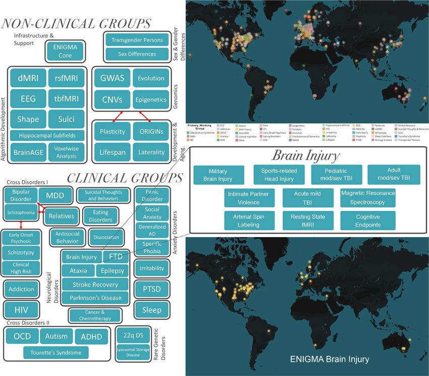
Brain Imaging and Behavior
This framework has proven to be a successful environment for (Teasdale and Jennett 1974) alone, or in combination with
studying other psychiatric and neurological populations, often other clinical signs or imaging (e.g. Stein and Spettell 1995),
with sample sizes 10–30 times larger than those in typical only partially capture the variability in cognitive, behavioral
brain imaging studies (Bearden and Thompson 2017; and social outcomes at acute and chronic stages following
Thompson et al. 2020). In this consensus statement, we de- injury. In the early phase after injury, lower GCS score, older
scribe the aims and goals of the ENIGMA Adult1 msTBI age, pupil dilatation, hypoxia, hypotension, and CT classifi-
(AMS-TBI) working group that was initiated in 2018 as part cations based on the size of lesions and the degree of midline
of ENIGMA Brain Injury, which is a collaboration of 10 TBI shift, provides some utility for predicting mortality and for
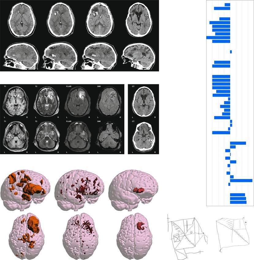
working groups (Wilde et al. 2019; Fig. 1). The group consists categorization of patients into very broad outcome groups
of scientists and clinicians from a wide-range of disciplines (Faried et al. 2018; Maas et al. 2013; Steyerberg et al. 2008).
and backgrounds, and we welcome new members from However, this information is less valuable for evaluating pa-
around the world to join our efforts. tients presenting with less severe injuries or more fine-tuned
The ENIGMA AMS-TBI working group aims to 1) be a prognostication of long-term neurobehavioral outcome.
driving force for new discoveries in AMS-TBI by providing Indeed, often individuals with similar indicators of severity
researchers with a comprehensive and effective framework and early clinical trajectories experience different outcomes
and platform for large-scale, cross-border collaboration and (Bigler et al. 2006; Lutkenhoff et al. 2019). Recovery and
data sharing. Moreover, we will 2) facilitate the development community re-integration are further complicated by a num-
and dissemination of multiscale and big data analysis pipe- ber of interacting premorbid, clinical, demographic, and ge-
lines for harmonized analyses in AMS-TBI using structural netic factors (Mollayeva et al. 2019).
and functional MRI in combination with other imaging mo- An integrated scientific endeavor is required in AMS-TBI
dalities, non-imaging biomarkers, genetics, as well as clinical research to address these challenges. This will require large
and behavioral measures. Ultimately, 3) we will offer investi- sample sizes drawn from a multi-modal approach which com-
gators an unprecedented opportunity to test important hypoth- bines neuroimaging, non-imaging biomarkers, psychological,
eses about injury neuropathology through recovery and mor- cognitive, and behavioral data. Recent developments in neu-
bidity in msTBI by taking advantage of our robust methods roimaging and computational algorithms offer powerful ap-
for large-scale data analysis. Below we outline the back- proaches to examine not only the overt behavior of individ-
ground and structure of ENIGMA AMS-TBI, the roles of uals, but also the brain structure, function, and neural compu-
investigators, and the working group’s short, intermediate, tations that can give rise to diverse outcomes (Amyot et al.
and long-term goals (Fig. 2). 2015). In addition, there is an unexploited potential of using
neuroimaging to inform clinicians about the optimal timing
and effects of interventions for individual patients.
Leveraging ENIGMA to address challenges In recent years, neuroscientists have encountered problems
in AMS-TBI research in the replication of published human neuroimaging studies,
especially those based on functional neuroimaging (Poldrack
TBI is a major and increasing global health challenge, with et al. 2017). Many believe that this may be due to small effect
more than 50 million new cases estimated to occur worldwide sizes; the median statistical power in neuroscience studies has
each year (5 to 20% are msTBI), and an ensuing disability that been estimated to be between 8 and 31% (Button et al. 2013).
is 2–3 times higher than the contribution from cerebrovascular One solution has been to increase sample sizes (Carter et al.
disorders or Alzheimer’s disease (GBD 2016 Neurology 2016; Szucs and Ioannidis 2017). Large-scale collaborative
Collaborators 2019; Maas et al. 2017). TBI is defined as an studies are therefore important to move the field of TBI for-
alteration in brain function, or other evidence of brain pathol- ward (Maas et al. 2017; Tosetti et al. 2013). Standing on the
ogy, caused by an external force (Menon et al. 2010), includ- shoulders of successful large-scale initiatives in TBI research
ing blunt or penetrating trauma, acceleration-deceleration such as the TRACK-TBI (https://tracktbi.ucsf.edu/)
forces or exposure to blast (Thurman and National Center consortium in the US, the CREACTIVE (http://creactive.
for Injury Prevention and Control (U.S.) 1995). marionegri.it/) and CENTER-TBI (www.center-tbi.eu; see
TBI is currently considered a chronic condition character- Steyerberg et al. 2019) collaborations in Europe, our
ized by evolving changes which require precise disease phe- ENIGMA working group will offer a new platform and
notyping, both in the acute stage and during the individual’s framework for researchers to make neuroimaging more
lifespan (Corrigan and Hammond 2013; Maas et al. 2017; useful for understanding AMS-TBI.
Masel and DeWitt 2010). Common measures of injury sever- By offering our framework and methods to the larger re-
ity, typically based on the Glasgow Coma Scale (GCS) score search community, we also aim to unlock the enormous po-
tential of analyzing dormant and unpublished “long tail” im-
1
Adult in this context is broadly defined as >16 years old. aging data (see Fig. 3a,b; Hawkins et al. 2019), which are not
Brain Imaging and Behavior

Fig. 1 The ENIGMA consortium and the Brain Injury working group. Organization and current geographical representation in the ENIGMA consortium
and the ENIGMA Brain Injury working group. Adapted from Thompson et al., 2020 and Wilde et al. 2019

being leveraged as part of any coordinated, collective effort            development in the biomedical industry. These advances
(Fig. 3a). Long-tail or “dark data” are data that accumulate in          could specifically enhance the use of neuroimaging in the
labs after a specific study finding is either interpreted and            clinical management of TBI through 1) the development and
published through a single study-specific lens or unpublished            validation of clinically useful methods for analysis that ac-
and then archived. Consequently, there is great potential to             commodate both high- and low-end imaging protocols (in-
integrate and harmonize such data in unique and creative                 cluding legacy data), and 2) by informing the development
ways. A central goal for our working group is to provide                 of a future core clinical imaging dataset for TBI, with acqui-
methodological tools to integrate these datasets from TBI labs           sition parameters and data structure established by broad con-
around the world (Fig. 3b).                                              sensus, that could be harmonized across vendors. Given that
   In the long term, methodologies developed as part of the              the clinical use of MRI dwarfs data acquired for research by
ENIGMA AMS-TBI initiative may have broader impact that                   several orders of magnitude, such harmonization (which is
go beyond research imaging. The lessons learned by integrat-             already occurring in some research contexts, e.g. Alfaro-
ing data and finding imaging biomarkers with diagnostic,                 Almagro et al. 2018; Wiberg et al. 2019) would make very
prognostic and therapeutic significance should inform the de-            large datasets accessible to research. Collation and integration
velopment of management protocols by clinicians and product              of such “non-research” clinical imaging for research could
Brain Imaging and Behavior
Fig. 2 Goals of ENIGMA AMS-
TBI. Schematic presentation of
the short, intermediate and long-
term goals of the ENIGMA
AMS-TBI working group
Establish ENIGMA AMS-TBI Consolidate ENIGMA AMS-TBI ENIGMA AMS-TBI
• Recruit researchers Support two main projects: • Be a driving force for new discover-
• Identify datasets • Improved methods for lesion map- ies in AMS-TBI
• Leadership and support ping and characterization • Development and dissemination of
• Harmonization of measures across multiscale and big data neuroimag-
• Regular meetings/calls
sites for improved clinical, cognitive ing analyses
• Memorandum of Underdstanding and behavioral phenotyping. • Offer investigators unprecedented
• Methods for data sharing and Motor Function
Finger Tapping Test Dominant Hand - R
Finger Tapping Test Non-Dominant Hand - L opportunity for hypothesis testing
Memory Function
handling regulatory issues RBANS Immediate Memory
RBANS Delayed Memory
RCFT Immediate Recall
RCFT Delayed Recall
CVLT II Trial 1-5 Total
in AMS-TBI
T2 FLAIR WM-Abnormalities SWI Identified Hemosiderin CVLT II Short Delay Free Recall
CVLT II Long Delay Free Recall
Language Function
RBANS Language PiB PET MRI ADC fMRI
Visuospatial/Constructional
RBANS Visuospatial/Constructional
RCFT Copy
Executive functioning
D-KEFS Visual Scanning
D-KEFS Number Sequencing
D-KEFS Letter Sequencing
D-KEFS Number-Letter Switching
D-KEFS Motor Speed
RBANS Attention
D-KEFS Letter Fluency
D-KEFS Category Fluency
D-KEFS Category Switching Response
D-KEFS Category Switching Accuracy
D-KEFS Color Naming
IMAGING
D-KEFS Word Reading
D-KEFS Inhibition
D-KEFS Inhibition/Switching
D-KEFS Error analysis Inhibition
D-KEFS Error analysis Inhibition/Switching
FEATURES
Patient Reported Symptoms (SCL-90)
Somatization
Obsessive-Compulsive
Interpersonal Sensitivity
Depression
Anxiety
Hostility
Phobic Anxiety
Paranoid Ideation
Psychoticism
Global Severity Index
Positive Symptom Distress Index
Positive Symptom Total
Informant Reported Executive Functioning
BRIEF-A Behavioral Regulation Index (BRI)
BRIEF-A Metacognitive Index (MI)
DTI MRI GRE
BRIEF-A General Executive Composite (BRI + MI)
-4 -3 -2 -1 0
Z-Scores
1 2 3 4
CT
Short-term (1st year) Intermediate (1-2 years) Long-term (>2 years)
deliver analyses that involve datasets with n > 100,000. There safeguards (The Royal College of Radiologists 2017), and
are clear regulatory barriers and consent hurdles that need to such use of data may be further facilitated by federated anal-
be addressed before such data were freely available for yses of data, where the research pipelines are brought to the
research (Anderson 2015; Benchimol et al. 2015). However, data (rather than vice versa), both for structural and functional
authoritative views suggest that fully anonymized clinical im- imaging (X. Li et al. 2020; Silva et al. 2019).
aging data can be used for research purposes with appropriate
Fig. 3 The long tail and dark data for traumatic brain injury (TBI) data that are inaccessible or archived. b The goal is to make TBI
research. The current state of TBI data consists of a relatively small imaging data Findable, Accessible, Interoperable, and Reusable (FAIR,
number of large, publicly accessible datasets reflected schematically as Wilkinson et al., 2016) thereby shortening the long tail of dark data, and
a right-skewed distribution (Panel a). The majority of data collected by making a greater proportion of the data in the TBI literature publicly
the field exists in the long tail of the distribution, with most datasets accessible to drive new discoveries and accelerate translation. (Adapted
consisting of relatively modest data sizes as either gray data that are from Hawkins et al., 2019)
difficult to access beyond summaries reported in publications; or darkBrain Imaging and Behavior
Short-term goal: Forming ENIGMA al. 2012; Boedhoe et al. 2017; Guadalupe et al. 2017; Hibar et
AMS-TBI—Its structure and methodological al. 2016; Hibar et al. 2017, 2015; Hoogman et al. 2017; Ikram et
framework al. 2012; Jahanshad et al. 2013; Schmaal et al. 2016; Stein et al.
2012; van Erp et al. 2016).
Our short-term goal (1st year) is to identify datasets and recruit One strength of ENIGMA is the focus of researchers within
researchers as members by providing an attractive platform and the consortium to develop standardized data processing pipe-
framework for global large-scale cross-border collaboration, data lines for handling distinct data types. Much of the variability
sharing, and analysis. A strength of ENIGMA AMS-TBI is our in research comes from investigators decisions in data pro-
emphasis on supporting the TBI research community with robust cessing and analysis, referred to as “researcher degrees of
methods and analyses, and the goal to advance brain imaging freedom” (see Nichols et al. 2017). In modern neuroimaging,
science in neurotrauma through the principles of transparency, these degrees of freedom can be readily found in analyses of
rigor, reproducibility, and collaboration. both structural and functional imaging data (Hallquist and
For ENIGMA AMS-TBI, there is a low threshold for par- Hillary 2019). To standardize approaches for data pre-pro-
ticipation (data sharing is not required), allowing individual cessing, in particular for functional imaging data pipelines,
researchers to choose to participate at different levels depending we plan to integrate members of the international community
on their interests and/or situation. There are a number of differ- conducting AMS-TBI work to investigate how to best harmo-
ent ways researchers can participate, these include:1) mega- nize and standardize such methods and provide quality con-
analyses (sharing raw data or numerical output from such data), trol. Overall, the goal for our ENIGMA working group is to
2) meta-analyses (no need to share raw data), and 3) methods act as a forum where AMS-TBI scientists can interact and
and protocol development (no need to participate with data). collaborate, and where consensus on methods can evolve
We welcome proposals from the TBI research community at and become suitable for the larger scientific community.
large, and we will serve as a hub for investigators who could Participating members are encouraged to adhere to the FAIR
benefit from the ENIGMA structure. In addition to working Data Principles (Findable, Accessible, Interoperable, Reusable),
with existing datasets, we will provide a platform for re- to enhance the usability of data (Wilkinson et al. 2016). Primary
searchers to collect and harmonize future studies. Most data data, derived data, and other research outputs such as protocols,
acquired to date have been collected using diverse protocols. source code and software, if well documented, accompanied by
Members of the group are developing protocols for future data descriptive metadata and organized in a standardized way, are
collection, to enable prospective harmonization within individ- likely to foster collaboration and reproducibility. An example of
ual cohort studies, thereby allowing members to participate in a relevant data repository for publishing data in neuroimaging is
future multicenter initiatives at low additional cost and effort. It OpenNeuro (https://openneuro.org), which uses the Brain
would be critically important, in this context, to ensure that we Imaging Data Structure (BIDS) format for organizing data
start with what is most universally implementable, and identify (https://bids.neuroimaging.io). By adhering to relevant
a core set of sequences and data collection for widespread use. standards, harmonizing analysis tools and sharing data as open
As with the NINDS Common Data Elements (CDEs; https:// as possible, possibilities for reuse, reproducibility, as well as
www.commondataelements.ninds.nih.gov/Traumatic% meta- and mega-analysis greatly increase, both within
20Brain%20Injury#pane-162) it may be useful to also provide ENIGMA and in the greater research community.
more aspirational imaging standards as basic and supplemental The ENIGMA AMS-TBI working group will provide sup-
- thus allowing optional use of more complex harmonized im- port to members on regulatory issues based on accumulated
age collection, if appropriate and possible. knowledge and available expertise within the network. When
The AMS-TBI working group will benefit greatly from the combining different data for analysis, there are many levels of
established procedures, methods, and analytic pipelines that sharing, ranging from sharing the raw data, to sharing quanti-
have engendered success across the larger ENIGMA consor- tative measures and features extracted from imaging scans, to
tium including more than 1400 scientists across 43 countries sharing only meta-data. It is important to consider the type of
and more than 20 psychiatric, neurological, and data to be shared and the local (institutional, national or inter-
neurodevelopmental disorders (Thompson et al. 2020). national) rules and regulations that need to be followed. There
Extending prior efforts, we will develop a comprehensive set is, therefore, not a single approach, and each participating site
of protocols, procedures and open source code for data analysis needs to abide by appropriate regulations. Our members have
tailored for tackling major challenges in msTBI imaging. Part of extensive experience in dealing with such issues, not only
this effort can be found in previous ENIGMA programs, which from participation in other ENIGMA groups, but also through
have developed imaging analysis pipelines to extract, homoge- participation in other large-scale international TBI collabora-
nize, and control the quality of data describing standardized tions (e.g., CENTER-TBI).
phenotypes from structural T1-weighted MRI, diffusion MRI, Working group chairs provide leadership to support re-
resting state functional MRI and EEG (Adams et al. 2016; Bis et searchers in achieving planned objectives. Our approach isBrain Imaging and Behavior
a d
1 Day 2 Weeks 5 Months
Day of Injury Neuropsychological Assessment ~8 Months Post-Injury
Post-Injury Post-Injury Post-Injury
Motor Function
Finger Tapping Test Dominant Hand - R
Finger Tapping Test Non-Dominant Hand - L
Memory Function
RBANS Immediate Memory
RBANS Delayed Memory
RCFT Immediate Recall
RCFT Delayed Recall
CVLT II Trial 1-5 Total
CVLT II Short Delay Free Recall
CVLT II Long Delay Free Recall
Language Function
RBANS Language
Visuospatial/Constructional
RBANS Visuospatial/Constructional
RCFT Copy
Executive functioning
D-KEFS Visual Scanning
b D-KEFS Number Sequencing
2 Weeks Post-Injury 5 Months Post-Injury D-KEFS Letter Sequencing
D-KEFS Number-Letter Switching
D-KEFS Motor Speed
RBANS Attention
D-KEFS Letter Fluency
D-KEFS Category Fluency
D-KEFS Category Switching Response
D-KEFS Category Switching Accuracy
D-KEFS Color Naming
D-KEFS Word Reading
D-KEFS Inhibition
D-KEFS Inhibition/Switching
D-KEFS Error analysis Inhibition
D-KEFS Error analysis Inhibition/Switching
Patient Reported Symptoms (SCL-90)
Somatization
Obsessive-Compulsive
Interpersonal Sensitivity
Depression
Anxiety
c Hostility
Phobic Anxiety
T2 FLAIR WM-Abnormalities SWI Identified Hemosiderin T1 Abnormalities
Paranoid Ideation
Psychoticism
Global Severity Index
Positive Symptom Distress Index
Positive Symptom Total
Informant Reported Executive Functioning
BRIEF-A Behavioral Regulation Index (BRI)
BRIEF-A Metacognitive Index (MI)
BRIEF-A General Executive Composite (BRI + MI)
-4 -3 -2 -1 0 1 2 3 4
Z-Scores
RCFT Copy RCFT Delayed Recall
based on the principles of team science and our success is Intermediate goal: Provide tools for improved
expected to be driven by the collective coordinated effort of lesion mapping and clinical, cognitive
participating researchers. Building on years of experience from and behavioral phenotyping in AMS-TBI
ENIGMA, we have developed a group-specific memorandum
of understanding (MOU), with policies for data sharing, author- Our intermediate goal (1–2 years) is to support two overarch-
ship, and for initiating new studies. Communication within the ing projects to address key challenges linked to the heteroge-
group will largely involve teleconferences with alternating neity of msTBI which will benefit all future ENIGMA AMS-
scheduling to accommodate members across different time- TBI projects. The first project will focus on developing im-
zones, in addition to face-to-face meetings, often in connection proved methods for lesion characterization, mapping, and
with international conferences. quantification. The second project will focus onBrain Imaging and Behavior
Fig. 4 The complexity of lesion characterization and behavioral phenotyping after AMS-TBI. From a structural neuroimaging perspective trauma-
R induced abnormalities differ by time post-injury as well as the imaging modality being used. a are all CT based showing that the size and location of the
hemorrhage, parenchymal displacement and edema dynamically change over time. b demonstrates that each MRI sequence has its own unique
sensitivity in assessing different aspects of neuroanatomy and neuropathology. c which presents the FLAIR, SWI and T1 signal abnormalities,
demonstrates the widespread pathology differently presented by these imaging methods. By 5 months’ post-injury, widespread volume loss, cortical
atrophy, ventriculomegaly and encephalomalacia have occurred. d show summary findings from a neuropsychological assessment at ~8 months post
injury. This case example depicts the neuropathological heterogeneity associated with TBI along with the dynamic changes over time and their influence
on neuropsychological test results. This patient sustained a severe TBI from a motorcycle collision with a vehicle. The patient was not helmeted at the
time of injury and, by witness accounts, was immediately rendered unconscious. Upon emergent care at the scene of the accident, the patient was
assessed to have a Glasgow Coma Scale (GCS) of 3, was life-flighted to a Level I emergency department (ED) with GCS remaining 3 throughout
transport and during ED assessment and treatment. In addition to the head injuries he sustained multiple systemic injuries including leg and rib fractures,
pulmonary contusion and liver laceration. An intracranial pressure monitor was inserted, the patient underwent tracheostomy for airway management and
shunted. The patient remained in a coma and received neurocritical care for almost 2 months, followed by 3 months of inpatient neurorehabilitation. a
Initial day-of-injury computed tomography was performed about 90 min’ post-injury. What is important to note in the initial scan is the original size of
the frontal intraparenchymal hemorrhage along with the size, symmetry and configuration of the ventricular system. Within 24 h, enlargement of the
intraparenchymal hemorrhage is observed along with distinct effacement of the anterior horn of the lateral ventricle and surrounding edema associated
with the hemorrhage. Subsequent to this scan he was shunted, with the shunt catheter clearly visible in the 2-week follow-up scan which depicts more
edema and midline shift. By 5 months’ post-injury, there is prominence of the ventricular system and cortical sulci in association with cortical atrophy
and frontal encephalomalacia associated with the location of the prior hemorrhage. b At 2 weeks post-injury, MRI studies were obtained. Each sequence
demonstrates a different aspect of the “Lesion.” The T1 sequence, which is the one commonly used for automated methods of image segmentation and
classification for quantitative analyses, depicts coarse anatomical features of the brain, but the focal intraparenchymal hemorrhage and surrounding
edema is not fully appreciated, being better distinguished by the T2 and FLAIR sequences. The SWI sequence depicts multiple, bilaterally scattered foci
of hemosiderin deposition reflective of shear injury, with particularly exquisite demarcation differentiating hemorrhage, parenchymal degradation along
with the surrounding edema. c Using a thresholding method for detecting white matter signal abnormality in FLAIR scans, the regions of white matter
hyperintensity are depicted three dimensionally in the images on the left. Each signal abnormality likely reflects localized white matter pathology. In the
middle are the regions of hemosiderin deposition detected on SWI, likewise reflecting specific foci of shear-lesion pathology constituting diffuse axonal
injury. On the right are the abnormalities found on T1. d Findings from neuropsychological assessment at almost 8 months post injury are presented as z-
score deviations from test manual normative data. The following tests were administered: Repeatable Battery for the Assessment of Neuropsychological
Status (RBANS, https://www.pearsonassessments.com/), Rey Complex Figure Test (RCFT, https://www.parinc.com), California Verbal Learning Test-
II (CVLT-II, https://www.pearsonassessments.com/), Delis-Kaplan Executive Function System (D-KFES, https://www.pearsonassessments.com/);
Symptom Checklist-90 (SCL-90, https://www.pearsonassessments.com/) and the Behavioral Rating Inventory of Executive Function (BRIEF, https://
www.parinc.com). Clinically, the 25-year-old presented with left side hemiparesis, emotional lability and major cognitive impairments, most notable in
terms of memory and executive functioning. Family and caregivers were most concerned about the patient’s irritability and inappropriate outbursts along
with impaired insight and judgment. Neuropsychological tests (lower z-scores = poorer function) demonstrated the expected left side reductions in motor
control (reduced finger tapping and grip strength) consistent with the location of the large intraparenchymal right frontal hemorrhagic injury (see Fig. 4a-
c). He was anosmic and unable to identify basic odors on the Smell Identification Test (https://sensonics.com/) along with diminished tactile
discrimination on the left side, but no visual field defect. Constructional praxis was diminished as evident in the copy of the Rey Complex
Figure Test (RCFT), with the more profound deficits most notable with impaired immediate as well as delayed memory. Memory and executive
impairments were evident on the RBANS, CVLT-II and DKFES tasks. Caregiver observation, based on the BRIEF (higher z-scores = more
problems) also confirmed real-world deficits in day-to-day impairments in planning, organization, decision making and problem solving.
Emotionally, as also reflected in the BRIEF results, the family caregiver reported marked dysfunction in emotional regulation with poor self-
monitoring and impaired insight. In contrast, on the SCL-90 (higher z-scores = more symptoms), which is a self-report measure, while somatic issues
that related to mobility and pain were prominently endorsed, the Global Severity Index (GSI) was only minimally elevated, with no significant
endorsement of symptoms related to depression or anxiety. This would be consistent with caregiver observations that the patient lacked insight into
changes in personality and emotional control, impairments often reported to be present in TBI patients with extensive frontotemporal pathology (Krudop
& Pijnenburg, 2015), as evident in this patient
harmonization of measures across sites to allow for improved mapping, pathology characterization, and clinical interpreta-
clinical, cognitive, and behavioral phenotyping. This will pro- tion (see Fig. 4). The heterogeneity of lesion profiles (e.g,
vide the research community with important methods to di- biomechanical cause, type of pathology, location, or volume)
rectly address two of the main challenges regarding clinico- frequently makes automatic MRI analysis pipelines break
pathological heterogeneity in msTBI. These projects will also down or fail due to causes that frequently include (but are
serve as important vehicles to motivate researchers to join our not exclusive to) inaccurate co-registration of scans across
early efforts, and for consolidating our working group. modalities and time points, faulty voxel-wise morphometric
analysis, and incorrect automatic parcellations of brain struc-
Standardization of image analysis protocols for AMS-TBI: tures (Irimia et al. 2014).
Improved methods for lesion characterization, mapping, Our working group will aim to propose, implement and
and quantification Our working group will aim to provide validate standards to facilitate such operations and to enhance
standardized best practices (e.g. Nichols et al. 2017) for mul- their reproducibility. Even relatively “simple” image process-
timodal neuroimaging analysis in AMS-TBI. This need is ing steps like “skull-stripping” - which is required for many
critical partly because analyzing MRI scans from AMS-TBI processing pipelines and brain co-registration - tend to fail
patients poses unique challenges from the standpoint of lesion when using conventional software on images from patientsBrain Imaging and Behavior
with TBI, and require customized pipelines (Lutkenhoff et al. understanding and interpretation of the available MRI sequence
2014). Conventional lesion mapping approaches - such as modalities and on their correct joint interpretation.
pathology masking - can fail, especially when multiple large
lesions are present (Wong et al. 2016). This is partly because Harmonizing protocols for improved clinical, cognitive, and
masks frequently classify voxels from different lesion types behavioral phenotyping through large-scale datasets
identically regardless of their presentation on MRI, and dis- Accurate patient diagnosis and prognostication, with respect
card potentially valuable information on lesion type and loca- to clinical, cognitive, and behavioral outcomes, is paramount
tion, factors that may have prognostic utility (B. Wang et al. within the TBI field. A sound clinical evaluation of an indi-
2013). Furthermore, lesion masks do not convey either the vidual patient includes information about premorbid factors,
pattern or the extent of injury-related brain deformations. As injury-related variables, and broad clinical and functional as-
a result, careful testing and validation—including visual in- sessments that are integrated and appropriately interpreted or
spection by neuroradiologists—can be necessary even when formulated (see Fig. 4). In the scientific literature, only general
masking techniques have been validated on systematic associations have emerged between premorbid, clinical, and
lesioning data sets. There has been growing interest in using demographic factors and subsequent outcomes (Ponsford et
machine learning (ML) to improve anatomical parcellation al. 2008; Spitz et al. 2012; Wood and Rutterford 2006). For
(Ledig et al. 2015) and lesion detection based on computed example, history of emotional disturbance, older age, and
tomography (CT) (Jain et al. 2019) and anatomical MRI higher severity of injury generally lead to poorer functional
(Kamnitsas et al. 2017) in AMS-TBI patients. Combining outcomes (Hoofien et al. 2001; Spitz et al. 2019). More re-
such methods with large databases of systematic lesions cently, there has been significant investment in identifying
(Wang et al. 2013) may be particularly advantageous for reliable biomarkers to aid in the initial diagnosis and charac-
connectome analysis (Irimia and Van Horn 2014) or when terization of TBI and prediction of future outcomes, ultimate-
the alternative involves laborious manual delineation. One ly, to enable tailored clinical interventions (“Precision
aim of our working group will be to propose detailed proce- Medicine”). This quest has included physiological and neuro-
dures to integrate information from different sources and imaging measures.
methods and to provide guidelines on their use. Group-level results suggest that anatomical and functional
As an example, we aim to provide distinct lesion mapping alterations to the brain generally correlate with changes in
decision trees that accommodate the availability—or partial cognition and behavior (e.g. Bonnelle et al. 2012; Brezova
lack—of MRI scans acquired using various sequences, includ- et al. 2014; Håberg et al. 2015; Kinnunen et al. 2011; Olsen
ing T1-weighted (T1w), susceptibility weighted imaging (SWI), et al. 2015). Brain changes have been characterized with re-
and fluid-attenuated inversion recovery (FLAIR) scans. TBI le- spect to loss in regional volume, altered white-matter micro-
sion characterization is a complex inferential process, which structure, functional connectivity and brain activation. Despite
aims to identify the lesion’s content, physical properties and the application of advanced neuroimaging techniques to TBI,
evolution based on complementary information from a variety including diffusion-weighted imaging and functional connec-
of MRI modalities. After image preprocessing, distinct MRI tivity analyses that can reveal subtle brain changes, the vast
modalities can be used to extract unique information on the majority of the variability in outcomes remains unexplained.
physical content, pathophysiological state or likely longitudinal This situation clearly highlights the problem of heterogeneity
trajectory of each lesion-confined voxel (Wang et al. 2013). For in TBI outcomes and raises the need for ENIGMA-type large-
example, the T1w MRI contrast is indicative of the content of fat, scale research projects. The lack of reliable predictive bio-
whereas FLAIR hyperintensities are linked to the localized tis- markers hamper the development of disease-modifying thera-
sue water content (e.g. suggesting vasogenic edema or pies. Moreover, there is difficulty in translating results obtain-
perivascular CSF). By contrast, SWI hypointensities results ed at the group-level to the individual, likely due to large
from the presence of (i) ferromagnetic hemoglobin in the lumina variability in regard to patient preinjury/genetic profile, demo-
of blood vessels and (ii) extravasated ferromagnetic material in graphics, injury mechanism, type and location and post-injury
the cerebral parenchyma. Consequently, lesion description can interval/phase (Fisher et al. 2018; Moen et al. 2016; Molenaar
be challenging in AMS-TBI because inferring MRI signal prov- et al. 2009). Failure to deduce facts from groups to individuals
enance does not equate straightforwardly to the characterization is probably a major factor explaining the failure of therapeutic
of pathobiology. For this reason, when lesion-related informa- interventions (L. M. Li et al. 2014; Saatman et al. 2008).
tion is made available from fewer—rather than more— Therefore, accurate individual-specific diagnosis must pre-
information channels, subtle yet consequential issues of inter- cede the development of effective treatments.
pretability and diagnosis may arise. To address such difficulties, The problem of heterogeneity is not unique to TBI. Many
our working group will aim to formulate a detailed protocol and other fields—for example, psychiatry—also face the ‘hetero-
implement conservative guidelines for lesion mapping, quanti- geneity problem’ (Feczko et al. 2019); 1) that any outcome or
fication and characterization based on the rigorous constellation of symptoms is not caused by a singleBrain Imaging and Behavior
mechanism, but is the result of variable combinations of transformative potential to more accurate patient classification
known and unknown factors; and 2) that our way of measur- opening avenues toward a more personalized medicine in
ing individual outcomes influences how we determine the AMS-TBI. Our ENIGMA AMS-TBI initiative will facilitate
relevant contribution of the potential mechanisms. For exam- these goals (a–d) and even allow for collection of new data as
ple, MRI measures that best diagnose TBI may differ from a consortium to fill gaps or deepen phenotyping.
those that best predict development of emotional disturbance,
or manifestation of any other behavior, following TBI.
Defining adequate, clinically relevant, and agreed-upon out- Long-term goal: ENIGMA as a sustainable
come measures poses a serious challenge. The interagency and driving force for new discovery
Traumatic Brain Injury Outcomes Workgroup addressed pri- in AMS-TBI
marily clinical research objectives (Hicks et al. 2013; Wilde et
al. 2010). The rationale behind the core measures was the need Our long-term goal (>2 years) is to be fully engaged with the
to create a primary set of well-established measures that ad- broader TBI research community and support researchers in
dress outcome domains in many studies. This group sought to tackling important research questions in AMS-TBI, focusing
identify a single measure or limited set of measures that best on the unique contributions of big data approaches. We expect
represented each domain. One of the primary objectives was the ENIGMA strategy to be ideally suited to particular re-
to facilitate comparability of outcome measurements across search questions and our early efforts will leverage our prima-
studies. Important efforts such as the Common Data ry strengths of data sharing and methods development. Here,
Elements (CDE) initiative have provided some direction for we outline a number of areas where we have current expertise
researchers for selecting CDEs linked to demographics, acute within ENIGMA AMS-TBI, and where we believe our ap-
clinical assessment, neuroimaging, biomarkers/specimens and proach has a lot of potential for high gains in the field.
outcome measures (Duhaime et al. 2010; Thurmond et al. Examples are provided recognizing that our group is in its
2010; Yue et al. 2013). Also, the Traumatic Brain Injury early phase, anticipating that the approaches and initiatives
Endpoints Development (TED) Initiative aims to provide har- will be shaped further by existing and new members.
monization of study measures across eight major TBI studies
(Manley et al. 2017). However, most existing msTBI studies Conducting international replication/reproducibility effort in
do not adhere to the CDEs or other standards. ENIGMA AMS-TBI With the replication crisis that emerged in the social
AMS-TBI will work with existing initiatives focusing on pro- sciences in 2015 (Maxwell et al. 2015; Open Science
spective or retrospective harmonization of measures and data Collaboration 2015) and expanded to nearly every corner of
across studies with an aim to contribute to a global solution to science, including the neurosciences (Button et al. 2013) there
this challenge. have been recent efforts to galvanize the community around
One avenue for tackling the challenge of heterogeneity is specific processing pipelines (see Esteban et al. 2019). In con-
by leveraging large-scale collaborative initiatives. The cert with these efforts we aim to work with the international
ENIGMA AMS-TBI working group offers: a) the ability to community to leverage the power of data sharing in order to
standardize quality assurance (QA) and imaging protocols identify the most robust findings in the TBI literature.
across sites; b) the potential for harmonization of current and To do so, the ENIGMA AMS-TBI aims to establish reli-
future demographic, clinical, and behavioral measures across able findings in the imaging and genetics community that can
sites. This will be accomplished by finding CDEs across serve as anchors to the field. From these vantage points, the
cohorts—what measures have most commonly been collected science of TBI can then advance on a firmer scientific footing.
and offer the most overlap across sites. Incorporated into this Given the range of possible premorbid and injury-related fac-
pipeline will be methods that handle, compare, and impute tors that influence the central neural system (CNS) and its
missing information from existing data; c) an open discussion functions (behavior), there remain great challenges in the
forum to establish a consensus regarding relevant and appro- study of reproducibility in TBI research. The promise this
priate measures for diagnosis as well as prognosis within effort holds, however, is to determine if key findings emerging
msTBI. The inclusion of clinicians and clinical researchers from the imaging literature are generalizable across sites and
in the ENIGMA AMS-TBI initiative will contribute to sound samples, thus providing investigators with a foundation from
discussions of what behavioral, cognitive and other psycho- which they can work. Establishing those reliable findings is
logical outcome measures are most likely to provide the most vital for the advancement of our understanding of the conse-
relevant optimal benchmark for imaging data; and d) given the quences of TBI on neural systems and patient outcomes. The
larger sample size, the ability to begin using new tools and ENIGMA AMS-TBI working group will vet the first genera-
techniques to better examine clinical, cognitive, and behavior- tion of replication studies with the TBI community and begin
al phenotypes or subgroups of patients. Advances in compu- designing analyses based upon data currently existing
tational and machine learning approaches may hold a amongst our collaborators. Moreover, we invite investigatorsBrain Imaging and Behavior
in the TBI research community to propose critical topics that animal studies, can also shed light on the mechanisms under-
require replication and can be supported by the ENIGMA lying msTBI (Lutkenhoff et al. 2019). Moreover, the value of
AMS-TBI working group. Establishing the reproducibility early MRI is not limited to structural scans. For example, a
of our science is a core agenda item for the ENIGMA AMS- functional MRI study in patients with post-traumatic amnesia
TBI working group. found evidence of disconnection between the medial temporal
lobes and the default mode network (De Simoni et al. 2016).
Acute/early MRI for guiding intervention and prognosis ENIGMA AMS-TBI will work on improved methods to de-
While CT imaging will continue to play an important role in lineate the optimal timing of MRI after AMS-TBI and to fur-
clinical decision-making in the acute treatment of AMS-TBI ther identify and refine lesion patterns yielding important
(Irimia et al. 2019), increased attention has been given to the prognostic information which can guide clinical decision-
clinical and prognostic value of acute/early MRI. Although making.
the optimal timing of MRI acquisition after AMS-TBI is still
unknown and may be both injury-specific and patient-specif- Imaging disorders of consciousness (DOC) after TBI Progress
ic, imaging does need to be performed early enough to inform in intensive care medicine has led to a large increase in the
clinical decision making. Taking an acute patient for an MRI proportion of patients who survive msTBI (Laureys and Boly
scan from an intensive care unit (ICU) while under ventilation 2008; Masel and DeWitt 2010). A majority of AMS-TBI sur-
can be challenging, but remains a vital means for assessment vivors enter a transient state of coma, which is generally con-
when precautions are taken to ensure MRI compatibility and sidered to resolve within 3 to 4 weeks (Young 2009), to then
safety (Carter et al. 2013; Newcombe et al. 2008; Newcombe spontaneously regain the two cardinal elements of conscious-
and Menon 2016). ness: arousal and (self-)awareness (Laureys 2005).
Early MRI has been successfully implemented to assess the Conventional structural MRI, DTI, and fMRI can provide
presence and evolution of brain lesions due to trauma added prognostic accuracy to the clinical observations and
(Newcombe et al. 2016, 2013). For example, the presence of CT imaging in predicting which patients will emerge from
brainstem lesions has been linked to increased mortality and coma (Snider et al. 2019; Stevens et al. 2014). A small
unfavorable Glasgow Outcome Scale at 6 months (risk ratio, number of patients with very severe TBI (Beaumont and
1.78; 95% CI, 1.01–3.15; I = 43%) (Haghbayan et al. 2017), Kenealy 2005; Løvstad et al. 2014; van Erp et al. 2015),
while lesions involving the ascending arousal network may be however, fail to fully regain consciousness and enter
critically predictive of poor outcome (Izzy et al. 2017; Moe et (transiently or for prolonged and sometimes life-long periods)
al. 2018). However, the accuracy and replicability of such into a vegetative (VS) or a minimally conscious state (MCS)
findings will benefit from the analysis of larger samples from (cf., Giacino et al. 2002; Jennett and Plum 1972; Monti et al.
multiple sites. Additionally, greater exploration of the func- 2010a). In the context of these three conditions (i.e., coma,
tional impact of injury to additional brain regions and the VS, MCS) - often referred to as Disorders of Consciousness
manner in which the same regions are impacted across imag- (DOC) - diagnosis and prognosis are a critical challenge
ing modalities is needed. (Monti et al. 2009; Owen and Coleman 2008). In the absence
In addition to prognosis, acute/early MRI may provide key of an objective means of determining level of consciousness,
information on the pathophysiological processes of specific differentiating an MCS from a VS is an inferential process
lesion types. Contusions in TBI tend to have distinct regions: (Giacino et al. 2014) which is known to be logically problem-
a core of restricted diffusion associated with hematoma, atic (cf., Monti and Owen 2010) and prone to misdiagnosis
surrounded by an area of raised apparent diffusion coefficient (Schnakers et al. 2006, 2009). However, accurate diagnosis of
(ADC) likely to be due to vasogenic edema, and in earlier DOC is essential for medical management, prognosis, moni-
scans (within 72 h) an outer rim of ADC hypointensity that toring of interventions, as well as the complex legal and eth-
is later subsumed by the vasogenic edema (Newcombe et al. ical ramifications concerning end-of-life decisions. Over the
2013). This outer rim may represent a region of microvascular last 20 years, neuroimaging has revolutionized our under-
failure resulting in cytotoxic edema, and may represent a standing of these conditions (Lutkenhoff and Monti 2016).
“traumatic penumbra” which may be rescued with effective Functional MRI has shown the ability to detect both residual
therapy. Indeed, in such “at-risk” regions of metabolically cortical processing and networks (e.g., Laureys et al. 2000;
compromised tissue, normobaric hyperoxia has been shown Menon et al. 1998; Monti, Pickard, and Owen 2013; Owen
to increase oxygen utilization using 15O PET and, thus, may et al. 2005) and voluntary (brain) behavior (e.g., Bardin et al.
help save the metabolically compromised tissue (Nortje et al. 2011; Edlow et al. 2017; Monti et al. 2015; Monti et al.
2008). This is consistent with a subsequent study which found 2010b) in a minority sub-group of otherwise unresponsive
that normobaric hyperoxia may pseudo-normalize the ADC in patients. 18F-FDG-PET has been shown, in a recent clinical
the cytotoxic rim (Veenith et al. 2014). Acute and early clin- validation study (Stender et al. 2014), to be able to detect the
ical MRI, in conjunction with carefully executed experimental presence of awareness in DOC with greater sensitivity thanBrain Imaging and Behavior
fMRI (93%) and to predict long-term outcome with high ac- improvements from methylphenidate treatment (Jenkins et
curacy (74%). al. 2019).
While traditional readings of structural imaging data (e.g., There is a need for large, well-controlled studies that in-
CT, MRI) have shown limited utility in DOC, more advanced clude neuroimaging data to better understand the neural un-
analytical and imaging techniques yield greater promise in derpinnings of treatment efficacy and individual injury-related
their ability to uncover patterns of damage in large-scale brain factors (Vander Linden et al. 2018) that contribute to success
networks (Monti 2012; Schiff 2010), considered hallmarks of or failure of a given intervention. One goal of ENIGMA
DOC, and to differentiate between diagnostic categories. AMS-TBI is to support analyses of effects of interventions
Advanced (i.e., “shape”) analysis of routine T1-weighted data, on broad cognitive processes even in the context of distinct
for example, has demonstrated a link between thalamic and imaging and rehabilitation protocols. This approach will seek
extra-thalamic subcortical atrophy and depth of impairment in to isolate the most robust main effects irrespective of between-
chronic DOC patients across etiologies (Lutkenhoff et al. study differences, which may guide more nuanced work to
2015) - a pattern of atrophy which, at least in TBI, might take examine mechanisms. Currently, the only method to examine
shape in the first months post injury (Lutkenhoff et al. 2019; main effects is to perform meta-analysis work limited to com-
Schnakers et al. 2019). Diffusion MRI can also help in quan- bining studies by cognitive modality (e.g., interventions that
tifying the structural integrity of white matter, and thus poten- aim at improving memory or attention). However, such an
tially the primary and secondary network damage encountered approach is still restricted by the absence of harmonization
in DOC (Voss et al. 2006). Several recent studies suggest that in scanning protocols and outcome variables, potentially call-
DTI-derived metrics of fractional anisotropy and diffusivity ing for the use of data reduction techniques (such as explor-
may be useful in differential diagnosis through the identifica- atory principal component analysis on disparate neuropsycho-
tion of the neural networks underlying the various levels of logical data) and use of multiple covariates (sample size per-
impairment seen in DOC (Wu et al. 2016; Xu et al. 2017; mitting). Second, to understand the efficacy of distinct reha-
Zheng et al. 2017). Nonetheless, several gaps in the literature bilitation protocols across a range of behaviors (e.g., improve-
and challenges in applying neuroimaging techniques to DOC ments in attention and/or memory), we will facilitate prospec-
still remain (Cavaliere et al. 2014). Patients with prolonged tive work by supporting data harmonization and analyses.
DOC are relatively few, and imaging these patients is chal- While there remain important challenges, data sharing offers
lenging. Most imaging group studies of DOC are performed the opportunity to orient a community of researchers around
on patients with mixed etiology (e.g., anoxia, stroke), despite common goals of understanding how to ideally study
known differences across etiologies (Adams et al. 2000; neuroplasticity in the context of neurorehabilitation; it will
Adams et al. 1999; Giacino and Kalmar 1997; Graham et al. be a goal of our working group to advance these efforts.
2005; Lutkenhoff et al. 2015; Multi-Society Task Force on
PVS 1994). Our working group will provide a platform to Testing specific hypotheses about functional brain plasticity
combine data from DOC patient groups across sites and de- after AMS-TBI Through the use of functional brain imaging
velop improved methods for using imaging in diagnosis and approaches (typically fMRI), investigators are frequently in-
outcome predictions which will be of great value for patients terested in the basis of brain plasticity, commonly referred to
and their caregivers. as neural “reorganization”, following AMS-TBI.
Reorganization is often loosely applied to refer to the broad
Imaging in treatment and rehabilitation after msTBI Most class of anatomical structural and functional alterations in the
intervention studies report results at the group average level, human brain when performing behavioral tasks after TBI. For
rendering little information on who might benefit from a re- some changes post-AMS-TBI, terms such as “compensation”
habilitation protocol or what might be the structural or func- are often used interchangeably with reorganization which has
tional underpinnings of treatment efficacy. There is, however, led to confusion and even controversy (see Hillary 2011;
a growing literature using neuroimaging methods to assess Turner et al. 2011). However, it is possible to define a priori
system-level plasticity as a result of specific rehabilitation hypotheses that predict what functional changes are associated
protocols (for a critical review, see Caeyenberghs et al. with which theories of anatomical and functional remodeling
2018), including efforts to develop biomarkers for motor in the brain (Hillary 2008; Hillary et al. 2006; Medaglia 2017;
(Lima et al. 2011) and cognitive (e.g. Arnemann et al. 2015; Medaglia et al. 2012; Olsen et al. 2015; Turkeltaub, in press;
Chen et al. 2011; Chiaravalloti et al. 2015) change. Moreover, Venkatesan and Hillary 2019) and even integrate alternative
using neuroimaging as decision aids in stratifying treatment methods including EEG and DTI to examine mechanisms of
response and supporting treatment selection has a great poten- increased frontal activation commonly observed in TBI (see
tial. As an example, a recent study combining MRI and I- Olsen et al. 2020). It is further important to use brain-behavior
ioflupane SPECT demonstrated that only TBI patients with analyses that distinguish competing theories of dysfunction
low caudate dopamine transporter levels had cognitive from those of adaptive neuroplasticity. What is required isYou can also read