MiR-377 inhibition enhances the survival of trophoblast cells via upregulation of FNDC5 in gestational diabetes mellitus
←
→
Page content transcription
If your browser does not render page correctly, please read the page content below
Open Medicine 2021; 16: 464–471
Research Article
Zhaozhao Hua*, Dana Li, Anqin Wu, Ting Cao, Shi Luo
miR-377 inhibition enhances the survival of
trophoblast cells via upregulation of FNDC5 in
gestational diabetes mellitus
https://doi.org/10.1515/med-2021-0247
received July 16, 2020; accepted February 9, 2021
1 Introduction
Abstract: Gestational diabetes mellitus (GDM) is a meta- Gestational diabetes mellitus (GDM) is a metabolic dys-
bolic dysregulation closely related to both obesity and type regulation closely related to both obesity and type 2 dia-
2 diabetes; however, the molecular mechanism underlying betes [1]. Pregnant women may suffer from impaired islet
GDM is still unclear. The purpose of this study was to inves- function, abnormal blood glucose metabolism, pregnancy
tigate the effects of microRNA-377 (miR-377-3p) and fibro- hypertension, leading to adverse pregnancy outcomes such
nectin type III domain containing 5 (FNDC5) in regulating as respiratory distress syndrome and wet lung [2]. Cardio-
the cell growth of trophoblasts under high glucose (HG) vascular abnormalities and metabolic syndrome may occur
conditions during the development of GDM. Serum miR- in the offspring of gestational diabetes patients, and the
377-3p was upregulated and positively correlated with incidence and mortality of respiratory distress syndrome
fasting blood glucose level in GDM patients. miR-377-3p are elevated. The intellectual and behavioral developments
downregulation increased the cell vitality and suppressed in neonatal and childhood stages are also affected, and the
the cell apoptosis of HG-treated HTR-8/SVneo and BeWo risk of obesity and diabetes in long-term adolescence is also
cells. Using TargetScan prediction, luciferase assay, and increased [3]. In recent years, the research on GDM has
western blot, it was found that miR-377-3p could target attracted great attention, but the molecular mechanism
FNDC5 and suppress its expression. However, FNDC5
underlying GDM is still unclear.
downregulation abolished the effect of miR-377-3p inhibitor
Using targeting gene’s 3′-untranslated region (UTR),
in HTR-8/SVneo cells. Together, miR-377 is a potential
microRNAs (miRNAs) usually post-transcriptionally reg-
target for GDM biomarker, which promotes cell growth
ulate gene expression. More and more miRNAs have been
and suppresses cell apoptosis, partly through the upregula-
found to play important roles in the pathogenesis of dia-
tion of FNDC5.
betes. In particular, the role of some miRNAs in gesta-
Keywords: gestational diabetes mellitus, miR-377-3p, tional diabetes has recently been found [4]. Studies have
FNDC5, cell survival, trophoblast cells shown that high glucose (HG) can induce human umbi-
lical vein endothelial cell dysfunction via upregulation
of miR-137 levels in gestational diabetes [5]. miR-657 pro-
moted macrophage polarization to M1 by targeting FAM46C
(family with sequence similarity 46, member C) in gesta-
tional diabetes [6]. miR-137 restricts the survival and
migration of human chorionic trophoblast cells HTR-8/
SVneo cells by decreasing fibronectin type III domain
* Corresponding author: Zhaozhao Hua, Department of Obstetrics,
containing 5 (FNDC5) in gestational diabetes [7]. miR-
The Second Affiliated Hospital of Guizhou University of Traditional 503 is upregulated in placental tissues and peripheral
Chinese Medicine, No. 83 Feishan Street, Yunyan District, blood of GDM patients, and miR-503 can target FNDC5
Guiyang City, Guizhou Province, 550003, China, to disrupt the function of islet cells [8]. Studies have
e-mail: huazhaozhao668@163.com, tel: +86-851-8555-7137
shown that miR-377-3p overexpression can upregulate
Dana Li, Anqin Wu, Ting Cao, Shi Luo: Department of Obstetrics,
The Second Affiliated Hospital of Guizhou University of Traditional
the level of fibronectin in diabetic nephropathy [9]. How-
Chinese Medicine, No. 83 Feishan Street, Yunyan District, ever, the role and mechanism of miR-377-3p in gesta-
Guiyang City, Guizhou Province, 550003, China tional diabetes have not been reported.
Open Access. © 2021 Zhaozhao Hua et al., published by De Gruyter. This work is licensed under the Creative Commons Attribution 4.0
International License.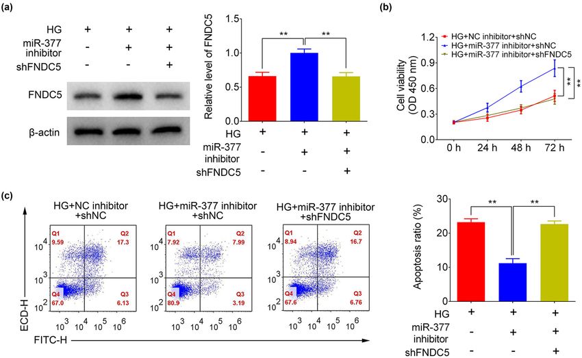
miR-377-3p in gestational diabetes mellitus 465
In this study, serum miR-377-3p level and its correla- Statement of informed consent: Written informed consent
tion with fasting blood glucose of the GDM patients were was obtained from a legally authorized representative(s)
examined and analyzed. miR-377-3p’s effects on tropho- for anonymized patient information to be published in this
blastic cell survival and apoptosis under the condition of article.
HG were investigated. The targeting relationship between
miR-377-3p and FNDC5 was studied using TargetScan
prediction and western blotting. In addition, the effects
of FNDC5 downregulation on cell growth and apoptosis 2.2 RNA extraction and quantitative reverse
of HTR-8/SVneo cells mediated by miR-377-3p inhibitor transcription PCR (qRT-PCR)
were analyzed.
Total RNAs in the serum and cells were extracted by TRIzol
(catalog number, 15596018, Thermo Fisher Scientific,
2 Methods Waltham, MA, USA). For reverse transcription, the Prime
Script RT Reagent Kit (catalog number, #RR037A, Takara)
was used. The cDNAs were obtained and then amplified
2.1 Clinical samples with the ExTaq Kit (catalog number, DRR001A, Takara) to
determine the miRNA levels. The U6 small nuclear RNA
All serum samples were collected from the Second Affiliated (snRNA) was used to normalize miR-377-3p expression.
Hospital of Guizhou University of Traditional Chinese The primer sequences were: miR-377-3p (forward): 5′-GAG
Medicine. A total of 30 GDM pregnant patients and 38 CAGAGGTTGCCCTTG-3′, miR-377-3p (reverse): 5′-ACAAAA
normal pregnant women (healthy) after C-section in the GTTGCCTTTGTGTGA-3′; U6 (forward): 5′-CTCGCTTCGGCA
third trimester of gestation were enrolled (Table 1). GDM GCACA-3′, U6 (reverse): 5′-AACGCTTCACGAATTTGCGT-3′.
was diagnosed according to the criteria published by the
International Association of the Diabetes and Pregnancy
Study Groups (IADPSG) [10]. Some cases were excluded,
which included women with in vitro fertilization (IVF), pre- 2.3 Cell culture
eclampsia, twins (multiple) pregnancy, maternal diabetes
history, or other pregnancy complications. All serum sam- Human trophoblast cell line HTR-8/SVneo and BeWo
ples were frozen by liquid nitrogen and stored at −80°C. were purchased from the Chinese Academy of Sciences
Cell Bank (Shanghai, China). HTR-8/SVneo cells were
Ethics approval: This study was approved by the Ethics derived by transfecting the cells that grew out of chorinic
Committee of the Second Affiliated Hospital of Guizhou villi explants of human first-trimester placenta with the
University of Traditional Chinese Medicine. gene encoding for simian virus 40 large T antigen. As a
Table 1: The expression of FoxO1 in placenta and omental adipose tissue of GDM
Characteristics Health GDM p-value
Number 38 30
Age (years) 33.21 ± 8.17 30.27 ± 6.38 0.1098
Gestational weeks 37.54 ± 1.31 38.12 ± 1.65 0.1119
Height (m) 1.61 ± 0.06 1.59 ± 0.56 0.2123
Pre-pregnancy weight (kg) 50.75 ± 5.85 50.1 ± 6.02 0.6526
Current weight (kg) 68.24 ± 6.40 70.35 ± 6.52 0.1848
Weight gain during pregnancy (kg) 17.48 ± 8.16 20.25 ± 7.12 0.1467
Pre-pregnancy BMI (kg/m2) 19.57 ± 2.63 19.78 ± 2.91 0.7713
Current BMI (kg/m2) 25.68 ± 2.52 28.00 ± 3.47 0.0503
Birth weight (g) 3518.98 ± 558.15 3545.05 ± 587.15 0.8523
Fasting plasma glucose (FPG) 3.72 ± 0.88 4.96 ± 1.59 0.0001*
Fasting insulin (FIN) 8.76 ± 2.10 15.70 ± 4.62 0.0001*
Homeostasis model of assessment-insulin 1.45 ± 0.30 5.50 ± 1.66 0.0001*
resistance (HOMA-IR)
miR-377 expression 1.53 ± 1.11 2.49 ± 1.70 0.0068*
*p < 0.05.
466 Zhaozhao Hua et al.
derivative of malignant choriocarcinoma, BeWo cells pre- Biosciences, San Jose, CA). After that, apoptosis rate was
served functional hormone synthesis in and cell mor- analyzed using a FACS Calibur system (BD Biosciences).
phology of the cytotrophoblast of the original tumor.
These two cell lines share a number of phenotypic pro-
perties with the parental trophoblast cells and are useful 2.7 Luciferase reporter assay
to study trophoblast and placental biology. Dulbecco’s
modified Eagle’s medium (catalog number, 30030, DMEM, To determine the direct interactions between miR-377-3p
Thermo Fisher Scientific) supplemented with 10% fetal and FNDC, luciferase reporter assay was performed. First,
bovine serum (catalog number, 16140071, FBS, Thermo DNA fragments of the FNDC 3′-UTR that contain miR-377-
Fisher Scientific) was used for cell culture, which was main- 3p binding sites were amplified. Then, the 3′-UTR of
tained in a humidified atmosphere with 5% CO2 at 37°C. The FNDC was cloned into the pMIR-Report luciferase vector
cells in the control group were cultured in the basal medium (catalog number, VT1399, Promega, Madison, WI, USA).
with a glucose concentration of 5 mM, whereas cells in the A vector with mutated miR-377-3p putative binding site
HG group were incubated in a HG medium (glucose con- was used as a control. After co-transfection with pMIR-
centration of 25 mM). Report vectors, β-gal (a reference for transfection effi-
ciency) and miR-377-3p mimic (NC mimic), luciferase
activity was determined by a dual luciferase reporter
assay system (catalog number, E1960, Promega).
2.4 Cell transfection
Cell transfection reagents including miR-377-3p inhibitor, 2.8 Protein extraction and western blot
miR-377-3p mimic, and the separate negative controls (NC
inhibitor or NC mimic) were synthesized (GenePharma, Western blotting was performed as previously reported [11].
Shanghai, China) to study the effect of miR-377-3p. Total protein from HTR-8/SVneo and BeWo cells was
These mimic and inhibitor were respectively transfected extracted by RIPA buffer (catalog number, 310003, BestBio,
into the trophoblast cells with the help of Lipofectamine Shanghai, China). Protein samples were separated using
2000 (catalog number, 11668019, Thermo Fisher Scientific) sodium dodecyl sulfate-polyacrylamide gel electrophoresis
according to the manufacturer’s instructions. After trans- (SDS-PAGE) and transferred onto polyvinylidene difluoride
fection, the cells were used for the qRT-PCR, cell viability, (PVDF) membranes (Bio-Rad Laboratories, Richmond, CA,
apoptosis assay, and western blot assays. USA). After blocking with 5% non-fat milk, the membranes
were incubated with the primary antibody of FNDC5 (cat-
alog number, 23995-1-AP, Thermo Fisher Scientific, dilution
2.5 Cell viability assay ratio 1:2,000) overnight at 4°C. Secondary goat anti-rabbit
IgG antibodies conjugated with horseradish peroxidase
The HTR-8/SVneo and BeWo cells were transfected with (catalog number, A27036, Thermo Fisher Scientific, dilution
miR-377-3p inhibitor or NC inhibitor for 24 h. Briefly, cells ratio 1:2,000) were used. β-actin (catalog number, MA1-140,
(5,000 per well) were seeded into 96-well plates with basal Thermo Fisher Scientific, dilution ratio 1:2,000) expression
or HG culture medium. After that, cell viability was deter- was used as control. For protein visualization, an ECL Kit
mined using cell counting kit-8 (catalog number, CK04, (catalog number, 345818, Millipore, Billerica, MA, USA) and
CCK8, Dojindo, Japan). The absorbance at 450 nm was a chemiluminescence imaging system were used. The wes-
detected using a microplate reader (Molecular Devises, tern blot bands were analyzed by Image J software (https://
CA, USA). imagej.net/Citing).
2.9 Statistical analysis
2.6 Apoptosis assay
Data are expressed as mean ± standard error of the mean
At 48 h post transfection, apoptosis assay was performed (SEM). Statistical differences between two groups were ana-
in HTR-8/SVneo and BeWo cells. The cells were first fixed lyzed using Student’s t-test. One-way ANOVA was used for
with 70% ethanol overnight and were treated with propi- the analysis of statistical differences among the experimental
dium iodide (PI), Annexin V (catalog number, 556570, BD groups. p < 0.05 was considered statistically significant.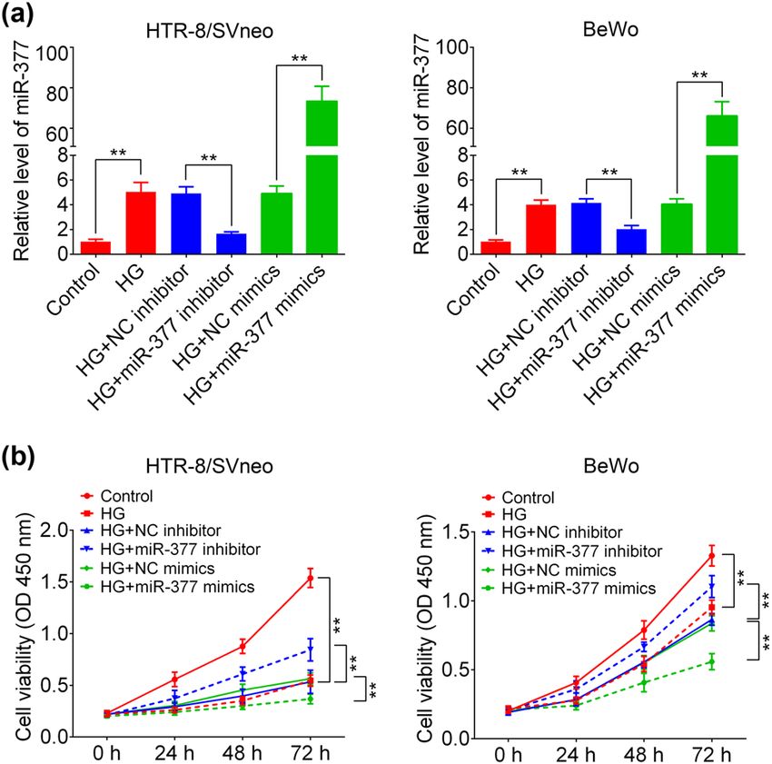
miR-377-3p in gestational diabetes mellitus 467
Figure 1: Expression of miR-377-3p and its correlation with fasting
blood glucose in GDM patients. (a) Serum expression of miR-377-3p
was upregulated in GDM patients compared with that in the healthy
women. (b) Serum miR-377-3p levels were positively correlated with
the fasting blood glucose in GDM patients (r = 0.6709, p < 0.0001).
The data are expressed as mean ± SEM; ** indicates p < 0.01.
3 Results
3.1 Serum miR-377-3p is significantly
increased in GDM patients Figure 2: Effect of miR-377-3p on HTR-8/SVneo and BeWo cells.
(a) miR-377-3p expression in HTR-8/SVneo and BeWo cells (control
As shown in Figure 1a, qRT-PCR results demonstrated or HG) with transfection of miR-377-3p inhibitor, miR-377-3p mimic,
that serum miR-377-3p levels were much higher in GDM NC mimic or NC inhibitor. (b) Cell growth curves of HTR-8/SVneo and
BeWo cells (control or HG) with transfection of miR-377-3p inhibitor,
patients (n = 30) than those in the healthy pregnant
miR-377-3p mimic, NC mimic or NC inhibitor. The data are expressed
women (n = 38). In addition, a positive correlation between as mean ± SEM. ** indicates p < 0.01.
relative level of miR-377-3p and fasting blood glucose level
in GDM patients was found (Figure 1b), which suggested
that miR-377-3p might play an important role in the patho-
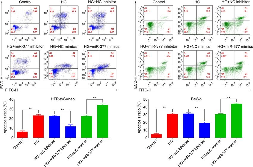
genesis of GDM. the apoptosis ratio of HTR-8/SVneo and BeWo cells. Mean-
while miR-377-3p inhibitor reduced the apoptosis ratio in
HG-treated HTR-8/SVneo and BeWo cells, whereas miR-
377-3p mimic led to the opposite result of cell growth and
3.2 miR-377-3p downregulation increases apoptosis.
cell vitality and suppresses apoptosis of
HG-treated HTR-8/SVneo and BeWo cells
To verify the role of miR-377-3p in regulating the bio- 3.3 miR-377-3p targets FNDC5 in HTR-8/
logical function of trophoblast cells under the condition of SVneo and BeWo cells
hyperglycemia, HTR-8/SVneo and BeWo cells were treated
with HG medium for 24 h in vitro. As shown in Figure 2a, The results above indicated that miR-377-3p inhibition
miR-377-3p level was significantly upregulated in the HG allows cell growth and decreases apoptosis level in tro-
group, in contrast with that in control groups, suggesting a phoblastic cells under the condition of hyperglycemia.
positive relation between miR-377-3p expression and glu- Thus, the molecular mechanism was investigated next.
cose. miR-377-3p inhibitor resulted in a significant down- As predicted by TargetScan, an online prediction soft-
regulation of miR-377-3p in HTR-8/SVneo and BeWo cells ware, FNDC5, was found to be a potential target gene of
with or without HG treatment, and miR-377-3p mimic miR-377-3p. Potential binding sites between miR-377-3p
increased miR-377-3p level. In addition, the cell viability and FNDC5 are shown in Figure 4a. The data of the luci-
curves in Figure 2b demonstrate that HG treatment inhib- ferase reporter experiment demonstrated that miR-377-3p
ited cell growth in HTR-8/SVneo and BeWo cells, whereas mimic reduced the luciferase activity of FNDC5 3′-UTR
miR-377-3p inhibitor increased cell growth in HG-treated reporter plasmid (Figure 4b). The protein expression of
HTR-8/SVneo and BeWo cells. Furthermore, the apoptosis FNDC5 in HG-treated HTR-8/SVneo and BeWo cells was
analysis in Figure 3 show that HG treatment upregulated significantly downregulated. In addition, FNDC5 expression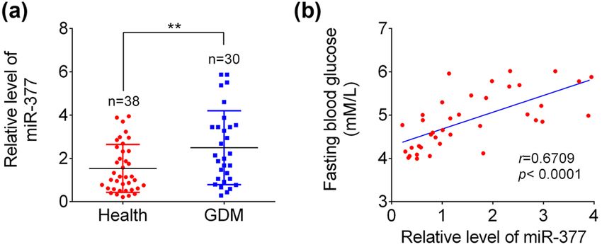
468 Zhaozhao Hua et al.
Figure 3: Apoptosis analysis of HTR-8/SVneo and BeWo cells (control or HG) and HG cells with the transfection of miR-377-3p inhibitor, miR-
377-3p mimic, NC mimic or NC inhibitor. Apoptosis rates were quantified and shown as histograms. The data are expressed as mean ± SEM;
** indicates p < 0.01.
was markedly increased in the HG + miR-377-3p inhibitor in HG-treated HTR-8/SVneo cells were suppressed in
group when compared to that in the HG + NC inhibitor cells of the HG + miR-377-3p inhibitor + shFNDC5 group
group, whereas miR-377-3p mimic further downregulated (Figure 5b). Furthermore, miR-377-3p inhibition signifi-
the expression of FNDC5 under HG treatment (Figure 4c). cantly downregulated the cell apoptosis ratio after HG
The data above indicated that miR-377-3p targeted FNDC5 stimulation. The reduced-cell apoptosis ratio was reversed
and suppressed its expression. in HG + miR-377-3p inhibitor + shFNDC5 group compared
to that in HG + miR-377-3p inhibitor + shNC group (Figure 5c).
Considering these findings, it can be concluded that upre-
gulation of FNDC5 was required in cell growth promotion
3.4 FNDC5 downregulation eliminates the and apoptosis suppression mediated by miR-377-3p inhi-
effect of miR-377-3p inhibitor bitor in trophoblast cells, indicating its involvement in the
development of GDM.
After establishing FNDC5 as a target gene of miR-377-3p,
the role of FNDC5 in growth and apoptosis of HTR-8/
SVneo cells was explored. As shown in Figure 5a, miR-
377-3p inhibition caused an upregulation of FNDC5 in 4 Discussion
HG-treated HTR-8/SVneo cells, and the FNDC5 expres-
sion was repressed again by transfection of shFNDC5. As a health problem among pregnant women, GDM exerts
The FNDC5 expression was further inhibited when trans- negative consequences in pregnancy. The normal biolo-
fecting shFNDC5 and miR-377-3p mimic. In addition, the gical function of trophoblast cells is critical to the devel-
increased cell viability caused by miR-377-3p inhibition opment of placenta. The reduced viability of trophoblastmiR-377-3p in gestational diabetes mellitus 469
Figure 4: miR-377-3p negatively regulated FNDC5 expression. (a) The predicted seed-recognition sites in the 3′-UTR of FNDC5 mRNA and the
miR-377-3p sequences were shown. (b) Relative luciferase activity of the FNDC5 3′-UTR reporter plasmid in 293 T and HTR-8/SVneo cells
after transfection with NC mimic or miR-377-3p mimic. The mutant FNDC5 3′-UTR reporter was also used as a control. (c) Protein expression
levels of FNDC5 in HTR-8/SVneo and BeWo cells from groups of control, HG, HG + NC inhibitor, HG + miR-377-3p inhibitor, HG + NC mimic,
HG + miR-377-3p mimic, as determined using western blotting. Bands were quantified and shown in histogram. The data are expressed as
mean ± SEM. ** indicates p < 0.01.
cells might cause dysplasia of pregnancy and even mis- HTR-8/SVneo cells was significantly suppressed by the
carriage [12]. miRNAs are sensitive and stable, and have HG treatment in the present study, along with the upre-
been demonstrated to be potential diagnostic markers gulation of miR-377-3p. Interestingly, considerable effects
and intervention targets for human diseases, including were observed in early and late apoptosis in BeWo treated
diabetes mellitus [13] and also GDM [14]. For example, with HG + miR-377 mimics in contrast to HTR-8/SVneo
serum miR-204 was found to be upregulated in type 1 cells where only late apoptosis was increased (Figure 3).
diabetes (T1D) patients and negatively correlated with The difference in early and late apoptosis ratios between
β cell function [15]. So far, decreased miR-132 was found early and late apoptosis percentages among HTR-8/SVneo
in GDM patients, and overexpression of miR-132 in and BeWo cells may account for their different sensitivity
HTR-8/SVneo cells could markedly rescue HG-induced to HG treatment and the rate of renewal iteration. While
suppression of cell proliferation [16]. More vital miRNAs the importance of miR-377-3p has been demonstrated in
and their roles in the pathogenesis of GDM deserve further some tumors, it was found that miR-377-3p plays an onco-
investigation. This study showed that serum miR-377-3p genic function in colorectal cancer development through
was increased in the GDM patients, and a positive correla- increasing GSK-3β expression and thereby activating
tion between miR-377-3p level and the fasting blood glu- NF-κB pathway [18]. In addition, tumor inhibitory func-
cose was indicated in GDM patients. tions of miR-377-3p were demonstrated in pancreatic
HG, as a characteristic of GDM, could inhibit prolif- cancer by regulating a serine/threonine kinase, namely
eration of trophoblast cells, thereby impairing the devel- Pim-3 proto-oncogene [19], and in gastric cancer through
opment of placenta [17]. Consistently, cell viability of reducing the level of vascular endothelial growth factor A470 Zhaozhao Hua et al.
Figure 5: FNDC5 downregulation abolished the effects of miR-377-3p on cell proliferation and apoptosis. (a) Protein expression levels of
FNDC5 in HTR-8/SVneo cells from groups of HG, HG + miR-377-3p inhibitor, HG + shFNDC5, or HG + miR-377-3p inhibitor + shFNDC5, as
determined using western blotting. Bands were quantified and shown in histogram. (b) Cell growth curves of HTR-8/SVneo cells from
groups of HG, HG + miR-377-3p inhibitor, HG + shFNDC5, HG + miR-377-3p inhibitor + shFNDC5. (c) Apoptosis analysis of HTR-8/SVneo cells
(control or HG) and HG cells from groups of HG, HG + miR-377-3p inhibitor, HG + shFNDC5, HG + miR-377-3p inhibitor + shFNDC5. Apoptosis
rates were quantified and shown as histograms. The data are expressed as mean ± SEM; ** indicates p < 0.01.
(VEGFA) [20]. However, the role of miR-377-3p in diabetes that regulates FNDC5 in GDM needs to be further explored.
is still poorly understood. Although serum miR-377-3p was In this study, it was found that miR-377-3p could directly
reported to be higher in pediatric patients with T1D [21], target FNDC5 and its inhibition reestablish cell growth in
upregulation of miR-377-3p in diabetic nephropathy indir- HG-treated HTR-8/SVneo and BeWo cells. In addition to
ectly resulted in the upregulation of fibronectin protein [9]. miR-377-3p, miR-137 was reported to suppress the viability
However, the function of miR-377-3p in GDM development and migration of trophoblasts through negatively regulating
still remains unclear. Herein, the first evidence for a pro- FNDC5 in GDM, which may result in adverse pregnancy out-
tective function of targeting miR-377-3p was presented: comes [7]. Furthermore, myostatin (Mstn) could increase
miR-377-3p inhibition can restore the protein expression miR-34a, leading to downregulation of FNDC5, which inhi-
of FNDC5, reestablish cell growth, and reduce apoptosis bits the browning of white adipocytes [24]. Therefore,
ratio in HTR-8/SVneo and BeWo cells. whether miR-377-3p, miR-137, and miR-34a could coordi-
As a transmembrane protein present in various tis- nately regulate FNDC5 expression in GDM, and how to
sues including heart, liver, skeletal muscle, and adipose synthetically adjust their levels to prevent and treat GDM
tissue, FNDC5, the precursor of irisin, was found to be a require future research.
novel player in metabolism and metabolic syndrome [22]. In brief, serum miR-377-3p is upregulated in GDM
Clinical studies combined with cellular experiments revealed patients and positively correlates with the fasting blood
that FNDC5 mRNA was decreased in adipose tissue of glucose, which might serve as a potential diagnostic
patients with type 2 diabetes [23]. In animal models of obe- biomarker for GDM patients. The downregulation of miR-
sity, upregulation of FNDC5 increased uncoupling protein 1 377-3p can relieve effects of HG on trophoblast cell pro-
(UCP1) expression and oxygen consumption, leading to high liferation and apoptosis, which is mediated through the
energy expenditure [22]. However, the potential mechanism regulation of FNDC5 expression. In addition, inhibition ofmiR-377-3p in gestational diabetes mellitus 471
FNDC5 could abrogate the effects caused by miR-377-3p fibronectin production in diabetic nephropathy. FASEB J.
inhibitor, indicating that miR-377-3p inhibition may have 2008;22(12):4126–35. PMID:PMC2614610.
a beneficial role in GDM remission by reestablishing cell [10] American Diabetes A. Diagnosis and classification of diabetes
mellitus. Diabetes Care. 2012;35(Suppl 1):S64–71.
growth and reducing apoptosis ratio. Together, miR-377
PMID:PMC3632174.
may be a potential target for GDM biomarker. [11] Zhu D, Sun C, Qian X. MST1 suppresses viability and promotes
apoptosis of glioma cells via upregulating SIRT6 expression.
Author contributions: Z. H. and D. L. designed the study, J Integr Neurosci. 2019;18(2):117–26.
supervised the data collection, and analyzed the data; [12] Tian FJ, Qin CM, Li XC, Wu F, Liu XR, Xu WM, et al. Decreased
stathmin-1 expression inhibits trophoblast proliferation and
A. W. interpreted the data and prepared the manuscript
invasion and is associated with recurrent miscarriage.
for publication; T. C. and S. L. supervised the data collec- Am J Pathol. 2015;185(10):2709–21.
tion, analyzed the data, and reviewed the draft of the manu- [13] Yang Z, Chen H, Si H, Li X, Ding X, Sheng Q, et al. Serum miR-
script. All authors have read and approved the manuscript. 23a, a potential biomarker for diagnosis of pre-diabetes and
type 2 diabetes. Acta Diabetol. 2014;51(5):823–31.
[14] Pheiffer C, Dias S, Rheeder P, Adam S. Decreased expression
Competing interest: The authors state that there are no
of circulating miR-20a-5p in South African women with
conflicts of interest to disclose.
gestational diabetes mellitus. Mol Diagn Ther.
2018;22(3):345–52.
Availability of data and materials: All data generated or [15] Xu G, Thielen LA, Chen J, Grayson TB, Grimes T, Bridges SL Jr,
analyzed during this study are included in this published et al. Serum miR-204 is an early biomarker of type 1 diabetes-
article. associated pancreatic beta-cell loss. Am J Physiol Endocrinol
Metab. 2019;317(4):E723–30. PMID:PMC6842918.
[16] Zhou X, Xiang C, Zheng X. miR-132 serves as a diagnostic
biomarker in gestational diabetes mellitus and its regulatory
effect on trophoblast cell viability. Diagn Pathol.
2019;14(1):119. PMID:PMC6814988.
References [17] Peng HY, Li MQ, Li HP. High glucose suppresses the viability
and proliferation of HTR8/SVneo cells through regulation of
[1] Barnes-Powell LL. Infants of diabetic mothers: the effects of the miR137/PRKAA1/IL6 axis. Int J Mol Med.
hyperglycemia on the fetus and neonate. Neonatal Netw. 2018;42(2):799–810. PMID:PMC6034938.
2007;26(5):283–90. [18] Liu WY, Yang Z, Sun Q, Yang X, Hu Y, Xie H, et al. miR-377-3p
[2] Miao M, Dai M, Zhang Y, Sun F, Guo X, Sun G. Influence of drives malignancy characteristics via upregulating GSK-3beta
maternal overweight, obesity and gestational weight gain on expression and activating NF-kappaB pathway in hCRC cells.
the perinatal outcomes in women with gestational diabetes J Cell Biochem. 2018;119(2):2124–34.
mellitus. Sci Rep. 2017;7(1):305. PMID:PMC5428436. [19] Chang W, Liu M, Xu J, Fu H, Zhou B, Yuan T, et al. miR-377
[3] Barbour LA, McCurdy CE, Hernandez TL, Kirwan JP, inhibits the proliferation of pancreatic cancer by targeting
Catalano PM, Friedman JE. Cellular mechanisms for insulin Pim-3. Tumour Biol. 2016;37(11):14813–24.
resistance in normal pregnancy and gestational diabetes. [20] Wang CQ, Chen L, Dong CL, Song Y, Shen ZP, Shen WM, et al.
Diabetes Care. 2007;30(Suppl 2):S112–9. miR-377 suppresses cell proliferation and metastasis in
[4] Filios SR, Shalev A. Beta-cell microRNAs: small but powerful. gastric cancer via repressing the expression of VEGFA. Eur Rev
Diabetes. 2015;64(11):3631–44. PMID:PMC4613982. Med Pharmacol Sci. 2017;21(22):5101–11.
[5] Peng HY, Li HP, Li MQ. High glucose induces dysfunction of [21] El-Samahy MH, Adly AA, Elhenawy YI, Ismail EA, Pessar SA,
human umbilical vein endothelial cells by upregulating Mowafy ME, et al. Urinary miRNA-377 and miRNA-216a as
miR-137 in gestational diabetes mellitus. Microvasc Res. biomarkers of nephropathy and subclinical atherosclerotic
2018;118:90–100. risk in pediatric patients with type 1 diabetes. J Diabetes
[6] Wang P, Wang Z, Liu G, Jin C, Zhang Q, Man S, et al. miR-657 Complications. 2018;32(2):185–92.
promotes macrophage polarization toward M1 by targeting [22] Bostrom P, Wu J, Jedrychowski MP, Korde A, Ye L, Lo JC, et al.
FAM46C in gestational diabetes mellitus. Mediators Inflamm. A PGC1-alpha-dependent myokine that drives brown-fat-like
2019;2019:4851214. PMID:PMC6930733. development of white fat and thermogenesis. Nature.
[7] Peng HY, Li MQ, Li HP. miR-137 restricts the viability and 2012;481(7382):463–8. PMID:PMC3522098.
migration of HTR-8/SVneo cells by downregulating FNDC5 in [23] Kurdiova T, Balaz M, Vician M, Maderova D, Vlcek M,
gestational diabetes mellitus. Curr Mol Med. Valkovic L, et al. Effects of obesity, diabetes and exercise on
2019;19(7):494–505. Fndc5 gene expression and irisin release in human skeletal
[8] Xu K, Bian D, Hao L, Huang F, Xu M, Qin J, et al. microRNA-503 muscle and adipose tissue: in vivo and in vitro studies.
contribute to pancreatic beta cell dysfunction by targeting the J Physiol. 2014;592(5):1091–107. PMID:PMC3948565.
mTOR pathway in gestational diabetes mellitus. EXCLI J. [24] Ge X, Sathiakumar D, Lua BJ, Kukreti H, Lee M, McFarlane C.
2017;16:1177–87. PMID:PMC5735340. Myostatin signals through miR-34a to regulate Fndc5
[9] Wang Q, Wang Y, Minto AW, Wang J, Shi Q, Li X, et al. expression and browning of white adipocytes. Int J Obes
MicroRNA-377 is up-regulated and can lead to increased (Lond). 2017;41(1):137–48. PMID:PMC5220162.You can also read