ASNS disruption shortens CTPS cytoophidia in Saccharomyces cerevisiae
←
→
Page content transcription
If your browser does not render page correctly, please read the page content below
G3, 2021, 11(2), jkaa060

    2
                                                                                                                              DOI: 10.1093/g3journal/jkaa060
                                                                                                              Advance Access Publication Date: 11 January 2021
                                                                                                                                               Investigation

ASNS disruption shortens CTPS cytoophidia in
Saccharomyces cerevisiae
Shanshan Zhang           ,1,2,3 Han-Chao Feng          ,1 and Ji-Long Liu        1,4,
                                                                                     *
1
  School of Life Science and Technology, ShanghaiTech University, Shanghai, 201210, China

                                                                                                                                                                 Downloaded from https://academic.oup.com/g3journal/article/11/1/jkaa060/6080684 by guest on 08 February 2021
2
  University of Chinese Academy of Sciences, Beijing, 100049, China
3
  Shanghai Institute of Biochemistry and Cell Biology, Chinese Academy of Sciences, Shanghai, 200031, China
4
  Department of Physiology, Anatomy and Genetics, University of Oxford, Oxford, OX1 3PT, UK

*Corresponding author: liujl3@shanghaitech.edu.cn; jilong.liu@dpag.ox.ac.uk

Abstract
Asparagine synthetase (ASNS) and CTP synthase (CTPS) are two metabolic enzymes that catalyze the biosynthesis of asparagine and CTP,
respectively. Both CTPS and ASNS have been identified to form cytoophidia in Saccharomyces cerevisiae. Glutamine is a common sub-
strate for both these enzymes, and they play an important role in glutamine homeostasis. Here, we find that the ASNS cytoophidia are
shorter than the CTPS cytoophidia, and that disruption of ASNS shortens the length of CTPS cytoophidia. However, the deletion of CTPS
has no effect on the formation and length of ASNS cytoophidia, or on the ASNS protein level. We also find that Asn1 overexpression indu-
ces the formation of a multi-dot structure in diauxic phase which suggests that the increased protein level may trigger cytoophidia forma-
tion. Collectively, our results reveal a connection between ASNS cytoophidia and CTPS cytoophidia.

Keywords: asparagine synthetase; CTP synthase; cytoophidium; glutamine metabolism; Saccharomyces cerevisiae

Introduction                                                                       to total auxotrophy (Jones 1978; Ramos and Wiame 1980; Dang
                                                                                   et al. 1996). Asn1 and Asn2 have high similarity in terms of pro-
In 2010, three laboratories independently reported that CTP syn-
                                                                                   tein size and amino acid sequence. The capabilities of Asn1 and
thase (CTPS) assembles into filamentous structures termed
                                                                                   Asn2 in cytoophidia formation are distinct, and Asn2 cytoophi-
cytoophidia in bacteria, yeast, and fruit fly (Ingerson-Mahar et al.
                                                                                   dium formation is dependent on the presence of Asn1 (Zhang
2010; Liu 2010; Noree et al. 2010). These newly discovered mem-
                                                                                   et al. 2018; Noree et al. 2019b).
braneless structures formed by metabolic enzymes were found to
                                                                                       CTP is a precursor of DNA and RNA biosynthesis and it partici-
be evolutionarily conserved (Chen et al. 2011; Azzam and Liu
                                                                                   pates in nucleotide metabolism and membrane phospholipid bio-
2013; Barry et al. 2014; Aughey et al. 2016; Liu 2016; Daumann et al.
                                                                                   synthesis. The rate-limiting step of de novo biosynthesis of CTP is
2018; Andreadis et al. 2019; Sun and Liu 2019a, 2019b; Wu and Liu
                                                                                   catalyzed by CTPS. URA7 and, to a lesser extent, URA8 are the
2019; Zhang and Liu 2019; Zhou et al. 2019; Zhang et al. 2020a,                    genes that code for CTPS in S. cerevisiae (Ozier-Kalogeropoulos
2020b; Zhou et al. 2020). To date, the understanding of cytoophi-                  et al. 1994), and Ura7 and Ura8 are very similar in protein size and
dia has been greatly expanded by genome-wide screening in                          amino acid sequence. The maximum expression of CTPS is ob-
Saccharomyces cerevisiae and it has been observed that most meta-                  served in the exponential phase (Nadkarni et al. 1995). It has been
bolic enzymes form filamentous or punctuated cytoophidia                           proven that CTPS cytoophidium formation in bacteria inhibits
(Shen et al. 2016; Noree et al. 2019a). These enzymes cover a vari-                the enzymatic activity (Barry et al. 2014), whereas in human cells
ety of biological reactions, such as glucose metabolism, transla-                  CTPS1 cytoophidium formation increases the enzymatic activity
tion initiation mechanisms, and purine biosynthesis (Shen et al.                   (Lynch et al. 2017). Drosophila CTPS can form conformationally dif-
2016; Noree et al. 2019a).                                                         ferent substrate-bound and product-bound filaments (Zhou et al.
    Asparagine synthetase (ASNS) catalyzes ATP-dependent de                        2019). CTPS2 cytoophidium conformation can switch between ac-
novo biosynthesis of asparagine from aspartic acid, and it has                     tive and inactive forms based on the substrate and product levels
been observed to form cytoophidia in budding yeast. In Escherichia                 (Lynch and Kollman 2020). These research results support that
coli, there are two types of ASNS, glutamine dependent and am-                     cytoophidium formation provides an additional layer of metabo-
monia dependent (Humbert and Simoni 1980; Ramos and Wiame                          lism regulation.
1980). ASN1 and ASN2 both code glutamine-dependent ASNS in                             The spatial relationship between ASNS and CTPS cytoophidia
budding yeast. Only a double mutant of ASN1 and ASN2 will lead                     has been observed as a head-to-head or side-by-side pattern

Received: May 15, 2021. Revised: December 9, 2020.
C The Author(s) 2021. Published by Oxford University Press on behalf of Genetics Society of America.
V
This is an Open Access article distributed under the terms of the Creative Commons Attribution License (http://creativecommons.org/licenses/by/4.0/), which
permits unrestricted reuse, distribution, and reproduction in any medium, provided the original work is properly cited.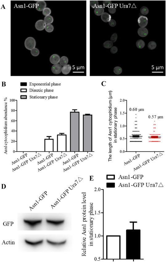
2   |   G3, 2021, Vol. 11, No. 2

(Zhang et al. 2018). To gain a better understanding of the associa-      sequencing. The expression of Asn1 was driven by endogenous
tion between these two types of cytoophidia, we explore the ef-          GAL promoter which was induced when cells were grown on ga-
fect of ASNS deletion on CTPS cytoophidia and vice versa. We             lactose.
also study changes in ASNS and CTPS cytoophidia in response to
different culture media.                                                 Glutamine analog treatment
                                                                         A glutamine analog 6-diazo-5-oxo-L-norleucine (DON) was used
                                                                         to treat ASN1-GFP ASN2-mCherry cells. We cultured the cells in
Materials and methods                                                    standard rich medium (YPD) with 0%, 2%, and 4% DON content,
Yeast strains and culture media                                          respectively. After 7 days of culture, when the cells were at
Yeast strains used in this study were derived by transformation          stationary phase, they were collected and fixed with 4% PFA.
of BY4741 (donated by Jinqiu Zhou from Shanghai Institute of                To count the abundance of cytoophidia, more than 1000 cells
Biochemistry and Cell Biology). Transformation was performed             were counted manually for each strain in each trial. Three groups
by the lithium acetate method. The generation of URA7-GFP

                                                                                                                                               Downloaded from https://academic.oup.com/g3journal/article/11/1/jkaa060/6080684 by guest on 08 February 2021
                                                                         of trials were mainly carried out manually. To count the length
ASN1-MCHERRY and ASN1-GFP ASN2-MCHERRY cells has been de-                of cytoophidia, more than 200 cells were counted manually for
scribed in our previous study (Zhang et al. 2018). The genotypes of      each strain in each of the three biological repeats. The experi-
all strains are listed in Table 1. Saccharomyces cerevisiae cells were   mental results were processed by ANOVA function of software
cultured in standard rich medium (YPD: 2% peptone, 1% yeast ex-          GraphPad Prism. *P < 0.05, **P < 0.01, and ***P < 0.001.
tract, 2% dextrose) or YPG (2% peptone, 1% yeast extract, 2% ga-
lactose) at 30 C unless otherwise indicated.                            Microscopy
                                                                         Cells were first fixed by 4% paraformaldehyde (PFA) after culture
Gene disruption                                                          for the indicated time at room temperature for 10 min (Zhang
ASN1 and ASN2 disruption strains containing URA7-GFP were                et al. 2021). Then cells were washed once with sterile water before
obtained by PCR-based gene targeting using pRS303 and pRS306             being mixed with PBS mountant (1.2% LMT agarose in PBS).
as described previously (Zhang et al. 2018). For URA7 disruption         Images for cytoophidium abundance and length quantification
cassette construction, the 5’ untranslated region and 3’ region          were captured using a Zeiss Axio Imager 2 microscope and Zeiss
were sub-cloned into pRS306. The disruption cassettes first line-        LSM 880 inverted laser-scanning confocal microscope, respec-
arized with EcoR I (New England Biolabs) and transformed into            tively. Then these images were processed with ZEN 2 lite (blue
ASN1-GFP cells to construct ASN1-GFP URA7䉭 strains. All the pri-         edition) and ImageJ Fiji software.
mers used for URA7 gene deletion are listed in Table 2. Single col-
onies obtained by transformation were screened by PCR to
                                                                         Table 2 Primers used for URA7 gene disruption
confirm the absence of the specific gene.
                                                                         Primer name                         Sequence (50 to 30 )
ASN1 overexpression
                                                                         URA7 UHA-F            gggGAATTCTTAAAGTTAGCCCTCCCATCTT
To construct ASN1 overexpression strain, ASN1 in URA7-GFP                URA7 UHA-R            gggGGGTACCGTTCTATTGACCAATTCACT
ASN1-MCHERRY and ASN1-GFP ASN2-MCHERRY cells was manip-                  URA7 DHA-F            gggTCTAGAATATTTGTAGTGCTTCTCTACAC
ulated by transformation of a linear PCR product consisting of           URA7 DHA-R            gggGAATTCTGATAAATAATCTCCCTGTTCA
                                                                         URA7 KO testing F     AACCACCTGTACGAACTGGCAC
GAL1 promoter sequence with 40 bp of flanking sequences up-
                                                                         URA7 KO testing R     CCAGTTGAAGATGCAAGAACAC
stream of the ASN1 coding sequence. pFA6a-LEU-GAL1 plasmid
originated from pFA6a-TRP-GAL1 plasmid was used for GAL se-
quence amplification. Primers used for pFA6a-LEU-GAL1 plasmid
construction and ASN1 overexpression strain construction are,
                                                                         Table 3 Primers used for pFA6a-Leu-GAL1 plasmid construction
respectively, listed in Tables 3 and 4. Transformants were se-
lected on SDþLeu and positive clones were validated by                   Primers                           Sequences (50 to 30 )

                                                                         Leu F               CTGATATCATCGATGAATTCATTGCGTATATA
Table 1 Saccharomyces cerevisiae strains used in this study                                    GTTTCGTCTACC
                                                                         Leu R               ATGGGGCTCTTTACAGATCTAACTGTGGGAATA
Strain name                                     Genotype                                       CTCAGGT
                                                                         Leu t F             CTGATCGCATACTCTTCTTACC
BY4741                             MATa his3D leu2D ura3D0 met15D        Leu t R             TAAGACCATGTAACTTTGCA
Ura7-GFP Asn1-mCherry              As BY4741, ura7-GFP(His3MX6), asn1-
                                     mCherry(KanMX6)
Asn1-GFP Asn2-mCherry              As BY4741, asn1-GFP(His3MX6), asn2-
                                     mCherry(KanMX6)
Ura7-GFP Gal-Asn1-mCherry          As BY4741, ura7-GFP(His3MX6), asn1-   Table 4 Primers used for ASN1 overexpression strain
                                     mCherry(KanMX6) PGAL10-ASN1         construction
Gal-Asn1-GFP Asn2-mCherry          As BY4741, asn1-GFP(His3MX6), asn2-
                                     mCherry(KanMX6) PGAL10-ASN1         Primer name                        Sequence (50 to 30 )
Ura7-GFP Asn1䉭                     As BY4741, ura7-GFP(His3MX6),
                                     asn1::URA3                          Asn1 OE F           GCACGTCTTCGTGCCTGAAAGCGGCGAAAATA
Ura7-GFP Asn2䉭                     As BY4741, ura7-GFP(His3MX6),                               CCACACATTTTGAGATCCGGGTTTT
                                     asn2::URA3                          Asn1 OE R           CTTGCTTTACGCTAAGGATATAAATCGGACGT
Asn1-GFP Ura7䉭                     As BY4741, Asn1-GFP(His3MX6),                               AACTTAAGCGCATAGGCCACTAGTGGAT
                                     ura7::URA3                          Asn1 OE t F         CTGATCGCATACTCTTCTTACC
Ura7WT-GFP                         As BY4741, ura7-GFP(His3MX6)          Asn1 OE t R         GCATTACCGGACCAATCTG
Ura7H360A-GFP                      As BY4741, ura7H360A-GFP(His3MX6)     Asn1 OE S1          GAAGAACCTCAGTGGCAAAT
S. Zhang et al.   |   3

                                                                                                                                                           Downloaded from https://academic.oup.com/g3journal/article/11/1/jkaa060/6080684 by guest on 08 February 2021
Figure 1 Ura7 and Asn1 cytoophidia in S. cerevisiae. Ura7-GFP Asn1-mCherry cells were grown in rich medium and cells were collected after 6, 24, and
168 h culture. The cells were subsequently fixed by 4% PFA at room temperature for 10 min. Ura7-GFP and Asn1-mCherry protein were observed by
fluorescent microscopy. The abundance of cytoplasmic cytoophidia were calculated and the average length of cytoplasmic cytoophidia were plotted.
(A) Representative confocal image of Ura7-GFP Asn1-mCherry cells. Ura7 cytoophidia were observed in all three growth phases. Scale bar 5 lm.
(B) Quantification of cells with visible cytoophidia was plotted and expressed as percentage of cells containing cytoophidia in three growth phases.
*P < 0.05 and ***P < 0.001 (C) The average length of Ura7 and Asn1 cytoophidia was measured and plotted in the stationary phase.

Data availability                                                             double tagging strain Ura7-GFP Asn1-mCherry. Cells of this strain
Strains and plasmids are available upon request. The authors af-              were collected in three growth phases: exponential phase, dia-
firm that all data necessary for confirming the conclusions of the            uxic phase and stationary phase (Figure 1A). The pattern of
article are present within the article, figures, and tables.                  cytoophidium formation of these two metabolic enzymes is simi-
                                                                              lar, in general. With cell growth, the abundance of Asn1 and Ura7
                                                                              cytoophidia gradually increases and reaches a peak in the sta-
Results                                                                       tionary phase (Figure 1B). In the exponential phase, dot-shaped
Comparison between Ura7 and Asn1 cytoophidia                                  Ura7 cytoophidia were observed. However, there are differences:
In our previous research, we observed that in most cells Ura7                 Ura7 starts to form cytoophidia in the exponential phase and for
cytoophidia and Asn1 cytoophidia overlapped in the middle or at               Asn1 it starts in the diauxic phase (Figure 1, A and B). Ura7 cytoo-
the ends (Zhang et al. 2018). For further study, we generated the             phidia are crooked and Asn1 cytoophidia are short and rod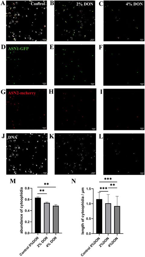
4   |   G3, 2021, Vol. 11, No. 2

                                                                                                                                                      Downloaded from https://academic.oup.com/g3journal/article/11/1/jkaa060/6080684 by guest on 08 February 2021

Figure 2 The effect of Asn1 or Asn2 disruption on Ura7 cytoophidia. Cells were grown in rich medium and collected in exponential, diauxic and
stationary phase. The cells were subsequently fixed by 4% PFA at room temperature for 10 min. Ura7-GFP and Asn1-mCherry protein were observed by
fluorescent microscopy. (A) Representative images of URA7-GFP, URA7-GFP ASN1䉭 AND URA7-GFP ASN2䉭 strain in stationary phase and details of Ura7
cytoophidium morphology. Scale bar 5 lm. (B) Quantification of cells with visible Ura7 cytoophidia was plotted and expressed as percentage of cells
containing cytoophidia in URA7-GFP, URA7-GFP ASN1䉭 AND URA7-GFP ASN2䉭 strains from exponential phase to stationary phase. (C) Ura7
cytoophidium length analysis of URA7-GFP, URA7-GFP ASN1䉭 AND URA7-GFP ASN2䉭 strains at stationary phase. ***P < 0.001. (D) Western blot analysis
of Ura7 protein level in URA7-GFP, URA7-GFP ASN1䉭 AND URA7-GFP ASN2䉭 strains at stationary phase. (E) Protein levels were plotted after
normalization over alpha-actin levels and the protein level of Ura7 in Ura7-GFP cells is referred to as 1.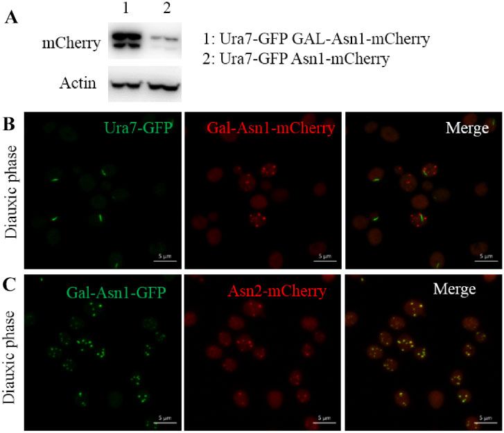
S. Zhang et al. | 5
Downloaded from https://academic.oup.com/g3journal/article/11/1/jkaa060/6080684 by guest on 08 February 2021
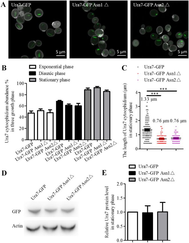
Figure 3 The effect of Ura7 disruption on Asn1 cytoophidia. Cells grown in rich medium were collected during three growth phases and observed by
fluorescence microscopy. (A) Representative confocal images of ASN1-GFP AND ASN1-GFP URA7䉭 strains in stationary phase. Scale bar 5 lm. (B)
Cytoophidium abundance in Asn1-GFP cells is plotted along with the abundance of cytoophidia in cells with URA7 knockout. (C) Asn1 cytoophidium
length measurement without and with URA7 deletion in stationary phase. (D) Western blot analysis of Asn1 protein level in ASN1-GFP AND ASN1-GFP
URA7䉭 strains at stationary phase. (E) Protein levels were plotted after normalization over alpha-actin levels and the protein level of Asn1 in Asn1-GFP
cells is referred to as 1.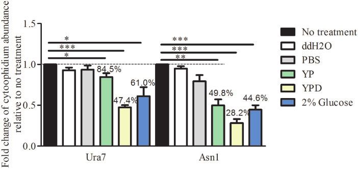
6   |   G3, 2021, Vol. 11, No. 2

                                                                                                                                                      Downloaded from https://academic.oup.com/g3journal/article/11/1/jkaa060/6080684 by guest on 08 February 2021
Figure 4 Ura7 and Asn1 cytoophidia respond to culture medium change. Cells were grown until stationary phase in rich medium which was then
replaced with a different medium for 4 h: PBS, ddH2O, YP, YPD, or 2% glucose. The cells were collected and fixed by 4% PFA. Ura7 and Asn1
cytoophidium abundance was calculated by ImageJ Fiji software and plotted after three biological repeats. More than 200 cells were manually counted
per strain per trial. *P < 0.05, **P < 0.01, and ***P < 0.001.

Figure 5 Stationary phase culture medium effect on Ura7 and Asn1 cytoophidia in exponential and diauxic phases. Stationary phase culture medium
was collected after 168 h culture. Cells were grown in rich medium until exponential and diauxic phase. The medium was replaced with stationary
phase culture medium when cells were at exponential phase and diauxic phase for 2 h. (A) Quantification of Ura7 and Asn1 cytoophidium abundance
before medium shift in exponential phase along with the cells grown under no treatment and under medium shift. (B) The same analysis as in A was
performed in diauxic phase. **P < 0.01.

shaped in the stationary phase (Figure 1B). The length quantifica-          or Asn2 disruption, the average length of the Ura7 cytoophidia
tion results showed that the average length of Ura7 cytoophidia             was 0.76 lm and all were less than 2 lm in length (Figure 2C). We
and Asn1 cytoophidia is 1.6 lm and 0.6 lm, respectively.                    also compared the Ura7 protein levels in Ura7-GFP, Ura7-GFP
                                                                            Asn1䉭 and Ura7-GFP Asn2䉭 cells to check whether the effect of
Either Asn1 or Asn2 disruption shortens Ura7                                ASNS disruption results from a change in protein level, but there
cytoophidia                                                                 was no significant difference between the three strains (Figure 2,
To have a clear understanding of the relationship between Ura7              D and E). ASNS disruption therefore affects Ura7 cytoophidium
and Asn1 cytoophidia, we tried to determine whether Ura7 cytoo-             length rather than its protein level in the stationary phase.
phidia are affected when Asn1 or Asn2 is disrupted. Hence, we
generated Ura7-GFP Asn1䉭 and Ura7-GFP Asn2䉭 strains based                   Ura7 knockout has no obvious effect on Asn1
on the Ura7-GFP strain (Figure 2A). The analysis of Ura7 cytoophi-          cytoophidia
dia in these three strains showed that there was no significant             To answer the question of whether Asn1 cytoophidium length is
change in abundance in the three growth phases (Figure 2B).                 affected by Ura7 disruption, we constructed Asn1-GFP Ura7䉭 cells
However, the average length of Ura7 was dramatically decreased              and used Asn1-GFP cells as the control strain. We collected cells in
when either Asn1 or Asn2 was disrupted. Before the ASNS disrup-             stationary phase (Figure 3A) and analyzed the effect of Ura7 dele-
tion, the Ura7 cytoophidia had a wider length range, and average            tion on Asn1 cytoophidium abundance, length and protein level.
length was 1.33 lm. There were cytoophidia more than 2 lm long              The quantification results show that Ura7 knockout had no impact
and we even observed some of more than 3 lm. After either Asn1              on abundance of Asn1 cytoophidia during the three growth phases
S. Zhang et al. | 7
Downloaded from https://academic.oup.com/g3journal/article/11/1/jkaa060/6080684 by guest on 08 February 2021
Figure 6 High Asn1 protein level results in Asn1 foci in diauxic phase. URA7-GFP GAL-ASN1-MCHERRY and GAL-ASN1-GFP ASN2-MCHERRY cells were
grown in medium containing galactose. URA7-GFP GAL-ASN1-MCHERRY strain was generated from URA7-GFP ASN1-MCHERRY strain by inserting Gal
promoter before the Asn1 coding sequence. The generation process of GAL-ASN1-GFP ASN2-MCHERRY strain was similar. (A) Asn1 protein level in
URA7-GFP GAL-ASN1-MCHERRY and URA7-GFP ASN1-MCHERRY cells. (B) Representative images of URA7-GFP GAL-ASN1-MCHERRY cells in diauxic phase.
(C) Representative images of GAL-ASN1-GFP ASN2-MCHERRY cells in diauxic phase. Scale bar: 5 lm.
(Figure 3B). The average length of Asn1 cytoophidia without and (Figure 4). Apparently, carbon source only or a mixture of carbon,
with Ura7 knockout was not significantly different, being, respec- nitrogen source and other nutrition facilitates the disassembly of
tively, 0.60 and 0.57 lm (Figure 3C). Meanwhile, the protein level of Ura7 and Asn1 cytoophidia.
Asn1 in the two cell strains was similar (Figure 3, D and E). Taken
together, these results reveal that Asn1 and Ura7 cytoophidia have Induction of Ura7 and Asn1 cytoophidia
divergent effects on each other. Ura7 and Asn1 cytoophidium abundance reached a peak in the
stationary phase. We wondered whether using stationary phase
Disassembly of the Ura7 and Asn1 cytoophidia culture medium could induce cytoophidium formation in the
With the growth phase transition from exponential to stationary, other two growth phases. We first collected the stationary phase
nutrients are gradually depleted. Ura7 cytoophidia and Asn1 culture medium in which cells had been grown for one week and
cytoophidia start to form in the exponential phase and diauxic used it to replace the culture medium of exponential and diauxic
phase, respectively. The greatest abundance of both types of phase cells for 4 hours. In the exponential growth phase, yeast
cytoophidia is during the stationary phase. This indicates that cells utilize glucose as a carbon source to produce ethanol. When
the level of nutrients in the culture medium has a close associa- glucose is limiting, cells start to use the available ethanol as an
tion with cytoophidium formation. energy source and enter into diauxic phase. In stationary phase,
We compared the effects on Ura7 and Asn1 cytoophidium for- there is no cell division and yeast growth reaches a plateau.
mation of changing to different culture media: ddH2O, PBS, YP, According to the quantification results, the shift to stationary
YPD, and 2% glucose (Figure 4). YPD is a nutritionally rich me- phase culture medium significantly increased both Ura7 and
dium for the growth of S. cerevisiae, containing glucose, peptone Asn1 cytoophidium formation in the exponential phase (Figure
and yeast extract. Peptone is a source of carbon, nitrogen, vita- 5A), from 59.23% to 80.94% for Ura7 and 0% to 22.08% for Asn1.
mins and minerals. Yeast extract supplies vitamins which stimu- To our surprise, Ura7 and Asn1 cytoophidia in the diauxic phase
late bacterial growth. Glucose is the carbohydrate source. were not influenced by this culture medium shift (Figure 5B).
Compared with no culture medium shift, ddH2O and PBS
treatment had no significant effect. But, treatment with the other Formation of Asn1 foci in diauxic phase
three media, YP, YPD and 2% glucose, resulted in significant dis- Asn1 cytoophidia begin to appear in diauxic phase and increase
assembly of the Ura7 cytoophidia and Asn1 cytoophidia. The in abundance from diauxic to stationary phase. It is reported that
abundance of Ura7 cytoophidia decreased to 84.5%, 47.4%, and the protein level of CTP could regulate CTPS cytoophidium for-
60.1% with YP, YPD and 2% glucose, respectively. For Asn1 cytoo- mation (Ingerson-Mahar et al. 2010; Chen et al. 2011). Hence, we
phidia, it decreased to 49.8%, 28.2%, and 44.6%, respectively increased the Asn1 protein level, by inserting the Gal promoter8 | G3, 2021, Vol. 11, No. 2
Downloaded from https://academic.oup.com/g3journal/article/11/1/jkaa060/6080684 by guest on 08 February 2021
Figure 7 Glutamine analog suppresses ASNS cytoophidium formation. (A–C) ASN1-GFP ASN2-mcherry cells treated by 6-diazo-5-oxo-L-norleucine
(DON), with concentrations at 0%, 2%, and 4%, respectively. (D–F) ASN1-GFP cytoophidia in ASN1-GFP ASN2-mcherry cells treated by DON, with
different concentrations. (G–I) ASN2-mcherry cytoophidia in ASN1-GFP ASN2-mcherry cells treated by DON, with various concentrations. (J–L) DNA in
ASN1-GFP ASN2-mcherry cells treated by DON. (M) The abundance of cytoophidia ASN1-GFP ASN2-mcherry cells treated by DON. (N) The average
length of cytoophidia in ASN1-GFP ASN2-mcherry cells treated by DON. The experimental results were processed by ANOVA function of software
GraphPad Prism. *P< 0.05, **P< 0.01, and ***P < 0.001.S. Zhang et al. | 9
before the Asn1 coding sequence, to investigate the effect on and Liu 2019). If the cells are treated by cold phosphate buffer sa-
Asn1 cytoophidium formation. The Western blot result shows line (a typical method) for a few minutes before fixation, cytoophi-
that Asn1 protein level is dramatically elevated in Ura7-GFP Gal- dia will disappear in most, if not all, cells.
Asn1-mCherry cells compared with Ura7-GFP Asn1-mCherry Here, we found that the formation of ASNS cytoophidia and
cells (Figure 6A). Besides, we found that Asn1 overexpression CTPS cytoophidia shows a similar pattern in S. cerevisiae. For
resulted in multi-dot foci in the diauxic phase (Figure 6B). We ASNS and CTPS, the process begins in the exponential phase and
also observed a similar phenotype in Gal-Asn1-GFP Asn2- diauxic phase, respectively, and reaches a peak in the stationary
mCherry cells (Figure 6C). Meanwhile, we found that the distribu- phase. There is a difference in morphology as CTPS cytoophidia
tion of Asn2 foci was the same as for Asn1 foci. are crooked and ASNS cytoophidia are stick shaped. The average
length of CTPS cytoophidia is more than twice as long as ASNS
A glutamine analog suppresses ASNS cytoophidia, and CTPS cytoophidia have a wider length range
cytoophidium formation than ASNS cytoophidia. The responses of the two types to me-
Downloaded from https://academic.oup.com/g3journal/article/11/1/jkaa060/6080684 by guest on 08 February 2021
Both CTPS and ASNS use glutamine as a nitrogen donor. In order dium switching is similar. A recent study from our lab shows that
to study the regulation of glutamine metabolism on the forma- hypoosmolality disrupts cytoophdium integrity during nitrogen
tion of ASNS cytoophidia, we used glutamine analog DON with starvation (Li and Liu 2020).
various concentrations to treat ASN1-GFP ASN2-mcherry cells. Our work also shows that disruption of either Asn1 or Asn2
The statistical results showed that the formation of ASNS shortens the average length of Ura7 (i.e., CTPS) cytoophidia,
cytoophidia was affected by the increase of DON concentration which suggests that there is interaction between these different
(Figure 7). Both ASN1 and ASN2 cytoophidia could be observed in types of cytoophidia. In addition, features of cytoophidium for-
more than 60% of cells without DON, and their average length mation and its pattern in response to growth phase and medium
was about 1.2 lm. When the DON content was 2%, the cytoophi- change were studied. The cells in stationary phase were stressed
dia abundance was about 53%, and the average length was only by the lack of nutrients. We find that the stationary phase culture
about 1 lm. When the DON concentration was increased to 4%, medium shift increases the abundance of Ura7 cytoophidia and
the cytoophidia abundance was less than 50%, and the average triggers the formation of Asn1 cytoophidia, suggesting that nutri-
length was less than 1 lM. These results suggest that DON has a ent deprivation has an effect on cytoophidium formation.
negative effect on the formation of ASNS cytoophidia. We previously showed that treatment of a glutamine analog
DON promotes CTPS cytoophdium assembly in fruit fly and human
Discussion cells (Chen et al. 2011), as well as in fission yeast (Zhang and Liu
2019). In the contrary, DON treatment induces the disassembly of
Awareness of the widespread presence of cytoophidia, the mem-
CTPS filaments in bacteria (Ingerson-Mahar et al. 2010). Here we find
braneless structures formed by metabolic enzymes, across spe-
that ASNS cytoophidia also respond to the treatment of DON.
cies is greatly raised. The study of the relationship between
However, DON treatment seems suppressing the formation of ASNS
cytoophidia and enzyme function attracts researchers’ attention.
cytoophidia. Together, these results suggest that the formation and
ASNS and CTPS are known to be glutamine-dependent metabolic
maintenance of CTPS and ASNS cytoophidia link to glutamine me-
enzymes that form cytoophidia. As the discovery of CTPS cytoo-
tabolism. As both CTPS and ASNS use glutamine as a substrate, it
phidia, intensive research in several species has been performed
would be interesting to analyze the dynamic behavior of CTPS cytoo-
in vivo and in vitro (Barry et al. 2014; Daumann et al. 2018; Li et al.
2018; Zhou et al. 2019), and CTPS cytoophidia have been men- phidia and ASNS cytoophidia with or without DON treatment.
tioned in other previous studies (Liu 2011; Gou et al. 2014; Zhang Further studies are required to increase our understanding of the re-
et al. 2014; Chang et al. 2015; Tastan and Liu 2015; Aughey and Liu lationship between ASNS cytoophidia and CTPS cytoophidia.
2016; Aughey et al. 2016; Liu 2016; Chang et al. 2017; Huang et al.
2017; Lynch et al. 2017; Chang et al. 2018; Lynch and Kollman
2020). It has been shown that the paralogs of ASNS (Asn1 and
Asn2) are divergent in terms of cytoophidium formation (Zhang Acknowledgments
et al. 2018; Noree et al. 2019b), and our previous study also found We are grateful to Professor Jinqiu Zhou and his lab members at
that ASNS cytoophidia and CTPS cytoophidia have a spatial asso- Shanghai Institute of Biochemistry and Cell Biology (SIBCB), Chinese
ciation. However, the understanding of ASNS cytoophidium for- Academy of Science (CAS) for their help. We thank Liu lab members
mation and its regulation is still limited. especially Yi-Lan Li for discussion. We thank the Molecular Imaging
Why the filamentous structures containing CTPS or ASNS have
Core Facility (MICF) and the Molecular and Cell Biology Core Facility
not been reported before. In our opinion, there are at least three
(MCBCF) at the School of Life Science and Technology,
possible explanations (Liu 2011; Aughey et al. 2014; Liu 2016).
ShanghaiTech University for providing technical support.
Firstly, growth phases matter. In budding yeast, for example, the
abundance of CTPS, ASNS, and glutamate synthase (GLT) changes
from below 5% at exponential phase to above 70% at stationary
phase (Shen et al. 2016). The filamentous structures of these pro-
Funding
teins could have been ignored if the large-scale screening is carried
at exponential phase. Secondly, some components in the culture This work was supported by ShanghaiTech University, National
medium have profound effects on the maintenance of cytoophidia. Natural Science Foundation of China (Grant No. 31771490) and
For example, the addition of glucose at the diauxic shift causes the UK Medical Research Council (Grant No. MC_UU_12021/3
ASNS cytoophidium disassembly (Zhang et al. 2018). Thirdly, the andMC_U137788471).
way to prepare cells for microscopic examination can be tricky. In
fission yeast, cytoophidia are very sensitive to temperature (Zhang Conflicts of interest: None declared.10 | G3, 2021, Vol. 11, No. 2
Literature cited Lynch EM, Hicks DR, Shepherd M, Endrizzi JA, Maker A, et al. 2017.
Human CTP synthase filament structure reveals the active en-
Andreadis C, Hulme L, Wensley K, Liu JL. 2019. The TOR pathway
zyme conformation. Nat Struct Mol Biol. 24:507–514.
modulates cytoophidium formation in Schizosaccharomyces pombe.
Lynch EM, Kollman JM. 2020. Coupled structural transitions enable
J Biol Chem. 294:14686–14703.
highly cooperative regulation of human CTPS2 filaments. Nat
Aughey GN, Grice SJ, Liu JL. 2016. The interplay between Myc and
Struct Mol Biol. 27:42–48.
CTP synthase in Drosophila. PLoS Genet. 12:e1005867.
Nadkarni AK, McDonough VM, Yang WL, Stukey JE, Ozier-
Aughey GN, Grice SJ, Shen QJ, Xu Y, Chang CC, et al. 2014. Nucleotide
Kalogeropoulos O, et al. 1995. Differential biochemical regulation
synthesis is regulated by cytoophidium formation during neuro-
development and adaptive metabolism. Biol Open. 3:1045–1056. of the URA7- and URA8-encoded CTP synthetases from
Aughey GN, Liu JL. 2016. Metabolic regulation via enzyme filamenta- Saccharomyces cerevisiae. J Biol Chem. 270:24982–24988.
tion. Crit Rev Biochem Mol Biol. 51:282–293. Noree C, Begovich K, Samilo D, Broyer R, Monfort E, et al. 2019a. A
Azzam G, Liu JL. 2013. Only one isoform of Drosophila melanogaster quantitative screen for metabolic enzyme structures reveals pat-
Downloaded from https://academic.oup.com/g3journal/article/11/1/jkaa060/6080684 by guest on 08 February 2021
CTP synthase forms the cytoophidium. PLoS Genet. 9:e1003256. terns of assembly across the yeast metabolic network. Mol Biol
Barry RM, Bitbol AF, Lorestani A, Charles EJ, Habrian CH, et al. 2014. Cell. 30:2721–2736.
Large-scale filament formation inhibits the activity of CTP syn- Noree C, Sato BK, Broyer RM, Wilhelm JE. 2010. Identification of novel
thetase. Elife. 3:e03638. filament-forming proteins in Saccharomyces cerevisiae and
Chang CC, Jeng YM, Peng M, Keppeke GD, Sung LY, et al. 2017. CTP Drosophila melanogaster. J Cell Biol. 190:541–551.
synthase forms the cytoophidium in human hepatocellular car- Noree C, Sirinonthanawech N, Wilhelm JE. 2019b. Saccharomyces cere-
cinoma. Exp Cell Res. 361:292–299. visiae ASN1 and ASN2 are asparagine synthetase paralogs that
Chang CC, Keppeke GD, Sung LY, Liu JL. 2018. Interfilament interac- have diverged in their ability to polymerize in response to nutri-
tion between IMPDH and CTPS cytoophidia. FEBS J. 285:
ent stress. Sci Rep. 9:278.
3753–3768.
Ozier-Kalogeropoulos O, Adeline MT, Yang WL, Carman GM,
Chang CC, Lin WC, Pai LM, Lee HS, Wu SC, et al. 2015. Cytoophidium
Lacroute F. 1994. Use of synthetic lethal mutants to clone and
assembly reflects upregulation of IMPDH activity. J Cell Sci. 128:
characterize a novel CTP synthetase gene in Saccharomyces cerevi-
3550–3555.
siae. Mol Gen Genet. 242:431–439.
Chen K, Zhang J, Tastan OY, Deussen ZA, Siswick MY, et al. 2011.
Ramos F, Wiame JM. 1980. Two asparagine synthetases in
Glutamine analogs promote cytoophidium assembly in human
and Drosophila cells. J Genet Genomics. 38:391–402. Saccharomyces cerevisiae. Eur J Biochem. 108:373–377.
Dang VD, Valens M, Bolotin-Fukuhara M, Daignan-Fornier B. 1996. Shen QJ, Kassim H, Huang Y, Li H, Zhang J, et al. 2016. Filamentation
Cloning of the ASN1 and ASN2 genes encoding asparagine syn- of metabolic enzymes in Saccharomyces cerevisiae. J Genet
thetases in Saccharomyces cerevisiae: differential regulation by the Genomics. 43:393–404.
CCAAT-box-binding factor. Mol Microbiol. 22:681–692. Sun Z, Liu JL. 2019a. Forming cytoophidia prolongs the half-life of
Daumann M, Hickl D, Zimmer D, DeTar RA, Kunz HH, et al. 2018. CTP synthase. Cell Discov. 5:32.
Characterization of filament-forming CTP synthases from Sun Z, Liu JL. 2019b. mTOR-S6K1 pathway mediates cytoophidium
Arabidopsis thaliana. Plant J. 96:316–328. assembly. J Genet Genomics. 46:65–74.
Gou KM, Chang CC, Shen QJ, Sung LY, Liu JL. 2014. CTP synthase Tastan OY, Liu JL. 2015. CTP synthase is required for optic lobe ho-
forms cytoophidia in the cytoplasm and nucleus. Exp Cell Res. meostasis in Drosophila. J Genet Genomics. 42:261–274.
323:242–253. Wu Z, Liu JL. 2019. Cytoophidia respond to nutrient stress in
Huang Y, Wang JJ, Ghosh S, Liu JL. 2017. Critical roles of CTP syn- Drosophila. Exp Cell Res. 376:159–167.
thase N-terminal in cytoophidium assembly. Exp Cell Res. 354: Zhang B, Tastan OY, Zhou X, Guo CJ, Liu X, et al. 2020a. The proline
122–133. synthesis enzyme P5CS forms cytoophidia in Drosophila. J Genet
Humbert R, Simoni RD. 1980. Genetic and biomedical studies dem- Genomics. 47:131–143
onstrating a second gene coding for asparagine synthetase in Zhang J, Hulme L, Liu JL. 2014. Asymmetric inheritance of cytoophi-
Escherichia coli. J Bacteriol. 142:212–220.
dia in Schizosaccharomyces pombe. Biol Open. 3:1092–1097.
Ingerson-Mahar M, Briegel A, Werner JN, Jensen GJ, Gitai Z. 2010. The
Zhang J, Liu JL. 2019. Temperature-sensitive cytoophidium assembly
metabolic enzyme CTP synthase forms cytoskeletal filaments.
in Schizosaccharomyces pombe. J Genet Genomics. 46:423–432.
Nat Cell Biol. 12:739–746.
Zhang S, Ding K, Shen QJ, Zhao S, Liu JL. 2018. Filamentation of as-
Jones GE. 1978. L-Asparagine auxotrophs of Saccharomyces cerevisiae:
paragine synthetase in Saccharomyces cerevisiae. PLoS Genet. 14:
genetic and phenotypic characterization. J Bacteriol. 134:200–207.
e1007737.
Li H, Ye F, Ren JY, Wang PY, Du LL, et al. 2018. Active transport of
Zhang S, Li H, Liu JL. 2021. Long-term imaging and dynamic analysis
cytoophidia in Schizosaccharomyces pombe. FASEB J. 32:5891–5898.
Li YL, Liu JL. 2020. Hypoosmolality impedes cytoophidium integrity of Cytoophidia in yeast. Methods Mol Biol. 2196:235–244.
during nitrogen starvation. Yeast. doi: 10.1002/yea.3542. Epub Zhang Y, Liu J, Liu JL. 2020b. The atlas of cytoophidia in Drosophila lar-
ahead of print; PMID: 33294993 vae. J Genet Genomics. 47:321–331.
Liu J-L. 2010. Intracellular compartmentation of CTP synthase in Zhou S, Xiang H, Liu JL. 2020. CTP synthase forms cytoophidia in ar-
Drosophila. J Genet Genomics. 37:281–296. chaea. J Genet Genomics. 47:213–223.
Liu JL. 2011. The enigmatic cytoophidium: compartmentation of CTP Zhou X, Guo CJ, Hu HH, Zhong J, Sun Q, et al. 2019. Drosophila CTP
synthase via filament formation. Bioessays. 33:159–164. synthase can form distinct substrate- and product-bound fila-
Liu JL. 2016. The Cytoophidium and its kind: filamentation and com- ments. J Genet Genomics. 46:537–545.
partmentation of metabolic enzymes. Annu Rev Cell Dev Biol. 32:
349–372. Communicating editor: C. HoffmanYou can also read