Review of COVID-19 Myocarditis in Competitive Athletes: Legitimate Concern or Fake News? - Frontiers
←
→
Page content transcription
If your browser does not render page correctly, please read the page content below
MINI REVIEW
                                                                                                                                             published: 14 July 2021
                                                                                                                                    doi: 10.3389/fcvm.2021.684780

                                            Review of COVID-19 Myocarditis in
                                            Competitive Athletes: Legitimate
                                            Concern or Fake News?
                                            Zulqarnain Khan 1*, Jonathan S. Na 1 and Scott Jerome 2
                                            1
                                             Department of Medicine, University of Maryland School of Medicine, Baltimore, MD, United States, 2 Division of
                                            Cardiovascular Medicine, Department of Medicine, University of Maryland School of Medicine, Baltimore, MD, United States

                                            Since the first reported case of COVID-19 in December 2019, the global landscape has
                                            shifted toward an unrecognizable paradigm. The sports world has not been immune
                                            to these ramifications; all major sports leagues have had abbreviated seasons, fan
                                            attendance has been eradicated, and athletes have opted out of entire seasons. For
                                            these athletes, cardiovascular complications of COVID-19 are particularly concerning,
                                            as myocarditis has been implicated in a significant portion of sudden cardiac death
                                            (SCD) in athletes (up to 22%). Multiple studies have attempted to evaluate post-COVID
                             Edited by:     myocarditis and develop consensus return-to-play (RTP) guidelines, which has led to
                     Andrew F. James,
                                            conflicting information for internists and primary care doctors advising these athletes.
 University of Bristol, United Kingdom
                                            We aim to review the pathophysiology and diagnosis of viral myocarditis, discuss the
                       Reviewed by:
                     Bernhard Maisch,       heterogeneity regarding incidence of COVID myocarditis among athletes, and summarize
      University of Marburg, Germany        the current expert recommendations for RTP. The goal is to provide guidance for
                      Andrew D’Silva,
               King’s College London,
                                            practitioners who will be managing and advising athletes in the COVID era.
                      United Kingdom
                                            Keywords: COVID myocarditis, COVID-19, cardiac complications of COVID, COVID athletes, return to play, sports
                 *Correspondence:           after COVID, pre-participation physicals, sudden cardiac death athletes
                    Zulqarnain Khan
 zulqarnain.khan@som.umaryland.edu
                                            INTRODUCTION
                    Specialty section:
          This article was submitted to     In December 2019, the first case of COVID-19 caused by severe acute respiratory syndrome
    General Cardiovascular Medicine,        coronavirus 2 (SARS-CoV-2) was reported in Wuhan, China. As the global landscape has shifted to
                a section of the journal
                                            reflect the pandemic, the sports world has not been immune to these ramifications. Professional and
  Frontiers in Cardiovascular Medicine
                                            college athletic seasons were abbreviated, fan attendance eliminated, and estimated losses of $92.6K
           Received: 24 March 2021          per minute for sports occupations, along with 1.3 million jobs lost (1). While COVID infections
            Accepted: 09 June 2021
                                            have affected competitive athletes in similar rates to the general population, the cardiovascular
            Published: 14 July 2021
                                            implications and their ability to resume athletic participation remains unclear. Of particular
                             Citation:      concern is viral myocarditis, cardiovascular inflammation associated with a significant portion of
  Khan Z, Na JS and Jerome S (2021)
                                            sudden cardiac death (SCD) in athletes (ranging from 5 to 22% pre-COVID) (2). In 2020, multiple
   Review of COVID-19 Myocarditis in
      Competitive Athletes: Legitimate
                                            athletes opted to forgo the season due to uncertainty about returning to play following the diagnosis
              Concern or Fake News?         of COVID myocarditis, including Boston Red Sox pitcher, Eduardo Rodriguez. We will briefly
   Front. Cardiovasc. Med. 8:684780.        review the pathophysiology and diagnosis of viral myocarditis, discuss the incidence of COVID
     doi: 10.3389/fcvm.2021.684780          myocarditis among athletes, and reconcile the current recommendations for return-to-play (RTP).

Frontiers in Cardiovascular Medicine | www.frontiersin.org                        1                                             July 2021 | Volume 8 | Article 684780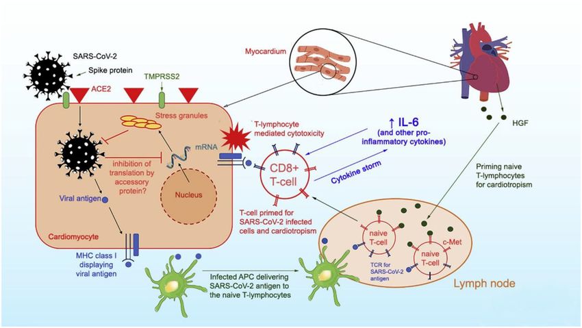
Khan et al.                                                                                                 COVID Myocarditis in Competitive Athletes

PATHOPHYSIOLOGY                                                             Exam
                                                                            The physical exam may demonstrate subtle positional or
Myocarditis is a nonischemic inflammatory process affecting the             reproducible chest pain. There may be signs of congestive
myocardium and inducing myocardial injury of varying clinical               heart failure, including jugular venous distension (JVD), ascites,
severity. The etiology of myocarditis may be infectious (viral,             abdominal pain, peripheral extremity edema, or crackles on a
bacterial) or noninfectious (toxins, hypersensitivity, autoimmune           lung exam. Given the propensity for dysrhythmia, examiners
disorders, and radiation). In viral myocarditis, which may or may           should keenly evaluate for rhythm irregularities, ectopic beats,
not directly translate to COVID, injury to the cardiac muscle is            or rate discrepancies (bradyarrhythmia and tachyarrhythmia).
attributable to direct virus-induced damage, as well as subsequent          Rarely, patients may present in fulminant cardiogenic shock as
autoimmune inflammation. The acute phase (within hours) of                  a result of COVID myocarditis with hypotension, narrow arterial
viral myocarditis is comprised of viral entry into myocytes                 pulse pressure, cool extremities, and altered mental status (8).
mediated by cell surface receptors (3). Once intracellular, the viral
genome is translated into viral proteins, which may disrupt key
dystrophin-glycoprotein interactions to impair cardiac function
                                                                            Biomarkers
                                                                            If viral myocarditis is suspected, clinicians should obtain markers
and injure myocyte cytoskeleton to cause myocyte death (4).
                                                                            of myocardial injury, including elevated troponin (I or T) and
During the second phase, there is an innate immune response
                                                                            creatinine kinase. Elevated brain natriuretic peptide (BNP) may
to the viral antigen mediated by humoral (B-cell) and cell-
                                                                            indicate ventricular dilation or strain from myocardial injury
mediated (T-cell) mechanisms. In the third phase, the host
                                                                            (9). Viral serology testing, although low sensitivity, may be
immune system may recognize intracellular components released
                                                                            reasonable if evaluating for viral myocarditis [including full
as a result of virus-induced injury as foreign antigens, which may
                                                                            respiratory viral panel, as well as SARS-CoV-2 polymerase chain
induce an immunologic response and autoantibodies against the
                                                                            reaction (PCR) testing or antibodies]. Particularly in athletes,
myocyte (via CD4+ cells stimulating B-cells, cytotoxic CD8+
                                                                            alternative etiologies of cardiomyopathy should be excluded,
cells, and cytokines). Over time, these autoantigens may cause
                                                                            such as substances (i.e., cocaine) and metabolic derangements
chronic myocardial inflammation, further myocyte necrosis, and
                                                                            (thyroid) with urine toxicology, and serum thyroid stimulating
progression of structural heart disease (dilated cardiomyopathy).
                                                                            hormone (TSH) (10). Inflammatory markers [e.g., C-reactive
   Per Siripanthong et al. (5), the pathophysiology of COVID
                                                                            protein (CRP)] can be obtained and trended with treatment.
myocarditis is postulated to be similar with SARS-CoV-2 entering
the cell by binding to angiotensin-converting enzyme 2 (ACE2)
receptors on cardiomyocyte surfaces, inducing viral replication,            Electrocardiogram
and setting off the lymphocytic inflammatory cascade augmented              In myocarditis, a 12-lead ECG may demonstrate changes such
by interleukin 6 (IL-6) mediated cytokine release (Figure 1).               as diffuse ST-segment elevations, T-wave inversions, low-voltage
Based on this animal model, the severity of COVID associated                QRS complexes, or even q-waves. As noted above, the infiltrative
myocarditis may reflect the immune response generated by the                nature of viral myocarditis may ultimately result in scarring,
host, so young, otherwise healthy, athletes may generate a more             which can impair the electrophysiological components of the
robust immunologic reaction to viral infection and experience               heart. Even transient myocardial inflammation may induce
greater lymphocytic proliferation and cytokine storm.                       intraventricular conduction delay, AV-block, supraventricular
                                                                            tachycardia (SVT), ventricular tachycardia (VT), ventricular
                                                                            fibrillation (VF), atrial fibrillation, or nonspecific ectopy. If
DIAGNOSIS                                                                   inflammation extends to the pericardium, the ECG may also
Clinical Presentation                                                       demonstrate PR-interval depressions (11).
The initial presentation of myocarditis is often nonspecific,
so a high index of suspicion is required by the clinician. A                Transthoracic Echocardiography
viral prodrome (congestion, rhinorrhea, cough, and/or fever)                The diagnostic workup for myocarditis should include a TTE,
may precede viral myocarditis. Young patients, particularly                 which can be useful in evaluating for myocarditis as well
athletes, without coronary artery disease (CAD) risk factors                as excluding alternative etiologies of cardiomyopathy, such as
may present with severe chest pain and ST-segment elevations                valvular pathology or other structural heart disease (11). In the
on an electrocardiogram (ECG), described as an “infarct-like”               acute phase of viral infection, myocardial inflammation may
pattern associated with viral myocarditis (6). Alternatively,               be characterized by impaired ventricular function, abnormal
patients may report various degrees of exertional dyspnea,                  ventricular dimensions (i.e., dilation or increased myocardial
atypical chest pain, palpitations, and/or generalized fatigue. In           wall thickness), and/or pericardial effusion. Specifically in this
extreme cases, previously healthy patients may present with                 scenario, increased wall thickness in the setting of low voltage
decompensated heart failure or cardiogenic shock (volume                    on the ECG is suggestive of myocardial edema or infiltrative
overload, depressed cardiac index, and cool extremities). The               disease. Chronic myocardial inflammation may cause ventricular
most morbid presentation is one of a patient with life-threatening          dilation, as well as hypokinesis, which may be global or regional
arrhythmia or SCD, as a result of the nonischemic ventricular               (12). Although TTE findings in myocarditis can be nonspecific,
scarring induced by myocarditis, which is a nidus for re-entrant            specialized modalities that attempt to quantify motion of specific
circuits (7).                                                               myocardial segments [such as strain rate imaging (SRI)] are

Frontiers in Cardiovascular Medicine | www.frontiersin.org              2                                        July 2021 | Volume 8 | Article 684780
Khan et al. COVID Myocarditis in Competitive Athletes
FIGURE 1 | Proposed mechanism of SARS-CoV-2 entry into myocytes and inflammatory pathways causing viral myocarditis. Reprinted with permission (5).
nonstandardized and have only been utilized in case reports imaging. To fulfill the updated LLC for acute myocardial
(13–15). inflammation (Supplementary Figure 1), CMR must identify
at least one criterion of both myocardial edema (T2-based)
AND nonischemic myocardial injury (T1-based). Moreover, the
Cardiac MRI
LLC boasts particularly high sensitivity and specificity in acute
Given the nonspecific nature of biomarkers, symptoms, ECG,
viral myocarditis, which is characterized by a CMR pattern of
and TTE in myocarditis, CMR has been heralded as the
subepicardial edema and patchy necrosis [often at the basal
noninvasive gold standard to evaluate myocardial inflammation,
inferolateral or lateral wall of the left ventricle (LV)], which may
including segments not ideal for biopsy (i.e., epicardium,
extend to mid-myocardial regions (12).
pericardium) (16). In 2018, the American College of Cardiology
In addition to diagnostic utility, CMR also has prognostication
(ACC) updated the CMR diagnostic criteria for myocarditis,
value, per Gräni et al. In their 2017 CMR evaluation (prior to
known as Lake Louise Criteria (LLC), to increase specificity
revision of LLC in 2018) of 670 suspected myocarditis patients,
(see Supplementary Figure 1) (12, 17). On CMR, there are
a 2–3 fold increase in hazard ratio was observed in development
three proposed diagnostic targets indicative of myocardial
of major adverse cardiovascular events (MACE) in patients who
inflammation: myocardial edema (mediated by inflammation),
had LGE on CMR (18). In a prognostic study more relevant
hyperemia (due to increased permeability of vascular beds), and
for COVID myocarditis, which can present with “infarct-like”
myocardial necrosis/scar (reflective of myocyte death).
findings (positive biomarkers, ST elevations on ECG, and LGE
According to Ferreira et al., these changes are reflected in
on CMR), Chopra et al. found a greater risk of MACE compared
signal intensity of various modalities within CMR imaging.
to noninfarct-like presentations (6).
Myocardial edema leads to prolonged myocardial relaxation
While CMR-based LLC is very accurate for diagnosis of
time, which can be measured on T1 or T2 weighted images,
acute inflammation, its sensitivity is reduced as myocardial
as well as hyperintensity on T2-weighted images. An expanded
inflammation becomes more diffuse. In a cost-conscious world,
extracellular space within myocardium is visualized by increased
CMR and trained radiologists also remain cost-prohibitive for
extracellular volume (ECV) or by administration of gadolinium-
nonacademic centers.
based contrast (GBCA), which localizes to inflamed myocardium
when measured in T1 weighted imaging, known as early
gadolinium enhancement (EGE). Finally, myocardial necrosis Endomyocardial Biopsy
leads to scarring, which allows delayed GBCA accumulation The gold standard for identifying myocarditis remains
known as late gadolinium enhancement (LGE) in T1-weighted endomyocardial biopsy (EMB) because it allows for
Frontiers in Cardiovascular Medicine | www.frontiersin.org 3 July 2021 | Volume 8 | Article 684780Khan et al. COVID Myocarditis in Competitive Athletes histopathological, immunohistochemically, and molecular as measurement of biomarkers, ECG, and TTE. Of the 145 biology analysis with few complications (12, 19, 20). Given athletes, only two (1.4%) had CMR evidence of myocarditis the patchy distribution of myocarditis, five or six EMB per updated LLC, as reviewed by two experienced radiologists samples are recommended to reduce false negative results, but (24). Notably, one athlete was largely asymptomatic with mild fewer may be obtained in practice (21). Histological analysis elevation of biomarkers (troponin-I peaked at 0.09 ng/mL), while of viral myocarditis demonstrates lymphocytic infiltration of the other had mild to moderate symptoms for 3 days in the myocardium. Suspected myocardial samples can also be analyzed setting of normal biomarkers, and both had normal LV function. via viral nucleic acid stains and quantitative PCR or RT-PCR As such, the authors questioned the use of CMR as a screening to evaluate for the presence of a viral genome. However, given tool for myocarditis in athletes without significant symptoms or the inherent risks of EMB (albeit cited as
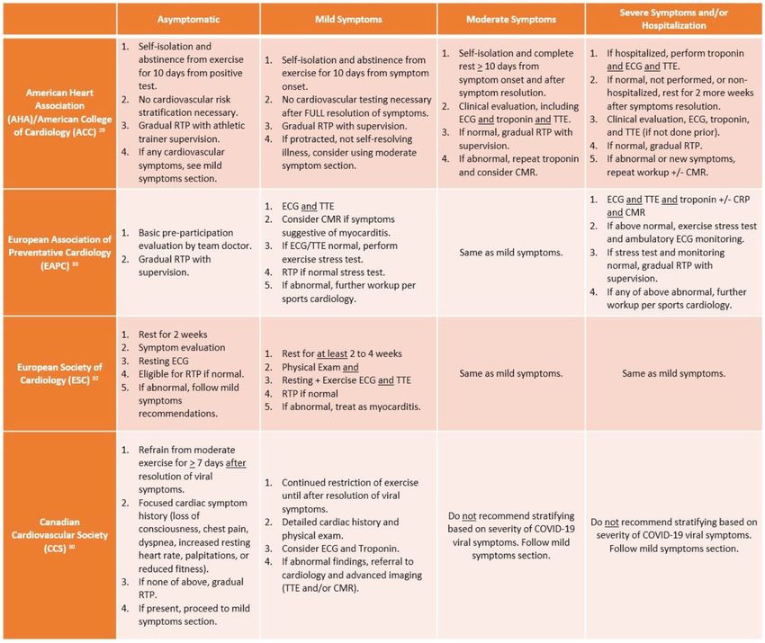
Khan et al.                                                                                                           COVID Myocarditis in Competitive Athletes

 FIGURE 2 | Summary of return to play (RTP) recommendations from major cardiology societies. ECG, electrocardiogram; TTE, transthoracic echocardiogram; CMR,
 cardiac magnetic resonance imaging; CRP, C-reactive protein.

mildly symptomatic athletes recovering from COVID-19 do not                       pertains to COVID-symptom based stratification. According to
require extensive risk stratification beyond history and physical                 McKinney et al., athletes should not be risk stratified based on
exam if their mild symptoms were self-limited. However, in                        their viral COVID illness symptoms, rather with the reporting
athletes with moderate to severe or not self-resolving symptoms,                  or development of cardiovascular symptoms following recovery
extensive cardiovascular risk stratification is needed, including                 from acute viral illness. The CCS recommendation is based on
ECG, biomarkers, and TTE (29). If testing is normal, then                         the lack of association between severity of COVID illness and
athletes may RTP gradually with supervision of athletic trainers,                 development of myocarditis, which is consistent with recent
while abnormal testing or development of new cardiovascular                       studies that have mostly identified myocarditis in asymptomatic
symptoms warrants repeat biomarkers and CMR.                                      or mildly symptomatic athletes. At that point, a cardiac symptom
   In contrast to the American recommendations, the                               questionnaire should be administered; if no cardiac symptoms
European and Canadian societies are more pragmatic                                are reported, athletes may gradually RTP following at least 7
with RTP screening, while acknowledging the inability to                          days of viral symptom resolution. COVID-infected athletes
offer universal cardiovascular testing in all COVID-infected                      who report having the aforementioned cardiac symptoms
athletes. Yet there are key differences between the Canadian                      require a focused history and physical exam, consideration of
Cardiovascular Society (CCS) and European Association                             ECG/troponin, and referral to cardiology (for TTE and/or CMR)
of Preventative Cardiology (EAPC)/European Society of                             if any abnormal findings noted (30). Meanwhile, the EAPC and
Cardiology (ESC) recommendations particularly when it                             ESC advocate for use of exercise stress testing in symptomatic

Frontiers in Cardiovascular Medicine | www.frontiersin.org                    5                                            July 2021 | Volume 8 | Article 684780
Khan et al.                                                                                                                   COVID Myocarditis in Competitive Athletes

athletes more than the Canadian or American societies; the                            at least in the Starekova et al. study, both athletes with
EAPC recommends athletes with mild to moderate symptoms                               COVID myocarditis had normal LV function, so they
should undergo ECG and TTE, then exercise stress testing for                          may have also evaded the EAPC/ESC recommendations for
eligibility to RTP if normal, while the ESC recommends exercise                       further workup. Nonetheless, as suggested by Moulson et al.
ECG in tandem with TTE. However, while the ESC maintains                              (27), primary screening via CMR is also low yield unless
the same recommendations for severe/hospitalization symptoms                          prompted by ECG, TTE, or biomarkers. Mitigating the low
as mild to moderate cases (akin to CCS), the EAPC is more                             prevalence of cardiac involvement in athletes with COVID
in line with the AHA/ACC in recommending a more rigorous                              with the risk of SCD, moving forward with a symptom-based
cardiovascular evaluation consisting of imaging, biomarkers,                          approach, suggested by most societies, to guide RTP seems
and stress testing.                                                                   most appropriate.
    In athletes diagnosed with COVID myocarditis, the 2015
recommendations for sports eligibility by “Task Force 3”                              AUTHOR CONTRIBUTIONS
(comprised of AHA and ACC) (31) should be adapted (see
Supplementary Figure 2).                                                              JN and SJ contributed to the development and writing of the
                                                                                      manuscript. All authors contributed to the article and approved
CONCLUSION                                                                            the submitted version.

Early pandemic studies in nonathletes reported higher                                 ACKNOWLEDGMENTS
incidence of COVID-related cardiac involvement, while recent
publications indicate that incidence of COVID myocarditis                             The authors would like to acknowledge the University of
in adult athletes is not robust as initially feared. While the                        Maryland School of Medicine, Department of Internal Medicine
recommendations by various cardiology societies are an                                and Division of Cardiology.
excellent resource, there remain limitations with regards to
stratifying athletes by symptoms of viral illness. In the cited                       SUPPLEMENTARY MATERIAL
cases of athletes with CMR-proven COVID myocarditis, the
affected athletes had mild to no symptoms, which means they                           The Supplementary Material for this article can be found
could have been eligible for RTP without further workup                               online at: https://www.frontiersin.org/articles/10.3389/fcvm.
per AHA/ACC and CCS guidelines (23, 24). Furthermore,                                 2021.684780/full#supplementary-material

REFERENCES                                                                             8. Kociol RD, Cooper LT, Fang JC, Moslehi JJ, Pang PS, Sabe MA, et al.
                                                                                          Recognition and initial management of fulminant myocarditis: a scientific
 1. Burrow G. The Economic Impact of COVID-19 on US Sports. Economic                      statement from the American Heart Association. Circulation. (2020) 141:e69–
    Modeling. Available online at: https://www.economicmodeling.com/2020/                 e92. doi: 10.1161/CIR.0000000000000745
    05/28/the-economic-impact-of-covid-19-on-us-sports-up-to-92-6k-lost-               9. Inciardi RM, Lupi L, Zaccone G, Italia L, Raffo M, Tomasoni D, et al. Cardiac
    every-minute/ (accessed January 02, 2021).                                            involvement in a patient with coronavirus disease 2019 (COVID-19). JAMA
 2. Shah N, Phelan DMJ. Myocarditis in the Athlete. American College of                   Cardiol. (2020) 5:819–24. doi: 10.1001/jamacardio.2020.1096
    Cardiology (2018). Available online at: https://www.acc.org/latest-in-            10. Trachtenberg BH, Hare JM. Inflammatory cardiomyopathic syndromes.
    cardiology/articles/2018/01/18/15/00/myocarditis-in-the-athlete (accessed             Circ Res. (2017) 121:803–18. doi: 10.1161/CIRCRESAHA.117.
    January 2, 2021).                                                                     310221
 3. Bergelson JM, Cunningham JA, Droguett G, Kurt-Jones EA,                           11. Caforio AL, Pankuweit S, Arbustini E, Basso C, Gimeno-Blanes J, Felix SB,
    Krithivas A, Hong JS, et al. Isolation of a common receptor for                       et al. Current state of knowledge on aetiology, diagnosis, management, and
    Coxsackie B viruses and adenoviruses 2 and 5. Science. (1997)                         therapy of myocarditis: a position statement of the European Society of
    275:1320–3. doi: 10.1126/science.275.5304.1320                                        Cardiology Working Group on Myocardial and Pericardial Diseases. Eur
 4. Badorff C, Lee GH, Lamphear BJ, Martone ME, Campbell KP, Rhoads RE,                   Heart J. (2013) 34:2636–48. 2648a–d. doi: 10.1093/eurheartj/eht210
    et al. Enteroviral protease 2A cleaves dystrophin: evidence of cytoskeletal       12. Ferreira VM, Schulz-Menger J, Holmvang G, Kramer CM, Carbone I, Sechtem
    disruption in an acquired cardiomyopathy. Nat Med. (1999) 5:320–                      U, et al. Cardiovascular magnetic resonance in nonischemic myocardial
    6. doi: 10.1038/6543                                                                  inflammation: expert recommendations. J Am Coll Cardiol. (2018) 72:3158–
 5. Siripanthong B, Nazarian S, Muser D, Deo R, Santangeli P, Khanji MY, et al.           76. doi: 10.1016/j.jacc.2018.09.072
    Recognizing COVID-19 related myocarditis: the possible pathophysiology            13. Kaluzynski K, Chen X, Emelianov SY, Skovoroda AR, O’Donnell M. Strain
    and proposed guideline for diagnosis and management. Heart Rhythm. (2020)             rate imaging using two-dimensional speckle tracking. IEEE Trans Ultrason
    17:1463–71. doi: 10.1016/j.hrthm.2020.05.001                                          Ferroelectr Freq Control. (2001) 48:1111–23. doi: 10.1109/58.935730
 6. Chopra H, Arangalage D, Bouleti C, Zarka S, Fayard F, Chillon S,                  14. Uziebło-Zyczkowska B, Mielniczuk M, Ryczek R, Krzesiński
    et al. Prognostic value of the infarct- and non-infarct like patterns                 P.     Myocarditis      successfully     diagnosed    and     controlled     with
    and cardiovascular magnetic resonance parameters on long-term                         speckle tracking echocardiography. Cardiovasc Ultrasound. (2020)
    outcome of patients after acute myocarditis. Int J Cardiol. (2016)                    18:19. doi: 10.1186/s12947-020-00203-4
    212:63–9. doi: 10.1016/j.ijcard.2016.03.004                                       15. Kostakou PM, Kostopoulos VS, Tryfou ES, Giannaris VD, Rodis IE, Olympios
 7. Peretto G, Sala S, Rizzo S, De Luca G, Campochiaro C, Sartorelli S, et al.            CD, et al. Subclinical left ventricular dysfunction and correlation with regional
    Arrhythmias in myocarditis: state of the art. Heart Rhythm. (2019) 16:793–            strain analysis in myocarditis with normal ejection fraction. A new diagnostic
    801. doi: 10.1016/j.hrthm.2018.11.024                                                 criterion. Int J Cardiol. (2018) 259:116–21. doi: 10.1016/j.ijcard.2018.01.058

Frontiers in Cardiovascular Medicine | www.frontiersin.org                        6                                                 July 2021 | Volume 8 | Article 684780
Khan et al. COVID Myocarditis in Competitive Athletes
16. Ponikowski P, Voors AA, Anker SD, Bueno H, Cleland JGF, Coats AJS, et al. 26. Martinez MW, Tucker AM, Bloom OJ, Green G, DiFiori JP, Solomon G, et al.
2016 ESC guidelines for the diagnosis treatment of acute chronic heart failure: Prevalence of inflammatory heart disease among professional athletes with
the Task Force for the Diagnosis Treatment of Acute Chronic Heart Failure prior COVID-19 infection who received systematic return-to-play cardiac
of the European Society of Cardiology (ESC) developed with the special screening. JAMA Cardiol. (2021). doi: 10.1001/jamacardio.2021.0565. [Epub
contribution of the Heart Failure Association (HFA) of the ESC. Eur Heart ahead of print].
J. (2016) 37:2129–200. doi: 10.1093/eurheartj/ehw128 27. Moulson N, Petek BJ, Drezner JA, Harmon KG, Kliethermes SA, Patel
17. Luetkens JA, Faron A, Isaak A, Dabir D, Kuetting D, Feisst A, et al. MR, et al. SARS-CoV-2 cardiac involvement in young competitive athletes.
Comparison of original and 2018 Lake Louise criteria for diagnosis of acute Circulation. (2021). doi: 10.1161/CIRCULATIONAHA.121.054824. [Epub
myocarditis: results of a validation cohort. Radiol Cardiothorac Imaging. ahead of print].
(2019) 1:e190010. doi: 10.1148/ryct.2019190010 28. Atlani-Duault L, Lina B, Malvy D, Yazdanpanah Y, Chauvin F, Delfraissy JF.
18. Gräni C, Eichhorn C, Bière L, Murthy VL, Agarwal V, Kaneko K, et al. COVID-19: France grapples with the pragmatics of isolation. Lancet Public
Prognostic value of cardiac magnetic resonance tissue characterization in risk Health. (2020) 5:e573–e4. doi: 10.1016/S2468-2667(20)30235-8
stratifying patients with suspected myocarditis. J Am Coll Cardiol. (2017) 29. Kim JH, Levine BD, Phelan D, Emery MS, Martinez MW, Chung
70:1964–76. doi: 10.1016/j.jacc.2017.08.050 EH, et al. Coronavirus disease 2019 and the athletic heart: emerging
19. Cooper LT, Baughman KL, Feldman AM, Frustaci A, Jessup M, Kuhl U, et al. perspectives on pathology, risks, and return to play. JAMA Cardiol. 6:219–27.
The role of endomyocardial biopsy in the management of cardiovascular (2020). doi: 10.1001/jamacardio.2020.5890
disease: a scientific statement from the American Heart Association, the 30. McKinney J, Connelly KA, Dorian P, Fournier A, Goodman JM, Grubic
American College of Cardiology, and the European Society of Cardiology. N, et al. COVID-19-myocarditis and return to play: reflections and
Endorsed by the Heart Failure Society of America and the Heart Failure recommendations from a Canadian Working Group. Can J Cardiol. (in
Association of the European Society of Cardiology. J Am Coll Cardiol. (2007) press). doi: 10.1016/j.cjca.2020.11.007
50:1914–31. doi: 10.1093/eurheartj/ehm456 31. Maron BJ, Udelson JE, Bonow RO, Nishimura RA, Ackerman MJ, Estes NA
20. Yilmaz A, Kindermann I, Kindermann M, Mahfoud F, Ukena C, Athanasiadis 3rd, et al. Eligibility and disqualification recommendations for competitive
A, et al. Comparative evaluation of left and right ventricular endomyocardial athletes with cardiovascular abnormalities: Task Force 3: hypertrophic
biopsy: differences in complication rate and diagnostic performance. cardiomyopathy, arrhythmogenic right ventricular cardiomyopathy and other
Circulation. (2010) 122:900–9. doi: 10.1161/CIRCULATIONAHA.109.924167 cardiomyopathies, and myocarditis: a scientific statement from the American
21. Tschöpe C, Ammirati E, Bozkurt B, Caforio ALP, Cooper LT, Felix Heart Association and American College of Cardiology. Circulation. (2015)
SB, et al. Myocarditis and inflammatory cardiomyopathy: current 132:e273–80. doi: 10.1161/CIR.0000000000000239
evidence and future directions. Nat Rev Cardiol. (2020) 18:169–93. 32. Schellhorn P, Klingel K, Burgstahler C. Return to sports after COVID-19
doi: 10.1038/s41569-020-00435-x infection. Eur Heart J. (2020) 41:4382–4. doi: 10.1093/eurheartj/ehaa448
22. Puntmann VO, Carerj ML, Wieters I, Fahim M, Arendt C, Hoffmann J, et al. 33. Bhatia RT, Marwaha S, Malhotra A, Iqbal Z, Hughes C, Börjesson M, et al.
Outcomes of cardiovascular magnetic resonance imaging in patients recently Exercise in the severe acute respiratory syndrome coronavirus-2 (SARS-
recovered from coronavirus disease 2019 (COVID-19). JAMA Cardiol. (2020) CoV-2) era: a question and answer session with the experts endorsed by
5:1265–73. doi: 10.1001/jamacardio.2020.3557 the section of sports cardiology & exercise of the European Association
23. Rajpal S, Tong MS, Borchers J, Zareba KM, Obarski TP, Simonetti OP, of Preventive Cardiology (EAPC). Eur J Prev Cardiol. (2020) 27:1242–
et al. Cardiovascular magnetic resonance findings in competitive athletes 51. doi: 10.1177/2047487320930596
recovering from COVID-19 infection. JAMA Cardiol. (2020) 6:116–8.
doi: 10.1001/jamacardio.2020.4916 Conflict of Interest: The authors declare that the research was conducted in the
24. Starekova J, Bluemke DA, Bradham WS, Eckhardt LL, Grist TM, Kusmirek absence of any commercial or financial relationships that could be construed as a
JE, et al. Evaluation for myocarditis in competitive student athletes recovering potential conflict of interest.
from coronavirus disease 2019 with cardiac magnetic resonance imaging.
JAMA Cardiol. (2021). doi: 10.1001/jamacardio.2020.7444. [Epub ahead of Copyright © 2021 Khan, Na and Jerome. This is an open-access article distributed
print]. under the terms of the Creative Commons Attribution License (CC BY). The use,
25. Kawakami R, Sakamoto A, Kawai K, Gianatti A, Pellegrini D, Nasr A, distribution or reproduction in other forums is permitted, provided the original
et al. Pathological evidence for SARS-CoV-2 as a cause of myocarditis: author(s) and the copyright owner(s) are credited and that the original publication
JACC review topic of the week. J Am Coll Cardiol. (2021) 77:314– in this journal is cited, in accordance with accepted academic practice. No use,
25. doi: 10.1016/j.jacc.2020.11.031 distribution or reproduction is permitted which does not comply with these terms.
Frontiers in Cardiovascular Medicine | www.frontiersin.org 7 July 2021 | Volume 8 | Article 684780You can also read