Dystocia in a Captive Reared Agouti (Dasyprocta leporina) in Trinidad and Tobago, West Indies
←
→
Page content transcription
If your browser does not render page correctly, please read the page content below
veterinary
           sciences
Case Report
Dystocia in a Captive Reared Agouti (Dasyprocta
leporina) in Trinidad and Tobago, West Indies
Kegan Romelle Jones 1,2, * , Kavita Ranjeeta Lall 2 and Gary Wayne Garcia 2
 1    Department of Basic Veterinary Sciences (DBVS), School of Veterinary Medicine (SVM),
      Faculty of Medical Sciences, The University of the West Indies (UWI), Mt. Hope, Trinidad and Tobago
 2    The Open Tropical Forage-Animal Production Laboratory (OTF-APL),
      Department of Food Production (DFP), Faculty of Food and Agriculture (FFA),
      The University of the West Indies (UWI), St. Augustine, Trinidad and Tobago;
      k_lee_24@yahoo.com (K.R.L.); prof.gary.garcia@gmail.com (G.W.G.)
 *    Correspondence: keganjones11@gmail.com; Tel.: +868-787-0833

 Received: 30 December 2019; Accepted: 11 February 2020; Published: 4 March 2020

 Abstract: Dystocia is a complication that occurs at parturition either due to foetal or maternal factors.
 This condition has been well studies in domesticated species. However, there is very little information
 on dystocia in the agouti (Dasyprocta leporina). The agouti is utilized for its meat in South America
 and the Caribbean. More recently, farming of these animals intensively is being practiced in the
 Neo-tropics. This case report attempted to provide some insight into dystocia in the agouti which
 has been rarely reported in animals in captivity. A female agouti weighing approximately 3 kg (kg),
 which was in the last stage of pregnancy, was found dead in its cage. The vulva of the animal had the
 hind-limbs of the offspring protruding. Upon necropsy the animal had little fat reserves and had
 two foetuses in the right horn of the uterus. The feet of on offspring were dislocated and exposed at
 the level of the vulva. Each foetus weighed approximately 200 g. The foetuses were well formed
 with fur, teeth and eyes. The placenta was attached to each of the foetuses. The pathological findings
 suggested that dystocia resulted in secondary uterine inertia, which was the cause of death of the
 adult female agouti. To prevent the recurrence of this situation the gestation should be staged (timed)
 using ultrasonography. Animals which are in their third stage of gestation should be monitored using
 cameras or with personnel at the facility to assist agoutis which are having difficulties at parturition.

 Keywords: Neo-tropics; uterine inertia; Caribbean; South America; reproductive diseases;
 domestic livestock

1. Introduction
     The agouti (D. leporina) is a rodent that belongs to the hysricomorphic group of rodents. It is one
of the most hunted species in the Caribbean and South America [1]. The use of this mini-livestock is
a major source of meat protein for rural communities in the Neo-tropics [2]. The exploitation of these
animals in the wild can lead to decreasing numbers in the wild population. However, if the agouti is
domesticated and reared in intensively, animals can be re-introduced into the natural environment as
a conservation strategy [1].
     Several articles have been published on the male anatomy of the agouti. The micro- and
macro-anatomy of the male reproductive tract [3,4], the electro-ejaculates of the male agouti has
been analyzed and these ejaculates have been extended and stored [4–8]. Finally, the stages of erection
in the male agouti have been investigated [9]. The age of weaning of this precocious rodent under
intensive production has been reported to be one week [10]. The female reproductive system has also
been studied and will be discussed in this paper.

Vet. Sci. 2020, 7, 30; doi:10.3390/vetsci7010030                                   www.mdpi.com/journal/vetsci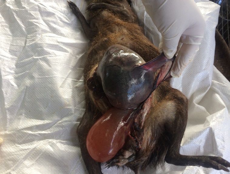
Vet. Sci. 2020, 7, 30                                                                               2 of 8

      The nutrition and feeding practices of agouti have been documented on several occasions.
The digestive tract of the agouti is similar to that of the rabbits. The agouti possessed a large caecum
and the small intestine was reported to be longer in comparison to that of the rabbit [11]. Some authors
have stated that the animal is a frugivore with a large majority of its diet consisting of fruit and
nuts [12,13]. Researchers have also found that the animal is a grubivore, consuming insects that were
present in the seed of some nuts [14]. Dookie et al. [15] reported a feed particle size preference of
12.7 mm × 25.4 mm. Lall et al. [16] reviewed the diets of these animals in captivity and in the wild.
It was summarised that the agouti was frugivorous, herbivorous and opportunistic in its feeding
behaviour. In captivity the consumption of animal matter has been documented with the agouti
consuming carrion, chicken eggs and dead chicken [17,18].
      The majority of diseases highlighted have been related to some infectious agent. Lall et al. [19]
reviewed pathogenic diseases present in the agouti and found that few animals showed clinical signs
of diseases. This made the agouti a major reservoir of diseases for domesticated animals. Additionally,
animals found in the neotropics were grouped into three categories. (1) Domesticated animals that
were introduced into the neo-tropics which were: cattle, sheep, goats, chickens, pigs and horses.
(2) Domesticated animals that originated from the neo-tropics (South American camelids, chinchillas,
ducks, turkeys and guinea pigs). (3) Non-domesticated animals that originated from the neotropics
(agouti, lappe, capybara, red brocket deer, collared peccary and manicou) [20–22].
      Investigation on the gastrointestinal parasites common to the agouti reared in captivity and the
wild have been documented [23–28]. Blood parasites have been found in wild agouti populations.
Organisms identified included Babesia, Leishmania and Trypanosoma cruzi [29,30]. However, in the
captive reared colonies of agoutis blood parasites were not identified [31]. Few reports were given on
the effects that the parasitic organisms had on the agouti. Authors who investigated the effects of the
parasites on the animals found that the agouti was clinically healthy, with a body condition of 3 out
of 5 [25].
      Some works on the detection of subclinical illness in the agouti have been reported, discussing the
haematology and serum biochemistry. Baas et al. [32] reported on blood reference values for agoutis
reared in captivity. Further work by Jones et al. [31] and Jones and Garcia [33,34] recorded blood
parameters for healthy agoutis reared intensively. However, little reports existed on non-infectious
diseases of the agouti. However, imbalances in vitamin D have been highlighted [35,36]. There is
a dearth of information on non-infectious reproductive diseases. Personal communication from the
third author of this paper stated that two diseases and dystocia, which is a consecutives situation that
seriously affects delivery. These were (1) pasteurellosis, (2) scabies and (3) dystocia. The objective of
this report was to highlight findings of one such dystocia.

2. Material and Method

2.1. Housing of the Agouti (Dasyprocta leporina)
     The maternity cages measured 0.61 m long × 0.91 m wide × 0.61 m high. Animals were separated
from the colony reared on floor pens when signs of pregnancy were noted. Confirmation of pregnancy
was done using physical signs such as a rounded abdomen and protruding nipples. The animal had
an approximate weight of 3 kg. The female agouti was part of a colony of agoutis, which were reared
intensively at the University of the West Indies Field Station, Trinidad. The colony housed one hundred
agoutis, which were reared in individual cages and also in pens on concrete floors.
     The diet of the animals consisted of locally available fruits such as mangoes (Mangifera indica),
pumpkins (Cucurbita pepo) and fat pork (Chrysobalanus icaco). Chicken eggs were also fed to the animals
in conjunction with commercial pelleted rabbit ration (Crude Protein 17%). Animals in the unit always
had a constant supply of water for drinking and thermoregulation. The concrete floor pen consisted of
breeding units with a female to male ration of 5:1. Animals which are kept at this station are under the
Vet. Sci. 2020, 7, 30 3 of 8
pen consisted of breeding units with a female to male ration of 5:1. Animals which are kept at this
station are under the sanction of animal welfare guideline of the University of the West Indies. In this
unit thereofare
sanction two veterinarians
animal whichofmonitor
welfare guideline and treat
the University ofanimals showing
the West Indies. overt
In thissigns
unit of disease.
there are two
veterinarians which monitor and treat animals showing overt signs of disease.
2.2. Clinical Signs
2.2. Clinical Signs
Pregnant agoutis were observed daily for any signs of physical abnormality, which included
abnormal discharges
Pregnant agoutisfrom
werethe vulva, diarrhea
observed daily forand
anyemaciation. Animals
signs of physical were also observed
abnormality, for any
which included
signs of physical
abnormal trauma
discharges fromorthe
wounds,
vulva,which may
diarrhea have
and occurred as
emaciation. a resultwere
Animals of fighting.
also observed for any
signs of physical trauma or wounds, which may have occurred as a result of fighting.
3. Results
3. Results
3.1. Post Mortem Examination
3.1. Post Mortem Examination
An adult female agouti (D. leporina) approximately 2 years of age was found dead in a maternity
cage.AnTheadult female agouti
hind-limbs of one (D. leporina)
offspring approximately
were protruding 2from
years ofmother’s
the age was found
vulva.dead
Uponinpresentation
a maternity
cage. The agouti
the dead hind-limbs
was of onetaken
then offspring
andwere protruding
a necropsy from theto
performed mother’s
identifyvulva. Upon presentation
any abnormalities the
in foetal
dead agouti
positioning. was then taken and a necropsy performed to identify any abnormalities in foetal positioning.
3.2. Treatment
3.2. Treatment
The animal was found dead in the cage. The animal had no prior signs of illness. This situation
The animal was found dead in the cage. The animal had no prior signs of illness. This situation
did not allow for the agouti to be treated for its dystocia.
did not allow for the agouti to be treated for its dystocia.
3.3. Necropsy Findings
3.3. Necropsy findings
The abdominal cavity of the agouti was opened and a right uterine horn was distended with two
The abdominal cavity of the agouti was opened and a right uterine horn was distended with two
foetuses and the left uterine horn was empty. The urinary bladder of the animal was also distended.
foetuses and the left uterine horn was empty. The urinary bladder of the animal was also distended.
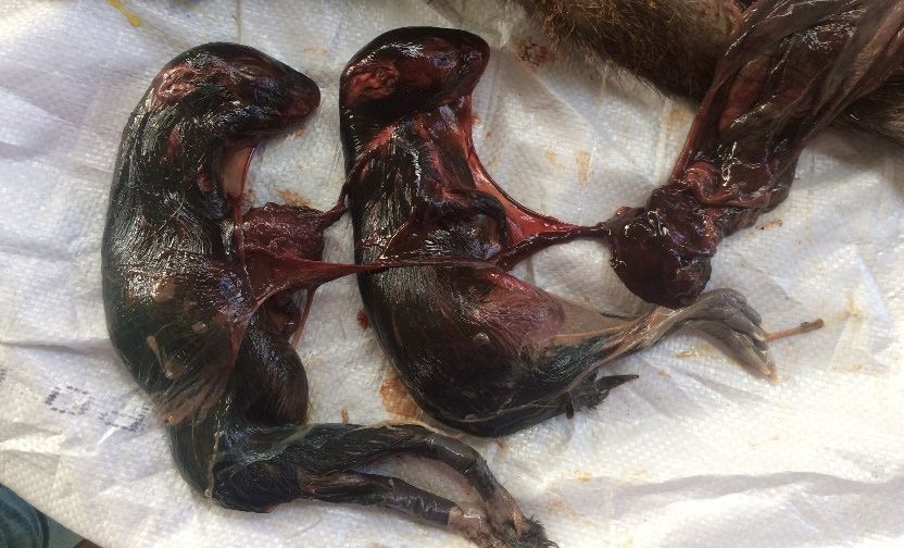
Figure 1 shows a part of an absolute large foetus, which resulted in the dydtocia, which consequently
Figure 1 shows a part of an absolute large foetus, which resulted in the dydtocia, which consequently
resulted in secondary inertia. The lack of medical attention to the secondary inertia resulted in the
resulted in secondary inertia. The lack of medical attention to the secondary inertia resulted in the
death of the offspring and the mother.
death of the offspring and the mother.
Figure 1. Necropsy of Adult Agouti with oversized foetus in the right uterine horn which resulted in
Figure 1. Necropsy of Adult Agouti with oversized foetus in the right uterine horn which resulted in
the death of the animal.
the death of the animal.
The right
The right uterine
uterine horn
horn was
was opened
opened and
and two
two foetuses,
foetuses, each
each weighing
weighing approximately
approximately 200
200 gg were
were
found inside. Both foetuses were in posterior longitudinal presentation, dorsal position and in
found inside. Both foetuses were in posterior longitudinal presentation, dorsal position and in normal normal
posture. The
posture. The offspring
offspring had
had hair, teeth and
hair, teeth and nails
nails with
with the
the placenta
placenta attached
attached to
to each
each animal
animal (Figure
(Figure 2).
2).
The carcass had little subcutaneous fat and the cause of death was attributed to secondary
The carcass had little subcutaneous fat and the cause of death was attributed to secondary uterine uterine
inertia due
inertia due to
to the
the dystocia.
dystocia.
3Vet. Sci. 2020, 7, 30 4 of 8
Figure
Figure 2.
2. Two
Two offspring
offspring weighing
weighing 200
200 gg each
each in
in the
the uterus.
uterus.
4.
4. Discussion
Discussion
Little
Little information
information has has been
been reported
reported on on the reproductive disorders in the agouti. Some Some authors
authors
have
have observed perinatal mortality in captive reared agoutis in Trinidad. There
perinatal mortality in captive reared agoutis in Trinidad. There was one reported case was one reported casein
in Trinidad,
Trinidad, in which
in which an agouti
an agouti dieddied
fromfrom secondary
secondary uterine
uterine inertia
inertia due todue to dystocia.
dystocia. In thatIncase
thatthe
caseadultthe
adult
femalefemale was reported
was reported to havetohad havesix had six offspring.
offspring. Three were Three were delivered
delivered via the vaginavia the
butvagina but the
the remaining
remaining
offspring wereoffspring
removed were viaremoved
caesarean viasection
caesarean[37]. section
In Brazil, [37]. In Brazil, pathological
pathological changes in the changes in the
reproductive
reproductive
system of captive system of captive
reared breeding reared
femalesbreeding females wereDystocia
were investigated. investigated. Dystociain
was reported was7.7% (n = 1) in
reported of
7.7% (n = females.
breeding 1) of breeding
In thatfemales. In that case
case of dystocia thereofwas dystocia thereoffspring
one large was onepresent
large offspring
in a uterine present
horn in a
[38].
uterine
Batista ethorn [38].also
al. [39] Batista et al. [39]
reported also reported
a prevalence of 6.24%a prevalence
for dystocia of 6.24% for dystocia
in captive in captive reared
reared agoutis.
agoutis.
Dystocia is the inability to expel the foetus from the uterus during parturition, which occasionally
occursDystocia is the inability
in domesticated animals. Ittocanexpel
be duethe foetus from
to maternal theabnormalities.
or foetal uterus during parturition,
Maternal which
abnormalities
occasionally
include; smalloccurspelvicin domesticated
size, narrow pelvic animals.
canal and It can be due
uterine to maternal
inertia. Foetal causes or foetal
are due abnormalities.
to abnormal
Maternal
presentation,abnormalities include;
position, posture, small pelvic
oversized foetus, size, narrow
foetal deathpelvic canal [40].
or monsters and uterine
In rabbits inertia.
treatmentFoetal of
causes
dystocia arehas
duebeen
to abnormal
done either presentation,
medically position,
or surgically.posture, oversized
In case of anyfoetus,
abnormal foetal death or monsters
presentations forms,
[40]. In rabbits
stimulating treatment
uterine of dystocia
contractility must not hasbebeen done either
performed before medically
correction of or presentation
surgically. In case of any
abmormalities
abnormal presentations
in order to avoid forms, cervical
uterine rupture, stimulating damage uterine contractility
or occlusion must in
of the foetus not
thebe performed
birth canal. Treatment before
correction of presentationofabmormalities
included administration calcium borogluconate,in order to avoid uterine
propylene glycol,rupture, cervicalCalcium
and oxytocin. damageand or
occlusion
oxytocin have of been
the foetus
reported intothe birththecanal.
increase Treatment
contractility of theincluded
uterus [41]. administration
Propylene glycol of provides
calcium
borogluconate,
energy for the smoothpropylene glycol,
muscles in and oxytocin. Calcium
the myometrium and oxytocin
[41]. Surgery has tohave been reported
be performed whentothe increase
above
the contractility
mentioned drugsofdo thenotuterus
result[41].
in thePropylene
solution glycol provides energy for the smooth muscles in the
for dystocia.
myometrium
In this case[41].the
Surgery
animalhas tofound
was be performed
dead in when the above
the morning. It mentioned drugs dothat
is being suggested notifresult
dystociain theis
solution
encounteredfor dystocia.
in the agouti in future the medical management used in the rabbit using a “dystocia
In this(oxytocin,
cocktail” case the animal
propylene was glycol
found dead in the morning.
and calcium It is being
borogluconate) suggested
could be given that toifthedystocia
affected is
encountered
animal [42]. In inmice
the agouti in future
with dystocia thetheuse medical management
of these drugs used in theasrabbit
are not considered using
the first line of a “dystocia
treatment.
cocktail”
The uses (oxytocin,
of these drugs propylene
have beenglycol and calcium
reported borogluconate)
to be ineffective could be
in treatment of given
dystociato the affected
in mice and
animal
authors[42].
haveIn mice with
suggested thatdystocia
surgicalthe use of be
treatment these
done drugs
as theare not considered
primary response toastreatment
the first [42].line of
treatment.
In thisThe
caseuses of these
the cause drugswas
of death have been reported
reported to be ineffective
to be secondary in treatment
uterine inertia of dystocia
due to dystocia. Therein
mice and authors
was normal have suggested
presentation, posture and thatposition
surgicaloftreatment
the foetuses.be done as thethe
However, primary response
both foetuses wereto
treatment
large and [42].
present in the right horn, which may have contributed to dystocia. Investigations showed
In thisthat
elsewhere casethethetwocause of death
uterine hornswaswerereported to be secondary
independent and each uterine
of them inertia
was due to dystocia.
connected There
to separate
was
cervixnormal presentation,
[43]. Therefore, theposture
two large andfoetuses
positionthat of the
werefoetuses.
presentHowever,
in the righttheuterine
both foetuses
horn would were large
have
and
had topresent in the right
pass through horn,cervix,
the right whichwhich may may havehave contributed
contributed to dystocia. Investigations
to the dystocia seen in this showedcase.
elsewhere
Further work thatdone
the two by uterine
Singh ethorns
al. [44]werewereindependent
in disagreement and each withof Mayor
them was connected
et al. [43] and to separate
stated that
cervix [43].had
the agouti Therefore,
one cervix the two
and large
the two foetuses
uterinethat were
horns present in
connected to the
the right
bodyuterine horn would
of the uterus, whichhave was
had to passtothrough
connected a common the right
cervix.cervix,
Otherwhich may have contributed
hystricomorphic rodents such to as
thethedystocia
capybaraseen(Hydrochaerus
in this case.
Further work done by Singh et al. [44] were in disagreement with Mayor et al. [43] and stated that
the agouti had one cervix and the two uterine horns connected to the body of the uterus, which was
4Vet. Sci. 2020, 7, 30 5 of 8
hydrochaeris) and the lappe (Agouti paca/Cuniculus paca) had uterine horns that were independent of
each other and were connected independently to two cervices [45,46].
Early observations on parturition in the agouti by Enders [46] stated that newborn animals were
fully haired, had open eyes and were patterned like the adult. The animal gave birth in a squatting
position having one hour intervals in delivery of offspring. Enders [47] also reported a dystocia that
resulted in the death of mother and offspring. The right hind limb of the lower foetus was engaged
resulting in failure to expel uterine contents. The limbs were dislocated due to trauma, which was
similar to this case of dystocia seen at the University Field Station. Weir [48] also reported a case of
dystocia where the foetus was reported as being oversized and was found in the right uterine horn.
In this case however, the foetuses had average birth weights [47] and were found in the right horn in
reports [48].
Prevention of dystocia can be done if accurate stages of gestation can be investigated.
Souza et al. [49] monitored gestation age and embryonic-foetal development in the agouti. It was
noted that gestational length was 103 days and, using ultrasonography the gestational sac can be
observed from day nine [49]. Brown [50] stated that the gestational period for the golden-rumped
agouti (Dasyprocta aguti) was 104 days, Pachaly et al. [51] reported a gestational period of 103 days
while Clarke and Olfert [52] gave a gestational period ranging from 104–120 days. Fortes el al. [53]
highlighted that at twenty-five days after mating the foetus was positioned in a “C Shape” with
primitive structures. To prevent the recurrence of this situation the gestation should be staged (timed)
using ultrasonography. Animals which are in their third stage of gestation should be monitored using
cameras or with personnel at the facility to assist agoutis which are having difficulties at parturition.
If difficulty is noticed at parturition, then the mother can be treated medically or surgically to alleviate
the situation. This will avoid have avoided the deaths of the mother and fetuses which occurred in
this report.
In this case the pregnant agouti carried two offspring, which was similar to the litter sizes reported
in the literature. Pachaly et al. [51] reported the agouti to have had an average litter size of 2.09 and
Mayor et al. [43] reported average litter sizes of 2.1. Brown-Uddenberg [54] reported average litter
sizes of 1.71 and Jones and Garcia [55] found average litter sizes of 1.7.
5. Conclusions
In this case the cause of death was attributed to secondary uterine inertia due to dystocia. The two
foetuses were found in one horn, which may have attributed to the dystocia. The absolute foetal
oversize and failing medical intervention were responsible for the death of the foetus and the dam.
It is generally not advised to leave periparturient animals unattended for several hours. To prevent the
recurrence of this situation the gestation should be staged (timed) using ultrasonography. Animals
which are in their third stage of gestation should be monitored using cameras or with personnel at the
facility to assist agoutis which are having difficulties at parturition.
There has been little information reported on the reproductive disorders of the agouti.
More information needs to be documented on reproductive disorders of agoutis reared in captivity.
Intensification of the agouti may reveal a higher incidence of dystocia and proper treatment of such
cases should be reported.
Author Contributions: Data collection was done by K.R.J. and K.R.L. Draft manuscript was written by K.R.J.
Editions to the manuscript were done by K.R.J., K.R.L. and G.W.G. The entire project was supervised by G.W.G.
All authors have read and agreed to the published version of the manuscript.
Funding: This project was funded by the Campus Research and Publication of the University of the West Indies,
St. Augustine Campus.
Conflicts of Interest: There was no conflict of interest between authors.Vet. Sci. 2020, 7, 30 6 of 8
References
1. Brown-Uddenberg, R.C.; Garcia, G.W.; Baptiste, Q.S.; Counand, T.; Adogwa, A.O.; Sampson, T. The Agouti
(Dasyprocta leporina, D. aguti) Booklet and Producers’ Manual; GWG Publications: Champs Fleurs, Trinidad and
Tobago, 2014; Available online: http://ostasp.brinkster.net/ (accessed on 17 September 2017).
2. Hardouin, J.; Thys, E.; Joiris, V.; Fielding, D. Mini-livestock breeding with indigenous species in the tropics.
Livest. Res. Rural Dev. 2003, 15, 4. Available online: http://www.lrrd.org/lrrd15/4/hard154.htm (accessed on
11 October 2019).
3. Mollineau, W.M.; Adogwa, A.O.; Young, K.; Jasper, N.; Garcia, G.W. The Gross anatomy of the male
reproductive system of a neo-tropical rodent: The Agouti (Dasyprocta leporina). Anat. Histol. Embryol. 2006,
35, 47–52. [CrossRef]
4. Mollineau, W.M.; Adogwa, A.O.; Garcia, G.W. The Gross and Micro Anatomy of the Accessory Sex Glands
of the Male Agouti (Dasyprocta leporina). Anat. Histol. Y Embryol. 2009, 38, 204–207. [CrossRef]
5. Mollineau, W.M.; Adogwa, A.O.; Garcia, G.W. A Preliminary technique for electro-ejaculation of agouti
(Dasyprocta leporina). Anim. Reprod. Sci. 2008, 108, 92–97. [CrossRef]
6. Mollineau, W.M.; Adogwa, A.O.; Garcia, G.W. Spermatozoa morphologies and fructose and citric acid
concentrations in agouti (Dasyprocta leporina) semen. Anim. Reprod. Sci. 2008, 105, 378–383. [CrossRef]
7. Mollineau, W.M.; Adogwa, A.O.; Garcia, G.W. Liquid and Frozen storage of Agouti (Dasyprocta leporina)
Semen extended with UHT Milk, Unpasteurized Coconut Water and Pasteurized Coconut Water. Vet. Med.
Int. 2011, 5, 702635. [CrossRef] [PubMed]
8. Mollineau, W.M.; Adogwa, A.O.; Garcia, G.W. Improving the efficiency of the preliminary electro-ejaculation
technique developed for semen collection from the Agouti (Dasyprocta leporina). J. Zoo Wildl. Med. 2010, 41,
633–637. [CrossRef] [PubMed]
9. Mollineau, W.M.; Adogwa, A.O.; Garcia, G.W. Anatomical stages of penile erection in the agouti (Dasyprocta
leporina) induced by electro-ejaculation. Anat. Histol. Embryol. 2012, 41, 392–394. [CrossRef] [PubMed]
10. Mohammed, R.; Legall, G.; Garcia, G.W. Towards the determination of a “Weaning Age” for the intensive
production of the Agouti (Dasyprocta leporina). Livest. Res. Rural Dev. 2018, 30. Available online:
http://www.lrrd.org/lrrd30/10/riyad30173.html (accessed on 11 October 2019).
11. Garcia, G.W.; Baptiste, Q.S.; Kakuni, M.; Arishima, K.; Makita, T. The Digestive System of the Agouti
(Dasyprocta leporina)—Gross Anatomy and Histology. Jpn. J. Zoo Wildl. Med. 2000, 5, 55–66. [CrossRef]
12. Henry, O. Frugivory and the Importance of Seeds in the Diet of the Orange-Rumped Agouti (Dasyprocta
leporina) in French Guiana. J. Trop. Ecol. 1999, 15, 291–300. [CrossRef]
13. Silvius, K.M.; Fragoso, J.M.V. Red-Rumped Agouti (Dasyprocta leporina) Home Range Use in an Amazonian
Forest: Implications for the Aggregated Distribution of Forest Trees. Biotropica 2003, 35, 74–83. [CrossRef]
14. Silvius, K.M. Spatio-Temporal Patterns of Palm Endocarp Use by Three Amazonian Forest Mammals:
Granivory or ‘Grubivory’? J. Trop. Ecol. 2002, 18, 707–723. [CrossRef]
15. Dookie, B.; Jones, K.R.; Mohammed, R.; Garcia, G.W. Feed particle size preference and feed wastage in
Agouti (Dasyprocta leporina) reared intensively in the Republic of Trinidad and Tobago. Livest. Res. Rural Dev.
2018, 30, 1–6.
16. Lall, K.R.; Jones, K.R.; Garcia, G.W. Nutrition of Six Selected Neo-Tropical Mammals in Trinidad and Tobago
with the Potential for Domestication. Vet. Sci. 2018, 5, 52. [CrossRef] [PubMed]
17. Figueira, L.; Zucaratto, R.; Pires, A.S.; Cid, B.; Fernandez, F.A.S. Carrion Consumption by Dasyprocta leporina
(Rodentia: Dasyproctidae) and a Review of Meat Use by Agoutis. Braz. J. Biol. 2014, 74, 585–587. [CrossRef]
18. Jones, K.R.; Lall, K.R.; Garcia, G.W. Omnivorous Behaviour of the Agouti (Dasyprocta leporina): A Neotropical
Rodent with the Potential for Domestication. Scientifica 2019, 5, 3759783. [CrossRef]
19. Lall, K.R.; Jones, K.R.; Garcia, G.W. Infectious Diseases of Six Non-Domesticated Neo-Tropical Animals in
Trinidad and Tobago. Int. J. Trop. Vet. Biomed. Res. 2018, 3, 1–31. [CrossRef]
20. Jones, K.R.; Garcia, G.W. Gastrointestinal parasites of domesticated animals introduced into the Neo-tropics
(New World Tropics). Concepts Dairy Vet. Sci. 2018, 1, 56–78.
21. Jones, K.R.; Garcia, G.W. Endoparasites of domesticated animals that originated in the neo-tropics (new world
tropics). Vet. Sci. 2019, 6, 24. [CrossRef]
22. Jones, K.R.; Garcia, G.W. Endoparasites of selective native non-domesticated mammals in the neo-tropics
(new world tropics). Vet. Sci. 2019, 6, 87.Vet. Sci. 2020, 7, 30 7 of 8
23. Suepaul, R.; Charles, R.; Dziva, F. Aerobic microflora and endoparasites of freshly shot wild Agouti (Dasyprocta
leporina) in Trindad, West Indies. J. Zoo Wildl. Med. 2016, 47, 1044–1048. [CrossRef] [PubMed]
24. Jones, K.R.; Garcia, G.W. A survey of the gastrointestinal parasites present in the Agouti (Dasyprocta leporina)
reared intensively in Trinidad. Livest. Res. Rural Dev. 2017, 29, 1–7.
25. Jones, K.R.; Garcia, G.W. Observations on endoparasitic load in captive reared agoutis (Dasyprocta leporina)
without anthelmintic exposure in Trinidad, Republic of Trinidad and Tobago. Livest. Res. Rural Dev. 2018, 30,
1–7.
26. Griffiths, H.J. Studies on Strongyloides agoutii from the agouti (Dasyptocta agouti). Can. J. Res. D 1940, 18,
173–190. [CrossRef]
27. Cameron, T.W.M.; Reesal, M.R. Studies on the endoparasitic fauna of Trinidad mammals. Can. J. Zool. 1951,
29, 276–289. [CrossRef]
28. Lainson, R.; Carneiro, L.; Silveira, F.T. Observations on the Eineria species of the Dasyprocta leporina (Linnaeus,
1758) (Rodentia: Dasyproctidae) for the state of Para, North Brazil. Mem. Inst. De Oswaldo Cruz 2007, 102,
183–189. [CrossRef]
29. de Thoisy, B.; Michel, J.-C.; Vogel, I.; Vie, J.-C. A survey of hemoparasite infection in free ranging mammals
and reptiles in French Guina. J. Parasitol. 2000, 86, 1035–1040. [CrossRef]
30. Ayala, S.C.; D’ Alessandro, A.; Mackenzie, R.; Angel, D. Hemoparasites in 830 wild animals from Eastern
Llanos of Colombia. J. Parasitol. 1973, 59, 52–59. [CrossRef]
31. Jones, K.R.; Lall, K.R.; Garcia, G.W. Haematological and Serum biochemical values of the agouti (Dasyprocta
leporina) reared intensively in Trinidad, Republic of Trinidad and Tobago. Livest. Res. Rural Dev. 2019, 31,
1–8.
32. Baas, E.J.; Potkay, S.; Bacher, J. The agouti (Dasyprocta sp.) in biomedical research and captivity. Lab. Anim. Sci.
1976, 26, 788–796. [PubMed]
33. Jones, K.R.; Garcia, G.W. Haematology and Serum Biochemistry in the Agouti (Dasyprocta spp.):
A Neo-Tropical Rodent with the Potential for Domestication. Concepts Dairy Vet. Sci. 2019, 3, 48–51.
34. Jones, K.R.; Garcia, G.W. Understanding of the Blood and Serum values of the Agouti (Dasyprocta spp.):
A Rodent of the Neo-Tropics with the potential to be domesticated. Trop. Agric. 2019, in press.
35. Kenny, D.; Cambre, R.C.; Lewandowski, A.; Pelto, J.A.; Iribeck, N.A.; Wilson, H.; Mierau, G.W.; Sill, M.G.;
Garcia, A.P. Suspected vitamin D3 toxicity in pacas (Cuniculus paca) and agoutis (Dasyprocta aguti). J. Zoo
Wildl. Med. 1993, 24, 129–139.
36. Anderson, K.M.; Lewandowski, A.; Dennis, P.M. Suspected hypervitaminosis in red-rumped agouti
(Dasyprocta leporina) receiving a commercial rodent diet. J. Zoo Wildl. Med. 2018, 49, 196–200. [CrossRef]
[PubMed]
37. Singh, M.D.; Garcia, G.W. Perimortality in a Captive Reared Agouti (Dasyprocta leporina). Wildl. Biol. Pract.
2015, 11, 70–74.
38. Batista, J.S.; Freitas, C.J.A.; Brilhante, F.S.; Viana, G.A.; Olinda, R.G.; Cavalcante, T.V.; de Paiva, K.A.R.;
de Oliveira, M.F. Pathological changes of the genital system of agoutis (Dasyprocta aguti Linnaeus, 1758)
females bred in captivity. Braz. Vet. J. 2016, 36, 634–641.
39. Batista, J.S.; Olinda, R.G.; Silva, T.M.F.; Rodriguez, C.M.F.; Oliveira, A.F.; Queiroz, S.A.C.; Morais, S.R.L.;
Oliveira, M.F. Diseases of agouti (Dasyprocta aguti) raised in captivity diagnosed by pathological examination.
Braz. Vet. J. 2010, 30, 497–502.
40. Sankar, P.; Mandal, D.; Kumar, V.; Mondal, M. Dystocia in rabbits and its surgical management. Explor. Anim.
Med Res. 2017, 7, 216–217.
41. Dickie, E. Dystocia in a rabbit (Oryctolagus cuniculus). Can. Vet. J. 2011, 52, 80–83.
42. Narver, H.L. Oxytocin in the treatment if dystocia in mice. J. Am. Assoc. Lab. Anim. Sci. 2012, 51, 10–17.
43. Mayor, P.; Bodmer, R.E.; Lopez-Bejar, M. Functional anatomy of the female genital organs of the wild
black agouti (Dasyprocta fuliginosa) female in the Peruvian Amazon. Anim. Reprod. Sci. 2011, 123, 249–257.
[CrossRef] [PubMed]
44. Singh, M.D.; Adogwa, A.O.; Mollineau, W.M.; Garcia, G.W. Gross and microscopic anatomy of the
reproductive tract of the female agouti (Dasyprocta leporina): A neotropical rodent with the potential for
domestication. Trop. Agric. (Trinidad) 2014, 91, 38–46.
45. Matamoros, Y. Anatomia e histologia del Sistema reproductor del tepezcuinte (Cuniculus paca). Rev. De
Biol. Trop. 1981, 29, 155–164.Vet. Sci. 2020, 7, 30 8 of 8
46. Miglino, M.A.; dos Santos, T.C.; Kanashiro, C.; dos Santos Ferraz, R.H. Morphology and Reproductive
Physiology of Female Capybaras. In Capybara: Biology, Use and Conservation of an Exceptional Neotropical
Species; Moreira, J.B., Ferraz, K.M.P.M.B., Herrera, E.A., Macdonald, D.W., Eds.; Springer Science + Media
Business: New York, NY, USA, 2013.
47. Enders, R.K. Parturition in the agouti with notes on several pregnant uteri. J. Mammal. 1931, 12, 390–396.
[CrossRef]
48. Weir, B.J. Some observations on reproduction in the female agouti, Dasyprocta aguti. J. Reprod. Fertil. 1971, 24,
203–211. [CrossRef]
49. Souza, F.C.A.; Alves, F.R.; Fortes, E.A.M.; Ferraz, M.S.; Machado Junior, A.A.N.; Menezes, D.J.A.;
de Carvalho, M.A.M. Pregnancy in Hystricomorpha: Gestational age and embryonic-fetal development
of agouti (Dasyprocta prymnolopha, Wagler 1831) estimated by ultrasonography. Theriogenology 2012, 78,
1278–1285. [CrossRef]
50. Brown, C.E. Rearing Wild Animals in Captivity, Gestational Periods. J. Mammal. 1936, 17, 10–13. [CrossRef]
51. Pachaly, J.R.; Acco, A.; Lange, R.R.; Noguiera, T.M.R.; Noguiera, M.F.; Ciffoni, E.M.G. Order Rodentia (Rodents),
Biology, Medicine and Surgery of South American Mammals, 2nd ed.; Fowler, M.E., Cubas, Z.S., Eds.; Iowa State
University Press: Ames, IA, USA, 2001; pp. 225–237.
52. Clark, J.D.; Olfert, E.D. Rodents (Rodentia). In Zoo and Wildlife Medicine, 2nd ed.; Fowler, M.E.,
Philadelphia, W.B., Eds.; Saunders: Philadelphia, PA, USA, 1986; pp. 728–737.
53. Fortes, E.A.M.; Ferraz, M.S.; Bezerra, D.O.; Conde Junior, A.M.; Cabral, R.M.; Sousa, F.C.A.; Almeida, H.M.;
Pessoa, G.T.; Menezes, D.J.A.; Guerra, S.P.L.; et al. Prenatal development of the agouti (Dasyprocta prymnolopha,
Wagler, 1831): External features and growth curves. Anim. Reprod. Sci. 2012, 140, 195–205. [CrossRef]
54. Brown-Uddenberg, R.C. Conceptualisation of an Intensive Production Model for the Agouti (Dasyprocta
leporina) a Neotropical Rodent in Trinidad. Ph.D. Thesis, University of the West Indies, West Indies, Kingston,
Jamaica, 2001.
55. Jones, K.R.; Garcia, G.W. Anthelmintic usage on the reproductive parameter in captive reared agoutis
(Dasyprocta leporina) in Trindad and Tobago, West Indies. Trop. Agric. 2020, 97. in press.
© 2020 by the authors. Licensee MDPI, Basel, Switzerland. This article is an open access
article distributed under the terms and conditions of the Creative Commons Attribution
(CC BY) license (http://creativecommons.org/licenses/by/4.0/).You can also read