Stepwise Evolution and Exceptional Conservation of ORF1a/b Overlap in Coronaviruses
←
→
Page content transcription
If your browser does not render page correctly, please read the page content below
Stepwise Evolution and Exceptional Conservation of ORF1a/b
Overlap in Coronaviruses
Han Mei,1 Sergei Kosakovsky Pond,2 and Anton Nekrutenko                                                *,1
1
 Department of Biochemistry and Molecular Biology, The Pennsylvania State University, University Park, PA, USA
2
 Department of Biology, Institute for Genomics and Evolutionary Medicine, Temple University, Philadelphia, PA, USA
*Corresponding author: E-mail: aun1@psu.edu.
Associate Editor: Aya Takahashi

                                                                                                                                                              Downloaded from https://academic.oup.com/mbe/advance-article/doi/10.1093/molbev/msab265/6368069 by guest on 02 December 2021
Abstract
The programmed frameshift element (PFE) rerouting translation from ORF1a to ORF1b is essential for propagation of
coronaviruses. The overlap between the two reading frames, a slippery sequence, and an ensemble of secondary structure
elements places severe constraints on this region as most possible nucleotide substitution may disrupt one or more of
these features. Here we performed a comparative analysis of all available coronaviral genomic data available to date to
demonstrate exceptional conservation and detect signatures of selection within the PFE region.
Key words: SARS-COV-2, frameshift, conservation.

Coronaviruses have large 26–32 kbp positive-strand RNA                                      Our group has been interested in the evolutionary dynam-
genomes. The initial 2=3 of the genome is occupied by an                                ics of overlapping coding regions (Nekrutenko et al. 2005;
open reading frame (ORF) ORF1ab encoding nonstructural                                  Chung et al. 2007; Szklarczyk et al. 2007). The vast amount
proteins essential for the coronaviral life cycle. As the desig-                        of newly generated sequence and functional data—a result of
nation “ab” suggests, it contains two reading frames with the                           the current SARS-CoV-2/COVID-19 pandemic—provides an
30 -end of ORF1a overlapping with the 50 -terminus of ORF1b.                            opportunity to re-examine our current knowledge. The
ORF1b is in 1 phase relative to ORF1a and translated via the                           length of the ORF1a and ORF1b overlap is phylogenetically
1 programmed ribosomal frameshifting controlled by the                                 conserved. It evolved in a stepwise manner, where the
PFE. As ORF1b encodes crucial components of coronavirus                                 changes in the overlap length are results of the loss of
transcription/replication machinery, including the RNA-                                 ORF1a stop codons leading to ORF1a extension, and the ac-
dependent RNA polymerase (RdRp), disrupting PFE abolishes                               quisition of insertions and deletions causing early stops of
viral replication completely (Brierley 1995; Plant et al. 2010;                         ORF1a.
Sola et al. 2015; Kelly et al. 2020). PFE consists of three con-                            Distance-based methods had shown that the d-coronavi-
secutive elements: 1) an attenuator loop, 2) the “NNN                                   rus genus was an early split-off lineage compared to a-, b-,
WWW H” slippery heptamer, and 3) a pseudoknot structure                                 and c-coronavirus (fig. 1). Comparisons of the RdRp, 3CLpro,
(Kelly et al. 2020; Huston et al. 2021). The sequence and                               HEL, M, and N proteins suggested that c- was more closely
structural conformation of these elements determine the                                 related to d-coronavirus, while a- and b-coronavirus cluster
efficiency of the frameshift event, which ranges from 15%                               together forming a distant clade (de Groot et al. 2012; Lau et
to 30% in SARS-CoV and SARS-CoV-2 (Baranov et al. 2005;                                 al. 2012; Woo et al. 2012; Coronaviridae Study Group of the
Kelly et al. 2020). Because disruption of PFE arrests viral rep-                        International Committee on Taxonomy of Viruses 2020).
lication, it is a promising therapeutic target. As a result, a                          However, comparing the S protein trees, a- and d-coronavirus
number of recent studies have scrutinized its characteristics                           share a higher amino acid identity, while b- and c-coronavirus
(reviewed in Rangan et al. (2021)) revealing a fluid secondary                          cluster together (Lau et al. 2012). Due to this, we initially
structure (Iserman et al. 2020; Ziv et al. 2020; Huston et al.                          assumed that a, b, and c formed an unresolved trifurcation
2021). In addition to secondary structures, PFE harbors the                             (fig. 1). To assess all possible configurations within this region,
overlap between ORF1a and ORF1b. It is defined as the                                   we surveyed all genomic sequences of family Coronaviridae
stretch of sequence from “H” in the slippery heptamer to                                available from the National Center for Biotechnology
the stop codon of ORF1a. The position of the ORF1a stop                                 Information (NCBI; see Materials and Methods section).
codon determines overlap length. For example, in SARS-                                  The distribution of overlap lengths among 4,904 coronaviral
CoV-2, it is 16 bp, while in mouse hepatitis virus (MHV) it                             genomes (supplementary table S1, Supplementary Material
is 22 nt (Plant et al. 2010).                                                           online) is shown in supplementary fig. 1, Supplementary

ß The Author(s) 2021. Published by Oxford University Press on behalf of the Society for Molecular Biology and Evolution.
This is an Open Access article distributed under the terms of the Creative Commons Attribution License (https://creativecommons.
org/licenses/by/4.0/), which permits unrestricted reuse, distribution, and reproduction in any medium, provided the original work is
properly cited.                                                                                                                        Open Access
Mol. Biol. Evol. doi:10.1093/molbev/msab265 Advance Access publication September 10, 2021                                                                1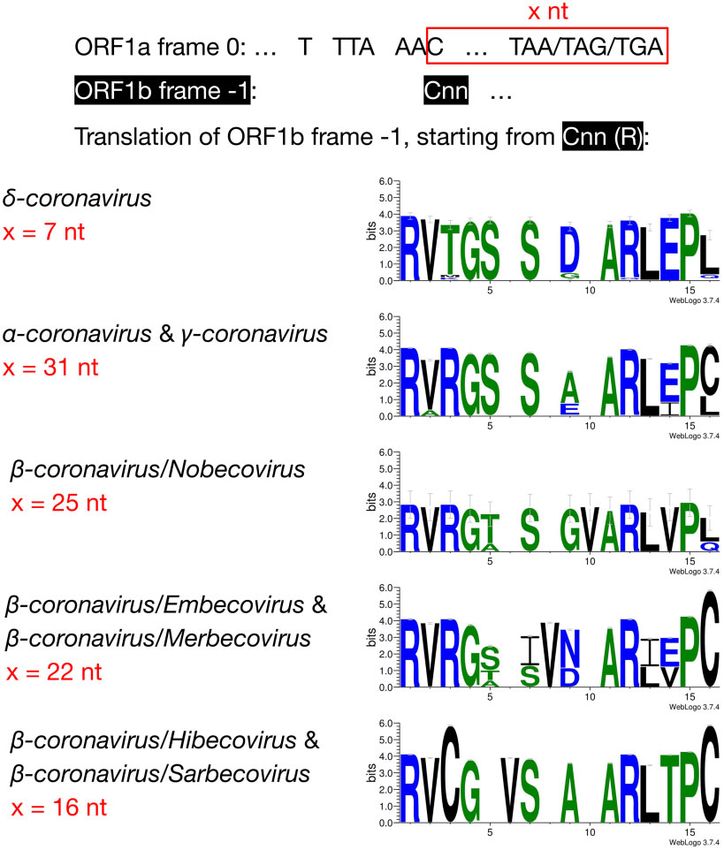
Mei et al. . doi:10.1093/molbev/msab265 MBE
Downloaded from https://academic.oup.com/mbe/advance-article/doi/10.1093/molbev/msab265/6368069 by guest on 02 December 2021
FIG. 1. PFE slippery sites and pseudoknot structures in coronaviruses. The slippery site “UUUAAAC” is shown in italic. The ORF1a stop codon is
shown in red. ORF1b frame starts from the “C” in the slippery site. a-, b-, and c-coronavirus were plotted as splitting from one common node (black
filled circle), with no phylogenetic order shown. The pseudoknot structures of SARS-CoV and MHV are redrawn based on Plant et al. (2010). The
pseudoknot structures of HCoV-229E and IBV are redrawn based on Plant et al. (2005). HCoV-229E, human coronavirus 229E; NC_002645.1. MHV,
mouse hepatitis virus; NC_001846.1. Bat Hp-b-coronavirus/Zhejiang2013, NC_025217.1. MERS-CoV, NC_019843.3. Ro-batCoV HKU9, rousettus
bat coronavirus HKU9, NC_009021.1. SARS-CoV, NC_004718.3. IBV, infectious bronchitis virus, NC_001451.1. BuCoV HKU11, bulbul coronavirus
HKU11, NC_011547.1.
Material online. There are five distinct overlap length groups the overlap, the stop codon which defines a 7 nt overlap is
(7, 16, 22, 25, and 31 nt) with clear taxonomic specificity. abolished at positions 5–7, through substitutions, which
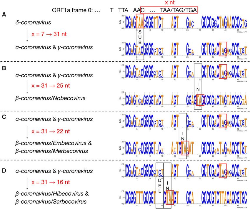
We then compared the first 15 amino acids of ORF1b in all extends ORF1a to the next available stop codon at positions
4,904 entries (fig. 2). The amino acid sequences are highly 38–40. This extension results in a new overlap with 31 nt in
conserved: positions 1 (R), 2 (V), 4 (G), 7 (S), 11–13 (ARL), length (fig. 3A). Comparing coronaviruses with 31 nt (a- and
and 15 (P) are almost invariable and highly redundant. Next, c-coronavirus) and 25 nt (b-coronavirus/Nobecovirus) over-
we compared the underlying nucleotide sequences of the PFE laps reveals a “GTA” insertion at positions 28–30. “TA” from
region (fig. 3). This suggests the following potential series of the “GTA” together with the following “G” forms a new stop
evolutionary events. d-Coronavirus with 7 nt overlap most codon leading to a 31 ! 25 nt shortening of the overlap. In a
likely represents the ancestral state. Comparing coronaviruses Nobecovirus with a 25 nt overlap, the 31 nt overlap stop co-
with 7 nt (d-coronavirus) and 31 nt (a- and c-coronavirus) in don (at positions 38–40) is still observable (fig. 3B). Further
2
ORF1a/b Overlap Evolution and Conservation . doi:10.1093/molbev/msab265                                                              MBE

                                                                                                                                                     Downloaded from https://academic.oup.com/mbe/advance-article/doi/10.1093/molbev/msab265/6368069 by guest on 02 December 2021
FIG. 2. Amino acid alignment of the first 13–14 amino acids in coronaviruses with different lengths in the overlap region. For each genus/subgenus
shown, all coronavirus entries belonging to it were used to generate the consensus amino acid sequences. Gaps are included to maintain
alignment.

comparison of coronaviruses with 31 nt (a- and c-coronavi-                  invariably represented by “T” (fig. 3D). The variable position
rus) and 22 nt (b-coronavirus/Embecovirus and                               of the stop codon likely has an implication to the frameshift
Merbecovirus) overlaps revealed a “GTA” insertion as well,                  efficiency in these taxa as was shown by Bhatt et al. (2021).
but at positions 22–24. “TA” at positions 23–24 and the fol-                These authors demonstrated that extension of the distance
lowing “A” or “G” at position 25 constitute a new stop codon.               between the slippery heptamer and the stop codon of 0-
In the 22 nt overlap, substitutions have been observed at the               frame decreases frameshifting frequency: an increase in the
original stop codon (at positions 38–40) from 31 nt overlap                 distance by 15 nucleotides, as is the case in a- and c-corona-
coronaviruses; more specifically, “C” appears at position 39                viruses (fig. 3), decreases efficiency by 20%, while removal of
(fig. 3C). Finally, we compared coronaviruses with 31 and                   the stop decreases it by half.
16 nt length in the overlap. The same “GTA” insertion foot-                     The abundance of SARS-CoV-2 sequencing data allows
print was found, at positions 16–18 ahead of the two “GTA”                  examining the substitution dynamics in population- and
insertions in 31 ! 25 nt and 31 ! 22 nt events. “TA” at                     individual-level sequencing data. For population-level analysis,
positions 17–18 and the following “A” at position 19 form                   we identified variants in the PFE region from >1,550,000 ge-
the stop codon in the 16 nt overlap coronaviruses. In addi-                 nome sequences available from GISAID (see Materials and
tion, deletions at positions 13–15 were observed (fig. 3D).                 Methods section). However, because GISAID contains only
These deletions are referred as “TCT”-like, since “TCT” are                 assembled genomes, these data do not provide information
the dominant nucleotides observed at positions 13–15 in                     about individual-level (intrasample) variation. Hence, we per-
the 7 and 31 nt overlap coronaviruses. At positions 38–40,                  formed an additional detailed analysis of >55,000 samples
the ancestral stop codon in the 31 nt overlap coronaviruses                 generated with the COG-UK (Lythgoe et al. 2021) consortium
cannot be seen, since the nucleotide at position 39 is                      (see Maier et al. (2021) for analysis details). A summary of

                                                                                                                                                3
Mei et al. . doi:10.1093/molbev/msab265 MBE
Downloaded from https://academic.oup.com/mbe/advance-article/doi/10.1093/molbev/msab265/6368069 by guest on 02 December 2021
FIG. 3. Nucleotide alignment of the overlap in coronaviruses with 7, 31, 25, 22, and 16 nt. The footprints of substitutions, insertions, and deletions
are shown in black boxes, and labeled as “SUB,” “IN,” and “DEL”, respectively. The stop codon of ORF1a in each of the 7, 31, 25, 22, and 16 nt overlap
coronaviruses is shown in a red box.
results from both analyses is shown in table 1. There is little samples). Functional significance, if any, for this substitution
variation in the PFE region as the fraction of samples contain- has not been reported.
ing individual substitutions appears to be small (the two Our results provide an alternative way to assess excep-
“Count” columns in table 1). Furthermore, the 30 out of 36 tional conservation of the PFE using publicly available se-
substitutions in table 1 are consistent with being a result of quence data highlighting the fact that the entire PFE region
RNA editing events from APOBEC (Chen and MacCarthy appears to be under strong purifying selection. These patterns
2017) or ADAR (Bazak et al. 2014) enzymatic complexes. are similar to observations obtained from deep mutational
The remaining six substitutions (all transitions) are predom- scanning where any alteration at the majority of PFE region
inantly located in the loop regions of the predicted PFE sec- sites have deleterious effects on the frameshift efficiency (e.g.,
ondary structure (Huston et al. 2021) and thus likely have Carmody et al. 2021).
minimal effect on the secondary structure.
Through a comparative analysis of GISAID sequences, we
found that several codons with non-negligible levels of vari- Materials and Methods
ation (table 2) were subject to purifying selection: RdRp: 1 Coronavirus Entries Retrieval and Filter
(A13,443>C/G), RdRp: 31 (13,532 A>G/C), RdRp: 32 (13,535 The 35,152 coronaviral entries in the NCBI taxonomy data-
C>T). This is consistent with a strong degree of functional base were sorted by length, and only those longer than
constraint. Interestingly, this analysis also identified a single 14,945 nt were kept, leaving a total of 4,939 genomes. The
codon: RdRp: T26I (13,516 T>C), which has been subject to slippery site and following overlap sequences were manually
pervasive positive selection since early 2021. Most of the inspected, in case the slippery site was incorrectly annotated.
sequences with this substitution are in the B.1.1.7 and We further filtered out those entries if they contained no
B.1.177.77 lineages (this is a consensus majority mutation in annotation information, or had gapped sequences in the
B.1.177.77 and B.1.614 lineages). RdRp: T26I is present at low overlap. 4,904 coronavirus entries were selected using this
frequencies in many viral lineages but is increasing in preva- approach (supplementary table S1, Supplementary Material
lence in recent months (0.5–1.0% global prevalence in recent online).
4ORF1a/b Overlap Evolution and Conservation . doi:10.1093/molbev/msab265 MBE
Table 1. Allelic Variants within the PFE Region are Called from Complete GISAID Genomes (population) and COG-UK (individual) Data
Site H B Reference Population Individual
Alternate Countc Alternate Min AF Max AF Countd
a
13,425 C T 1,812 — — — —
13,429a C T 460 — — — —
13,430a C T 169 — — — —
13,431a C T 517 — — — —
13,432b A G 110 — — — —
Downloaded from https://academic.oup.com/mbe/advance-article/doi/10.1093/molbev/msab265/6368069 by guest on 02 December 2021
13,434a G A 213 — — — —
13,43a C T/A 1,328/120 T 0.116 0.971 14
13,437b T C 195 C 0.985 0.988 5
13,440 S S G A 116 — — — —
13,443b# A G/T 134/22 — — — —
13,445a C T 680 T 0.068 0.970 25
13,447a G A 16 — — — —
13,451a C T 393 T 0.941 0.977 19
13,457a C T 3,663 T 0.052 0.963 19
13,458a G — — A 0.069 0.970 6
13,458 L L G T 1,220 T 0.080 0.976 6
13,481b A G 9 — — — —
13,486a C T 1,656 T 0.055 0.965 7
13,487b A G 151 G 0.901 0.949 12
13,497b A G 434 — — — —
13,498a C T 189 — — — —
13,500a C T 243 — — — —
13,504 S G T 102 — — — —
13,505a C T 314 T 0.887 0.917 5
13,511 S A T/G/C 121/58/11 — — — —
13,512 S G T 114 — — — —
13,513a G A 342 — — — —
13,514a C T 495 T 0.065 0.889 6
13,516a" C T 4,272 T 0.101 0.840 49
13,525 S S A C 104 — — — —
13,526b T C 117 — — — —
13,532b# A G 742 — — — —
13,535a# C T 11,942 T 0.215 0.841 23
13,541b T C 26 — — — —
13,547a C T 675 T 0.067 0.898 8
13,550a C T 2,346 T 0.878 0.921 11
a
Potential APOBEC-edited sites;
b
Potential ADAR-edited sites.
Site numbering is in 0-based coordinates.
c
Out of 1,525,442 complete genome.
d
Out of 55,163 individual samples. Locations of substitutions in a stem (S) or a loop (L) are based on structures predicted by Huston et al. (H) and Bhatt et al. (B). # and "
highlight sites showing signatures of negative and positive selection, respectively (see table 2).
Table 2. Sites with Selection Signatures Identified using a Fixed Effects WebLogo (Crooks et al. 2004). The same was done to a-co-
Likelihood Method on Internal Branches using SARS-CoV-2 Phylogeny ronavirus and c-coronavirus. Within b-coronavirus, for
Built from GISAID Sequences (FEL; [Kosakovsky Pond and Frost 2005])a:
Nobecovirus, Embecovirus, and Merbecovirus, the first 14
synonymous substitution rate (maximum likelihood estimate, MLE), b:
non-synonymous substitution rate (MLE), x:b/a amino acids were used to build the consensus; for
Hibecovirus and Sarbecovirus, the first 13 amino acids were
Codon Nucleotide a b x LRT P value used. In terms of the nucleotide sequence alignments, for
1 13,443 0 0 4.286 0.002 each genus/subgenus, the nucleotide sequences used to gen-
31 13,352 7.040 0 0 0.015 erate the amino acids mentioned above were taken to make
26 13,516 0 4.722 ‘ 0.004 the nucleotide consensus sequence using WebLogo.
32 13,535 5.205 0 0 0.035
Here, a < b signifies positive selection, while a > b is indicative of negative
selection. Processing of GISAID Data
Each genome was subjected to codon-aware alignment with
Amino Acid Alignment and Nucleotide Alignment of the NCBI reference genome (accession number NC_045512)
the Overlap Region and then subdivided into ten regions based on CDS features:
For all d-coronavirus entries in supplementary table S1, ORF1a (including nsp10), ORF1b (starting with nsp12), S,
Supplementary Material online, the first 13 amino acids of ORF3a, E, M, ORF6, ORF7a, ORF8, N, and ORF10. For each
ORF1b were taken to generate a consensus sequence using region, we scanned and discarded sequences containing too
5Mei et al. . doi:10.1093/molbev/msab265 MBE
many ambiguous nucleotides to remove data with possible Faria NR, Mellan TA, Whittaker C, Claro IM, da S, Candido D, Mishra S,
sequencing errors. Thresholds were 0.5% for the S gene, 0.1% Crispim MAE, Sales FCS, Hawryluk I, McCrone JT, et al. 2021.
Genomics and epidemiology of the P.1 SARS-CoV-2 lineage in
for ORF1a and ORF1b genes, and 1% for all other genes. We Manaus, Brazil. Science 372(6544):815–821.
mapped individual sequences to the NCBI reference genome Gianella S, Delport W, Pacold ME, Young JA, Choi JY, Little SJ, Richman
(NC_045512) using a codon-aware extension to the Smith- DD, Kosakovsky Pond SL, Smith DM. 2011. Detection of minority
Waterman algorithm implemented in the BioExt package resistance during early HIV-1 infection: natural variation and spuri-
(Pond et al. 2005; Gianella et al. 2011) and translated mapped ous detection rather than transmission and evolution of multiple
viral variants. J Virol. 85(16):8359–8367.
sequences to amino-acids. Codon sequences were next Huston NC, Wan H, Strine MS, de Cesaris Araujo Tavares R, Wilen CB,
mapped onto the amino-acid alignment. Variants were called
Downloaded from https://academic.oup.com/mbe/advance-article/doi/10.1093/molbev/msab265/6368069 by guest on 02 December 2021
Pyle AM. 2021. Comprehensive in vivo secondary structure of the
directly. Selection analyses were performed using the proto- SARS-CoV-2 genome reveals novel regulatory motifs and mecha-
cols used previously (Faria et al. 2021; Tegally et al. 2021) based nisms. Mol Cell. 81(3):584–598.e5.
on the FEL analysis (Kosakovsky Pond and Frost 2005) within Iserman C, Roden CA, Boerneke MA, Sealfon RSG, McLaughlin GA,
Jungreis I, Fritch EJ, Hou YJ, Ekena J, Weidmann CA, et al. 2020.
the HyPhy package (Kosakovsky Pond et al. 2019). Genomic RNA elements drive phase separation of the SARS-CoV-
2 nucleocapsid. Mol Cell. 80(6):1078–1091.e6.
Supplementary Material Kelly JA, Olson AN, Neupane K, Munshi S, San Emeterio J, Pollack L,
Woodside MT, Dinman JD. 2020. Structural and functional conser-
Supplementary data are available at Molecular Biology and vation of the programmed 1 ribosomal frameshift signal of SARS
Evolution online. coronavirus 2 (SARS-CoV-2). J. Biol. Chem. 295(31):10741–10748.
Kosakovsky Pond SL, Frost SDW. 2005. Not so different after all: a com-
Acknowledgments parison of methods for detecting amino acid sites under selection.
Mol Biol Evol. 22(5):1208–1222.
This work is funded by NIH Grants U41 HG006620, R01 Kosakovsky Pond SL, Poon AFY, Velazquez R, Weaver S, Hepler NL,
AI134384 (NIH/NIAID), and NSF ABI Grant 1661497 and Murrell B, Shank SD, Magalis BR, Bouvier D, Nekrutenko A, et al.
2027196 (NSF/DBI, BIO). The funders had no role in study 2020. HyPhy 2.5—a customizable platform for evolutionary hypoth-
design, data collection and analysis, decision to publish, or esis testing using phylogenies. Mol Biol Evol. 37(1):295–299.
Lau SKP, Woo PCY, Yip CCY, Fan RYY, Huang Y, Wang M, Guo R, Lam
preparation of the manuscript.
CSF, Tsang AKL, Lai KKY, et al. 2012. Isolation and characterization of
a novel Betacoronavirus subgroup A coronavirus, rabbit coronavirus
References HKU14, from domestic rabbits. J. Virol. 86(10):5481–5496.
Lythgoe KA, Hall M, Ferretti L, de Cesare M, MacIntyre-Cockett G, Trebes
Baranov PV, Henderson CM, Anderson CB, Gesteland RF, Atkins JF,
A, Andersson M, Otecko N, Wise EL, Moore N, et al.; on behalf of the
Howard MT. 2005. Programmed ribosomal frameshifting in decod-
Oxford Virus Sequencing Analysis Group (OVSG). 2021. SARS-CoV-2
ing the SARS-CoV genome. Virology 332(2):498–510.
within-host diversity and transmission. Science. 372(6539):eabg0821.
Bazak L, Haviv A, Barak M, Jacob-Hirsch J, Deng P, Zhang R, Isaacs FJ,
Maier W, Bray S, van den Beek M, Bouvier D, Coraor N, Miladi M, Singh B,
Rechavi G, Li JB, Eisenberg E, et al. 2014. A-to-I RNA editing occurs at
De Argila JR, Baker D, Roach N, et al. 2021. Freely accessible ready to use
over a hundred million genomic sites, located in a majority of hu-
global infrastructure for SARS-CoV-2 monitoring. bioRxiv [Preprint].
man genes. Genome Res. 24(3):365–376.
2021 Mar 25:2021.03.25.437046. doi: 10.1101/2021.03.25.437046.
Bhatt PR, Scaiola A, Loughran G, Leibundgut M, Kratzel A, Meurs R,
Nekrutenko A, Wadhawan S, Goetting-Minesky P, Makova KD. 2005.
Dreos R, O’Connor KM, McMillan A, Bode JW, et al. 2021. Structural
Oscillating evolution of a mammalian locus with overlapping read-
basis of ribosomal frameshifting during translation of the SARS-CoV-
ing frames: an XLalphas/ALEX relay. PLoS Genet. 1(2):e18.
2 RNA genome. Science 372(6548):1306–1313.
Plant EP, Rakauskaite R, Taylor DR, Dinman JD. 2010. Achieving a golden
Brierley I. 1995. Ribosomal frameshifting on viral RNAs. J Gen Virol. 76 ( Pt
mean: mechanisms by which coronaviruses ensure synthesis of the
8):1885–1892.
correct stoichiometric ratios of viral proteins. J Virol. 84(9):4330–4340.
Carmody PJ, Zimmer MH, Kuntz CP, Harrington HR, Duckworth KE,
Pond SL, Frost SD, Muse SV. 2005. HyPhy: hypothesis testing using phy-
Penn WD, Mukhopadhyay S, Miller TF, Schlebach JP. 2021.
logenies. Bioinformatics. 21(5):676–679.
Coordination of -1 programmed ribosomal frameshifting by tran-
Rangan R, Watkins AM, Chacon J, Kretsch R, Kladwang W, Zheludev IN,
script and nascent chain features revealed by deep mutational scan-
Townley J, Rynge M, Thain G, Das R. 2021. De novo 3D models of
ning. bioRxiv [Preprint]. 2021 Mar 25:2021.03.25.437046. doi:
SARS-CoV-2 RNA elements from consensus experimental secondary
10.1101/2021.03.11.435011v1.
structures. Nucleic Acids Res. 49(6):3092–3108.
Chen J, MacCarthy T. 2017. The preferred nucleotide contexts of the
Sola I, Almazan F, Z ~iga S, Enjuanes L. 2015. Continuous and discontin-
un
AID/APOBEC cytidine deaminases have differential effects when
uous RNA synthesis in coronaviruses. Annu Rev Virol. 2(1):265–288.
mutating retrotransposon and virus sequences compared to host
Szklarczyk R, Heringa J, Pond SK, Nekrutenko A. 2007. Rapid asymmetric
genes. PLoS Comput Biol. 13(3):e1005471.
evolution of a dual-coding tumor suppressor INK4a/ARF locus con-
Chung W-Y, Wadhawan S, Szklarczyk R, Pond SK, Nekrutenko A. 2007. A
tradicts its function. Proc Natl Acad Sci U S A. 104(31):12807–12812.
first look at ARFome: dual-coding genes in mammalian genomes.
Tegally H, Wilkinson E, Giovanetti M, Iranzadeh A, Fonseca V, Giandhari J,
PLoS Comput Biol. 3(5):e91.
Doolabh D, Pillay S, San EJ, Msomi N, et al. 2021. Detection of a SARS-
Coronaviridae Study Group of the International Committee on
CoV-2 variant of concern in South Africa. Nature 592(7854):438–443.
Taxonomy of Viruses. 2020. The species severe acute respiratory
Woo PCY, Lau SKP, Lam CSF, Lau CCY, Tsang AKL, Lau JHN, Bai R, Teng
syndrome-related coronavirus: classifying 2019-nCoV and naming
JLL, Tsang CCC, Wang M, et al. 2012. Discovery of seven novel
it SARS-CoV-2. Nat Microbiol. 5:536–544.
Mammalian and avian coronaviruses in the genus deltacoronavirus
Crooks GE, Hon G, Chandonia J-M, Brenner SE. 2004. WebLogo: a se-
supports bat coronaviruses as the gene source of alphacoronavirus
quence logo generator. Genome Res. 14(6):1188–1190.
and betacoronavirus and avian coronaviruses as the gene source of
de Groot RJ, Baker SC, Baric R, Enjuanes L, Gorbalenya AE, Holmes KV,
gammacoronavirus and deltacoronavirus. J Virol. 86(7):3995–4008.
Perlman S, Poon L, Rottier PJM, Talbot PJ, et al. 2012. Family coro-
Ziv O, Price J, Shalamova L, Kamenova T, Goodfellow I, Weber F, Miska
naviridae. In: King AMQ, Lefkowitz E, Adams MJ, Carstens EB, editors.
EA. 2020. The short- and long-range RNA-RNA interactome of
Virus taxonomy: ninth report of the International Committee on
SARS-CoV-2. Mol Cell. 80(6):1067–1077.e5.
Taxonomy of Viruses. Amsterdam: Elsevier. p. 806–828.
6You can also read